(3) types of cytoskeletal fibers microtubule intermediate...
TRANSCRIPT
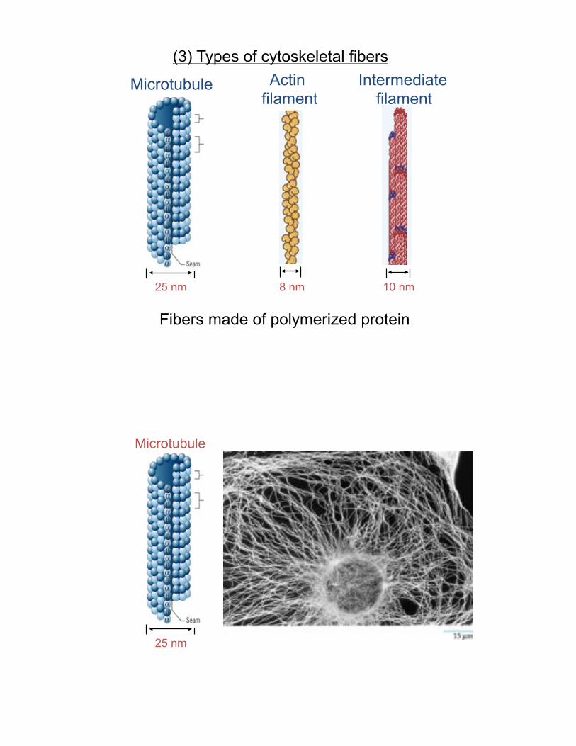
Actin filament
Microtubule Intermediate filament
25 nm 8 nm 10 nm
Fibers made of polymerized protein
(3) Types of cytoskeletal fibers
Microtubule
25 nm

Cell Shape and Transportation:
(Cells, Figure 7.14)
Organization of the Cell
Cell is highly structured Organelles are not passive blobs?

Do these organelles move on microtubules?
A snapshot
(Figure 9.2)

Drugs can be used to study the functions of Microtubules
Nocodazole Taxol
!"#$%&$'()*$+(,$ $ $$$$$$$$
GTP/GDP
Figure 9.9
What is the structure?
(13) protofilaments align to form a hollow tube

Self-assembly into polymer/microtubule
What is the structure? Microtubules are polymers -)%$."#$%&$'&&$(/+',$ $$$$$$$$$
Microtubule Organizing Center (MTOC) Centrosome
PCM
Centrioles

Transportation and Polarity:
(Cells, Figure 7.14)
!"#$%&'$()*)+*)$,,*-+.*/0$%$*)0$*1234*&56*
Colcemid washout experiment
Figure 9.19

(13) protofilaments align to form a hollow tube = microtubule lateral bonds give tubule strength
Microtubule Assembly

(13) protofilaments align to form a hollow tube = microtubule lateral bonds give tubule strength
Microtubule Assembly

Figure 9.26
Where are subunits added?
Injected with biotin-Tubulin (1 minute)
Figure 9.8
FRAP

Figure 9.8
FRAP:
Can tell you about the dynamics of molecules in the polymer
Figure 9.4
Injected with rhod-Tubulin
We can watch MT dynamics

Figure 9.27
Inject with GFP-Tubulin
Dynamic Instability (Speckles)
GTP hydrolysis changes conformation and stability of MTs

(+) end cap regulates stability of MT
GTP cap:
GTP-bound structure is different than GDP-bound
(+) end !-tubulin
Stability of microtubules
1) GTP vs GDP bound cap
2) Microtubule associated proteins (MAPs)
GTP
GDP

From website: flipper e nuvola
From website: flipper e nuvola

Stability of microtubules
1) GTP vs GDP bound cap
2) Microtubule associated proteins (MAPs)
GTP
GDP

MAPs can destabilize MTs
katanin MCAK (GAP) Colchicine (drug)
How do (+) TIPs stabilize the (+) tip?
XMAP215 CLIP170 (function)
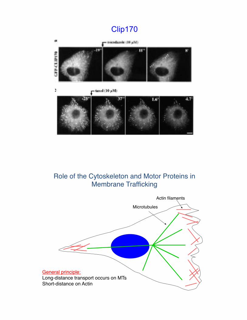
Clip170
Role of the Cytoskeleton and Motor Proteins in Membrane Trafficking#
General principle: #Long-distance transport occurs on MTs #Short-distance on Actin#
Microtubules#
Actin filaments#

Transportation and Polarity:
(Cells, Figure 7.14)
Figure 9.13
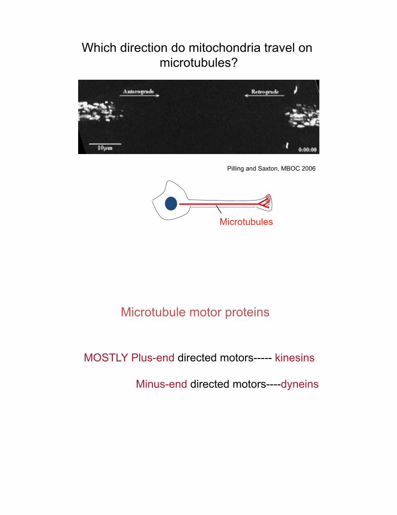
Which direction do mitochondria travel on microtubules?
Pilling and Saxton, MBOC 2006
Microtubules
Microtubule motor proteins
MOSTLY Plus-end directed motors----- kinesins Minus-end directed motors----dyneins
dyneins (-, only) kinesins (+, mostly)
*
*
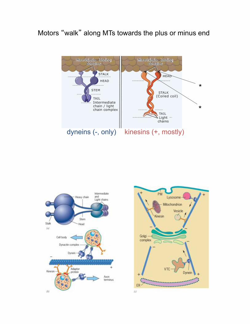
Motors “walk” along MTs towards the plus or minus end

How cargoes are loaded onto motors:
Tail domain binds cargo via adaptor protein
AP1
vesicle
Kinesin:

What can measure from this movie?
Microtubules Kinesin-1
Watching Kinesin-1 move –speed, processive
Figure 9.6

mito
chon
dria
How do you determine which motor is involved?
dyneins (-, only) kinesins (+, mostly)
Motors “walk” along MTs towards the plus or minus end

kinesins: -MT (+) end -motor domain at N-terminus -ATP-dependent
dyneins: -MT (-) end -motor domain at C-terminus -cytoplasmic form (homodimer) -ATP-dependent
Ron Vale and colleagues


Figure 9.66
7&8).%$*+9*:$.%+(*,;<$,,$=*9+%*;8>(*;(=*&)5*50;#$*
?+(;)0;(*@%&$=';(*
General principle 2: Large structural changes occur with microtubules, small changes are with actin filaments#

Role of the Cytoskeleton and Motor Proteins in Membrane Trafficking#
General principle 1: long-distance transport occurs on microtubules, short distance on actin filaments#
Microtubules#
Actin filaments#
Actin filaments building block = actin#
Functions: structural support, contraction, migration##
Lodish Fig. 19-3#
Figure 9.44 (-) end

Figure 9.46
Figure 9.46
Rate and direction of growth depends on free actin concentration
Critical concentration

A$B.,;>+(*<-*;8>(C<&(=&(B*#%+)$&(5*
D8>(*#+,-'$%&E;>+(*

Proteins that regulate actin polymerization
Therefore, the direction of growth is regulated
7%+)$&(5*)0;)*=$#+,-'$%&E$*;8>(*F,;'$()5*

Small G protein activation regulates actin organization
Rac Rho Cdc42
Filopodia Stress fibers
lamellipodia
Actin

Actin

Figure 9.71
Directed cell motility
Actin

Myosin: variety of tail domains
*
*
Structure of myosin proteins
Binds tightly in the absence of ATP ATP hydrolysis - power stroke - lever arm

A$B.,;>+(*+9*'-+5&(*<-*#0+5#0+%-,;>+(*
Figure 9.52
Rab proteins on vesicles are linked to cytoskeleton Also regulated by GTP-Rab state


Myosin: variety of tail domains

1-+5&(*GG*
H&($5&(*
Myosin: mostly (+) end directed
Figure 9.49


Myosin: variety of tail domains
1-+5&(*GG*
H&($5&(*
Myosin: mostly (+) end directed
Figure 9.49


Myosin: variety of tail domains
A$B.,;>+(*+9*'-+5&(*<-*#0+5#0+%-,;>+(*

93
The Cytoskeleton: Intermediate Filaments!
IJ*

IK*

IL*

II*
MNN*

101
The Cytoskeleton: Intermediate Filaments!
MNO*
Mutant Normal
epidermolysis bullosa simplex

7,;8$5*/0$%$*-+.’,,*F(=*$#&)0$,&;,*8$,,5*
H$%;>(5*9+%'*G()$%'$=&;)$*@&,;'$()5*)0;)*+%B;(&E$*>55.$5*
Intermediate filaments building block = variable
(keratin, vimentin, nuclear lamins, others)#
Functions: mechanical integrity of tissue, cell, or subcellular organelle !
Lodish Fig. 19-33#

Laminopathies (HGPS, progeria)
Some IFs are found in all cells.#Nuclear lamins, a special type of IF, form a basket underlying
the nuclear membrane, giving it strength and organization.#
Lodish Fig. 19-33# *Phosphorylated at head and tail during mitosis!


4$,,*4-8,$*:!PQ;'&(*40;(B$5*
CDK1/cyclinB (kinase)
PP1 (phosphatase)
Regulation of Nuclear Lamins
LBR
Lamin B

11/7/08 10:34 AM19.png 480!360 pixels
Page 1 of 1http://www.soi.wide.ad.jp/class/20050028/slides/03/img/19.png