305 - red latinoamericana de reproducción asistidaredlara.com/images/arq/rla2010_jbra.pdf · uso...
TRANSCRIPT
305
JBRA Assist. Reprod. | V. 16 | nº6 | Nov-Dec / 2012
JBRA6_150213.indb 305 15/02/2013 17:17:08

JBRA - Assisted Reproduction
INDEXADO NAS SEGUINTES BASES DE DADOS – Indexed on the following databases:CompendexEMBASEExcepta MédicaGeobase
PERIODICA (México)Plataforma SCImago Journal & Country RankPORTAL DE PERIÓDICOS DA CAPESScopus (Holanda)
Jornalista Responsável: Heber Maia – MTb 31.660
Produção Editorial e Gráfica: AlamTec Ciência Médica Editorial LTDARua das Roseiras, 464CEP 03144-090 - São Paulo-SPTel/Fax: (11) 2341-8045e-mail: [email protected]
Endereço para Correspondência: Dra. Maria do Carmo Borges de Souza Av. das Américas, 4666 - Sl. 312 / 313Barra da Tijuca - RJ CEP 22649-900E-mail: [email protected]: (21) 2430-9060Fax: (21) 2430-9070
ÓRGÃO DE DIVULGAÇÃO DA SOCIEDADE BRASILEIRA DE REPRODUÇÃO ASSISTIDA E DA REDE LATINOAMERICANA DE REPRODUÇÃO ASSISTIDA
ISSN: 1517-5693 - V. 16 | nº1 | Jan-Feb / 2012
Você. Nós. Somos os pais da fertilidade
Veja a obra de arte que fizemosjuntos.
SAC Merck Serono: 0800.113320
Anúncio veiculado em Maio de 2011.
Merck Serono é uma divisão da Merck.
JBRA_01_2012_AlamTec_260712.indb 3 27/07/12 16:19
Você. Nós. Somos os pais da fertilidade
Veja a obra de arte que fizemosjuntos.
SAC Merck Serono: 0800.113320
Anúncio veiculado em Maio de 2011.
Merck Serono é uma divisão da Merck.
JBRA6_150213.indb 306 15/02/2013 17:17:09
JBRA - Assisted Reproduction
INDEXADO NAS SEGUINTES BASES DE DADOS – Indexed on the following databases:CompendexEMBASEExcepta MédicaGeobase
PERIODICA (México)Plataforma SCImago Journal & Country RankPORTAL DE PERIÓDICOS DA CAPESScopus (Holanda)
Jornalista Responsável: Heber Maia – MTb 31.660
Produção Editorial e Gráfica: AlamTec Ciência Médica Editorial LTDARua das Roseiras, 464CEP 03144-090 - São Paulo-SPTel/Fax: (11) 2341-8045e-mail: [email protected]
Endereço para Correspondência: Dra. Maria do Carmo Borges de Souza Av. das Américas, 4666 - Sl. 312 / 313Barra da Tijuca - RJ CEP 22649-900E-mail: [email protected]: (21) 2430-9060Fax: (21) 2430-9070
ÓRGÃO DE DIVULGAÇÃO DA SOCIEDADE BRASILEIRA DE REPRODUÇÃO ASSISTIDA E DA REDE LATINOAMERICANA DE REPRODUÇÃO ASSISTIDA
ISSN: 1517-5693 - V. 16 | nº1 | Jan-Feb / 2012
Você. Nós. Somos os pais da fertilidade
Veja a obra de arte que fizemosjuntos.
SAC Merck Serono: 0800.113320
Anúncio veiculado em Maio de 2011.
Merck Serono é uma divisão da Merck.
JBRA_01_2012_AlamTec_260712.indb 3 27/07/12 16:19
INDEXADO NAS SEGUINTES BASES DE DADOS – Indexed on the following databases:CompendexEMBASEExcepta MédicaGeobase
PERIODICA (México)Plataforma SCImago Journal & Country RankPORTAL DE PERIÓDICOS DA CAPESScopus (Holanda)
ÓRGÃO DE DIVULGAÇÃO DA SOCIEDADE BRASILEIRA DE REPRODUÇÃO ASSISTIDA E DA REDE LATINOAMERICANA DE REPRODUÇÃO ASSISTIDA
ISSN: 1517-5693 - V. 16 | nº6 | Nov-Dec / 2012
Você. Nós. Somos os pais da fertilidade
Veja a obra de arte que fizemosjuntos.
SAC Merck Serono: 0800.113320
Anúncio veiculado em Maio de 2011.
Merck Serono é uma divisão da Merck.
Edição Especial - Dados de Registro Latinoamericano
JBRA - Assisted Reproduction
Jornalista Responsável: Heber Maia – MTb 31.660
Comercialização, Produção Editorial e Gráfica: AlamTec Ciência Médica Editorial LTDARua das Roseiras, 464CEP 03144-090 - São Paulo-SPTel/Fax: (11) 2341-8045e-mail: [email protected]
Endereço para Correspondência:Dra. Maria do Carmo Borges de Souza Av. das Américas, 4666 - Sl. 312 / 313Barra da Tijuca - RJ CEP 22649-900E-mail: [email protected]: (21) 2430-9060Fax: (21) 2430-9070
JBRA6_150213.indb 307 15/02/2013 17:17:10

Referências bibliográficas: 1. Basset R.M. et al. Continued improvements in the quality and consistency of follitropin alfa, recombinant human FSH. Reproductive BioMedicine Online. 2005; 10(2): 169-177(9). 2. Gervais A, et al. Glycosylation of recombinant gonadotrophins: characterization and batch-to-batch consistency. Glycobiology 2003; 13(3):179-189.Bula do produto no interior desta publicação. Destinado exclusivamente à classe médica. Veiculado em junho de 2012.
Precisão,pureza econsistênciana associação das duas gonadotrofinas1,2
Merck Serono é uma divisão da Merck.
JBRA_01_2012_AlamTec_260712.indb 5 27/07/12 16:19
CORPO EDITORIAL – EDITORIAL BOARD
Editor – EditorMaria do Carmo Borges de Souza (Fertipraxis/ RJ Brasil)
Editor Adjunto – Assistant EditorPaulo Franco Taitson (ARQ / PUCMG Brasil)
Consultor Editorial - Editorial ConsultantJosé Gonçalves Franco Jr (UNESP – Botucatu / CRH SP Brasil)
Editores Associados – Associate EditorsEdson Borges Jr (Fertility / Faculdade de Medicina de Jundiaí - Inst Sapientiae SP Brasil)
João Batista A Oliveira (CRH SP Brasil)
Selmo Geber (Origen / UFMG Brasil)
Weydson Barros Leal (UFPE Brasil)
CONSULTORES CIENTÍFICOS – Scientific Reviewers
Adelino Amaral Silva (Gênesis / Escola Superior de Ciências da Saúde DF Brasil)
Agnaldo Lopes da Silva Filho (UFMG Brasil)
Alessandro Schuffner (Conceber PR Brasil)
Álvaro Petracco (Fertilitat/ PUC RS Brasil)
Ana Cristina Allemand Mancebo (G&O Barra RJ Brasil)
Anne R Greenlee (OHSH EUA)
Antonio Requena (IVI Madrid Espanha)
Aroldo Camargos (UFMG Brasil)
Bela Zausner (Gênese BA Brasil)
Bruno Scheffer (IBRRA MG Brasil)
Buenaventura Coroleu (Instituto Universidade Dexeus, Barcelona, Espanha)
Carlos André Henriques (UFRJ / G&O Barra RJ Brasil)
Carlos María Romeo-Casabona (Universidade de Deusto e do Pais Basco Espanha)
Cesar Cafatti (Clin Los Dominicos Chile)
Claudia Borrero (Conceptum Colombia)
Claudia G Petersen (CRH SP Brasil)
Cláudio Chillik (CEGYR Argentina)
Condesmar Marcondes Filho (Nucl Santista RH SP Brasil)
David Vantman (CER Chile)
Dirceu H Mendes Pereira (Profert SP Brasil)
Eduardo Pandolfi Passos (SEGIR / UFRGS RS Brasil)
Ernesto Gallardo Lozano (IMER México)
Fabio Firmbach Pasqualotto (Conception / UCS RS Brasil)
Fernando Zegers-Hochschild (Clin Las Condes Chile)
Francisco Risquez (Clin La Trinidad Venezuela)
Francisco J.B. Sampaio (UERJ Brasil)
Humberto Ikuo Shibasaki (UFMT Brasil)
Jorge Blaquier (Fertilab Argentina)
João Pedro Junqueira Caetano (Pró-Criar/ Mater Dei MG Brasil)
Joaquim Roberto C Lopes (Cenafert BA Brasil)
Jonathas Borges Soares (Faculdade Medicina do ABC / Projeto Alfa SP Brasil)
Juan Manuel Montoya (Conceptum Colombia)
Ivan Valencia Madera (CEMEFES Equador)
Karen Sermon (VUB Bélgica)
Leila Montenegro S Farah (Fertility / Faculdade de Medicina de Jundiaí - Inst Sapientiae SP Brasil)
Leticia Urdapilleta (Cegyr Argentina)
Lídio Jair Ribas Centa (Androlab/ UFPR Brasil)
Luiz Fernando Dale (C Medicina RJ Brasil)
Madalena Caldas (GERAR PE Brasil)
Marcos Sampaio (Origen MG Brasil)
Mariângela Badalotti (Fertilitat PUC RS Brasil)
Marilena Correa (IMS-UERJ Brasil)
Mario Cavagna (H Perola B/ CRH SP Brasil)
Maria Silva Approbato (UFG Brasil)
Marisa Decat de Moura (IBBRA/Universidade FUMEC BH Brasil)
Miguel Angel Checa Vizcaino (Universidade Autönoma de Bracelona, Espanha)
Newton Eduardo Busso (Fac. CM Santa Casa de SP / Unifert SP Brasil)
Paulo Serafini (Huntington/ USP SP Brasil)
Ricardo Melo Marinho (FCMMG MG Brasil)
Roberta Wonchockier (Projeto Alfa SP Brasil)
Roberto Coco (Fecunditas Argentina)
Rose Marie M Melamed (Fertility SP Brasil)
Sidney Glina (Fac. Medicina do ABC / Hosp Albert Einstein SP Brasil)
Silvana Chedid Chedid-Grieco (SP Brasil)
Sergio Reis Soares (IVI Lisboa Portugal)
Tania Maria Ruffoni Ortiga Ortiga ( Inst Biofisica da UFRJ )
Renato Fanchin (Hôpital A. Béclère, University Paris-Sud 11 França)
JBRA6_150213.indb 308 15/02/2013 17:17:10

Referências bibliográficas: 1. Basset R.M. et al. Continued improvements in the quality and consistency of follitropin alfa, recombinant human FSH. Reproductive BioMedicine Online. 2005; 10(2): 169-177(9). 2. Gervais A, et al. Glycosylation of recombinant gonadotrophins: characterization and batch-to-batch consistency. Glycobiology 2003; 13(3):179-189.Bula do produto no interior desta publicação. Destinado exclusivamente à classe médica. Veiculado em junho de 2012.
Precisão,pureza econsistênciana associação das duas gonadotrofinas1,2
Merck Serono é uma divisão da Merck.
JBRA_01_2012_AlamTec_260712.indb 5 27/07/12 16:19JBRA6_150213.indb 309 15/02/2013 17:17:11
Pergoveris® alfafolitropina (r-hFSH) 150UI (11μg) alfalutropina (r-hLH) 75UI (3μg). USO SUBCUTÂNEO USO ADULTO Indicações: Pergoveris® é indicado para a estimulação do desenvolvimento folicular em mulheres com insuficiência grave de LH e FSH. Nos ensaios clínicos, estas pacientes foram definidas por um nível sérico de LH <1,2 UI/L. Contra-indicações: Hipersensibilidade à alfafolitropina, alfalutropina ou a qualquer dos excipientes; tumores do hipotálamo ou da hipófise; hipertrofia ou cistos ovarianos não originados por doença do ovário policístico; hemorragias ginecológicas de etiologia desconhecida; carcinoma do útero, ovário ou mama e nas situações em que não é possível a obtenção de uma resposta efetiva (insuficiência ovariana primária, malformações dos órgãos sexuais incompatíveis com gravidez e fibromiomas uterinos incompatíveis com gravidez). Cuidados e Advertências: Na mulher, a utilização segura e eficaz de Pergoveris® requer monitorização ecográfica regular da resposta ovariana, de preferência em conjunto com a determinação dos níveis séricos de estradiol. Deve ser utilizada em mulheres a dose mínima eficaz em relação ao objetivo do tratamento. As pacientes devem ser avaliadas para as seguintes situações, devendo ser instituído um tratamento específico apropriado: hipotiroidismo; insuficiência da supra-renal; hiperprolactinemia; tumores do hipotálamo ou da hipófise. A síndrome de hiperestimulação ovariana (OHSS) é uma situação clínica distinta da hipertrofia ovariana assintomática, podendo se manifestar em graus crescentes de gravidade. Uma resposta excessiva ovariana ao tratamento com gonatropinas raramente origina uma OHSS, exceto quando se administra hCG para induzir a ovulação. Portanto, em casos de hiperestimulação ovariana é prudente não administrar hCG e recomendar à paciente que se abstenha de ter relações sexuais ou utilize métodos anticoncepcionais de barreira, durante pelo menos 4 dias. Em mulheres submetidas à indução da ovulação, a incidência de gravidez e nascimentos múltiplos é aumentada quando comparada à concepção natural. A maioria das concepções múltiplas é de gêmeos. Gravidez e aleitamento: Pergoveris® não deve ser administrado durante a gravidez ou o aleitamento. Reações adversas: Cefaléia, exacerbação de asma, dor abdominal e sintomas gastro-intestinais, tromboembolismo normalmente associado com síndrome de hiperestimulação ovariana grave (OHSS), reações no local da injeção. Reações alérgicas sistêmicas leves (eritema cutâneo, edema, urticária, dificuldades respiratórias) e graves (reações anafiláticas). Cistos ovarianos, dor mamária, dor pélvica, OHSS, torção do ovário. Interações medicamentosas: Pergoveris® não deve ser administrado com outros medicamentos na mesma seringa, exceto com alfafolitropina. Posologia: O tratamento deve ser adaptado à resposta individual da paciente. Um regime posológico recomendado inicia-se com a administração diária de um frasco de Pergoveris®. Caso um aumento da dose de FSH seja considerado apropriado, o ajuste da dose deve ser efetuado preferencialmente após intervalos de 7-14 dias e com incrementos de 37,5 a 75 UI, utilizando um medicamento contendo alfafolitropina. Pode ser aceitável prolongar a duração da estimulação em qualquer um dos ciclos por até 5 semanas. Quando se obtém uma resposta ótima, deve ser administrada uma única injeção de 5.000 UI a 10.000 UI de hCG, 24 a 48 horas após a última injeção de Pergoveris®. Recomenda-se que a paciente tenha relações sexuais no dia da administração de hCG e no dia seguinte. Caso se obtenha uma resposta excessiva, o tratamento deve ser interrompido e hCG não deve ser administrado. O tratamento deve ser reiniciado no ciclo seguinte, com uma dose de FSH inferior à do ciclo anterior. Cuidados de conservação: Prazo de validade: 24 meses. Este medicamento é de uso único e deve ser utilizado imediatamente após abertura e reconstituição. Conservar em temperatura entre 15 e 30°C. Conservar na embalagem original para proteger da luz. VENDA SOB PRESCRIÇÃO MÉDICA. SAC Merck Serono: 0800-113320. Registro MS: 1.0089.0360.
Contraindicação: carcinoma do útero, ovário ou mama. Interação medicamentosa: Pergoveris® não deve ser administrado com outros medicamentos na mesma seringa, exceto com alfafolitropina.
AO PERSISTIREM OS SINTOMAS, O MÉDICO DEVERÁ SER CONSULTADO.
Destinado exclusivamente à classe médica. Veiculado em junho de 2012.
Merck Serono é uma divisão da Merck.
JBRA_01_2012_AlamTec_260712.indb 6 27/07/12 16:19JBRA6_150213.indb 310 15/02/2013 17:17:11
311
JBRA Assist. Reprod. | V. 16 | nº6 | Nov-Dec / 2012
Pergoveris® alfafolitropina (r-hFSH) 150UI (11μg) alfalutropina (r-hLH) 75UI (3μg). USO SUBCUTÂNEO USO ADULTO Indicações: Pergoveris® é indicado para a estimulação do desenvolvimento folicular em mulheres com insuficiência grave de LH e FSH. Nos ensaios clínicos, estas pacientes foram definidas por um nível sérico de LH <1,2 UI/L. Contra-indicações: Hipersensibilidade à alfafolitropina, alfalutropina ou a qualquer dos excipientes; tumores do hipotálamo ou da hipófise; hipertrofia ou cistos ovarianos não originados por doença do ovário policístico; hemorragias ginecológicas de etiologia desconhecida; carcinoma do útero, ovário ou mama e nas situações em que não é possível a obtenção de uma resposta efetiva (insuficiência ovariana primária, malformações dos órgãos sexuais incompatíveis com gravidez e fibromiomas uterinos incompatíveis com gravidez). Cuidados e Advertências: Na mulher, a utilização segura e eficaz de Pergoveris® requer monitorização ecográfica regular da resposta ovariana, de preferência em conjunto com a determinação dos níveis séricos de estradiol. Deve ser utilizada em mulheres a dose mínima eficaz em relação ao objetivo do tratamento. As pacientes devem ser avaliadas para as seguintes situações, devendo ser instituído um tratamento específico apropriado: hipotiroidismo; insuficiência da supra-renal; hiperprolactinemia; tumores do hipotálamo ou da hipófise. A síndrome de hiperestimulação ovariana (OHSS) é uma situação clínica distinta da hipertrofia ovariana assintomática, podendo se manifestar em graus crescentes de gravidade. Uma resposta excessiva ovariana ao tratamento com gonatropinas raramente origina uma OHSS, exceto quando se administra hCG para induzir a ovulação. Portanto, em casos de hiperestimulação ovariana é prudente não administrar hCG e recomendar à paciente que se abstenha de ter relações sexuais ou utilize métodos anticoncepcionais de barreira, durante pelo menos 4 dias. Em mulheres submetidas à indução da ovulação, a incidência de gravidez e nascimentos múltiplos é aumentada quando comparada à concepção natural. A maioria das concepções múltiplas é de gêmeos. Gravidez e aleitamento: Pergoveris® não deve ser administrado durante a gravidez ou o aleitamento. Reações adversas: Cefaléia, exacerbação de asma, dor abdominal e sintomas gastro-intestinais, tromboembolismo normalmente associado com síndrome de hiperestimulação ovariana grave (OHSS), reações no local da injeção. Reações alérgicas sistêmicas leves (eritema cutâneo, edema, urticária, dificuldades respiratórias) e graves (reações anafiláticas). Cistos ovarianos, dor mamária, dor pélvica, OHSS, torção do ovário. Interações medicamentosas: Pergoveris® não deve ser administrado com outros medicamentos na mesma seringa, exceto com alfafolitropina. Posologia: O tratamento deve ser adaptado à resposta individual da paciente. Um regime posológico recomendado inicia-se com a administração diária de um frasco de Pergoveris®. Caso um aumento da dose de FSH seja considerado apropriado, o ajuste da dose deve ser efetuado preferencialmente após intervalos de 7-14 dias e com incrementos de 37,5 a 75 UI, utilizando um medicamento contendo alfafolitropina. Pode ser aceitável prolongar a duração da estimulação em qualquer um dos ciclos por até 5 semanas. Quando se obtém uma resposta ótima, deve ser administrada uma única injeção de 5.000 UI a 10.000 UI de hCG, 24 a 48 horas após a última injeção de Pergoveris®. Recomenda-se que a paciente tenha relações sexuais no dia da administração de hCG e no dia seguinte. Caso se obtenha uma resposta excessiva, o tratamento deve ser interrompido e hCG não deve ser administrado. O tratamento deve ser reiniciado no ciclo seguinte, com uma dose de FSH inferior à do ciclo anterior. Cuidados de conservação: Prazo de validade: 24 meses. Este medicamento é de uso único e deve ser utilizado imediatamente após abertura e reconstituição. Conservar em temperatura entre 15 e 30°C. Conservar na embalagem original para proteger da luz. VENDA SOB PRESCRIÇÃO MÉDICA. SAC Merck Serono: 0800-113320. Registro MS: 1.0089.0360.
Contraindicação: carcinoma do útero, ovário ou mama. Interação medicamentosa: Pergoveris® não deve ser administrado com outros medicamentos na mesma seringa, exceto com alfafolitropina.
AO PERSISTIREM OS SINTOMAS, O MÉDICO DEVERÁ SER CONSULTADO.
Destinado exclusivamente à classe médica. Veiculado em junho de 2012.
Merck Serono é uma divisão da Merck.
JBRA_01_2012_AlamTec_260712.indb 6 27/07/12 16:19
DIRETORIA DA SBRA - 2011/2012
Presidente
Adelino Amaral Silva
Vice Presidente
Edson Borges Júnior
secretário
Paulo Franco Taitson
tesoureira
Hitomi Miura Nakagava
dePartamento de Publicações
editora
Maria do Carmo Borges de Souzaeditor adjunto
Paulo Franco Taitsone-mail: [email protected]
Diretoria da REDLARA - 2011-2014
diretora executiVa
Maria do Carmo Borges de SouzaBrasilE-mail: [email protected] [email protected]
Vice diretor executiVo
Roberto CocoArgentinaE-mail: [email protected]
diretores regionais
região: Costa Rica, El Salvador, Guatemala, México, Panamá, República DominicanaCarlos Félix ArceMé[email protected]
região: Bolívia, Chile & PeruFabrizio Vizcarra Alosilla [email protected]
região: Colômbia, Equador & VenezuelaMaría Teresa UrbinaVenezuelaE-mail: [email protected]
região: Argentina, Paraguai & UruguaiGabriel FiszbajnArgentinaE-mail: [email protected]
região: BrasilSelmo GeberBrasil.E-mail: [email protected]
secretária executiVa
Marina DiazMéxico E-mail: [email protected]
JBRA6_150213.indb 311 15/02/2013 17:17:11
312
JBRA Assist. Reprod. | V. 16 | nº6 | Nov-Dec / 2012
Instruções para AutoresINFORMAÇÕES GERAIS1. O JBRA Assisted Reproduction (JBRA Assist. Reprod) é publicação oficial da Sociedade Brasileira de Reprodução Assistida (SBRA – www.sbra.com.br) e da Rede Latino-americana de Reprodução Assistida (www.redlara.com) para conteúdos científicos, com periodicidade bimestral. É dirigido a especialistas e pesquisadores em saúde, parti-cularmente ginecologistas, andrologistas, biólogos, urolo-gistas e embriologistas. São aceitos para avaliação estu-dos básicos e clínicos nas áreas de reprodução assistida, infertilidade, genética reprodutiva, imunologia reproduti-va, andrologia, microbiologia reprodutiva, laboratório em reprodução assistida e endocrinologia ginecológica, sob a forma de artigos originais, artigos de revisão, artigos de atualização e relatos de caso (conforme detalhamento a seguir). Os artigos podem ser submetidos nos idiomas por-tuguês, espanhol ou inglês. Autores interessados em tra-duzir seu artigo para inglês podem solicitar um orçamento de tradução ao J Bras Rep Assist.
2. Artigos submetidos ao JBRA Assisted Reproduction devem ser inéditos, isto é, não devem ter sido publicados nem sub-metidos para análise por outras revistas, no todo ou parcial-mente. Em casos de figuras já publicadas, autorização deve ser obtida e a fonte deve ser citada. Uma vez publicados, os artigos passam a ser de propriedade da SBRA.
3. As Instruções para Autores do JBRA Assisted Reproduc-tion incorporam as recomendações dos Uniform Require-ments for Manuscripts Submitted to Biomedical Journals. A versão completa do texto está disponível em www.icmje.org. Manuscritos que estiverem em desacordo com as instruções aqui apresentadas serão devolvidos para a incorporação de ajustes antes da avaliação pelo Conselho Editorial.
4. Todo artigo publicado no JBRA Assisted Reproduction passa pelo processo de revisão por especialistas (peer re-view). Os artigos submetidos são primeiramente encami-nhados aos editores para uma avaliação inicial quanto ao escopo do trabalho e às exigências editoriais do Jornal. Se a avaliação é positiva, o artigo é enviado a dois revisores especialistas na área pertinente. Todo o processo é anôni-mo, ou seja, os revisores são cegos quanto à identidade dos autores e seu local de origem e vice-versa. Após a avaliação do artigo pelos revisores, os artigos podem ser aceitos sem modificações, recusados ou devolvidos aos autores com su-gestões de modificações, sendo que cada artigo pode retor-nar várias vezes aos autores para esclarecimentos e modifi-cações, sem que isso implique necessariamente a aceitação futura do trabalho.
5. O número de autores de cada manuscrito fica limitado a seis. O conceito de co-autoria implica contribuição substan-cial na concepção e planejamento do trabalho, análise e in-terpretação dos dados e redação ou revisão crítica do texto. Contribuições significativas feitas ao estudo, mas que não se enquadram nesses critérios, podem ser citadas na seção de agradecimentos.
6. Artigos de pesquisas clínicas (clinical trials) devem ser registrados em um dos Registros de Ensaios Clínicos valida-dos pelos critérios estabelecidos pela Organização Mundial da Saúde e pelo International Committee of Medical Journal Editors (por exemplo, www.actr.org.au, www.clinicaltrials.gov, www.ISRCTN.org, www.umin.ac.jp/ctr/index/htm e www.trialregister.nl). O número de identificação do estudo deverá ser apresentado ao final do resumo.
7. Para textos que forem aceitos para publicação, uma de-claração, assinada por todos os autores deverá ser enviada à revista, contendo as seguintes informações: a) o manus-crito é original; b) o manuscrito não foi publicado nem sub-metido a outra revista, nem o será se vier a ser publicado no JBRA Assisted Reproduction; c) todos os autores parti-ciparam ativamente na elaboração do estudo e aprovaram a versão final do texto; d) situações de potencial conflito de interesse (financeiro ou de outra natureza) estão sendo informadas; e) foi obtida aprovação do estudo pelo comi-tê de ética da instituição à qual o trabalho está vinculado
(para artigos que relatam dados de pesquisa experimental; f) foi obtido consentimento informado dos pacientes inclu-ídos no estudo (quando aplicável). As informações sobre a aprovação do estudo por comitê de ética e a obtenção de consentimento informado também devem constar na seção Métodos do artigo.
8. Antes da publicação dos artigos aceitos, os autores cor-respondentes receberão, via e-mail, em arquivo PDF, o ar-tigo editorado para aprovação. Nessa fase, as correções devem limitar-se a erros tipográficos, sem alteração do conteúdo do estudo. Os autores deverão devolver as provas aprovadas via e-mail ou fax até 48 horas após o recebimen-to da mensagem.
TIPOS DE ARTIGOS PUBLICADOSArtigos originais. Trabalhos resultantes de pesquisa cien-tífica que apresentam dados originais sobre aspectos ex-perimentais ou observacionais de caráter médico, biológi-co, bioquímico e psicossocial e incluem análise estatística descritiva e/ou inferências de dados próprios. Esses arti-gos têm prioridade para publicação. Devem ser compostos de: página de rosto, resumo e palavras-chave, abstract e keywords, texto (dividido nas seções Introdução, Métodos, Resultados, Discussão ou equivalentes, Conclusões), agra-decimentos (se aplicável), lista de referências (máximo de 40), tabelas (se houver), legendas de figuras (se houver) e figuras (se houver).Artigos de revisão. Trabalhos que têm por objetivo resu-mir, analisar, avaliar ou sintetizar trabalhos de investigação já publicados em revistas científicas. Devem incluir síntese e análise crítica da literatura levantada e não ser confun-didos com artigos de atualização. Devem ser compostos de: página de rosto, resumo e palavras-chave, abstract e keywords, texto, lista de referências, tabelas (se houver), legendas de figuras (se houver) e figuras (se houver).Artigos de atualização ou opinião. Trabalhos que relatam informações geralmente atuais sobre tema de interesse para determinadas especialidades (por exemplo, uma nova técnica ou método). Têm características distintas de um ar-tigo de revisão, visto que não apresentam análise crítica da literatura. Devem ser compostos de: página de rosto, resumo e palavras-chave, abstract e keywords, texto, lista de referências, tabelas (se houver), legendas de figuras (se houver) e figuras (se houver).Relatos de caso. Artigos que representam dados descri-tivos de um ou mais casos, explorando um método ou pro-blema através de exemplo(s). Os casos escolhidos devem ser de grande interesse, com doença ou evolução incomuns ou submetidos a tratamentos inusitados ou alternativos. Podem envolver humanos ou animais e devem apresentar as características do indivíduo estudado (sexo, idade, etc.). Devem ser compostos de: página de rosto, resumo e pala-vras-chave, abstract e keywords, texto (dividido nas seções Introdução, Descrição do caso e Discussão ou equivalen-tes), lista de referências, legendas de figuras (se houver) e figuras (se houver).Cartas ao leitor. Cartas ao editor comentando, discutindo ou criticando os artigos publicados no JBRA Assisted Repro-duction serão bem recebidas e publicadas desde que aceitas pelo Conselho Editorial. Devem ser compostas de: título, nome do autor, identificação da publicação que está sendo comentada e lista de referências (se houver). Recomenda-se um máximo de 500 palavras, incluindo referências. Sem-pre que possível, uma resposta dos autores será publicada juntamente com a carta.
PREPARAÇÃO DOS ORIGINAISUtilize preferencialmente o processador de texto Microsoft Word®. Os trabalhos devem ser digitados em fonte Times New Roman tamanho 12, espaço simples, alinhados à es-querda, iniciando cada seção em página nova, na seguinte ordem: página de rosto, resumo e palavras-chave, abstract e keywords, texto, agradecimentos, lista de referências, ta-belas, legendas de figuras e figuras. Todas as páginas de-vem ser numeradas.
JBRA6_150213.indb 312 15/02/2013 17:17:11
313
JBRA Assist. Reprod. | V. 16 | nº6 | Nov-Dec / 2012
Siglas devem ser definidas por extenso na primeira ocorrência no texto; após a primeira ocorrência, somente a sigla deverá ser utilizada. No resumo, o uso de siglas deve ser evitado.Substâncias devem ser apresentadas utilizando seu nome genérico. Se relevante, o nome comercial da substância e o fabricante podem ser informados entre parênteses. A apresentação de unidades de medida deve seguir o siste-ma internacional (SI).Genes de animais devem ser apresentados em itálico com ini-cial maiúscula (exemplo: Sox2); genes de seres humanos tam-bém devem ser apresentados em itálico, porém com todas as letras maiúsculas (exemplo: SOX2). Proteínas devem seguir o mesmo padrão de maiúsculas/minúsculas, porém sem itálico.
PÁGINA DE ROSTOA página de rosto deve conter: - Título conciso e explicativo, representando o conteúdo do
trabalho, em português e inglês- Título resumido (máximo de 40 caracteres)- Nomes dos autores- Afiliação dos autores, indicando departamento/unidade,
instituição e região geográfica- Nome da instituição onde o trabalho foi executado- Informações sobre auxílios recebidos sob a forma de fi-
nanciamento, equipamentos ou medicamentos- Congressos onde o estudo foi apresentado- Nome, endereço, telefone, fax e email do autor correspon-
dente
RESUMO E ABSTRACTTodos os trabalhos devem apresentar um resumo em portu-guês e um abstract em inglês. Trabalhos escritos em espa-nhol devem apresentar, além do resumo no idioma original, também um resumo em português e um abstract em inglês. O conteúdo dos textos deve ser idêntico, e não deve ultra-passar 250 palavras. Para artigos originais, o resumo deve ser estruturado como segue: Objetivo, Métodos, Resultados e Conclusões. Para relatos de caso, artigos de revisão e artigos de atualização, o resumo não deve ser estruturado. Deve-se evitar o uso de abreviações no resumo, e não de-vem ser citadas referências.Logo após o resumo/abstract/resumen, deverão ser apre-sentadas de três a seis palavras-chave que sejam integran-tes da lista de Descritores em Ciências da Saúde (http://decs.bvs.br).
AGRADECIMENTOSEsta seção é dedicada a reconhecer o trabalho de pessoas que tenham colaborado intelectualmente, mas cuja contri-buição não justifica co-autoria, ou de pessoas ou institui-ções que tenham dado apoio material.
REFERÊNCIASNo texto, as citações serão identificadas entre parênteses, pelo sobrenome do autor seguido do ano de publicação. Exemplos: um autor (Steptoe, 1978), dois autores (Edwards & Steptoe, 1980), mais de dois autores (Van Steirteghem et al., 1988). A lista de referências deve ser apresentada em ordem alfa-bética (último sobrenome de cada autor seguido das duas primeiras iniciais), e não deve ser numerada. Trabalhos do mesmo autor devem ser ordenados cronologicamente; tra-balhos de mesmo autor e ano devem ser identificados com letras após o ano (2000a, 2000b, etc.). A apresentação das referências seguirá os modelos propostos nos Uniform Re-quirements for Manuscripts Submitted to Biomedical Jour-nals (ver exemplos a seguir). Todas as referências citadas na lista devem ser mencionadas no texto e vice-versa.
1. Artigo de periódicoEdwards RG, Steptoe PC, Purdy JM. Establishing full-term human pregnancies using cleaving embryos grown in vitro. Br J Obstet Gynaecol. 1980;87:737-56.
2. LivroWolf DP, Quigley MM, eds. Human in vitro fertilization and embryo transfer. New York: Plenum Press; 1984.
3. Capítulo de livroSimpson JL. Gonadal dysgenesial and sex abnormalities: phenotypic-karyotypic correlations. In: Vallet HL, Porter IH, eds. Genetic mechanisms of sexual development. New York: Academic Press; 1979. p. 365-77.
4. Artigo de revista eletrônicaAbood S. Quality improvement initiative in nursing homes: the ANA acts in an advisory role. Am J Nurs [revista eletrônica]. 2002 Jun [citado 2002 ago 12];102(6):[aproximadamente 3 p.]. Disponível em: http://www.nursingworld.org/AJN/2002/june/Wawatch.htm.
5. Artigo publicado na Internet:Wantland DJ, Portillo CJ, Holzemer WL, Slaughter R, McGhee EM. The effectiveness of web-based vs. non-web-based interventions: a meta-analysis of behavioral change out-comes. J Med Internet Res. 2004;6(4):e40. Disponível em: http://www.jmir.org/2004/4/e40/. Acessado: 29/11/2004.
6. SiteOncoLink [site na Internet]. Philadelphia: University of Penn-sylvania; c1994-2006. [atualizado 2004 set 24; citado 2006 mar 14]. Disponível em: http://cancer.med.upenn.edu/.
7. SoftwareSmallwaters Corporation. Analysis of moment structures: AMOS [software]. Version 5.0.1. Chicago: Smallwaters; 2003.
TABELAS E FIGURASTabelas e figuras (gráficos, fotografias, etc.) devem ser numeradas em algarismos arábicos conforme a ordem de aparecimento no texto e devem ter legendas individuais, apresentadas ao final do trabalho. Cada tabela e figura deve ser submetida em folha separada.Nas tabelas, deverão ser utilizadas apenas linhas horizon-tais, e cada dado deverá constar em uma célula indepen-dente. Explicações sobre itens das tabelas devem ser apre-sentadas em notas de rodapé identificadas pelos seguintes símbolos, nesta seqüência: *,†, ‡, §, ||,¶,**,††,‡‡.Figuras em geral (gráficos, fotografias, etc.) serão publicadas em preto e branco. Despesas com a eventual reprodução de fotografias em cor serão de responsabilidade do autor.Figuras podem ser submetidas eletronicamente, nas ex-tensões .jpg, .gif ou .tif, com resolução mínima de 300 dpi (para possibilitar uma impressão nítida), ou por correio (ver instruções de envio mais adiante). Todas as figuras enviadas pelo correio devem ser identificadas no verso com o uso de etiqueta colante contendo o nome do primeiro autor, o nú-mero da figura e uma seta indicando o lado para cima.Fotografias escaneadas não serão aceitas; fotografias em papel devem ser encaminhadas pelo correio. Fotografias de pacientes não devem permitir sua identificação. Gráficos devem ser apresentados somente em duas dimensões. Figuras já publicadas e incluídas em artigos submetidos de-vem indicar a fonte original na legenda e devem ser acom-panhadas por uma carta de permissão do detentor dos di-reitos (editora ou revista).
ENVIO/SUBMISSÃO DE ARTIGOS
Os artigos devem ser submetidos preferencialmente por email ([email protected]). Texto e figuras devem ser enviadas como um anexo à mensagem. Figuras (exclusiva-mente gráficos e fotografias digitais) podem ser enviadas nas extensões .jpg, .gif ou .tif, com resolução mínima de 300 dpi e tamanho máximo total (do conjunto de figuras) de 3 MB.Se a submissão por email não for possível, duas cópias do tex-to e figuras devem ser enviadas para o endereço a seguir:
Profa. Dra. Maria do Carmo Borges de SouzaEditora do Jornal Brasileiro de Reprodução AssistidaCentro Médico BarraShopping Av. das Américas, 4666, salas 312/313 CEP 22649-900 ‒ Rio de Janeiro, RJFone: (21) 2430.9060 Fax: (21) 2430.9070http://www.sbra.com.br
JBRA6_150213.indb 313 15/02/2013 17:17:11
314
JBRA Assist. Reprod. | V. 16 | nº6 | Nov-Dec / 2012
GENERAL INFORMATION1. JBRA Assisted Reproduction (JBRA Assist. Reprod) is the official publication by both the Brazilian Society of Assisted Reproduction (SBRA – www.sbra.com.br) and the Latin America Network of Assisted Reproduction (www.redlara.com) destined to scientific-based and bimonthly issued papers. It is designated to specialists and researchers in the health area, in particular to gynecologists, andrologists, biologists, urologists and embryologists. Basic and clinical studies in the areas of assisted reproduction, infertility, reproductive genetics, reproductive immunology, andrology, reproductive microbiology, laboratory in assisted reproduction and gynecological endocrinology will be accepted for evaluation in the form of original articles, reviews, update articles and case reports (as detailed below). Articles may be submitted in Portuguese, Spanish or English. Authors interested in having their articles translated into English may request an estimate at J Bras Rep Assist.
2. Papers submitted to JBRA Assisted Reproduction must be original, that is, they cannot have been either published or submitted for analysis by other journals, partially or in the whole. In cases where the illustrations have been published previously, an authorization must be granted and the source cited. Once published, the copyright of the articles belongs to SBRA.
3. The Instructions for Authors by JBRA Assisted Reproduction is comprised of the recommendations given by the Uniform Requirements for Manuscripts Submitted to Biomedical Journals. The complete version of the text is available at www.icmje.org. Manuscripts not in accordance with the instructions presented herein will be returned for modifications to be made before the Editorial Board has evaluated them.
4. Every article published in JBRA Assisted Reproduction undergoes a review process by specialists (peer review). Submitted articles are primarily sent to editors for an initial evaluation as to the scope of the work and the editorial demands of the journal. In case of a positive evaluation, the article is then sent to two reviewers specialized in the appropriate area. Every process is anonymous, that is, reviewers are not aware of author’s identity and place of origin and vice versa. After the articles are evaluated by reviewers, they can be accepted without alterations, refused or returned to authors along with suggestions for modifications. Each article may return to its author several times for clarification and alteration, without necessarily meaning a future acceptance of the article.
5. The number of authors for each manuscript is limited to six. The co-authorship concept connotes substantial contribution in the creation and planning of the paper, analysis and interpretation of data not to mention the writing and critical revision of the text. Significant contributions given to the study which do not fit these criteria may be cited in the acknowledgements section.
6. Clinical trials articles should be registered in the Clinical Trials Registry validated by the criteria established by the World Health Organization and by the International Committee of Medical Journal Editors (for instance, www.actr.org.au, www.clinicaltrials.gov, www.ISRCTN.org, www.umin.ac.jp/ctr/index/htm and www.trialregister.nl). The study identification number shall be presented at the end of the abstract.
7. For texts accepted for publication, a statement signed by all authors shall be sent to the journal, including the following information: a) the manuscript is original; b) the manuscript has not been previously published nor submitted to any other journal, and will not be published in case it is accepted by JBRA Assisted Reproduction; c) all authors have actively taken part in the preparation of the study and have approved of the final version of the text; d) situations on potential conflict of interests (either financial or of any other nature) are being informed; e) an approval of the study by the Ethics Committee of the institution to which the paper is linked was obtained (for articles reporting experimental research data; f) an informed consent by the patients included in the
study was obtained (when applicable) . All information on the approval of the study by the Ethics Committee and the possession of an informed consent should also be mentioned in the Methods section of the article.
8. Before the publication of accepted articles, the corresponding authors will receive the published article via e-mail attachment in a PDF archive for approval. At this point, corrections should be limited to typographic mistakes, without altering the content of the study. Authors should return approved papers by e-mail or fax 48 hours after receiving the message.
TYPES OF PUBLISHED ARTICLESOriginal articles. Pieces of work resulting from scientific research presenting original data about experimental or observational aspects of medical, biological, biochemical and psychosocial character and including descriptive statistical analysis and/or inferences of own data. These articles have priority for publication. They must be composed of: title page, resumo e palavras-chave (in Portuguese )abstract and keywords, text (divided in Introduction, Methods, Results, Discussion or equivalent, Conclusion), acknowledgments (if applicable), references (40 at the most), tables (if available) figure legends (if available) and figures (if available).Reviews. Papers whose aim is to summarize, analyze, evaluate or synthesize investigative papers already published in scientific journals. They must include a synthesis and critical analysis of the researched literature and cannot be confused with update articles. They must be composed of: title page, resumo e palavras-chave ( in Portuguese ), abstract and keywords, text, references, tables (if available) ,figure legends (if available) and figures (if available).Update or opinion articles. Papers reporting usually current information on themes of interest to certain specialties (such as a new technique or method). They have different characteristics from reviews , since they do not display critical analysis of the literature. They must be composed of: title page, resumo e palavras-chave ( in Portuguese ), abstract and keywords, text, references, tables (if available) ,figure legends (if available) and figures (if available).Case reports. Articles representing descriptive data of one or more cases, exploiting a method or problem through example(s). The selected cases should be of great interest, with unusual disease or evolution or submitted to unexpected or alternative treatments. They may involve humans or animals and should present the studied individual’s characteristics (gender, age, etc.). They must be composed of: title page, resumo e palavras-chave ( in Portuguese ), abstract and keywords, text (divided in: Introduction, Case Description and Discussion or equivalent ), references, figure legends (if available) and figures (if available).Letters to the reader. Letters to the editor commenting, discussing or criticizing articles published in JBRA Assisted Reproduction will be welcome and published as long as they are accepted by the Editorial Board. They must be composed of: title, name of author, identification of the publication being commented on and references (if available). It is recommended to include 500 words at the most, references inclusive. Whenever possible, a reply by the authors will be published alongside with the letter.
PREPARATION OF ORIGINAL PAPERS
Preferably use Microsoft Word® processor. Papers should be typed in Times New Roman font sized 12, single-spaced and aligned to the left. Every section should be started on a new page in the following order: title page, resumo e palavras-chave ( in Portuguese ), abstract and keywords, text, acknowledgements, references, tables, figure legends and figures. All of the pages should be numbered consecutively.Abbreviations should be spelled out in the first mention in the text; and after the first appearance, only the abbreviation should be used. In the abstract, the use of abbreviations should be avoided.Chemicals should be presented by their generic name. If relevant, commercial name of the substance and the manufacturer’s name may be informed in parentheses.
Instructions for Authors
JBRA6_150213.indb 314 15/02/2013 17:17:11
315
JBRA Assist. Reprod. | V. 16 | nº6 | Nov-Dec / 2012
The presentation of units of measurements should follow the International System (IS).Genes of animals should be presented in italics with capital letter initials (example: Sox2); genes of human beings should also be presented in italics; however, with all capital letters (example: SOX2). Proteins should follow the same pattern: capital/small, without italics, though.
TITLE PAGEThe title page should carry the following information: - Concise and comprehensive title, representing the content of the article, both in Portuguese and English- Short running head (no more than 40 characters including letters and spaces)- Authors’ names- Authors’ institutional affiliation, showing department/unit, institution and geographic region- Name of the institution where the work was carried- Information about support given in the form of loan, equipment or drugs- Congresses where the study was presented- Name, mailing address, telephone and fax numbers, and e-mail address of the corresponding author
RESUMO AND ABSTRACTAll articles should present an abstract both in Portuguese and in English. Papers written in Spanish should present, besides their abstracts in the original language, one abstract in Portuguese and another one in English. The content of both texts should be identical, and should not exceed 250 words. For original articles, the abstract should be structured as follows: Objective, Methods, Results and Conclusion. For case reports, reviews and update articles, the abstract should not be structured. The use of abbreviations should be avoided in the abstract, and references should not be cited.Right after the resumo/abstract/resumen, three to six keywords belonging to the list of Health Sciences Descriptors (http://decs.bvs.br) should be presented.
ACKNOWLEDGEMENTSThis part is dedicated to acknowledging the work of those who have helped intellectually, but whose contribution does not justify co-authorship or those people or institutions who have given material support.
REFERENCESIn the text, the citations will be identified by the author’s last name in parentheses followed by the publication year. Examples: one author (Steptoe, 1978), two authors (Edwards & Steptoe, 1980), and more than two authors (Van Steirteghem et al., 1988). The references should be presented in alphabetical order (each author’s surname followed by his/her first two initials), and should not be numbered. Papers by the same author should be chronologically organized; papers by the same author in the same year should be identified with letters after each year (2000a, 2000b, etc.). The presentation of references will follow the format proposed in the Uniform Requirements for Manuscripts Submitted to Biomedical Journals (see examples below). All references cited in the list should be mentioned in the text and vice-versa.
1. Journal ArticleEdwards RG, Steptoe PC, Purdy JM. Establishing full-term human pregnancies using cleaving embryos grown in vitro. Br J Obstet Gynaecol. 1980;87:737-56.
2. BookWolf DP, Quigley MM, eds. Human in vitro fertilization and embryo transfer. New York: Plenum Press; 1984.
3. Book ChapterSimpson JL. Gonadal dysgenesial and sex abnormalities: phenotypic-karyotypic correlations. In: Vallet HL, Porter IH,
eds. Genetic mechanisms of sexual development. New York: Academic Press; 1979. p. 365-77.
4. Electronic Journal ArticleAbood S. Quality improvement initiative in nursing homes: the ANA acts in an advisory role. Am J Nurs [electronic journal]. 2002 June [cited 2002 aug 12];102(6):[approximately 3 p.]. Available at: http://www.nursingworld.org/AJN/2002/june/Wawatch.htm.
5. Article published in the Internet:Wantland DJ, Portillo CJ, Holzemer WL, Slaughter R, McGhee EM. The effectiveness of web-based vs. non-web-based interventions: a meta-analysis of behavioral change outcomes. J Med Internet Res. 2004;6(4):e40. Available at: http://www.jmir.org/2004/4/e40/. Accessed: 29/11/2004.
6. SiteOncoLink [site in the Internet]. Philadelphia: University of Pennsylvania; c1994-2006. [updated 2004 Sept 24; cited 2006 March 14]. Available at: http://cancer.med.upenn.edu/.
7. SoftwareSmallwaters Corporation. Analysis of moment structures: AMOS [software]. Version 5.0.1. Chicago: Smallwaters; 2003.
TABLES AND FIGURESTables and figures (graphs, photographs, etc.) should be numbered in Arabic numerals according to the order in which they appear in the text and should have individual legends, presented at the end of the paper. Each table and figure should be submitted on a separate sheet of paper.In the tables, use horizontal lines only, and each piece of information should be in an independent cell. Explanations about items in the tables should be presented in footnotes identified by the following symbols, in this sequence: *,†, ‡, §, ||,¶,**,††,‡‡.Figures in general (graphs, photographs, etc.) will be published in black and white. Expenses due to the eventual reproduction of photographs in color will be the author’s responsibility.Figures may be submitted in electronic formats such as .jpg, .gif or .tif, with a minimum resolution of 300 dpi (in order to guarantee clear printing), or by mail (see further mailing instructions). All figures sent by mail should be identified on the back with an adherent sticker containing author’s first name, number of the figure and an arrow indicating which side is up.Scanned photographs will not be accepted; photographs in paper must be sent by mail. Photographs of patients should not allow their identification. Graphs should be two-dimensional only. Figures previously published and included in submitted articles should include the original source in the legend and should be accompanied by a permission letter from the copyright’s holder (publisher or journal).
MAILING/SUBMISSION OF ARTICLESArticles should be submitted preferably by e-mail ([email protected]). Text and figures should be sent as attachments together with the message. Figures (graphs and digital photographs exclusively) may be sent in the formats .jpg, .gif ou .tif, with minimum resolution of 300 dpi and total maximum size of 3 MB (all figures).If submission by e-mail is not possible, two copies of the text must be sent to the address below:
Profa. Dra. Maria do Carmo Borges de SouzaEditora do Jornal Brasileiro de Reprodução AssistidaCentro Médico Barra Shopping Av. das Américas, 4666, salas 312/313 CEP 22649-900 ‒ Rio de Janeiro, RJFone: (55)(21) 2430.9060 Fax: (55)(21) 2430.9070http://www.sbra.com.br
JBRA6_150213.indb 315 15/02/2013 17:17:11
316
JBRA Assist. Reprod. | V. 16 | nº6 | Nov-Dec / 2012
INFORMACIONES GENERALES1. El JBRA Assisted Reproduction (JBRA Assist. Reprod) és una publicación oficial de la Sociedad Brasileña de Reproducción Asistida (SBRA – www.sbra.com.br) y de la Red Latinoamericana de Reproducción Asistida (www.redlara.com) para contenidos científicos, con periodicidad bimestral. És dirigido a especialistas e investigadores en salud, particularmente ginecólogos, andrólogos, biólogos, urólogos y embriólogos. Se recibe para evaluación estudios básicos y clínicos en los siguientes áreas: reproducción asistida, infertilidad, genética reproductiva, inmunología reproductiva, andrología, microbiología reproductiva, laboratorio en reproducción asistida y endocrinología ginecológica, bajo la forma de artículos originales, de revisión, de actualización y relatos de caso (conforme detallamos a continuación). Se reciben artículos en portugués, español o inglés. Autores interesados en traducir sus artículos al inglés pueden solicitar un presupuesto de traducción al J Bras Rep Assist.
2. Artículos sometidos al JBRA Assisted Reproduction deben ser inéditos, o sea, no deben haber sido publicados ni sometidos para análisis por otras revistas, en su totalidad o parcialmente. En casos de imágenes ya publicadas, se debe obtener autorización y nombrar la fuente. Una vez que su artículo(s) haya(n) sido publicado(s), pasa(n) a ser propiedad de la SBRA.
3. Las Instrucciones para Autores del JBRA Assisted Reproduction incorporan las recomendaciones de los Uniform Requirements for Manuscripts Submitted to Biomedical Journals. La versión completa del texto está disponible en www.icmje.org. Manuscritos que no estén conforme las instrucciones aquí presentadas serán devueltos para la incorporación de ajustes antes de la evaluación por el Consejo Editorial.
4. Todo artículo publicado en el JBRA Assisted Reproduction pasa por un proceso de revisión por especialistas (peer review). Los artículos sometidos son primeramente enviados a los editores para una evaluación inicial respecto al objetivo del trabajo y a las exigencias editoriales del JBRA. Si la evaluación es positiva, el artículo es enviado a dos revisores especialistas del área pertinente. Todo el proceso es anónimo, o sea, los revisores desconocen la identidad de los autores y su local de origen y viceversa. Después de la evaluación del artículo por los revisores, se puede: a-aceptar el artículo sin modificaciones, b-rechazar el artículo, c-devolverlo a los autores con sugerencias de modificaciones; en el último caso, un artículo puede regresar varias veces a sus autores para aclaraciones y modificaciones, sin que eso implique necesariamente la aceptación futura del trabajo.
5. Se limita a seis el número de autores de cada manuscrito. El concepto de coautoría implica contribución substancial en la concepción y planeamiento del trabajo, análisis e interpretación de los datos y redacción o revisión crítica del texto. Contribuciones significativas hechas al estudio, pero que no se cuadran en esos criterios, pueden ser descritas en la sección de agradecimientos.
6. Artículos de investigaciones clínicas (clinical trials) deben ser registrados en uno de los Registros de Ensayos Clínicos validados por los criterios establecidos por la Organización Mundial de la Salud y por el International Committee of Medical Journal Editors (por ejemplo, www.actr.org.au, www.clinicaltrials.gov, www.ISRCTN.org, www.umin.ac.jp/ctr/index/htm y www.trialregister.nl). El número de identificación del estudio deberá ser presentado al final del resumen.
7. Caso se acepte su trabajo para publicación, débese enviar al JBRA una declaración firmada por todos los autores, con la siguiente información: a) el manuscrito es original; b) el manuscrito no fue publicado ni sometido a otra revista, ni será, en el caso de su publicación por el JBRA Assisted Reproduction; c) todos los autores participaron activamente en la elaboración del estudio y aprobaron la versión final del texto; d) situaciones de potencial conflicto de interés (financiero o de otra naturaleza) serán informadas; e) se
obtuvo aprobación del estudio por el comité de ética de la institución a la cual el trabajo está vinculado (para artículos que relatan datos de pesquisa experimental); f) se obtuvo consentimiento informado de los pacientes incluidos en el estudio (cuando se aplica). Se debe informar en la sección Métodos del artículo los datos sobre la aprobación del estudio por el comité de ética y la obtención de consentimiento informado.
8. Antes de la publicación de los artículos aprobados, los autores correspondientes recibirán, por e-mail, en documento PDF, el artículo listo para publicación, para aprobación. En esta etapa, las correcciones deben limitarse a errores tipográficos, sin cambios de contenido del estudio. Los autores deberán devolver las pruebas aprobadas por e-mail o fax antes de 48 horas después de haberlo recibido.
TIPOS DE ARTÍCULOS PUBLICADOSArtículos originales. Trabajos resultantes de pesquisa científica que presentan datos originales sobre aspectos experimentales u observacionales de carácter médico, biológico, bioquímico y psicosocial e incluyen análisis estadística descriptiva y/o inferencias de datos propios. Estos artículos tienen prioridad para publicación. Deben contener: hoja frontal, resumen y palabras-llave, abstract y keywords, texto (dividido en las secciones Introducción, Métodos, Resultados, Discusión o equivalentes, Conclusiones), agradecimientos (si se aplica), listado de referencias (máximo de 40), tablas (si hay), notas al pié de imágenes (si hay) e imágenes (si hay).Artículos de revisión. Trabajos que tienen por objetivo resumir, analizar, evaluar o sintetizar trabajos de investigación ya publicados en revistas científicas. Deben incluir síntesis y análisis crítica de la literatura levantada y no ser confundidos con artículos de actualización. Deben contener: hoja frontal, resumen y palabras-llave, abstract y keywords, texto, listado de referencias, tablas (si hay), notas al pié de imágenes (si hay) e imágenes (si hay).Artículos de actualización u opinión. Trabajos que reportan informaciones generalmente actuales sobre tema de interés para determinadas especialidades (por ejemplo, una nueva técnica o método). Tienen características diferentes de un artículo de revisión, pues no presenta análisis crítica de la literatura. Deben contener: hoja frontal, resumen y palabras-llave, abstract y keywords, texto, listado de referencias, tablas (si hay), notas al pié de imágenes (si hay) e imágenes (si hay).Relatos de caso. Artículos que representan datos descriptivos de uno o más casos, explorando un método o problema a través de ejemplo(s). Los casos elegidos deben ser de gran interés, con enfermedad o evolución anormal o sometidos a tratamientos inusitados o alternativos. Pueden involucrar humanos o animales y deben presentar las características del individuo en estudio (sexo, edad, etc.). Deben contener: hoja frontal, resumen y palabras-llave, abstract y keywords, texto (dividido en las sesiones Introducción, Descripción del caso y Discusión o equivalentes), listado de referencias, notas al pié de imágenes (si hay) e imágenes (si hay).Cartas al lector. Con gusto recibiremos cartas al editor comentando, discutiendo o criticando los artículos publicados en el JBRA Assisted Reproduction; estas serán publicadas desde que el Consejo Editorial las apruebe. Deben contener: título, nombre del autor, identificación de la publicación que se comenta y listado de referencias (si hay). Recomendase un máximo de 500 palabras, incluyendo referencias. Siempre que posible, se publicará una respuesta de los autores junto a la carta.
PREPARO DE LOS ORIGINALESUtilice preferentemente Microsoft Word®. Los trabajos deben ser tecleados en Times New Roman tamaño 12, espacio sencillo, alineados a la izquierda, iniciando cada sección en página nueva, en el siguiente orden: hoja frontal, resumen y palabras-llave, abstract y keywords, texto, agradecimientos, listado de referencias, tablas, notas al pié de imágenes e imágenes. Todas las páginas deben de ser numeradas.Siglas deben ser definidas por extenso en la primera ocurrencia en el texto; después de la primera ocurrencia, solamente la sigla deberá ser utilizada. En el resumen, el uso de siglas debe ser evitado.
Instrucciones para Autores
JBRA6_150213.indb 316 15/02/2013 17:17:12
317
JBRA Assist. Reprod. | V. 16 | nº6 | Nov-Dec / 2012
Substancias deben ser presentadas utilizando su nombre genérico. Si es relevante, el nombre comercial de la substancia y el fabricante pueden ser informados entre paréntesis. La presentación de unidades de medida debe seguir el sistema internacional (SI).Genes de animales deben ser presentados en itálico con inicial mayúscula (ejemplo: Sox2); genes de seres humanos también deben ser presentados en itálico, pero con todas las letras mayúsculas (ejemplo: SOX2). Proteínas deben seguir el mismo patrón de mayúsculas / minúsculas, pero sin itálico.
HOJA FRONTALLa hoja frontal debe contener: - Título conciso y explicativo, representando el contenido del trabajo, en portugués e inglés. (no seria: portugués, inglés y español ¿)- Título resumido (máximo de 40 caracteres).- Nombres de los autores.- Afiliación de los autores, indicando departamento/unidad, institución y región geográfica.- Nombre de la institución donde el trabajo fue ejecutado.- Informaciones sobre ayudas recibidas bajo la forma de financiamiento, equipamientos o medicamentos.- Congresos donde el estudio fue presentado.- Nombre, dirección, teléfono, fax y e-mail del autor correspondiente.
RESUMEN Y ABSTRACTTodos los trabajos deben presentar un resumen en portugués y un abstract en inglés. Trabajos escritos en español deben presentar, además del resumen en su idioma original, también un resumen en portugués y un abstract en inglés. El contenido de los textos debe ser idéntico, y no debe sobrepasar 250 palabras. Para artículos originales, el resumen debe ser estructurado como detallamos a continuación: Objetivo, Métodos, Resultados y Conclusiones. Para relatos de caso, artículos de revisión y artículos de actualización, el resumen no debe ser estructurado. Débese evitar el uso de abreviaciones en el resumen, y no deben ser mencionadas referencias.Luego después del resumo/abstract/resumen, deberán ser presentadas de tres a seis palabras-llave que sean integrantes de la lista de Descriptores en Ciencias de la Salud (http://decs.bvs.br).
AGRADECIMIENTOSEsta sección es dedicada a reconocer el trabajo de personas que hayan colaborado intelectualmente, pero cuya contribución no justifica coautoría, o personas o instituciones que hayan dado apoyo material.
REFERENCIASEn el texto, las citaciones serán identificadas entre paréntesis, por el apellido del autor seguido del año de publicación. Ejemplos: un autor (Steptoe, 1978), dos autores (Edwards & Steptoe, 1980), más de dos autores (Van Steirteghem et al., 1988). El listado de referencias debe ser presentado en orden alfabética (último apellido de cada autor seguido de las dos primeras iniciales), y no debe ser numerada. Trabajos del mismo autor deben ser ordenados cronológicamente; trabajos del mismo autor y año deben ser identificados con letras después el año (2000a, 2000b, etc.). La presentación de las referencias seguirá los modelos propuestos en los Uniform Requirements for Manuscripts Submitted to Biomedical Journals (ver ejemplos a continuación). Todas las referencias citadas en la lista deben ser mencionadas en el texto y viceversa.
1. Artículo de periódicoEdwards RG, Steptoe PC, Purdy JM. Establishing full-term human pregnancies using cleaving embryos grown in vitro. Br J Obstet Gynaecol. 1980;87:737-56.
2. LibroWolf DP, Quigley MM, eds. Human in vitro fertilization and embryo transfer. New York: Plenum Press; 1984.
3. Capítulo de libroSimpson JL. Gonadal dysgenesial and sex abnormalities: phenotypic-karyotypic correlations. In: Vallet HL, Porter IH, eds. Genetic mechanisms of sexual development. New York: Academic Press; 1979. p. 365-77.
4. Artículo de revista electrónicaAbood S. Quality improvement initiative in nursing homes: the ANA acts in an advisory role. Am J Nurs [revista electrónica]. 2002 Jun [citado 2002 ago 12];102(6):[aproximadamente 3 p.]. Disponíble en: http://www.nursingworld.org/AJN/2002/june/Wawatch.htm.
5. Artículo publicado en Internet:Wantland DJ, Portillo CJ, Holzemer WL, Slaughter R, McGhee EM. The effectiveness of web-based vs. non-web-based interventions: a meta-analysis of behavioral change outcomes. J Med Internet Res. 2004;6(4):e40. Disponible en: http://www.jmir.org/2004/4/e40/. Acceso en: 29/11/2004.
6. Sitio webOncoLink [sitio web en Internet]. Philadelphia: University of Pennsylvania; c1994-2006. [actualizado 2004 set 24; citado 2006 mar 14]. Disponible en: http://cancer.med.upenn.edu/.
7. SoftwareSmallwaters Corporation. Analysis of moment structures: AMOS [software]. Versión 5.0.1. Chicago: Smallwaters; 2003.
Tablas y figurasTablas y figuras (gráficos, fotografías, etc.) deben ser numeradas en arábigo conforme el orden que aparezca en el texto y deben tener explicaciones individuales, presentadas al final del trabajo. Cada tabla y figura debe ser sometida en hoja separada.En las tablas, deben ser utilizadas solamente lineas horizontales, y cada dato deberá de tener una celda independiente. Explicaciones sobre ítems de las tablas deben ser presentadas en notas de rodapié identificadas por los siguientes símbolos, en esa secuencia: *,†, ‡, §, ||,¶,**,††,‡‡.Figuras en general (gráficos, fotografías, etc.) serán publicadas en negro y blanco. Gastos con eventual reproducción de fotografías en color serán de responsabilidad del autor.Figuras pueden ser sometidas electrónicamente, en las extensiones .jpg, .gif ou .tif, con resolución mínima de 300 dpi (para hacer posible una impresión nítida), o por correo (ver instrucciones de envío más adelante). Todas las figuras enviadas por correo deben ser identificadas en el anverso con el uso de etiqueta que contenga el nombre del primero autor, el número de la figura y una flecha que indique el lado para arriba.No se aceptan fotografías escaneados; fotografías en papel deben ser enviadas por correo. Fotografías de pacientes no deben permitir su identificación. Gráficos deben ser presentados solamente en dos dimensiones. Figuras ya publicadas e incluidas en artículos sometidos deben indicar la fuente original en la explicación y deben venir con una carta de permiso del dueño de los derechos (editora o revista).
ENVÍO DE ARTÍCULOSLos artículos deben ser sometidos preferentemente por e-mail ([email protected]). Texto y figuras deben ser enviadas como un adjunto al mensaje. Figuras (exclusivamente gráficos y fotografías digitales) pueden ser enviadas en las extensiones .jpg, .gif ou .tif, con resolución mínima de 300 dpi y tamaño máximo total (del conjunto de figuras) de 3 MB.Si el envío por e-mail no es posible, dos copias del texto y figuras deben ser enviadas para la siguiente dirección:
Profa. Dra. Maria do Carmo Borges de SouzaEditora do Jornal Brasileiro de Reprodução AssistidaCentro Médico BarraShopping Av. das Américas, 4666, salas 312/313 CEP 22649-900 • Rio de Janeiro, RJFone: (21) 2430.9060 Fax: (21) 2430.9070http://www.sbra.com.br
JBRA6_150213.indb 317 15/02/2013 17:17:12

318
Sumário
Editorial
Meet the RLA 2010Maria do Carmo Borges de Souza, Paulo F. Taitson .....................................................................................................................319
Artigo Original
Assisted reproductive technologies in Latin America: The Latin American Registry, 2010Fernando Zegers-Hochschild , Juan Enrique Schwarze ; Javier Crosby ; Carolina Musri , and Maria do Carmo Borges de Souza.....................................................................................................................320
Elective embryo transferJuan-Enrique Schwarze; Javier Crosby; Carolina Musri; Fernando Zegers-Hochschild.....................................................................................................................329
Trends in the number of transferred embryos, clinical pregnancy rate and multiple pregnancy rate in Latin America : 1998 through 2008Juan-Enrique Schwarz; Javier Crosby; Carolina Musri; Fernando Zegers-Hochschild.....................................................................................................................332
Experimental evaluation of bovine pericardium efficacy on adhesion pelvic prevention in the dog model. Rodopiano de Souza Florêncio, Pablo Rassi Florencio, Manoel João Batista Castello Girão, Edmund Chada Baracat . .....................................................................................................................337
Point of View
Historical document: Inter-American Court of Human Rights: the front page of the sentence against Costa Rica. San Jose, Dec 20,2012......................................................................................................................343
COSTA RICA, again part of the Latin American Community of ART.Maria do Carmo Borges de Souza and Roberto Coco.....................................................................................................................344
Eventos.....................................................................................................................345
JBRA6_150213.indb 318 15/02/2013 17:17:12
319
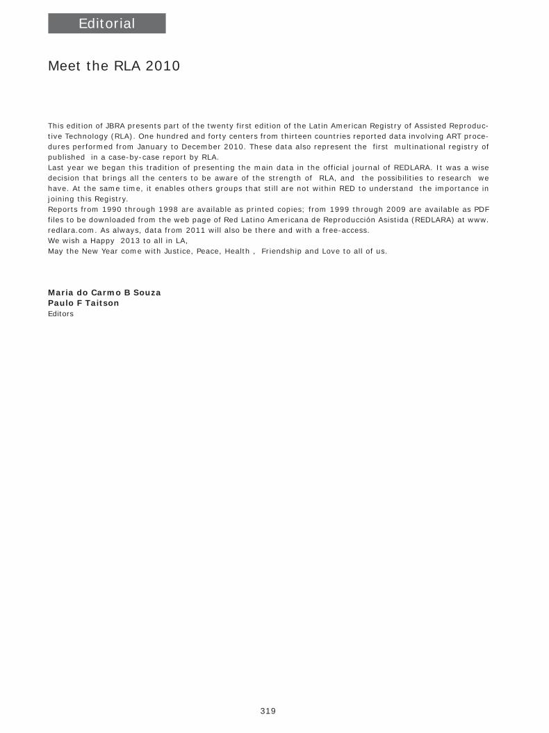
Editorial
Meet the RLA 2010
This edition of JBRA presents part of the twenty first edition of the Latin American Registry of Assisted Reproduc-tive Technology (RLA). One hundred and forty centers from thirteen countries reported data involving ART proce-dures performed from January to December 2010. These data also represent the first multinational registry of published in a case-by-case report by RLA.Last year we began this tradition of presenting the main data in the official journal of REDLARA. It was a wise decision that brings all the centers to be aware of the strength of RLA, and the possibilities to research we have. At the same time, it enables others groups that still are not within RED to understand the importance in joining this Registry. Reports from 1990 through 1998 are available as printed copies; from 1999 through 2009 are available as PDF files to be downloaded from the web page of Red Latino Americana de Reproducción Asistida (REDLARA) at www.redlara.com. As always, data from 2011 will also be there and with a free-access.We wish a Happy 2013 to all in LA, May the New Year come with Justice, Peace, Health , Friendship and Love to all of us.
Maria do Carmo B SouzaPaulo F TaitsonEditors
JBRA6_150213.indb 319 15/02/2013 17:17:12
320
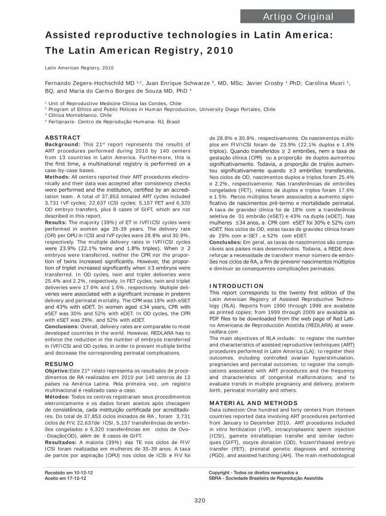
Artigo Original
ABSTRACTBackground: This 21st report represents the results of ART procedures performed during 2010 by 140 centers from 13 countries in Latin America. Furthermore, this is the first time, a multinational registry is performed on a case-by-case bases.Methods: All centers reported their ART procedures electro-nically and their data was accepted after consistency checks were performed and the institution, certified by an accredi-tation team. A total of 37,853 initiated ART cycles included 3,731 IVF cycles; 22,637 ICSI cycles; 5,157 FET and 6,320 OD embryo transfers, plus 8 cases of GIFT, which are not described in this report.Results: The majority (39%) of ET in IVF/ICSI cycles were performed in women age 35-39 years. The delivery rate (DR) per OPU in ICSI and IVF cycles were 28.8% and 30.9%, respectively. The multiple delivery rates in IVF/ICSI cycles were 23.9% (22.1% twins and 1.8% triples). When ≥ 2 embryos were transferred, neither the CPR nor the propor-tion of twins increased significantly. However, the propor-tion of triplet increased significantly when ≥3 embryos were transferred. In OD cycles, twin and triplet deliveries were 25.4% and 2.2%, respectively. In FET cycles, twin and triplet deliveries were 17.6% and 1.5%, respectively. Multiple deli-veries were associated with a significant increase in preterm delivery and perinatal mortality. The CPR was 18% with eSET and 43% with eDET. In women aged ≤34 years, CPR with eSET was 30% and 52% with eDET. In OD cycles, the CPR with eSET was 29%, and 52% with eDET.Conclusions: Overall, delivery rates are comparable to most developed countries in the world. However, REDLARA has to enforce the reduction in the number of embryos transferred in IVF/ICSI and OD cycles, in order to prevent multiple births and decrease the corresponding perinatal complications.
RESUMOObjetivo:Este 21º relato representa os resultados de proce-dimentos de RA realizados em 2010 por 140 centros de 13 países na América Latina. Pela primeira vez, um registro multinacional é realizado caso-a-caso.Métodos: Todos os centros registraram seus procedimentos eletronicamente e os dados foram aceitos após checagem de consistência, cada instituição certificada por acreditado-res. Do total de 37,853 ciclos iniciados de RA , foram 3,731 ciclos de FIV, 22,637de ICSI, 5,157 transferências de embri-ões congelados e 6,320 transferências em ciclos de Ovo--Doação(OD), além de 8 casos de GIFT.Resultados: A maioria (39%) das TE nos ciclos de FIV/ICSI foram realizadas em mulheres de 35-39 anos. A taxa de partos por aspiração (OPU) nos ciclos de ICSI e FIV foi
de 28.8% e 30.9%, respectivamente. Os nascimentos múlti-plos em FIV/ICSI foram de 23.9% (22.1% duplos e 1.8% triplos). Quando transferidos ≥ 2 embriões, nem a taxa de gestação clínica (CPR) ou a proporção de duplos aumentou significativamente. Todavia, a proporção de triplos aumen-tou significativamente quando ≥3 embriões transferidos. Nos ciclos de OD, nascimentos duplos e triplos foram 25.4% e 2.2%, respectivamente. Nas transferências de embriões congelados (FET), relatos de duplos e triplos foram 17.6% e 1.5%. Partos múltiplos foram associados a aumento signi-ficativo de nascimentos pré-termo e mortalidade perinatal. A taxa de gravidez clínica foi de 18% com a transferência seletiva de 01 embrião (eSET) e 43% na dupla (eDET). Nas mulheres ≤34 anos, a CPR com eSET foi 30% e 52% com eDET. Nos ciclos de OD, estas taxas de gravidez clínica foram de 29% com e-SET , e 52% com eDET.Conclusões: Em geral, as taxas de nascimentos são compa-ráveis aos países mais desenvolvidos. Todavia, a REDE deve reforçar a necessidade de transferir menor número de embri-ões nos ciclos de RA, a fim de prevenir nascimentos múltiplos e diminuir as consequentes complicações perinatais.
INTRODUCTIONThis report corresponds to the twenty first edition of the Latin American Registry of Assisted Reproductive Techno-logy (RLA). Reports from 1990 through 1998 are available as printed copies; from 1999 through 2009 are available as PDF files to be downloaded from the web page of Red Lati-no Americana de Reproducción Asistida (REDLARA) at www.redlara.com .The main objectives of RLA include: to register the number and characteristics of assisted reproductive techniques (ART) procedures performed in Latin America (LA); to register their outcomes, including controlled ovarian hyperstimulation, pregnancies and perinatal outcomes; to register the compli-cations associated with ART procedures and the frequency and characteristics of congenital malformations; and to evaluate trends in multiple pregnancy and delivery, preterm birth, perinatal mortality and others.
MATERIAL AND METHODSData collection:One hundred and forty centers from thirteen countries reported data involving ART procedures performed from January to December 2010. ART procedures included in vitro fertilization (IVF), intracytoplasmic sperm injection (ICSI), gamete intrafallopian transfer and similar techni-ques (GIFT), oocyte donation (OD), frozen/thawed embryo transfer (FET), prenatal genetic diagnosis and screening (PGD), and assisted hatching (AH). The main methodological
Assisted reproductive technologies in Latin America: The Latin American Registry, 2010Latin American Registry, 2010
Fernando Zegers-Hochschild MD 1,2, Juan Enrique Schwarze 3, MD, MSc; Javier Crosby 1 PhD; Carolina Musri 1, BQ, and Maria do Carmo Borges de Souza MD, PhD 4
1 Unit of Reproductive Medicine Clínica las Condes, Chile2 Program of Ethics and Public Policies in Human Reproduction, University Diego Portales, Chile3 Clínica Monteblanco, Chile4 Fertipraxis- Centro de Reprodução Humana- RJ, Brasil
Recebido em 10-12-12Aceito em 17-12-12
Copyright - Todos os direitos reservados a SBRA - Sociedade Brasileira de Reprodução Assistida
JBRA6_150213.indb 320 15/02/2013 17:17:12

321321Assisted reproductive technologies in Latin America:The Latin American Registry, 2010 - Zegers-Hochschild F. et all.
JBRA Assist. Reprod. | V. 16 | nº6 | Nov-Dec / 2012
characteristic of the current report is that for the first time the data of each treatment cycle was recorded independen-tly, instead of a summary of cases, as it was reported until now, thus, this is the first multinational case by case regis-try. This way of collecting data has two main advantages. First, it reduced the work of those responsible for reporting data from each center, and second, a case-by-case multina-tional register, allows for more sophisticated biostatistics and epidemiological analysis. As in the past, each center provided their data on voluntary bases. Furthermore, before the data is accepted, each center has to undergo periodical accredi-tation visits, where a clinician and an embryologist from a different country, evaluate the professionals, the infrastruc-ture and equipment of the center, together with their quali-ty control programs and their consent forms. Furthermore, the data provided by the center to the RLA is carefully and thoroughly analyzed. Each center has an individual password in order to access the RLA-server, where the center can uplo-ad the data of each cycle. The data can be uploaded either by filling a specially designed page each time a new case is performed, or by uploading an Excel file whenever possible. The central office of RLA gains immediately access to the data, and checks for inconsistencies and resolve any further question with the center.Data validation: The data provided by each centers is checked for inconsistency by the program; and any error is discussed with the center, and the data is rectified if necessa-ry. The truthfulness of the data reported by each institution is checked as part of the periodic accreditation process conduc-ted by a biologist and a clinician from different countries.Limitations of data collection: Some centers do not have complete follow-up of each pregnancy. This is especially so in institutions not associated with obstetric units. Our calculations are that missing data is in the order of 5% of pregnancies. From a different perspective, not all centers performing ART belong to REDLARA. We estimate that the RLA registers more than 80% of ART procedures performed in Latin America. Statistical analysis: Chi square test was used to analyze independence of categorical variables. When multiple variable analyses were performed, i.e. logistic regression or lineal regression, the dependent variables were consi-dered significant if the confidence interval of the odd ratio
(OR), or regression coefficient did not cross the non-signi-ficant value. A p-value <0.05 was considered as statisti-cally significant. When comparing two outcomes, the risk ratio (RR), and its corresponding 95% confidence interval (95%CI) are presented.
RESULTSParticipating centers: One hundred and forty (140) centers belonging to 13 countries reported their ART proce-dures performed during 2010 (Annex I). These represent five more centers than those reporting in 2009. The new institutions belong to Argentina, Brazil, Ecuador and Mexico. Size of participating institutions:The number of initiated cycles corresponds to the sum of initiated cycles of IVF/ICSI/GIFT, and embryo transfers, both FET and OD. The avera-ge number of initiated cycles registered by the clinics was 268. More than half of the centers registered less than 150 cycles, whereas only three centers registered more than one thousand cycles. The distribution of the clinics according to the number of cycles registered is as follows: 27%, ≤ 100 cycles; 36% between 100 and 250 cycles; 24% between 251 and 500 cycles; 11% between 500 and 1,000 cycles; and only 2%, ≥ 1,000 cycles.ART procedure and access:The total number of ART procedures registered by the RLA was 37,853. Of these, 47% (n=17,673) were reported by Brazil; 22% (n=8,336) by Argentina; and 12% (n=4,433) by Mexico (table 1). Out of 26,3736 initiated autologous--cycles, 3,731 (14%) corresponded to IVF, and 22,637 (86%) to ICSI cycles. One hundred and twenty six clinics registered 5,157 FET cycles. And one 127 clinics reported 6,320 OD cycles. In 56% of TABELA these cycles, the eggs were donated from pure donors, i.e. women that underwent controlled ovarian hyperstimu-lation (COS) and oocyte pick up with the only purpose of donating their oocytes; and 44% were egg-sharing, i.e. patients undergoing COS and oocyte pick-up, for an autologous treatment and simultaneously dona-ted a proportion of their gametes. Table 1 also shows access to ART procedures in LA, expressed as the total number of initiated cycles per million women aged 15 to 45 years.
Table 1. ART procedures and access in 2010
Country Number of clinics
Assisted reproductive techniquesAccess (***)
IVF(*) ICSI(*) FET OD(**) Total
Argentina 22 810 4,462 1,231 1,832 8,336 921.2
Brazil 56 542 12,913 2,515 1,703 17,673 372.2
Chile 7 143 1,088 255 159 1,652 426.4
Colombia 8 358 420 127 284 1,189 112.5
Ecuador 5 61 248 79 104 492 139.2
Guatemala 1 42 43 12 17 114 35.9
Mexico 25 828 1,991 637 977 4,433 160.2
Nicaragua 1 66 25 0 5 96 66.1
Panama 2 38 163 27 50 278 354.8
Peru 4 436 713 104 805 2,058 289.6
Dominican R. 1 55 15 2 5 77 34.2
Uruguay 2 32 226 46 48 352 501.8
Venezuela 6 320 330 122 331 1,103 168.7
Total 140 3,731 22,637 5,157 6,320 37,853 304.8
include 7 cycles of GIFT/TOMI in Chile and 1 in Argentina(*) initiated cycles; (**) includes the transfer of fresh and frozen embryos ; (***) number of cycles/million of women 15-45 years
JBRA6_150213.indb 321 15/02/2013 17:17:12
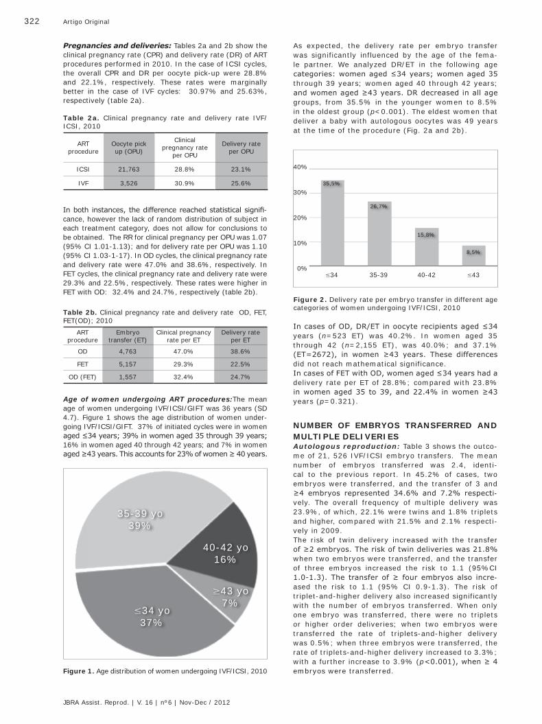
322 Artigo Original
JBRA Assist. Reprod. | V. 16 | nº6 | Nov-Dec / 2012
In both instances, the difference reached statistical signifi-cance, however the lack of random distribution of subject in each treatment category, does not allow for conclusions to be obtained. The RR for clinical pregnancy per OPU was 1.07 (95% CI 1.01-1.13); and for delivery rate per OPU was 1.10 (95% CI 1.03-1-17). In OD cycles, the clinical pregnancy rate and delivery rate were 47.0% and 38.6%, respectively. In FET cycles, the clinical pregnancy rate and delivery rate were 29.3% and 22.5%, respectively. These rates were higher in FET with OD: 32.4% and 24.7%, respectively (table 2b).
Pregnancies and deliveries: Tables 2a and 2b show the clinical pregnancy rate (CPR) and delivery rate (DR) of ART procedures performed in 2010. In the case of ICSI cycles, the overall CPR and DR per oocyte pick-up were 28.8% and 22.1%, respectively. These rates were marginally better in the case of IVF cycles: 30.97% and 25.63%, respectively (table 2a).
As expected, the delivery rate per embryo transfer was significantly influenced by the age of the fema-le partner. We analyzed DR/ET in the following age categories: women aged ≤34 years; women aged 35 through 39 years; women aged 40 through 42 years; and women aged ≥43 years. DR decreased in all age groups, from 35.5% in the younger women to 8.5% in the oldest group (p<0.001). The eldest women that deliver a baby with autologous oocytes was 49 years at the time of the procedure (Fig. 2a and 2b).
Table 2a. Clinical pregnancy rate and delivery rate IVF/ICSI, 2010
ARTprocedure
Oocyte pick up (OPU)
Clinicalpregnancy rate
per OPU
Delivery rate per OPU
ICSI 21,763 28.8% 23.1%
IVF 3,526 30.9% 25.6%
Table 2b. Clinical pregnancy rate and delivery rate OD, FET, FET(OD); 2010
ARTprocedure
Embryotransfer (ET)
Clinical pregnancyrate per ET
Delivery rateper ET
OD 4,763 47.0% 38.6%
FET 5,157 29.3% 22.5%
OD (FET) 1,557 32.4% 24.7%
Age of women undergoing ART procedures:The mean age of women undergoing IVF/ICSI/GIFT was 36 years (SD 4.7). Figure 1 shows the age distribution of women under-going IVF/ICSI/GIFT. 37% of initiated cycles were in women aged ≤34 years; 39% in women aged 35 through 39 years; 16% in women aged 40 through 42 years; and 7% in women aged ≥43 years. This accounts for 23% of women ≥ 40 years.
Figure 1. Age distribution of women undergoing IVF/ICSI, 2010
35-39 yo39%
≤34 yo37%
≥43 yo7%
40-42 yo16%
Figure 2. Delivery rate per embryo transfer in different age categories of women undergoing IVF/ICSI, 2010
40%
30%
20%
10%
0%≤34
35,5%
35-39
26,7%
40-42
15,8%
≤43
8,5%
In cases of OD, DR/ET in oocyte recipients aged ≤34 years (n=523 ET) was 40.2%. In women aged 35 through 42 (n=2,155 ET), was 40.0%; and 37.1% (ET=2672), in women ≥43 years. These differences did not reach mathematical significance. In cases of FET with OD, women aged ≤34 years had a delivery rate per ET of 28.8%; compared with 23.8% in women aged 35 to 39, and 22.4% in women ≥43 years (p=0.321).
NUMBER OF EMBRYOS TRANSFERRED AND MULTIPLE DELIVERIESAutologous reproduction: Table 3 shows the outco-me of 21, 526 IVF/ICSI embryo transfers. The mean number of embryos transferred was 2.4, identi-cal to the previous report. In 45.2% of cases, two embryos were transferred, and the transfer of 3 and ≥4 embryos represented 34.6% and 7.2% respecti-vely. The overall frequency of multiple delivery was 23.9%, of which, 22.1% were twins and 1.8% triplets and higher, compared with 21.5% and 2.1% respecti-vely in 2009. The risk of twin delivery increased with the transfer of ≥2 embryos. The risk of twin deliveries was 21.8% when two embryos were transferred, and the transfer of three embryos increased the risk to 1.1 (95%CI 1.0-1.3). The transfer of ≥ four embryos also incre-ased the risk to 1.1 (95% CI 0.9-1.3). The risk of triplet-and-higher delivery also increased significantly with the number of embryos transferred. When only one embryo was transferred, there were no triplets or higher order deliveries; when two embryos were transferred the rate of triplets-and-higher delivery was 0.5%; when three embryos were transferred, the rate of triplets-and-higher delivery increased to 3.3%; with a further increase to 3.9% (p<0.001), when ≥ 4 embryos were transferred.
JBRA6_150213.indb 322 15/02/2013 17:17:13
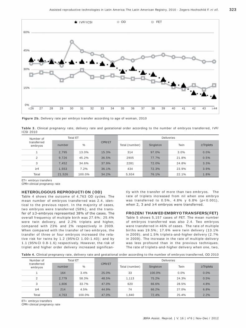
323323Assisted reproductive technologies in Latin America:The Latin American Registry, 2010 - Zegers-Hochschild F. et all.
JBRA Assist. Reprod. | V. 16 | nº6 | Nov-Dec / 2012
≥44
Figure 2b. Delivery rate per embryo transfer according to age of woman, 2010
IVF/ICSI OD FET
60%
45%
30%
15%
0%31 3932 4033 41≤26 34 4227 35 4328 3629 3730 38
HETEROLOGOUS REPRODUCTION (OD) Table 4 shows the outcome of 4,763 OD cycles. The mean number of embryos transferred was 2.4, iden-tical to the previous report. In the majority of cases, two embryos were transferred (58%), and the trans-fer of ≥3-embryos represented 38% of the cases. The overall frequency of multiple birth was 27.6%; 25.4% were twin delivery, and 2.2% triplets and higher, compared with 23% and 2% respectively in 2009. When compared with the transfer of two embryos, the transfer of three or four embryos increased the rela-tive risk for twins by 1.2 (95%CI 1.00-1.40); and by 1.1 (95%CI 0.8-1.6) respectively. However, the risk of triplet and higher order delivery increased significan-
Table 3. Clinical pregnancy rate, delivery rate and gestational order according to the number of embryos transferred, IVF/ICSI 2010
Number oftransferredembryos
Total ETCPR/ET
Deliveries
number % Total (number) Singleton Twin ≥Triplets
1 2,795 13.0% 15.3% 314 97.0% 3.0% 0.0%
2 9,726 45.2% 36.5% 2905 77.7% 21.8% 0.5%
3 7,452 34.6% 37.9% 2281 72.0% 24.8% 3.3%
≥4 1,553 7.2% 36.1% 434 72.3% 23.9% 3.9%
Total 21,526 100.0% 34.2% 5,934 76.1% 22.1% 1.8%
ET= embryo transfersCPR=clinical pregnancy rate
tly with the transfer of more than two embryos. The rate of triplets increased from nil when one embryo was transferred to 0.5%, 4.8% y 6.8% (p<0.001), when 2, 3 and ≥4 embryos were transferred.
FROZEN/THAWED EMBRYO TRANSFERS(FET)Table 5 shows 5,157 cases of FET. The mean number of embryos transferred was also 2.4. Two embryos were transferred in 46% of cases. The rate of multiple births was 19.5%; 17.6% were twin delivery (13.1% in 2009); and 1.5% triplets-and-higher delivery (2.7% in 2009). The increase in the rate of multiple-delivery was less profound than in the previous techniques. The rate of triplets-and-higher delivery when one, two,
Table 4. Clinical pregnancy rate, delivery rate and gestational order according to the number of embryos transferred, OD 2010
Number oftransferredembryos
Total ETCPR/ET
Deliveries
number % Total (number) Singleton Twin ≥Triplets
1 164 3.4% 25.0% 33 100.0% 0.0% 0.0%
2 2,779 58.3% 48.5% 1,113 75.2% 24.3% 0.5%
3 1,606 33.7% 47.0% 620 66.6% 28.5% 4.8%
≥4 214 4.5% 44.9% 74 66.2% 27.0% 6.8%
Total 4,763 100.0% 47.0% 1,840 72.4% 25.4% 2.2%
ET= embryo transfersCPR=clinical pregnancy rate
JBRA6_150213.indb 323 15/02/2013 17:17:13
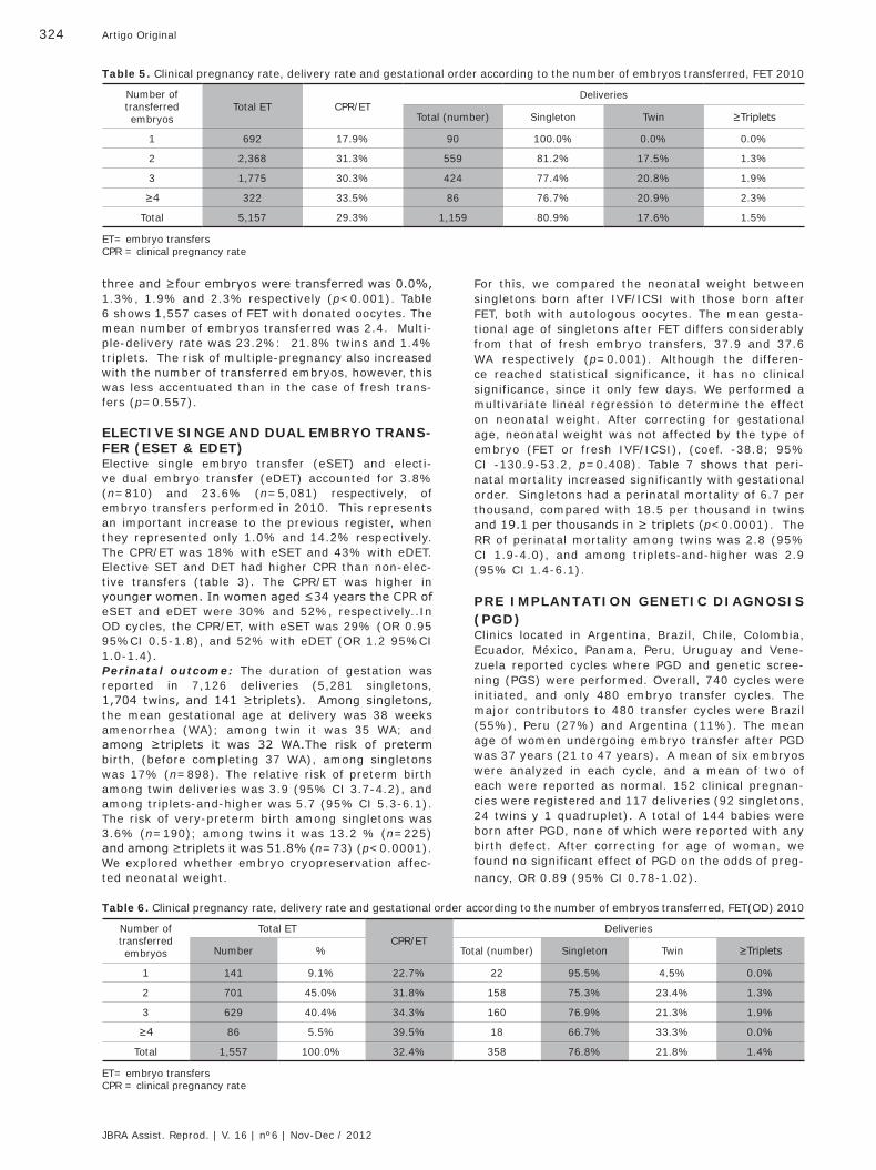
324 Artigo Original
JBRA Assist. Reprod. | V. 16 | nº6 | Nov-Dec / 2012
three and ≥four embryos were transferred was 0.0%, 1.3%, 1.9% and 2.3% respectively (p<0.001). Table 6 shows 1,557 cases of FET with donated oocytes. The mean number of embryos transferred was 2.4. Multi-ple-delivery rate was 23.2%: 21.8% twins and 1.4% triplets. The risk of multiple-pregnancy also increased with the number of transferred embryos, however, this was less accentuated than in the case of fresh trans-fers (p=0.557).
ELECTIVE SINGE AND DUAL EMBRYO TRANS-FER (ESET & EDET)Elective single embryo transfer (eSET) and electi-ve dual embryo transfer (eDET) accounted for 3.8% (n=810) and 23.6% (n=5,081) respectively, of embryo transfers performed in 2010. This represents an important increase to the previous register, when they represented only 1.0% and 14.2% respectively. The CPR/ET was 18% with eSET and 43% with eDET. Elective SET and DET had higher CPR than non-elec-tive transfers (table 3). The CPR/ET was higher in younger women. In women aged ≤34 years the CPR of eSET and eDET were 30% and 52%, respectively..In OD cycles, the CPR/ET, with eSET was 29% (OR 0.95 95%CI 0.5-1.8), and 52% with eDET (OR 1.2 95%CI 1.0-1.4).Perinatal outcome: The duration of gestation was reported in 7,126 deliveries (5,281 singletons, 1,704 twins, and 141 ≥triplets). Among singletons, the mean gestational age at delivery was 38 weeks amenorrhea (WA); among twin it was 35 WA; and among ≥triplets it was 32 WA.The risk of preterm birth, (before completing 37 WA), among singletons was 17% (n=898). The relative risk of preterm birth among twin deliveries was 3.9 (95% CI 3.7-4.2), and among triplets-and-higher was 5.7 (95% CI 5.3-6.1). The risk of very-preterm birth among singletons was 3.6% (n=190); among twins it was 13.2 % (n=225) and among ≥triplets it was 51.8% (n=73) (p<0.0001). We explored whether embryo cryopreservation affec-ted neonatal weight.
Table 6. Clinical pregnancy rate, delivery rate and gestational order according to the number of embryos transferred, FET(OD) 2010
Number oftransferredembryos
Total ETCPR/ET
Deliveries
Number % Total (number) Singleton Twin ≥Triplets
1 141 9.1% 22.7% 22 95.5% 4.5% 0.0%
2 701 45.0% 31.8% 158 75.3% 23.4% 1.3%
3 629 40.4% 34.3% 160 76.9% 21.3% 1.9%
≥4 86 5.5% 39.5% 18 66.7% 33.3% 0.0%
Total 1,557 100.0% 32.4% 358 76.8% 21.8% 1.4%
ET= embryo transfersCPR = clinical pregnancy rate
Table 5. Clinical pregnancy rate, delivery rate and gestational order according to the number of embryos transferred, FET 2010
Number oftransferred embryos
Total ET CPR/ETDeliveries
Total (number) Singleton Twin ≥Triplets
1 692 17.9% 90 100.0% 0.0% 0.0%
2 2,368 31.3% 559 81.2% 17.5% 1.3%
3 1,775 30.3% 424 77.4% 20.8% 1.9%
≥4 322 33.5% 86 76.7% 20.9% 2.3%
Total 5,157 29.3% 1,159 80.9% 17.6% 1.5%
ET= embryo transfersCPR = clinical pregnancy rate
For this, we compared the neonatal weight between singletons born after IVF/ICSI with those born after FET, both with autologous oocytes. The mean gesta-tional age of singletons after FET differs considerably from that of fresh embryo transfers, 37.9 and 37.6 WA respectively (p=0.001). Although the differen-ce reached statistical significance, it has no clinical significance, since it only few days. We performed a multivariate lineal regression to determine the effect on neonatal weight. After correcting for gestational age, neonatal weight was not affected by the type of embryo (FET or fresh IVF/ICSI), (coef. -38.8; 95% CI -130.9-53.2, p=0.408). Table 7 shows that peri-natal mortality increased significantly with gestational order. Singletons had a perinatal mortality of 6.7 per thousand, compared with 18.5 per thousand in twins and 19.1 per thousands in ≥ triplets (p<0.0001). The RR of perinatal mortality among twins was 2.8 (95% CI 1.9-4.0), and among triplets-and-higher was 2.9 (95% CI 1.4-6.1).
PRE IMPLANTATION GENETIC DIAGNOSIS (PGD)Clinics located in Argentina, Brazil, Chile, Colombia, Ecuador, México, Panama, Peru, Uruguay and Vene-zuela reported cycles where PGD and genetic scree-ning (PGS) were performed. Overall, 740 cycles were initiated, and only 480 embryo transfer cycles. The major contributors to 480 transfer cycles were Brazil (55%), Peru (27%) and Argentina (11%). The mean age of women undergoing embryo transfer after PGD was 37 years (21 to 47 years). A mean of six embryos were analyzed in each cycle, and a mean of two of each were reported as normal. 152 clinical pregnan-cies were registered and 117 deliveries (92 singletons, 24 twins y 1 quadruplet). A total of 144 babies were born after PGD, none of which were reported with any birth defect. After correcting for age of woman, we found no significant effect of PGD on the odds of preg-nancy, OR 0.89 (95% CI 0.78-1.02).
JBRA6_150213.indb 324 15/02/2013 17:17:13
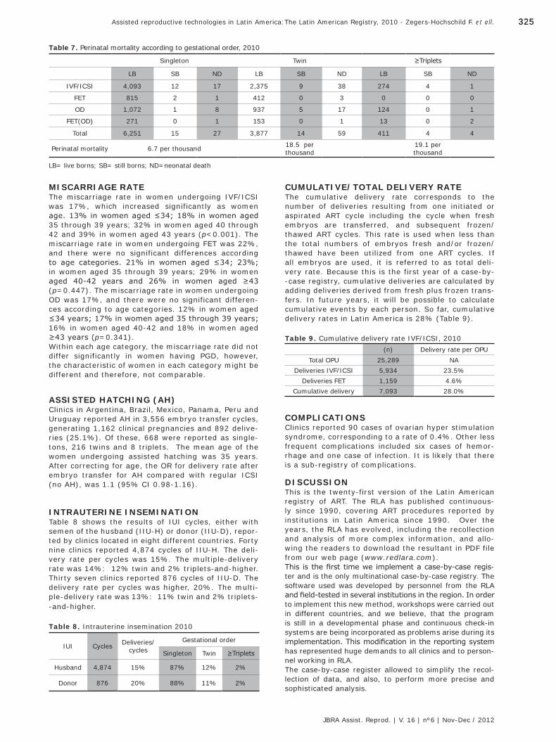
325325Assisted reproductive technologies in Latin America:The Latin American Registry, 2010 - Zegers-Hochschild F. et all.
JBRA Assist. Reprod. | V. 16 | nº6 | Nov-Dec / 2012
MISCARRIAGE RATEThe miscarriage rate in women undergoing IVF/ICSI was 17%, which increased significantly as women age. 13% in women aged ≤34; 18% in women aged 35 through 39 years; 32% in women aged 40 through 42 and 39% in women aged 43 years (p<0.001). The miscarriage rate in women undergoing FET was 22%, and there were no significant differences according to age categories. 21% in women aged ≤34; 23%; in women aged 35 through 39 years; 29% in women aged 40-42 years and 26% in women aged ≥43 (p=0.447). The miscarriage rate in women undergoing OD was 17%, and there were no significant differen-ces according to age categories. 12% in women aged ≤34 years; 17% in women aged 35 through 39 years; 16% in women aged 40-42 and 18% in women aged ≥43 years (p=0.341).Within each age category, the miscarriage rate did not differ significantly in women having PGD, however, the characteristic of women in each category might be different and therefore, not comparable.
ASSISTED HATCHING (AH)Clinics in Argentina, Brazil, Mexico, Panama, Peru and Uruguay reported AH in 3,556 embryo transfer cycles, generating 1,162 clinical pregnancies and 892 delive-ries (25.1%). Of these, 668 were reported as single-tons, 216 twins and 8 triplets. The mean age of the women undergoing assisted hatching was 35 years. After correcting for age, the OR for delivery rate after embryo transfer for AH compared with regular ICSI (no AH), was 1.1 (95% CI 0.98-1.16).
INTRAUTERINE INSEMINATION Table 8 shows the results of IUI cycles, either with semen of the husband (IIU-H) or donor (IIU-D), repor-ted by clinics located in eight different countries. Forty nine clinics reported 4,874 cycles of IIU-H. The deli-very rate per cycles was 15%. The multiple-delivery rate was 14%: 12% twin and 2% triplets-and-higher. Thirty seven clinics reported 876 cycles of IIU-D. The delivery rate per cycles was higher, 20%. The multi-ple-delivery rate was 13%: 11% twin and 2% triplets--and-higher.
Table 8. Intrauterine insemination 2010
IUI Cycles Deliveries/cycles
Gestational order
Singleton Twin ≥Triplets
Husband 4,874 15% 87% 12% 2%
Donor 876 20% 88% 11% 2%
Table 9. Cumulative delivery rate IVF/ICSI, 2010(n) Delivery rate per OPU
Total OPU 25,289 NADeliveries IVF/ICSI 5,934 23.5%
Deliveries FET 1,159 4.6%Cumulative delivery 7,093 28.0%
Table 7. Perinatal mortality according to gestational order, 2010
Singleton Twin ≥Triplets
LB SB ND LB SB ND LB SB ND
IVF/ICSI 4,093 12 17 2,375 9 38 274 4 1
FET 815 2 1 412 0 3 0 0 0
OD 1,072 1 8 937 5 17 124 0 1
FET(OD) 271 0 1 153 0 1 13 0 2
Total 6,251 15 27 3,877 14 59 411 4 4
Perinatal mortality 6.7 per thousand 18.5 perthousand
19.1 perthousand
LB= live borns; SB= still borns; ND=neonatal death
CUMULATIVE/TOTAL DELIVERY RATEThe cumulative delivery rate corresponds to the number of deliveries resulting from one initiated or aspirated ART cycle including the cycle when fresh embryos are transferred, and subsequent frozen/thawed ART cycles. This rate is used when less than the total numbers of embryos fresh and/or frozen/thawed have been utilized from one ART cycles. If all embryos are used, it is referred to as total deli-very rate. Because this is the first year of a case-by--case registry, cumulative deliveries are calculated by adding deliveries derived from fresh plus frozen trans-fers. In future years, it will be possible to calculate cumulative events by each person. So far, cumulative delivery rates in Latin America is 28% (Table 9).
COMPLICATIONSClinics reported 90 cases of ovarian hyper stimulation syndrome, corresponding to a rate of 0.4%. Other less frequent complications included six cases of hemor-rhage and one case of infection. It is likely that there is a sub-registry of complications.
DISCUSSIONThis is the twenty-first version of the Latin American registry of ART. The RLA has published continuous-ly since 1990, covering ART procedures reported by institutions in Latin America since 1990. Over the years, the RLA has evolved, including the recollection and analysis of more complex information, and allo-wing the readers to download the resultant in PDF file from our web page (www.redlara.com).This is the first time we implement a case-by-case regis-ter and is the only multinational case-by-case registry. The software used was developed by personnel from the RLA and field-tested in several institutions in the region. In order to implement this new method, workshops were carried out in different countries, and we believe, that the program is still in a developmental phase and continuous check-in systems are being incorporated as problems arise during its implementation. This modification in the reporting system has represented huge demands to all clinics and to person-nel working in RLA. The case-by-case register allowed to simplify the recol-lection of data, and also, to perform more precise and sophisticated analysis.
JBRA6_150213.indb 325 15/02/2013 17:17:14

326 Artigo Original
JBRA Assist. Reprod. | V. 16 | nº6 | Nov-Dec / 2012
Another strength of this register is the uniformity of terminology. All clinics reporting to RLA, use the glos-sary defined in 2009 by the International Committee for Monitoring Assisted Reproductive Technologies (ICMART) and the World Health Organization (WHO) 1;
2. In 2010, 140 clinics from thirteen countries reported the data of 37,853 ART cycles. This represents a small drop of 0.5 % compared to 2009. In 2010 the mean number of cycles registered by each clinic reached 268 cycles, whereas in 2009 it was 281. This drop in the mean number of cycles performed by each clinic can in part result from the lack of public support for infertile couples and therefore, low access to ART procedures 3. Only one center from Argentina, which reported 500 cycles in 2009 and one center from Venezuela, which reported 700 cycles in 2009, did not report this year. The rest of centers, which having reported in 2009, did not report in 2010 had only 30 and 150 initiated in 2009. Therefore, it is likely that the number of proce-dures per center is not increasing as much as new centers reporting.The use of ICSI instead of conventional IVF conti-nues to increase. In 2010 represented 86% of oocyte pick-ups, while in 2009 and 2008, ICSI represented 85%; and in 2007 the 83% (www.redlara.com). This tendency is also seen in Europe, where in the last report, 69% of autologous cycles were ICSI 4. The age of women undergoing IVF/ICSI cycles conti-nues to increase. In 2009, the proportion of IVF/ICSI cycles performed in women aged 35 through 39, repre-sented 40%; and 18% of women were ≥40 years. In 2010, women age ≥40 years represent 23% of fresh IVF/ICSI cycles. Furthermore, 7% of IVF/ICSI cycles were performed in women aged ≥43 years. Since the age of the woman is one of the most important prog-nosis factors, this demographic reality is important to consider when analyzing regional outcomes.The delivery rate per oocyte pick-up in IVF/ICSI reached 23.4%, and the cumulative delivery rate reached 28%. These results are higher than the data published by ESHRE, where cumulative delivery rate reached 22% 5. Part of this difference is explained by the higher mean number of embryos transferred in Latin America compared with the majority of Europe. The mean number of transferred embryos in IVF/ICSI did not decrease in comparison with previous years. However, the frequency of eSET and eDET did increa-se significantly. This might be due to a more accurate and thorough register of cases. In spite of this encou-raging news, it is worrisome that in 42% of embryo transfers more than three embryos were transferred, and in 7% of cases, more than four embryos were transferred. According to ESHRE´s last report, only 24% of their embryo transfers corresponded to three and more embryos 5.Both in IVF/ICSI and OD cycles, the transfer of more embryos resulted in an increase in the risk of triplets--and-higher order deliveries. Interestingly, the incre-ase in the risk of twin-deliveries was marginal, and barely reached statistical significance. Thus, 24% of deliveries in IVF/ICSI cycles were multiple (2% triplets-and-higher), and 28% deliveries in OD cycles were multiple (2% triplets and higher). Increasing the number of ET above two, does not significantly impact delivery rates nor twin rates. What it does, is increase the high order multiples which are so detrimental for perinatal mortality and morbidity.As shown in this as well as previous reports, even twin deliveries increase the risk of preterm birth and
perinatal mortality. And as discussed previously, the transfer of ≥two embryos is associated to an increase in the risk of multiple delivery. Probably, the main reason to transfer more embryos is the desire of both clinicians and patients to improve the outcome of each ART cycle, without considering the risk of multi-ple deliveries and associated prematurity. The data showed in this report is quite reassuring, since the results associated with eSET and eDET, especially in younger patients undergoing IVF/ICSI, and OD cycles- are higher than reported in previous reports. Since the present report correspond to the analysis of observational data, and not the results of randomized controlled trials, the results cannot be considered as a evidence or support for a decreased benefit in some procedures. For example, PGD was not associated with neither a significant increase in the delivery rate nor a reduction in the miscarriage rate. This might be explained by the fact, that the number of procedu-res is still low and RLA does not register differently pre-natal genetic diagnosis and pre-natal screening. Furthermore, the selection of women having PGD can be very different to the rest of the population, even when stratified by age. On the other hand, assisted hatching does not increase delivery rate, since no statistical significance was reached, however, caution must be expressed when analyzing this data. The frequency of complications associated to ART proce-dures was rather low, only 90 cases of OHSS were repor-ted, which represented a risk of 0.3% of initiated cycles. Furthermore, only 6 cases of genital hemorrhage and 1 case of infections were reported. This might represent a recollection bias, that needs to be improved.This is the fourth report of IUI cycles. Clinics reported 4,874 IUI with husband´s semen, and 876 cycles with donor´s semen. This represent a clear drop compared to 2009, when 13,410 IIU-H and 2,430 IIU-D cycles were reported. This might be explained by the labor--consuming work that represented the change into a case-by-case register.In summary, this is the first case-by-case register published by the RLA. It is reassuring for patients and clinics that the results of ART procedures performed in the region are similar or even better than in most European and Asian countries 6,5. However, REDLARA has to enforce the reduction in the number of embryos transferred in IVF/ICSI and OD cycles, in order to prevent multiple births, or at least, high order multiples and decrease the corresponding perinatal complications.
Correspondence: Oficina del Registro LatinoAmericano ReproducciónLo Fontecilla 441, Santiago, Chile. Teléfono: 56-2-6108181 Fax: 56-2-6108167E-mail: [email protected]
REFERENCES1. Zegers-Hochschild F, Adamson GD, de Mouzon J, Ishi-hara O, Mansour R, Nygren K, et al. The International Committee for Monitoring Assisted Reproductive Technology (ICMART) and the World Health Organization (WHO) Revised Glossary on ART Terminology, 2009. Hum Reprod. 2009, Nov;24(11):2683-7.2. Zegers-Hochschild F, Nygren KG, Adamson GD, de Mouzon J, Lancaster P, Mansour R, et al. The International Committee Monitoring Assisted Reproductive Technologies (ICMART) glossary on ART terminology. Fertil Steril. 2006, Jul;86(1):16-9.
JBRA6_150213.indb 326 15/02/2013 17:17:14
327327Assisted reproductive technologies in Latin America:The Latin American Registry, 2010 - Zegers-Hochschild F. et all.
JBRA Assist. Reprod. | V. 16 | nº6 | Nov-Dec / 2012
3. Zegers-Hochschild F, Nygren K, and Ishihara O. The impact of legislation and socioeconomic factors in the access to and glob-al practice of assisted reproduction. In: Textbook of Assisted Reproductive Techniques. London, UK: 2012. p. 441-450.4. Ferraretti AP, Goossens V, de Mouzon J, Bhattacharya S, Castilla JA, Korsak V, et al. Assisted reproductive technology in Europe, 2008: results generated from European registers by ESHRE. Hum Reprod. 2012, Sep;27(9):2571-2584.5. Ferraretti AP, Goossens V, de Mouzon J, Bhattacharya S, Castilla JA, Korsak V, et al. Assisted reproductive technology in Europe, 2008: results generated from European registers by ESHRE. Hum Reprod. 2012, Sep;27(9):2571-2584.6. Nygren KG, Sullivan E, Zegers-Hochschild F, Mansour R, Ishihara O, Adamson GD, and de Mouzon J. International Committee for Monitoring Assisted Reproductive Technolo-gy (ICMART) world report: assisted reproductive technology 2003. Fertil Steril. 2011, Jun;95(7):2209-22, 2222.e1-17.
ANNEX IParticipating Institutions
ARGENTINABuenos Aires
1. Centro de estudios en Ginecología y Repro-ducción (CEGYR)
2. Centro de Salud Reproductiva (CER)3. Centro de Investigaciones en Medicina Repro-
ductiva –CIMER4. FECUNDITAS5. Centro especializado en tratamientos para la
mujer-GENS6. Hospital de Clínicas7. Centro de Reproducción-Servicio de Ginecolo-
gía-Hospital Italiano8. HALITUS, Instituto Médico9. PREGNA, Medicina reproductiva10. Procrearte11. Fertilidad San Isidro12. SEREMAS13. FERTILAB
Córdova1. Centro Integral de Ginecología, Obstetricia y
Reproducción (CIGOR)2. FECUNDART
La Plata1. Centro de Reproducción
Mar del Plata1. Centro de estudios en Reproducción y Proce-
dimientos de Fertilización Asistida (CRECER)Mendoza
1. Centro de estudios en Reproducción Huma-na (CERH)
2. Instituto de Medicina ReproductivaSalta
1. MATER Medicina Reproductiva2. SARESA, Salud Reproductiva Salta
Bahía blanca1. AMERIS, Centro de Fertilidad, Ginecología
y Urología (nuevo reportó sólo 13 casos)
BRASILBelo Horizonte
1. Instituto de Saúde Mulher2. Clínica ORIGEN3. Clínica Pro-criar/Mater Dei
Brasilia1. Instituto Verhum - Video Endoscopia e Repro-
duçao Humana2. GÊNESIS – Centro de Assistência em Reprodu-
çao Humana Ltda.3. Centro de Ensino e Pesquisa em Reprodu-
cao AssistidaCampo Grande-Mato Grosso
1. Fertility Centro de Fertilização Humana Assisti-da de Campo Grande
Campinas-SP1. Centro de Reprodução Humana de Campinas2. Clínica Androfert
Cuiabá-Mato Grosso1. Instituto Pérola de Reprodução Humana2. LIFE Reproducción Humana
Curitiba1. ANDROLAB – Clínica e Laboratorio de Andrología2. CONCEBER, Centro de Medicina Reproductiva3. Clínica FERTWAY
Florianópolis1. CLINIFERT – Centro de Reprodução Humana-
Fortaleza2. BIOS - Centro de Medicina Reprodutiva3. FERTVIDA (anteriormente “CRIAR - Centro de
Reproducao Humana”)4. CONCEPTUS – Centro de Reprodução Huma-
na do CearáGoiania Goiás
1. Fértile Diagnósticos - Reprodução HumanaJuiz de Fora-Minas Gerais
1. Pro-criar, Monte Sinai, Clinica de Reprodu-ção Humana
Londrina-Paraná1. CEDILON Serviços Médicos S.C. Ltda.
Paso Fundo1. GÉNESIS – Clínica de Reprodução Humana
Porto Alegre1. Centro de Reprodução Humana Nilo Frantz2. FERTILITAT – Centro de Medicina Reprodutiva3. INSEMINE - Centro de Reprodução Humana4. Núcleo de Reprodução Humana do Hospital
Moínhos de Vento GERAR5. PROGEST6. SEGIR – Servicio de Ecografía, Genética e
Reprodução HumanaRecife-Pernambuco
1. NASCER Medicina Reprodutiva2. Clínica de Fertilidade GERAR
Ribeirao Preto-SP1. Centro de Reprodução Humana Prof. Franco Junior2. Clinica Matrix3. Laboratorio de Reprodução Humana, Hospital
das Clínicas de Ribeirao PretoRio de Janeiro-RJ
1. Centro de Medicina da Reprodução Ltda.2. Centro de Fertilidade Rede D´Or3. Clínica Origen4. Clinica Pró Nascer5. G&O Ginecología e Obstetricia da Barra (now
Fertipraxis)6. HUNTINGTON – Centro de Medicina Reproduc-
tiva (now Primordia)Salvador Bahía
1. Centro de Reprodução Humana, Endoscopia e Medicina Fetal
Sao José Dos Campos-SP1. Clínica REPROFERTY2. Embryolife-Instituto de Medicina Reprodutiva
Sao José do Rio Preto1. IMR - Centro Instituto de Medicina Repro-
dutiva e FetalSao Paulo-SP
1. Centro de Reprodução Humana FERTIVITRO Ltda.2. ORIGINARE, Centro de Investigação em
Reprodução Humana3. Centro de Reprodução Humana Monteleone4. CEERH – Centro Especializado em Reprodu-
ção Humana5. FERTILITY – Centro de Fertilização Assistida6. FERTICLIN – Clínica de Fertilidade Humana7. Chedid Grieco Medicina Reprodutiva8. HUNTINGTON – Centro de Medicina Reprodutiva9. Serviço de Reprodução Humana, Hospital e
Maternidade Santa JoanaUberlandia –Minas Gerais
10. FECUNDA - Instituto de Reprodução Humana
JBRA6_150213.indb 327 15/02/2013 17:17:14

328 Artigo Original
JBRA Assist. Reprod. | V. 16 | nº6 | Nov-Dec / 2012
Vitória11. Jules White Medicina Reproductiva (ante-
riormente, “HUNTINGTON - Centro de Medi-cina Reproductiva”)
Santos1. Clínica PRO GENESIS (new)
Presidente Prudente1. REPRODUCTION, Clínica Urológica e Centro
de Reproducao Humana LTDA. (new)
CHILESantiago
1. Centro de estudios Reproductivos (CER)2. Unidad de Medicina reproductiva, Clinica
Alemana3. Unidad de Medicina Reproductiva, Clinica
las Condes4. Unidad de Medicina Reproductiva, Clinica las
Nieves5. Programa de Fertilización Asistida I.D.I.M.I.
Viña del Mar1. Unidad de Medicina reproductiva, Clinica de
la MujerConcepción
1. Centro de Fertilidad y Medicina Reproductiva CONCEPCION S.A.
COLOMBIACali
1. Centro FECUNDAR Cali2. Centro Médico IMBANACO
Bogotá1. Unidad de Fertilidad del Couytry Ltda2. Asociados en Fertilidad Humana3. PROFAMILIA-Fertil4. Unidad de Fertilidad, Procreación Médicamen-
te AsistidaMedellín
1. IN SER, Instituto Antioqueño de ReproducciónBarranquilla
1. PROCREAR
ECUADORQuito
1. Centro Ecuatoriano de Reproducción Humana2. CONCEBIR, Unidad de Fertilidad y esterilidad
Guayaquil 1. Instituto Nacional de Investigación de Fertili-
dad Esterilidad (INNAIFEST) (new)2. Unidad de Fertilidad Hospital Alcivar Cuenca3. Clinica de Medicina Reproductiva BIOGEPA
GUATEMALA1. Centro de Reproducción humana S.A.
MEXICOCiudad de Juarez-Chihuahua
1. Unidad de Reproducción Humana y GenéticaGuadalajara-Jalisco
1. Centro de Reproducción Asistida del Occidente2. Instituto de Ciencias en Reproducción
Humana – VIDA3. Instituto de Medicina Reproductiva del Bajío (IMER)
Hermosillo1. Clínica de Biología de la Reproducción, Hospi-
tal CIMA - León –Guanajuato1. Instituto de Ciencias en Reproducción
Humana – VIDAMatamoros
1. Instituto de Ciencias en Reproducción Humana – VIDA
México DF1. Centro Especializado en Esterilidad y Repro-
ducción Humana2. INGENES3. Centro Médico Nacional 20 de Noviembre4. Centro de Reproducción Asistida del Hospital
Español5. Instituto Médico de la Mujer6. Instituto Mexicano de alta tecnología repro-
ductiva S.C. (new)7. Centro especializado para la atención de
la mujer8. Instituto Valenciano de Infertilidad (IVI) (new)
Monterrey1. Centro Universitario de Medicina Reproduc-
tiva, Universidad Autónoma de Nuevo León2. Instituto para el Estudio de la Concep-
ción HumanaPuebla
1. Centro de Ginecología y Reproducción Asistida S.C. GYRA
Querétaro1. Médica Fértil
San Luis de Potosí1. Médica Fértil2. Filius (anteriormente, OBGIN S.C., SLP) Tijua-
na Baja California1. Instituto de Medicina Reproductiva del Bajío
– IMERVeracruz
1. Centro de Diagnóstico Ginecológico Saltillo1. Centro de Reproducción asistida de Saltillo (new)
NICARAGUAManagua
1. Centro de Fertilidad de Nicaragua, NICFERT
PANAMA1. IVI, Panamá S.A.2. Women´s Health in IVF
PERÚLima
1. Clínica CEFRA, Centro de fertilidad y Repro-ducción Asistida
2. Clínica Miraflores, Instituto de Ginecología y Fertilidad
3. Grupo PRANOR, Clinica Concebir4. Grupo PRANOR, Instituto de Ginecología y
reproducción.
REPUBLICA DOMINICANASanto Domingo
1. 1.-PROFERT, Programa de Fertilización Asistida y Medicina Perinatal.
URUGUAYMontevideo
1. Centro de Esterilidad Montevideo (CEM)2. Centro de Reproducción Humana del Interior
VENEZUELACaracas
1. FERTILAB, Clinica el Avila2. UNIFERTES, Clínica el Avila3. EMBRIOS, Centro de Fertilidad y Reproducción
humana, Hospital de Clínicas Caracas4. GENESIS, Unidad de Fertilidad y Reproducción
Valencia1. Instituto Venezolano de Fertilidad
Maracaibo1. Laboratorios In Vitro de Venezuela
JBRA6_150213.indb 328 15/02/2013 17:17:14

329
Artigo Original
ABSTRACTMultiple birth, due the transfer of more than one embryo, constitutes the most serious complication for both mother and children after assisted reproductive technology. Embryo cryopreservation allows the sequential transfer of the whole cohort of embryos generated in one ART cycle, either by electively transferring one embryo (eSET) or two embryos (eDET). We reviewed the database of ART procedures reported to the Latin American Network of ART (REDLARA) by 140 institutions in 2010. We iden-tify eSET, eDET, and 3ET and 4ET when three and four or more embryos were transferred, respectively, and none was cryopreserved. We analysed the outcome of 808 eSET, 5,978 eDET, 4,398 3ET and 968 4ET. Women that underwent eSET and 4ET were older than women that underwent eDET and 3ET (p<0.0001). The mean number of oocytes recovered was higher in women undergoing eDET (12.3) than in women undergoing eSET (6.7); 3ET (9.0); and 4ET (9.8) (p<0.0001). The clinical pregnancy rate reached 18% with eSET; 43% with eDET; 34% with 3ET; and 35% with 4ET. the proportion of twin delivery was 0.9% with eSET; when more than one embryo were transferred, the twin rate did not differ significantly: 22% with eDET; 21% with 3ET and 23% with 4ET. However, high order multiple births (≥3newborns) increased signifi-cantly with both 3ET (3.0%) and 4ET (4.4%). that neither the weight nor gestational age at delivery of singletons were associated to the number of embryos transferred. With the proper counselling regarding cumulative preg-nancy rate and risks associated with twin pregnancy, and a fixed payment for all ET performed, we expect more couples -and physicians- would undergo eSET.
INTRODUCTIONMultiple birth constitute the most serious complication for both mother and children after Assisted Reproduc-tive Technology (ART). For the mother, multiple preg-nancies are associated with higher risks of hypertensive disorders, anaemia and haemorrhage during pregnan-cy 1;2. For the foetuses, multiple pregnancy increases significantly the risk of preterm birth and perinatal morbidity and mortality3.Since our ability to predict the capacity of the embryo to implant is limited, the transfer of more than one embryo increases the possibility of transferring one normal embryo, and thus obtaining a pregnancy. It is for this reason that clinicians, many times, influenced by couples, transfer more than one embryo, hoping to maximise the outcome of a single cycle. Couples in countries with little or no reimbursement for infertility-related procedures are even more prone to such attitude 4.Embryo cryopreservation allows the sequential transfer of the whole cohort of embryos generated in one ART cycle,
either by electively transferring one embryo (eSET) or two embryos (eDET). Elective SET has been shown to be an effective strategy to minimise the risk of twins without compromising IVF cumulative success rates5, while eDET have shown to maintain a higher pregnancy rate by expo-sing women to high twin-pregnancy rates 6-8.The objective of this study is to compare the outcome of elective single embryo transfer (eSET); elective dual embryo transfer (eDET); three-embryo transfer (3ET); and four-and-more embryo transfer (4ET).
MATERIALS AND METHODSWe reviewed the database of ART procedures reported to the Latin American Network of ART (REDLARA) by 140 institutions in 2010. We identify eSET and eDET, when one or two embryos were transferred, and the rest of the cohort of embryos were cryopreserved; an 3ET and 4ET when three and four or more embryos were transferred, respectively, and none was cryopreserved. The data analysed included age of the female partner, number of oocytes recovered, number of embryos trans-ferred, stage of development at embryo transfer (ET), and clinical outcome. Clinical outcome was defined as clinical pregnancy rate per ET (CPR/ET), abortion rate, newborn weight in grams, and gestational age in comple-ted weeks at the time of delivery. Precise definitions can be found in the ICMART glossary (ref).
STATISTICAL ANALYSISCategorical variables were compared with Fisher´s exact test. Interval variables were compared either with Student´s t-test or one-way ANOVA, depending on the number of strata of the categorical variable. Multiple variable analysis was performed either with lineal regres-sion or logistic regression, depending on the nature of the outcome variable. We considered a p-value les than 0.05 to be statistical significant.
RESULTSWe analysed the outcome of 808 eSET, 5,978 eDET, 4,398 3ET and 968 4ET. Table 1 shows the demogra-phic data of these ET. Women that underwent eSET and 4ET were older than women that underwent eDET and 3ET (p<0.0001). The mean number of oocytes recovered was higher in women undergoing eDET (12.3) than in women undergoing eSET (6.7); 3ET (9.0); and 4ET (9.8) (p<0.0001). Also, the mean number of embryos cryopreserved was higher in cases of eSET (4.4) than in eDET (4.0) (p=0.0468)- The majority of embryos were cryopreserved at clea-ving stage, both in eSET (56%) and eDET (63%).
Elective embryo transfer
Juan-Enrique Schwarze, MD MSc1; Javier Crosby, PhD2; Carolina Musri, BQ2; Fernando Zegers-Hochschild, MD2,3.
1 Clínica Monteblanco, Chile2 Unit of Reproductive Medicine Clínica las Condes, Chile3 Program of Ethics and Public Policies in Human Reproduction, University Diego Portales, Chile
Recebido em 10-11-12Aceito em 17-12-12
Copyright - Todos os direitos reservados a SBRA - Sociedade Brasileira de Reprodução Assistida
JBRA6_150213.indb 329 15/02/2013 17:17:14
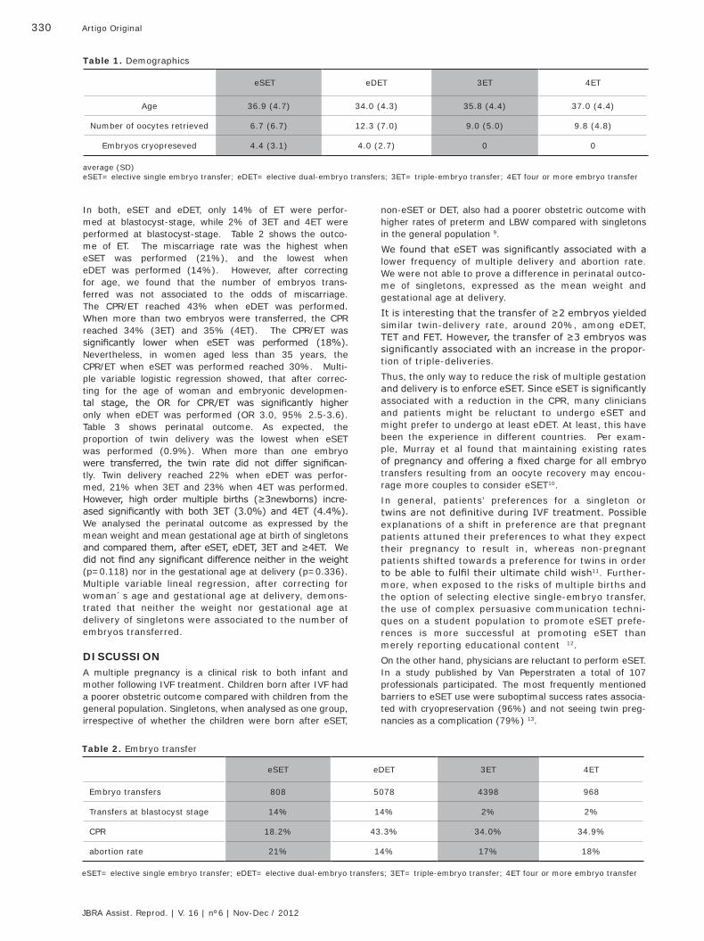
330 Artigo Original
JBRA Assist. Reprod. | V. 16 | nº6 | Nov-Dec / 2012
non-eSET or DET, also had a poorer obstetric outcome with higher rates of preterm and LBW compared with singletons in the general population 9. We found that eSET was significantly associated with a lower frequency of multiple delivery and abortion rate. We were not able to prove a difference in perinatal outco-me of singletons, expressed as the mean weight and gestational age at delivery.It is interesting that the transfer of ≥2 embryos yielded similar twin-delivery rate, around 20%, among eDET, TET and FET. However, the transfer of ≥3 embryos was significantly associated with an increase in the propor-tion of triple-deliveries. Thus, the only way to reduce the risk of multiple gestation and delivery is to enforce eSET. Since eSET is significantly associated with a reduction in the CPR, many clinicians and patients might be reluctant to undergo eSET and might prefer to undergo at least eDET. At least, this have been the experience in different countries. Per exam-ple, Murray et al found that maintaining existing rates of pregnancy and offering a fixed charge for all embryo transfers resulting from an oocyte recovery may encou-rage more couples to consider eSET10.In general, patients’ preferences for a singleton or twins are not definitive during IVF treatment. Possible explanations of a shift in preference are that pregnant patients attuned their preferences to what they expect their pregnancy to result in, whereas non-pregnant patients shifted towards a preference for twins in order to be able to fulfil their ultimate child wish11. Further-more, when exposed to the risks of multiple births and the option of selecting elective single-embryo transfer, the use of complex persuasive communication techni-ques on a student population to promote eSET prefe-rences is more successful at promoting eSET than merely reporting educational content 12.On the other hand, physicians are reluctant to perform eSET. In a study published by Van Peperstraten a total of 107 professionals participated. The most frequently mentioned barriers to eSET use were suboptimal success rates associa-ted with cryopreservation (96%) and not seeing twin preg-nancies as a complication (79%) 13.
In both, eSET and eDET, only 14% of ET were perfor-med at blastocyst-stage, while 2% of 3ET and 4ET were performed at blastocyst-stage. Table 2 shows the outco-me of ET. The miscarriage rate was the highest when eSET was performed (21%), and the lowest when eDET was performed (14%). However, after correcting for age, we found that the number of embryos trans-ferred was not associated to the odds of miscarriage. The CPR/ET reached 43% when eDET was performed. When more than two embryos were transferred, the CPR reached 34% (3ET) and 35% (4ET). The CPR/ET was significantly lower when eSET was performed (18%). Nevertheless, in women aged less than 35 years, the CPR/ET when eSET was performed reached 30%. Multi-ple variable logistic regression showed, that after correc-ting for the age of woman and embryonic developmen-tal stage, the OR for CPR/ET was significantly higher only when eDET was performed (OR 3.0, 95% 2.5-3.6). Table 3 shows perinatal outcome. As expected, the proportion of twin delivery was the lowest when eSET was performed (0.9%). When more than one embryo were transferred, the twin rate did not differ significan-tly. Twin delivery reached 22% when eDET was perfor-med, 21% when 3ET and 23% when 4ET was performed. However, high order multiple births (≥3newborns) incre-ased significantly with both 3ET (3.0%) and 4ET (4.4%). We analysed the perinatal outcome as expressed by the mean weight and mean gestational age at birth of singletons and compared them, after eSET, eDET, 3ET and ≥4ET. We did not find any significant difference neither in the weight (p=0.118) nor in the gestational age at delivery (p=0.336). Multiple variable lineal regression, after correcting for woman´s age and gestational age at delivery, demons-trated that neither the weight nor gestational age at delivery of singletons were associated to the number of embryos transferred.
DISCUSSIONA multiple pregnancy is a clinical risk to both infant and mother following IVF treatment. Children born after IVF had a poorer obstetric outcome compared with children from the general population. Singletons, when analysed as one group, irrespective of whether the children were born after eSET,
Table 1. Demographics
eSET eDET 3ET 4ET
Age 36.9 (4.7) 34.0 (4.3) 35.8 (4.4) 37.0 (4.4)
Number of oocytes retrieved 6.7 (6.7) 12.3 (7.0) 9.0 (5.0) 9.8 (4.8)
Embryos cryopreseved 4.4 (3.1) 4.0 (2.7) 0 0
average (SD)eSET= elective single embryo transfer; eDET= elective dual-embryo transfers; 3ET= triple-embryo transfer; 4ET four or more embryo transfer
Table 2. Embryo transfer
eSET eDET 3ET 4ET
Embryo transfers 808 5078 4398 968
Transfers at blastocyst stage 14% 14% 2% 2%
CPR 18.2% 43.3% 34.0% 34.9%
abortion rate 21% 14% 17% 18%
eSET= elective single embryo transfer; eDET= elective dual-embryo transfers; 3ET= triple-embryo transfer; 4ET four or more embryo transfer
JBRA6_150213.indb 330 15/02/2013 17:17:15
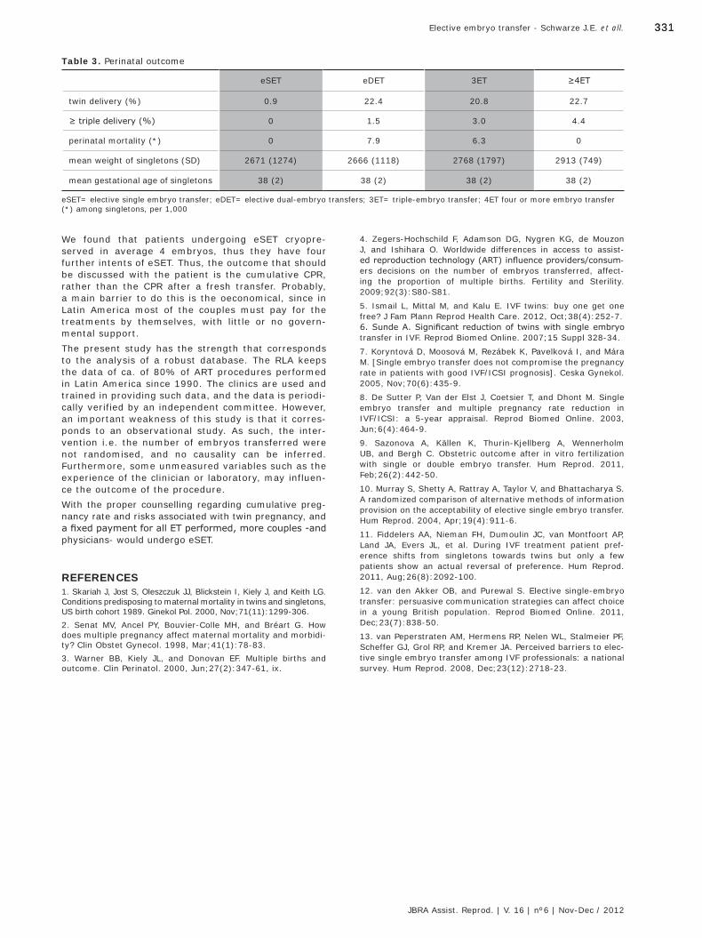
331331Elective embryo transfer - Schwarze J.E. et all.
JBRA Assist. Reprod. | V. 16 | nº6 | Nov-Dec / 2012
4. Zegers-Hochschild F, Adamson DG, Nygren KG, de Mouzon J, and Ishihara O. Worldwide differences in access to assist-ed reproduction technology (ART) influence providers/consum-ers decisions on the number of embryos transferred, affect-ing the proportion of multiple births. Fertility and Sterility. 2009;92(3):S80-S81.
5. Ismail L, Mittal M, and Kalu E. IVF twins: buy one get one free? J Fam Plann Reprod Health Care. 2012, Oct;38(4):252-7.6. Sunde A. Significant reduction of twins with single embryo transfer in IVF. Reprod Biomed Online. 2007;15 Suppl 328-34.
7. Koryntová D, Moosová M, Rezábek K, Pavelková I, and Mára M. [Single embryo transfer does not compromise the pregnancy rate in patients with good IVF/ICSI prognosis]. Ceska Gynekol. 2005, Nov;70(6):435-9.
8. De Sutter P, Van der Elst J, Coetsier T, and Dhont M. Single embryo transfer and multiple pregnancy rate reduction in IVF/ICSI: a 5-year appraisal. Reprod Biomed Online. 2003, Jun;6(4):464-9.
9. Sazonova A, Källen K, Thurin-Kjellberg A, Wennerholm UB, and Bergh C. Obstetric outcome after in vitro fertilization with single or double embryo transfer. Hum Reprod. 2011, Feb;26(2):442-50.
10. Murray S, Shetty A, Rattray A, Taylor V, and Bhattacharya S. A randomized comparison of alternative methods of information provision on the acceptability of elective single embryo transfer. Hum Reprod. 2004, Apr;19(4):911-6.
11. Fiddelers AA, Nieman FH, Dumoulin JC, van Montfoort AP, Land JA, Evers JL, et al. During IVF treatment patient pref-erence shifts from singletons towards twins but only a few patients show an actual reversal of preference. Hum Reprod. 2011, Aug;26(8):2092-100.
12. van den Akker OB, and Purewal S. Elective single-embryo transfer: persuasive communication strategies can affect choice in a young British population. Reprod Biomed Online. 2011, Dec;23(7):838-50.
13. van Peperstraten AM, Hermens RP, Nelen WL, Stalmeier PF, Scheffer GJ, Grol RP, and Kremer JA. Perceived barriers to elec-tive single embryo transfer among IVF professionals: a national survey. Hum Reprod. 2008, Dec;23(12):2718-23.
We found that patients undergoing eSET cryopre-served in average 4 embryos, thus they have four further intents of eSET. Thus, the outcome that should be discussed with the patient is the cumulative CPR, rather than the CPR after a fresh transfer. Probably, a main barrier to do this is the oeconomical, since in Latin America most of the couples must pay for the treatments by themselves, with little or no govern-mental support. The present study has the strength that corresponds to the analysis of a robust database. The RLA keeps the data of ca. of 80% of ART procedures performed in Latin America since 1990. The clinics are used and trained in providing such data, and the data is periodi-cally verified by an independent committee. However, an important weakness of this study is that it corres-ponds to an observational study. As such, the inter-vention i.e. the number of embryos transferred were not randomised, and no causality can be inferred. Furthermore, some unmeasured variables such as the experience of the clinician or laboratory, may influen-ce the outcome of the procedure.With the proper counselling regarding cumulative preg-nancy rate and risks associated with twin pregnancy, and a fixed payment for all ET performed, more couples -and physicians- would undergo eSET.
REFERENCES1. Skariah J, Jost S, Oleszczuk JJ, Blickstein I, Kiely J, and Keith LG. Conditions predisposing to maternal mortality in twins and singletons, US birth cohort 1989. Ginekol Pol. 2000, Nov;71(11):1299-306.2. Senat MV, Ancel PY, Bouvier-Colle MH, and Bréart G. How does multiple pregnancy affect maternal mortality and morbidi-ty? Clin Obstet Gynecol. 1998, Mar;41(1):78-83.3. Warner BB, Kiely JL, and Donovan EF. Multiple births and outcome. Clin Perinatol. 2000, Jun;27(2):347-61, ix.
Table 3. Perinatal outcome
eSET eDET 3ET ≥4ET
twin delivery (%) 0.9 22.4 20.8 22.7
≥ triple delivery (%) 0 1.5 3.0 4.4
perinatal mortality (*) 0 7.9 6.3 0
mean weight of singletons (SD) 2671 (1274) 2666 (1118) 2768 (1797) 2913 (749)
mean gestational age of singletons 38 (2) 38 (2) 38 (2) 38 (2)
eSET= elective single embryo transfer; eDET= elective dual-embryo transfers; 3ET= triple-embryo transfer; 4ET four or more embryo transfer(*) among singletons, per 1,000
JBRA6_150213.indb 331 15/02/2013 17:17:15
332
Artigo Original
Trends in the number of transferred embryos, clinical pregnancy rate and multiple pregnancy rate in Latin America: 1998 through 2008
Juan-Enrique Schwarze, MD MSc 1; Javier Crosby, PhD 2; Carolina Musri, BQ 2; Fernando Zegers-Hochschild, MD 2,3
1 Clínica Monteblanco, Chile2 Unit of Reproductive Medicine Clínica las Condes, Chile3 Program of Ethics and Public Policies in Human Reproduction, University Diego Portales, Chile
ABSTRACTBackground. Reducing the rate in IVF of multiple births and associated perinatal complications has become the major challenge in Latin America, due to is association with a significant increase in the prema-turity, and perinatal mortality.Methods. We selected all procedures involving the transfer of IVF embryos, reported to the Latin Ameri-ca Registry of ART, initiated between January 1998 and December 2008. A Poisson regression analysis was performed, stratified by woman´s age to analyse the trend in the transfer of 2, 3 and ≥4-embryos and ≥triple-pregnancy rate. Results. A total of 164,874 IVF/ICSI transfers were considered. Overall, there was a decline in the propor-tion of procedures performed in women aged ≤34 years old from 51% to 43%; and an increase of ICSI proce-dures from 61% to 86%. All age groups experienced a reduction in the proportion of ≥4-embryos transfers and an increase in that of 2-embryo transfers. Overall, the clinical pregnancy rate improved from 29% to 35% (p=0.01); and the incidence of ≥triple-pregnancy decreased from 8% to 3% in 2008 (p<0.001), while twin pregnancy remained stable (~20%)Conclusions. Our data support the policy of trans-ferring less embryos. It shows a decline in the mean number of embryos transferred and proportion ≥ 4-embryos transfers. Moreover, it reports a clinical pregnancy rate which instead of being reduced, has shown an increase over the 10-year period.
INTRODUCTIONThe first IVF-baby in Latin America was born in Colombia in 1984. This technology spread rapidly all over Latin America generating a substantial increase in the number of patients gaining access to Assisted Repro-ductive Technologies (ART). The current need for ART procedures, however, is by far underserved, and great disparity persists in the access to such technology1.The latest report by the International Committee Monitoring Assisted Reproductive Technology presents worldwide information about ART procedures perfor-med in 20032. At that time, the number of cycles repor-ted by Latin-American centers represented only 2.3% of the 932,415 cycles performed worldwide. Expres-sed as the ratio of ART cycles per million inhabitants, the disparity in access to ART is even more striking: countries like Sweden, Denmark and Belgium perform
between 1,000 to 2,000 cycles per million inhabitants, whilst countries in Latin America perform between 33 and 109 cycles per million inhabitants2. In countries where ART is reimbursed or covered by public health insurance, the mean number of embryos transferred is much smaller than the number of embryos transferred in countries where ART is funded by each couple3.Current economic policies in Latin America cause that only a small percentage of the gross domestic product is allocated to health related expenditures. Furthermore, the majority is diverted to private heal-th expenditure such as insurance companies, who do not cover infertility-related procedures4. Thus couples have to rely on their own resources to fund their treat-ments which limits the number of infertile couples with access to treatment and the number of opportunities that couples have to receive ART treatments. In order to achieve successful outcomes while minimi-sing the number of ART procedures attempts, multiple embryos are transferred into the uterus which in term increases the chances of pregnancy5. However, this strategy also increases in the rate of twin and high order multiple pregnancies6.At a regional level, Latin America shows comparable clinical outcomes to those of developed countries. Simi-lar clinical pregnancy rates and delivery rates, are repor-ted, although with a larger number of embryos transfer-red, and regardless of the age of the female partner2. Reducing the number of embryos transferred with a reduction in the rate of multiple births and associated perinatal complications has become the major chal-lenge in Latin America, since multiple pregnancies are associated with a significant increase in the prematu-rity, and perinatal mortality1. Furthermore, the increa-se in family disorders mainly absorbed by women, and high individual and insurance costs to cover neonatal expenses are also issues that need to be addressed.This study examines trends in embryo transfer prac-tices and clinical and multiple pregnancy rates for assisted reproductive technologies performed in Latin America between 1998 and 2008. Given the strong association between number of embryos transferred and multiple birth risk7,8, assessment of embryo trans-fer practices is essential to understanding the multi-ple gestation and birth risk associated with assisted reproduction. Our aim is to document the decline in the number of embryos transferred, and the impact such shift might have had on clinical pregnancy and multiple gestation rates.
Recebido em 10-11-12Aceito em 17-12-12
Copyright - Todos os direitos reservados a SBRA - Sociedade Brasileira de Reprodução Assistida
JBRA6_150213.indb 332 15/02/2013 17:17:15
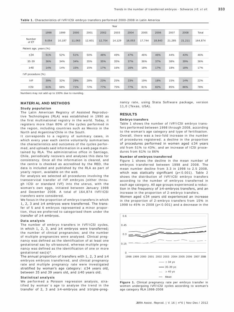
333333Trends in the number of transferred embryos - Schwarze J-E. et all.
JBRA Assist. Reprod. | V. 16 | nº6 | Nov-Dec / 2012
Table 1. Characteristics of IVF/ICSI embryo transfers performed 2000-2008 in Latin America
Year
1998 1999 2000 2001 2002 2003 2004 2005 2006 2007 2008 Total
Number of ET 9,054 10,187 11,063 12,601 12,704 14,129 16,053 17,744 18,843 21,285 21,211 164,874
Patient age, years (%)
≤34 51% 52% 51% 50% 48% 49% 47% 46% 46% 44% 43% 46%
35-39 36% 34% 34% 35% 35% 35% 37% 36% 37% 38% 39% 36%
≥40 14% 14% 15% 15% 17% 16% 16% 18% 17% 18% 18% 17%
IVF procedure (%)
IVF 39% 32% 29% 28% 23% 25% 23% 19% 18% 15% 14% 22%
ICSI 61% 68% 71% 72% 77% 75% 77% 81% 82% 85% 86% 78%
Numbers may not add up to 100% due to rounding.
MATERIAL AND METHODSStudy populationThe Latin American Registry of Assisted Reproduc-tive Technologies (RLA) was established in 1990 as the first multinational registry in the world. Today, it registers more than 80% of the cycles performed in the region, including countries such as Mexico in the North and Argentina/Chile in the South. It corresponds to a registry of summary cases, in which every year each centre voluntarily summarises the characteristics and outcomes of the cycles perfor-med, and uploads said information in a web page main-tained by RLA. The administrative office in Santiago, Chile, periodically recovers and analyses this data for consistency. Once all the information is cleared, and the centre is checked as accredited by the RED, the data is included and published by the RLA as part of yearly report, available on the web.For analysis we selected all procedures involving the transcervical transfer of IVF embryos (either throu-gh ICSI or standard IVF) into the uterus, with the woman’s own eggs, initiated between January 1998 and December 2008. A total of 164,874 IVF/ICSI transfers were considered.We focus in the proportion of embryo transfers in which 1, 2, 3 and ≥4 embryos were transferred. The trans-fer of 5 and 6 embryos represented a minor propor-tion, thus we preferred to categorised them under the transfer of ≥4 embryos.Data analysisThe number of embryo transfers in IVF/ICSI cycles, in which 1, 2, 3, and ≥4 embryos were transferred; the number of clinical pregnancies; and the number of multiple pregnancies were analysed. Clinical preg-nancy was defined as the identification of at least one gestational sac by ultrasound, whereas multiple preg-nancy was defined as the identification of one or more gestational sac(s)9. The annual proportion of transfers with 1, 2, 3 and ≥4 embryos embryos transferred, and clinical pregnancy rate and multiple pregnancy rate were investigated stratified by woman’s age category: ≤34 years old, between 35 and 39 years old, and ≥40 years old.Statistical analysisWe performed a Poisson regression analysis, stra-tified by woman´s age to analyse the trend in the transfer of 2, 3 and ≥4-embryos and ≥triple-preg-
nancy rate, using Stata Software package, version 11,0 (Texas, USA).
RESULTSEmbryo transfersTable 1 shows the number of IVF/ICSI embryo trans-fers performed between 1998 through 2008, according to the woman’s age category and type of fertilisation. Overall, there was a two-fold increase in the number of procedures registered; a decline in the proportion of procedures performed in women aged ≤34 years old from 51% to 43%; and an increase of ICSI proce-dures from 61% to 86% Number of embryos transferredFigure 1 shows the decline in the mean number of embryos transferred between 1998 and 2008. The mean number decline from 3.3 in 1998 to 2.5 2008, which was statically significant (p<0.001). Table 2 shows the distribution of IVF/ICSI embryo transfers according to the number of embryos transferred in each age category. All age groups experienced a reduc-tion in the frequency of ≥4-embryos transfers, and an increase in the proportion of 2-embryo transfers.Women aged ≤34 years old experienced an increase in the proportion of 2-embryo transfers from 15% in 1998 to 45% in 2008 (p<0.001) and a decrease in the
0.6
0.45
0.3
0.15
0
≤ 34 yo
35-39 yo
≥ 40 yo
Mean
1998 20042001 20071999 20052002 20082000 20062003
Figure 1. Clinical pregnancy rate per embryo transfer in women undergoing IVF/ICSI cycles according to woman’s age category RLA 1998-2008
JBRA6_150213.indb 333 15/02/2013 17:17:16
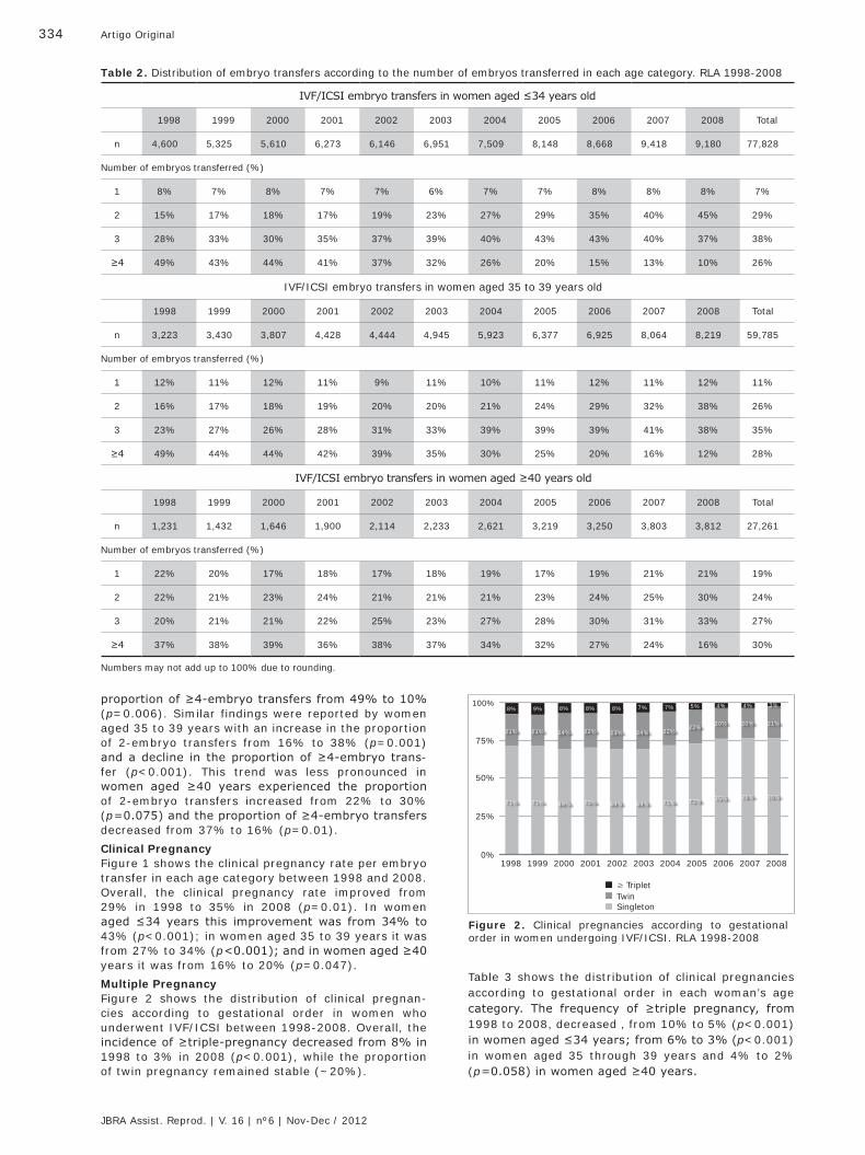
334 Artigo Original
JBRA Assist. Reprod. | V. 16 | nº6 | Nov-Dec / 2012
≥ TripletTwinSingleton
100%
75%
25%
0%
50%
1998 20042001 20071999 20052002 20082000 20062003
Figure 2. Clinical pregnancies according to gestational order in women undergoing IVF/ICSI. RLA 1998-2008
71%
21%
8%
71%
22%
7%
71%
21%
9%
72%
22%
5%
75%
20%
4%
76%
20%
4%
76%
21%
3%
69%
24%
8%
69%
23%
8%
69%
24%
7%
70%
22%
8%
Table 2. Distribution of embryo transfers according to the number of embryos transferred in each age category. RLA 1998-2008
IVF/ICSI embryo transfers in women aged ≤34 years old
1998 1999 2000 2001 2002 2003 2004 2005 2006 2007 2008 Total
n 4,600 5,325 5,610 6,273 6,146 6,951 7,509 8,148 8,668 9,418 9,180 77,828
Number of embryos transferred (%)
1 8% 7% 8% 7% 7% 6% 7% 7% 8% 8% 8% 7%
2 15% 17% 18% 17% 19% 23% 27% 29% 35% 40% 45% 29%
3 28% 33% 30% 35% 37% 39% 40% 43% 43% 40% 37% 38%
≥4 49% 43% 44% 41% 37% 32% 26% 20% 15% 13% 10% 26%
IVF/ICSI embryo transfers in women aged 35 to 39 years old
1998 1999 2000 2001 2002 2003 2004 2005 2006 2007 2008 Total
n 3,223 3,430 3,807 4,428 4,444 4,945 5,923 6,377 6,925 8,064 8,219 59,785
Number of embryos transferred (%)
1 12% 11% 12% 11% 9% 11% 10% 11% 12% 11% 12% 11%
2 16% 17% 18% 19% 20% 20% 21% 24% 29% 32% 38% 26%
3 23% 27% 26% 28% 31% 33% 39% 39% 39% 41% 38% 35%
≥4 49% 44% 44% 42% 39% 35% 30% 25% 20% 16% 12% 28%
IVF/ICSI embryo transfers in women aged ≥40 years old
1998 1999 2000 2001 2002 2003 2004 2005 2006 2007 2008 Total
n 1,231 1,432 1,646 1,900 2,114 2,233 2,621 3,219 3,250 3,803 3,812 27,261
Number of embryos transferred (%)
1 22% 20% 17% 18% 17% 18% 19% 17% 19% 21% 21% 19%
2 22% 21% 23% 24% 21% 21% 21% 23% 24% 25% 30% 24%
3 20% 21% 21% 22% 25% 23% 27% 28% 30% 31% 33% 27%
≥4 37% 38% 39% 36% 38% 37% 34% 32% 27% 24% 16% 30%
Numbers may not add up to 100% due to rounding.
proportion of ≥4-embryo transfers from 49% to 10% (p=0.006). Similar findings were reported by women aged 35 to 39 years with an increase in the proportion of 2-embryo transfers from 16% to 38% (p=0.001) and a decline in the proportion of ≥4-embryo trans-fer (p<0.001). This trend was less pronounced in women aged ≥40 years experienced the proportion of 2-embryo transfers increased from 22% to 30% (p=0.075) and the proportion of ≥4-embryo transfers decreased from 37% to 16% (p=0.01).Clinical PregnancyFigure 1 shows the clinical pregnancy rate per embryo transfer in each age category between 1998 and 2008. Overall, the clinical pregnancy rate improved from 29% in 1998 to 35% in 2008 (p=0.01). In women aged ≤34 years this improvement was from 34% to 43% (p<0.001); in women aged 35 to 39 years it was from 27% to 34% (p<0.001); and in women aged ≥40 years it was from 16% to 20% (p=0.047). Multiple PregnancyFigure 2 shows the distribution of clinical pregnan-cies according to gestational order in women who underwent IVF/ICSI between 1998-2008. Overall, the incidence of ≥triple-pregnancy decreased from 8% in 1998 to 3% in 2008 (p<0.001), while the proportion of twin pregnancy remained stable (~20%).
Table 3 shows the distribution of clinical pregnancies according to gestational order in each woman’s age category. The frequency of ≥triple pregnancy, from 1998 to 2008, decreased , from 10% to 5% (p<0.001) in women aged ≤34 years; from 6% to 3% (p<0.001) in women aged 35 through 39 years and 4% to 2% (p=0.058) in women aged ≥40 years.
JBRA6_150213.indb 334 15/02/2013 17:17:16
335335Trends in the number of transferred embryos - Schwarze J-E. et all.
JBRA Assist. Reprod. | V. 16 | nº6 | Nov-Dec / 2012
Table 3. Multiple pregnancy rate according to woman’s age category. RLA 1998-2008
Clinical pregnancies according to gestational order in women aged ≤34 years old
1998 1999 2000 2001 2002 2003 2004 2005 2006 2007 2008
n 1,568 1,843 1,815 2,276 2,431 2,659 2,895 3,127 3,434 3,853 3,946
Gestational order (%)
Single-ton 66% 65% 66% 66% 66% 68% 69% 71% 72% 72% 73%
Twin 23% 23% 26% 24% 25% 26% 25% 24% 24% 24% 22%
≥Triplet 10% 11% 9% 9% 9% 8% 7% 6% 5% 5% 6%
Clinical pregnancies according to gestational order in women aged 35 to 39 years old
1998 1999 2000 2001 2002 2003 2004 2005 2006 2007 2008
n 869 944 963 1,272 1,439 1,531 1,840 1,982 2,268 2,674 2,819
Gestational order (%)
Single-ton 75% 76% 73% 72% 71% 72% 73% 74% 80% 80% 79%
Twin 19% 19% 21% 21% 21% 22% 20% 22% 18% 17% 18%
≥Triplet 6% 6% 6% 7% 7% 5% 7% 4% 3% 3% 2%
Clinical pregnancies according to gestational order in women aged ≥40 years old
1998 1999 2000 2001 2002 2003 2004 2005 2006 2007 2008
n 197 226 248 315 448 437 504 681 604 720 751
Gestational order (%)
Single-ton 82% 84% 79% 83% 78% 75% 79% 81% 85% 88% 87%
Twin 14% 14% 18% 14% 18% 18% 16% 16% 14% 10% 11%
≥Triplet 4% 2% 3% 3% 5% 7% 5% 4% 2% 2% 1%
Numbers may not add up to 100% due to rounding.
Table 5. Perinatal outcome according to gestational order of 73,857 babies born due to IVF/ICSI cycles performed between 1998 and 2008
Gestational Order Perinatal mortality RR (95%IC)
single 0.86% 1
twin 2.92% 3.05 (2.69-3.45)
triplets 6.49% 6.79 (5.94-7.76)
≥quadruplets 19.29% 20.19 (17.02-23.95)
Table 4. Incidence Risk Ratio for ≥ triple pregnancy stra-tified by quartile of mean number of embryos transferred1
Quartile (mean number of transferred embryos) IRR year (95% CI) p
1- (2.47-2-70) 0.87 (0.80-0.969 0.003
2- (2.71-2.97) 0.41 (0.19-0.86) 0.019
3- (2.98-3.13) 0.98 (0.93-1.04) 0.638
4- (3.14-3.42) 0.97 (0.92-1.04) 0.477
1 Adjusted for age category
Table 4 shows the incidence risk ratio for ≥ triple pregnancy adjusted for woman´s age category, stra-tified by quartile of the mean number of transferred embryos. As seen, the incidence risk ratio for ≥triple pregnancy diminished significantly over time in the groups where less embryos were transferred. In the two groups where more embryos were transferred, the decrease in the incidence risk ratio did not change significantly over time. Table 5 shows the perinatal outcome of babies born due to FIV/ICSI performed between 1998 and 2008 according to the gestational order. The risk of perina-tal mortality increased significantly with higher gesta-tional orders.
DISCUSSIONThe number of infertile couple undergoing assisted reproductive technologies increased significantly in the period analysed, as did the clinical pregnancy rate per embryo transfer, regardless of the woman’s age-cate-gory. When focusing on the success such procedures, attention should also be given to its complications. The most important being that of multiple pregnancy which is significantly associated with preterm delivery and perinatal morbidity and mortality.In Latin America, the frequency of ≥triple pregnancy rate has halved in all woman’s age categories over this 10-year period, while the proportion of twin pregnancy
JBRA6_150213.indb 335 15/02/2013 17:17:17
336 Artigo Original
JBRA Assist. Reprod. | V. 16 | nº6 | Nov-Dec / 2012
remained stable. Multivariate analysis showed that the most important factor that accounted for this decli-ne is the significant decrease in the mean number of embryos transferred. The constant, yet minor, decrea-se in the mean number of embryos transferred signifi-cantly lowered the frequency of ≥triple pregnancy, but showed no significant effect in the proportion of twin pregnancies. The increase seen in the proportion of three-embryo transfers may have countered,the effect caused by the reduction in the proportion of ≥four--embryo transfers.Importantly, this decline in the number of embryos transferred was not associated with a decline in the clinical pregnancy rate, on the contrary, the clinical pregnancy rate per embryo transfer improved signi-ficantly between 1998-2008 in all age categories. This suggests that other unmeasured factors impro-ved over time. One of these might be the transfer of embryos in later stages of development, allowing for a better selection and improved implantation rates.In an attempt to diminish the ART-related multi-ple pregnancies epidemic, countries like Sweden, Belgium and Japan have implemented strict policies and support the elective transfer of one embryo, with a clear reduction in the multiple pregnancy rate (10-14). This is feasible in countries where ART proce-dures are covered by health insurance and thus have some control over ART practice. This is difficult in Latin America, where ART procedures are mainly covered by infertile couples, and the open market economy dicta-tes strong competition between different ART centres, which have the tendency to maximise results, regard-less of multiple pregnancy rate.Nevertheless, we found that practice in the region is moving towards the right direction with more work needed for reducing the number of embryos transfer-red. Our data support the policy of transferring less embryos. It shows a decline in the mean number of embryos transferred and proportion ≥ 4-embryos transfers. Moreover, it reports a clinical pregnancy rate which instead of being reduced, has shown an increase over the 10-year period.
Correspondência:Oficina del Registro Latino Americano de Reproducción Asis-tida - Camino Lo Fontecilla 421, Las Condes- Oficina del Registro LatinoAmericano Reproducción - Lo Fontecilla 441, Santiago, Chile. Teléfono: 56-2-6108181 / Fax: 56-2-6108167E-mail: [email protected]
REFERENCES1. Zegers-Hochschild, Schwarze, Crosby, and do Carmo. Twenty years of Assisted Reproductive Technology (ART) in Latin Ameri-ca. Jornal Brasileiro de Reproducao Assistida. 2011;15(2):15-30.2. Nygren KG, Sullivan E, Zegers-Hochschild F, Mansour R, Ishiha-ra O, Adamson GD, and de Mouzon J. International Committee for Monitoring Assisted Reproductive Technology (ICMART) world report: assisted reproductive technology 2003. Fertil Steril. 2011, Jun;95(7):2209-22, 2222.e1-17.3. Zegers-Hochschild F, Adamson DG, Nygren KG, de Mouzon J, and Ishihara O. Worldwide differences in access to assis-ted reproduction technology (ART) influence providers/consu-mers decisions on the number of embryos transferred, affec-ting the proportion of multiple births. Fertility and Sterility. 2009;92(3):S80-S81.4. Zegers-Hochschild, Schwarze, and Galdames. Assisted repro-ductive technology in Latin America: an example of regional cooperation and development . ESHRE Monographs issue:ESHRE Special Task Force on ‘Developing Countries and Infertility’. 2008;142-47.5. JOINT SOGC-CFAS. Guidelines for the number of embryos to transfer following in vitro fertilization No. 182, September 2006. Int J Gynaecol Obstet. 2008, Aug;102(2):203-16.6. Van Voorhis BJ, and Ryan GL. Ethical obligation for restricting the number of embryos transferred to women: combating the multiple-birth epidemic from in vitro fertilization. Semin Reprod Med. 2010, Jul;28(4):287-94.7. Sunderam S, Chang J, Flowers L, Kulkarni A, Sentelle G, Jeng G, et al. Assisted reproductive technology surveillance--United States, 2006. MMWR Surveill Summ. 2009, Jun 12;58(5):1-25.8. Zegers-Hochschild F, Schwarze JE, and Galdames V. XIX Regis-tro Latinoamericano de Reproduccion Asistida 2008. 2010;9. Zegers-Hochschild F, Adamson GD, de Mouzon J, Ishihara O, Mansour R, Nygren K, et al. International Committee for Monito-ring Assisted Reproductive Technology (ICMART) and the World Health Organization (WHO) revised glossary of ART terminology, 2009. Fertil Steril. 2009, Nov;92(5):1520-4.10. Karlström PO, and Bergh C. Reducing the number of embryos transferred in Sweden-impact on delivery and multiple birth rates. Hum Reprod. 2007, Aug;22(8):2202-7.1. De Neubourg D, Gerris J, Van Royen E, Mangelschots K, and Vercruyssen M. Impact of a restriction in the number of embryos transferred on the multiple pregnancy rate. Eur J Obstet Gynecol Reprod Biol. 2006, Feb 1;124(2):212-5.12. Germond M, Primi MP, Urner F, Chanson A, Wirthner D, and Senn A. Number of transferred embryos: how to reduce multiple pregnancies. Ann N Y Acad Sci. 2004, Dec;103493-100.13. Komori S, Kasumi H, Horiuchi I, Hamada Y, Suzuki C, Shigeta M, and Koyama K. Prevention of multiple pregnancies by restric-ting the number of transferred embryos: randomized control study. Arch Gynecol Obstet. 2004, Sep;270(2):91-3.14. Saldeen P, and Sundström P. Would legislation imposing single embryo transfer be a feasible way to reduce the rate of multiple pregnancies after IVF treatment? Hum Reprod. 2005, Jan;20(1):4-8.
JBRA6_150213.indb 336 15/02/2013 17:17:17
337
Artigo Original
RESUMOObjetivo: avaliar a eficácia do pericárdio bovino (PB) na prevenção de aderências em cães submetidos a duas lesões indutoras de aderências no grupo controle (GC) e no grupo experimental (GE). Métodos: dezes-sete cadelas, sem raça definida e não prenhas, foram anestesiadas e submetidas a lesões indutoras de aderências, sendo a primeira a miometrectomia ante-rior com sutura isquêmica, e a segunda, o fechamento do peritônio com sutura ancorada isquêmica. No GE, com nove cadelas, colocou-se o PB recobrindo inte-gralmente a primeira lesão; na segunda lesão, reco-briu-se a área de sutura isquêmica na extremidade inferior. Após seis dias, no mínimo, as cadelas foram sacrificadas e as aderências quantificadas, segundo a sistematização sugerida pela Adhesion Sudy Group denominada MCASM*(more comprehensive adhesion score method), e pela avaliação do percentual de área aderida, excluindo a força da aderência, denomina-da AEEL*(adhesion extension experimental lesion). Resultados: O GC e GE não tiveram diferenças signi-ficativas na média de peso (kg) e tempo cirúrgico em minutos (12,62/15,66 p= 0, 3868) e (78,25/77, 88, p= 0, 9086). Lesão 1: o GC apresentou escore médio 6,50 versus 6,00 no GE, p= 0,718 (MCASM) e 4,87/4,33 com p= 0,615 (AEEL), diferenças não signi-ficativas. Lesão 2: o GC teve escore médio 4,62 versus 7,55 no GE e p= 0,005 (MCASM) e 2,75/4,77 com p= 0,015 (AEEL), diferenças significantes. Conclusões: As lesões foram fortes indutoras de aderências, e o PB não preveniu a formação de aderências. Além disso, o PB produziu mais aderências na lesão 2.Palavras-chave: aderências pós-operatorias, métodos de barreira cirúrgicos, pericárdio bovino, lesões experi-mentais de aderências, prevenção de aderências.
ABSTRACT Purpose: to investigate the efficacy of bovine pericar-dium to prevent pelvic adhesion in dogs. Methods: Seventeen female non pregnant dogs were anesthetized and submitted to two adhesion-inducing lesions – the first lesion was performed in the anterior portion of uterus (elliptical incision) and sutured with an ischemic line of chromic catgut and the second, when closing the peritoneum, also with an ischemic suture. BP was used as the barrier agent in nine dogs, fully covering the first lesion and applied on the inferior part of the second lesion. Six days later, the dogs were sacrificed and the adhesions were examined according to the ¨More Comprehensive Adhesion Score Method¨ (MCASM, Adhesion Study Group-EUA) and to the score developed by the author in which only the percentage of the adhesion is examined, ¨Adhesion Extension in Experimental Lesion¨ (AEEL).Results: There were no significant differences betwe-en the two groups regarding weight and surgical time in minutes . No significant differences were observed between the MCASM scores of the two groups (6.50 vs. 6.00, p= 0.718) and the AEEL (4.87 vs. 4.33, p= 0.615) for lesion 1. There were significant differen-ces in the second lesion in the two groups, regarding the mean scores given by the MCASM system (4.62 vs.7.55, p=0.005) and the adhesion extension score (2.75 vs. 4.77, p=0.015). Conclusions: BP did not prevent the formation of adhesions caused by the lesions performed. Further-more, BP enhanced the formation of adhesions in lesion 2.Key words: Postoperative adhesion prevention, surgi-cal barriers, bovine pericardium, experimental lesions of adhesion, prevention of adhesion.
Experimental evaluation of bovine pericardium efficacy on adhesion pelvic prevention in the dog model.
Avaliação experimental da eficácia do pericardio bovino na prevenção de aderências pélvicas em modelo canino.
Rodopiano de Souza Florêncio¹, Pablo Rassi Florencio², Manoel João Batista Castello Girão³, Edmund Chada Baracat .4
Department of Gynecology and Obstetrics and Department of Surgery (Experimental Surgery), Faculty of Medicine, Federal University of Goiás; Department of Gynecology, Federal University of São Paulo - School of Medicine.
¹ Department of Gynecology and Obstetrics, School of Medicine (Laboratory of Human Reproduction), Federal University of Goiás – UFG – Goiania (GO), Brazil² Plastic Surgeon. Former Resident in the Department of Surgery, Federal University of São Paulo - School of Medicine- UNIFESP - São Paulo (SP), Brazil. ³ Department of Gynecology, Federal University of São Paulo – School of Medicine – UNIFESP – São Paulo (SP), Brazil.4 Department of Gynecology, Federal University of São Paulo – School of Medicine – UNIFESP and in the Faculty of Medicine, University of São Paulo – USP – São Paulo, Brazil.
Recebido em 10-12-12Aceito em 31-12-12
Copyright - Todos os direitos reservados a SBRA - Sociedade Brasileira de Reprodução Assistida
JBRA6_150213.indb 337 15/02/2013 17:17:17

338 Artigo Original
JBRA Assist. Reprod. | V. 16 | nº6 | Nov-Dec / 2012
ACKNOWLEDGEMENTThis work was carried out at the discipline of Operative Technique and Experimental Surgery in the Depart-ment of Surgery, Faculty of Medicine, UFG, with the participation of gynecologists: Vidal Augusto Cortizo, Denis Ribeiro de Oliveira, José Hidalgo Donizeth, Vini-cius Alves Oliveira and residents of the Department of Surgery, Faculty of Medicine, UFG and Department of Surgery, Paulista School of Medicine(UNIFESP), and medical students.
INTRODUCTIONOver the past years abdominopelvic adhesion has been studied in a very detailed and professional way. These studies analyze the importance of adhesion formation to the human being once it is related to chronic pelvic pain, infertility, surgery complication in reoperation and the relevant costs in the health care systems.Ellis et al., 1999 studied the percentage of hospital admittance caused directly or indirectly by abdomi-nopelvic adhesion in Scotland in the year of 1994. The studied population consisted of patients that underwent surgery in 1986 who had no record of abdominopelvic surgery in the preceding 5 years. Among 27.790 patients 34.6% were readmitted to hospital on an average of 2.1 times in ten years of observation; 22.1% of patients were readmitted in the first year after the surgery. In developed countries the main cause of bowel obstruction can be attributed to abdominopelvic surgery (Beck et al., 2000).Svanes Fevang et al., 2004 studied 500 patients in Norway between 1961 and 1995 . They underwent surgery because of bowel occlusion related to adhesion and they were followed for an average of ten years, but some patients were followed for fourty years. The cumulative recurrence rate was 18% at ten years and 29% at thirty years. Patients with high recidivism rate showed higher recurrence rate and for those who underwent four previous surgeries, for example, the recidive rate was 81%. Wilson et al., 2002 estimated that in Scotland the re-admission cost for post-surgi-cal abdominal adhesion, mainly colon-rectal, during ten years follow-up observation was about 569 million pounds. Scientists speculate that if adhesion preven-tion products costing 50 pounds per patient were used, the hospital re-admittance would diminish 16% and if the cost was 200 pounds the hospital re-admittance would diminish 64% in three years. The barrier method presents preventive properties against adhesion and this method is the most well researched over the past years. Among the common barrier methods, apparently effective, there is the polytetrafluoroethylene membrane (PTFE) commercia-lized by Gore Tex Surgical Membrane, Interceed and Seprafilm. Many multicentre studies were published about Interceed (oxidized regenerated cellulose and Absorbable Adhesion Barrier) after studies in animals (Interceed Adhesion Study Group, 1998; Nordic Adhe-sion Prevention Study Group, 1996; Franklin, 1995). Such studies showed a reduction in the incidence, extension and severity of post-surgery adhesion in human patients but, on the other hand, some negati-ve results were showed by different researches (Sara-vielos & Li, 1996; Reid et al.,1997; Baysal, 2001). Diamond & The Seprafilm Adhesion Study Group., 1996 showed positive results of Seprafilm (sodium hyaluronate-carboxymethylcellulose membrane) against pelvic adhesion.
Boyers et al., 1988 studying the effect of the expan-ded membrane (PTFE) in rabbits achieved a positive result. This result motivated a multicentre study in humans which was published in 1992 by The Surgical Membrane Study Group. They tested the effectiveness of the membrane against adhesion reformation. After this study some conclusions could be drawn: a-few training procedures are needed; b-the membrane must be located at least 1cm beyond the cruenta area; c- laparoscopic removal was not a difficult task; d-the product did not cause the death of the user. Studies in animals (Montz et al., 1992; Grow et al., 1994; Hellebrekers et al., 2000) showed the positive adhe-sion prevention effect of Gore Tex Surgical Membrane. Other authors (Haney et al., 1995; The Miomectomy Adhesion Multicenter Study Group, 1995) confirmed this positive result in humans. Hurst et al., 1999 coor-dinated an observational multicentre study to deter-mine if permanent implantation of the PTFE membra-ne in the pelvis caused any adverse effects and they concluded that removal of the barrier was not neces-sary. The membrane was implanted permanently and no long-term complications occurred in any of the 146 women subjected to the study.Farquhar et al., 2006 conducted a metanalysis study to assess the effect of mechanical barriers such as PTFE membrane, Interceed and Seprafilm. Their results suggest a positive adhesion prevention effect and Gore-Tex may be slightly superior to Interceed in preventing adhesion.These products are expensi-ve in our country and it has motivated scientists to look out for different options which would be compa-tible with our environment and less expensive. In our country the bovine pericardium(BP) is intensively used in heart surgery and that could be the right option. Such membrane is stored in formaldehyde and it is thicker than the Surgical Membrane which could be an inconvenience. On the other hand the bovine pericar-dium is resistant and malleable (Braile et al., 1998). Mavritas et al., 2005 , studied the dynamic mechanical behavior of BP in its native form and under specific enzymatic degradation with chondroitinase ABC. They confirmed the viscoelastic properties of BP. Gomes et al., 1994 studied the effect of this product in abdominal hernias correction in pigs and there was no adhesion formation. Therefore we decided to evalu-ate the bovine pericardium in adhesion prevention in female dogs with no specific pedigree. An efficient adhesion inducing model was used and it was previou-sly tested in a pilot project approval by Animal and Human Research Ethics Committee in Clinical Hospital of Federal University of Goiás.
METHODS AND MATERIALSTwenty non-pregnant female dogs with no specific pedi-gree were assigned to two groups each containing ten animals – control group (CG) and experimental group (EG). For the CG were used only the lesions described as it follows, no anti-adhesion method was used. For the EG, bovine pericardium was used.In order to induce adhesion formation two lesions were performed. The first lesion consisted of one elliptic incision of 3 to 4 centimeters (Figure 1) in the ante-rior portion of uterus sutured with an ischemic line of chromic catgut 00 and associated with obliteration to the bilateral uterine artery branch. The second lesion consisted at the closing of peritoneum also with an ischemic suture of chromic catgut 00 (Figure 2). It was performed after placing the bovine pericardium
JBRA6_150213.indb 338 15/02/2013 17:17:17
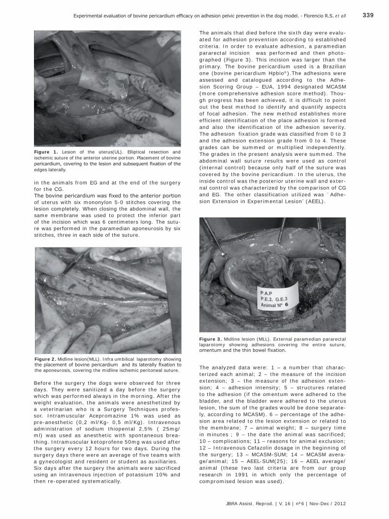
339339
JBRA Assist. Reprod. | V. 16 | nº6 | Nov-Dec / 2012
Experimental evaluation of bovine pericardium efficacy on adhesion pelvic prevention in the dog model. - Florencio R.S. et all
Figure 2. Midline lesion(MLL). Infra umbilical laparotomy showing the placement of bovine pericardium and its laterally fixation to the aponeurosis, covering the midline ischemic peritoneal suture.
Figure 3. Midline lesion (MLL). External paramedian pararectal laparotomy showing adhesions covering the entire suture, omentum and the thin bowel fixation.
Figure 1. Lesion of the uterus(UL). Elliptical resection and ischemic suture of the anterior uterine portion. Placement of bovine pericardium, covering to the lesion and subsequent fixation of the edges laterally.
in the animals from EG and at the end of the surgery for the CG.The bovine pericardium was fixed to the anterior portion of uterus with six mononylon 5-0 stitches covering the lesion completely. When closing the abdominal wall, the same membrane was used to protect the inferior part of the incision which was 6 centimeters long. The sutu-re was performed in the paramedian aponeurosis by six stitches, three in each side of the suture.
The animals that died before the sixth day were evalu-ated for adhesion prevention according to established criteria. In order to evaluate adhesion, a paramedian pararectal incision was performed and then photo-graphed (Figure 3). This incision was larger than the primary. The bovine pericardium used is a Brazilian one (bovine pericardium Hpbio®).The adhesions were assessed and catalogued according to the Adhe-sion Scoring Group – EUA, 1994 designated MCASM (more comprehensive adhesion score method). Thou-gh progress has been achieved, it is difficult to point out the best method to identify and quantify aspects of focal adhesion. The new method establishes more efficient identification of the place adhesion is formed and also the identification of the adhesion severity. The adhesion fixation grade was classified from 0 to 3 and the adhesion extension grade from 0 to 4. These grades can be summed or multiplied independently. The grades in the present analysis were summed. The abdominal wall suture results were used as control (internal control) because only half of the suture was covered by the bovine pericardium. In the uterus, the inside control was the posterior uterine wall and exter-nal control was characterized by the comparison of CG and EG. The other classification utilized was ¨Adhe-sion Extension in Experimental Lesion¨(AEEL).
Before the surgery the dogs were observed for three days. They were sanitized a day before the surgery which was performed always in the morning. After the weight evaluation, the animals were anesthetized by a veterinarian who is a Surgery Techniques profes-sor. Intramuscular Acepromazine 1% was used as pre-anesthetic (0,2 ml/Kg- 0,5 ml/Kg). Intravenous administration of sodium thiopental 2,5% ( 25mg/ml) was used as anesthetic with spontaneous brea-thing. Intramuscular ketoprofene 50mg was used after the surgery every 12 hours for two days. During the surgery days there were an average of five teams with a gynecologist and resident or student as auxiliaries. Six days after the surgery the animals were sacrificed using an intravenous injection of potassium 10% and then re-operated systematically.
The analyzed data were: 1 – a number that charac-terized each animal; 2 – the measure of the incision extension; 3 – the measure of the adhesion exten-sion; 4 – adhesion intensity; 5 – structures related to the adhesion (if the omentum were adhered to the bladder, and the bladder were adhered to the uterus lesion, the sum of the grades would be done separate-ly, according to MCASM). 6 – percentage of the adhe-sion area related to the lesion extension or related to the membrane; 7 – animal weight; 8 – surgery time in minutes ; 9 – the date the animal was sacrificed; 10 – complications; 11 – reasons for animal exclusion; 12 – Intravenous Cefazolin dosage in the beginning of the surgery; 13 – MCASM-SUM; 14 – MCASM avera-ge/animal; 15 – AEEL-SUM(25); 16 – AEEL average/animal (these two last criteria are from our group research in 1991 in which only the percentage of compromised lesion was used).
JBRA6_150213.indb 339 15/02/2013 17:17:19
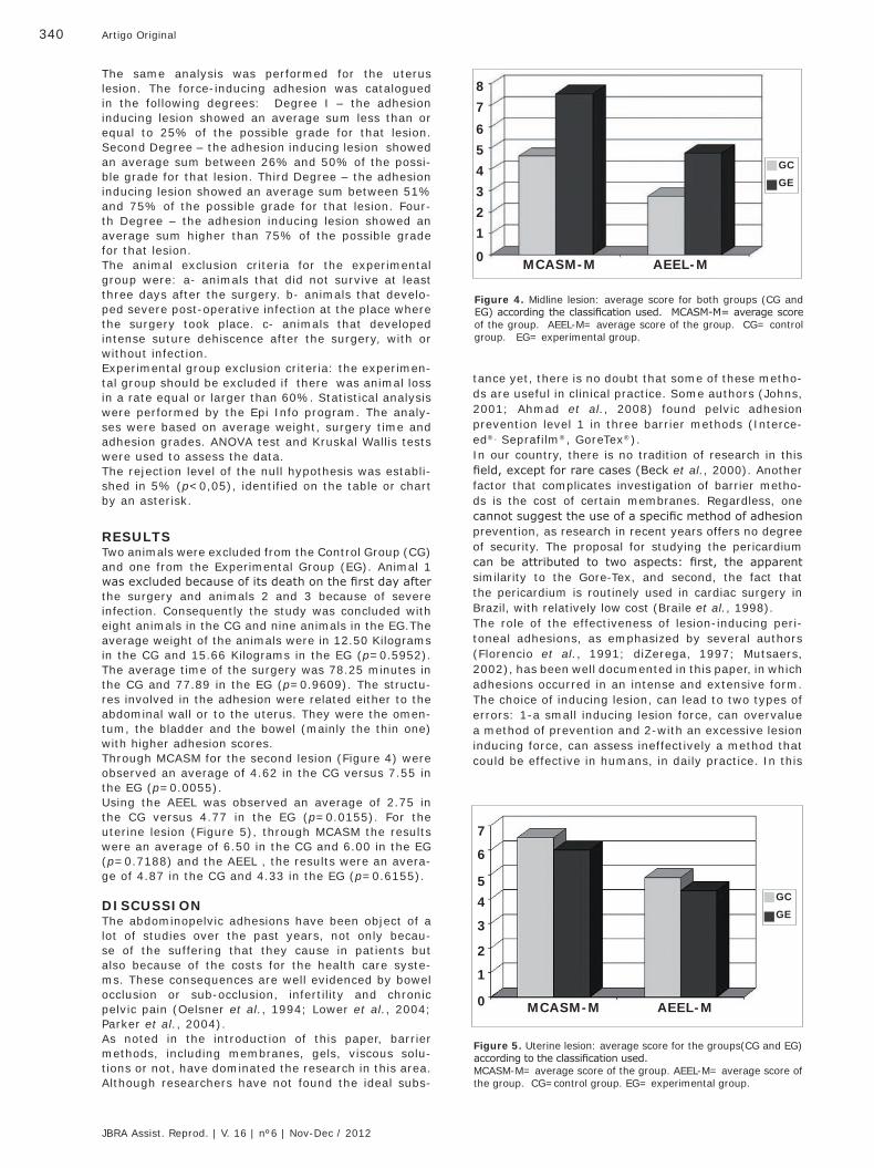
340 Artigo Original
JBRA Assist. Reprod. | V. 16 | nº6 | Nov-Dec / 2012
The same analysis was performed for the uterus lesion. The force-inducing adhesion was catalogued in the following degrees: Degree I – the adhesion inducing lesion showed an average sum less than or equal to 25% of the possible grade for that lesion. Second Degree – the adhesion inducing lesion showed an average sum between 26% and 50% of the possi-ble grade for that lesion. Third Degree – the adhesion inducing lesion showed an average sum between 51% and 75% of the possible grade for that lesion. Four-th Degree – the adhesion inducing lesion showed an average sum higher than 75% of the possible grade for that lesion.The animal exclusion criteria for the experimental group were: a- animals that did not survive at least three days after the surgery. b- animals that develo-ped severe post-operative infection at the place where the surgery took place. c- animals that developed intense suture dehiscence after the surgery, with or without infection.Experimental group exclusion criteria: the experimen-tal group should be excluded if there was animal loss in a rate equal or larger than 60%. Statistical analysis were performed by the Epi Info program. The analy-ses were based on average weight, surgery time and adhesion grades. ANOVA test and Kruskal Wallis tests were used to assess the data.The rejection level of the null hypothesis was establi-shed in 5% (p<0,05), identified on the table or chart by an asterisk.
RESULTSTwo animals were excluded from the Control Group (CG) and one from the Experimental Group (EG). Animal 1 was excluded because of its death on the first day after the surgery and animals 2 and 3 because of severe infection. Consequently the study was concluded with eight animals in the CG and nine animals in the EG.The average weight of the animals were in 12.50 Kilograms in the CG and 15.66 Kilograms in the EG (p=0.5952). The average time of the surgery was 78.25 minutes in the CG and 77.89 in the EG (p=0.9609). The structu-res involved in the adhesion were related either to the abdominal wall or to the uterus. They were the omen-tum, the bladder and the bowel (mainly the thin one) with higher adhesion scores.Through MCASM for the second lesion (Figure 4) were observed an average of 4.62 in the CG versus 7.55 in the EG (p=0.0055). Using the AEEL was observed an average of 2.75 in the CG versus 4.77 in the EG (p=0.0155). For the uterine lesion (Figure 5), through MCASM the results were an average of 6.50 in the CG and 6.00 in the EG (p=0.7188) and the AEEL , the results were an avera-ge of 4.87 in the CG and 4.33 in the EG (p=0.6155).
DISCUSSIONThe abdominopelvic adhesions have been object of a lot of studies over the past years, not only becau-se of the suffering that they cause in patients but also because of the costs for the health care syste-ms. These consequences are well evidenced by bowel occlusion or sub-occlusion, infertility and chronic pelvic pain (Oelsner et al., 1994; Lower et al., 2004; Parker et al., 2004).As noted in the introduction of this paper, barrier methods, including membranes, gels, viscous solu-tions or not, have dominated the research in this area. Although researchers have not found the ideal subs-
tance yet, there is no doubt that some of these metho-ds are useful in clinical practice. Some authors (Johns, 2001; Ahmad et al., 2008) found pelvic adhesion prevention level 1 in three barrier methods (Interce-ed®, Seprafilm®, GoreTex®).In our country, there is no tradition of research in this field, except for rare cases (Beck et al., 2000). Another factor that complicates investigation of barrier metho-ds is the cost of certain membranes. Regardless, one cannot suggest the use of a specific method of adhesion prevention, as research in recent years offers no degree of security. The proposal for studying the pericardium can be attributed to two aspects: first, the apparent similarity to the Gore-Tex, and second, the fact that the pericardium is routinely used in cardiac surgery in Brazil, with relatively low cost (Braile et al., 1998).The role of the effectiveness of lesion-inducing peri-toneal adhesions, as emphasized by several authors (Florencio et al., 1991; diZerega, 1997; Mutsaers, 2002), has been well documented in this paper, in which adhesions occurred in an intense and extensive form. The choice of inducing lesion, can lead to two types of errors: 1-a small inducing lesion force, can overvalue a method of prevention and 2-with an excessive lesion inducing force, can assess ineffectively a method that could be effective in humans, in daily practice. In this
Figure 4. Midline lesion: average score for both groups (CG and EG) according the classification used. MCASM-M= average score of the group. AEEL-M= average score of the group. CG= control group. EG= experimental group.
Figure 5. Uterine lesion: average score for the groups(CG and EG) according to the classification used. MCASM-M= average score of the group. AEEL-M= average score of the group. CG=control group. EG= experimental group.
87
654321
0 MCASM-M AEEL-M
GC
GE
7
6
54
3
2
1
0
GC
GE
MCASM-M AEEL-M
JBRA6_150213.indb 340 15/02/2013 17:17:20
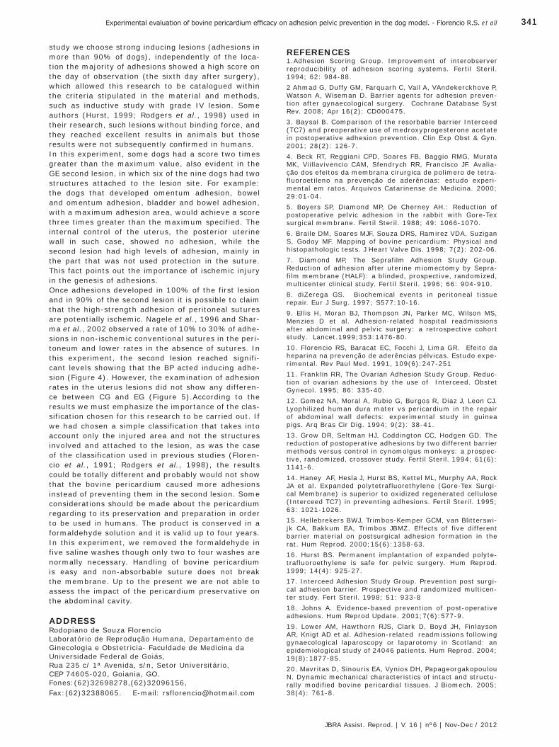
341341
JBRA Assist. Reprod. | V. 16 | nº6 | Nov-Dec / 2012
Experimental evaluation of bovine pericardium efficacy on adhesion pelvic prevention in the dog model. - Florencio R.S. et all
study we choose strong inducing lesions (adhesions in more than 90% of dogs), independently of the loca-tion the majority of adhesions showed a high score on the day of observation (the sixth day after surgery), which allowed this research to be catalogued within the criteria stipulated in the material and methods, such as inductive study with grade IV lesion. Some authors (Hurst, 1999; Rodgers et al., 1998) used in their research, such lesions without binding force, and they reached excellent results in animals but those results were not subsequently confirmed in humans.In this experiment, some dogs had a score two times greater than the maximum value, also evident in the GE second lesion, in which six of the nine dogs had two structures attached to the lesion site. For example: the dogs that developed omentum adhesion, bowel and omentum adhesion, bladder and bowel adhesion, with a maximum adhesion area, would achieve a score three times greater than the maximum specified. The internal control of the uterus, the posterior uterine wall in such case, showed no adhesion, while the second lesion had high levels of adhesion, mainly in the part that was not used protection in the suture. This fact points out the importance of ischemic injury in the genesis of adhesions. Once adhesions developed in 100% of the first lesion and in 90% of the second lesion it is possible to claim that the high-strength adhesion of peritoneal sutures are potentially ischemic. Nagele et al., 1996 and Shar-ma et al., 2002 observed a rate of 10% to 30% of adhe-sions in non-ischemic conventional sutures in the peri-toneum and lower rates in the absence of sutures. In this experiment, the second lesion reached signifi-cant levels showing that the BP acted inducing adhe-sion (Figure 4). However, the examination of adhesion rates in the uterus lesions did not show any differen-ce between CG and EG (Figure 5).According to the results we must emphasize the importance of the clas-sification chosen for this research to be carried out. If we had chosen a simple classification that takes into account only the injured area and not the structures involved and attached to the lesion, as was the case of the classification used in previous studies (Floren-cio et al., 1991; Rodgers et al., 1998), the results could be totally different and probably would not show that the bovine pericardium caused more adhesions instead of preventing them in the second lesion. Some considerations should be made about the pericardium regarding to its preservation and preparation in order to be used in humans. The product is conserved in a formaldehyde solution and it is valid up to four years. In this experiment, we removed the formaldehyde in five saline washes though only two to four washes are normally necessary. Handling of bovine pericardium is easy and non-absorbable suture does not break the membrane. Up to the present we are not able to assess the impact of the pericardium preservative on the abdominal cavity.
ADDRESSRodopiano de Souza Florencio Laboratório de Reprodução Humana, Departamento de Ginecologia e Obstetricia- Faculdade de Medicina da Universidade Federal de Goiás, Rua 235 c/ 1ª Avenida, s/n, Setor Universitário,CEP 74605-020, Goiania, GO. Fones:(62)32698278,(62)32096156, Fax:(62)32388065. E-mail: [email protected]
REFERENCES1.Adhesion Scoring Group. Improvement of interobserver reproducibility of adhesion scoring systems. Fertil Steril. 1994; 62: 984-88.2 Ahmad G, Duffy GM, Farquarh C, Vail A, VAndekerckhove P, Watson A, Wiseman D. Barrier agents for adhesion preven-tion after gynaecological surgery. Cochrane Database Syst Rev. 2008; Apr 16(2): CD000475.3. Baysal B. Comparison of the resorbable barrier Interceed (TC7) and preoperative use of medroxyprogesterone acetate in postoperative adhesion prevention. Clin Exp Obst & Gyn. 2001; 28(2): 126-7.4. Beck RT, Reggiani CPD, Soares FB, Baggio RMG, Murata MK, Viillavivencio CAM, Sfendrych RR, Francisco JF. Avalia-ção dos efeitos da membrana cirurgica de polimero de tetra-fluoroetileno na prevenção de aderências: estudo experi-mental em ratos. Arquivos Catarinense de Medicina. 2000; 29:01-04.5. Boyers SP, Diamond MP, De Cherney AH.: Reduction of postoperative pelvic adhesion in the rabbit with Gore-Tex surgical membrane. Fertil Steril. 1988; 49: 1066-1070.6. Braile DM, Soares MJF, Souza DRS, Ramirez VDA, Suzigan S, Godoy MF. Mapping of bovine pericardium: Physical and histopathologic tests. J Heart Valve Dis. 1998; 7(2): 202-06.7. Diamond MP, The Seprafilm Adhesion Study Group. Reduction of adhesion after uterine miomectomy by Sepra-film membrane (HALF): a blinded, prospective, randomized, multicenter clinical study. Fertil Steril. 1996; 66: 904-910.8. diZerega GS. Biochemical events in peritoneal tissue repair. Eur J Surg. 1997; S577:10-16. 9. Ellis H, Moran BJ, Thompson JN, Parker MC, Wilson MS, Menzies D et al. Adhesion-related hospital readmissions after abdominal and pelvic surgery: a retrospective cohort study. Lancet.1999;353:1476-80. 10. Florencio RS, Baracat EC, Focchi J, Lima GR. Efeito da heparina na prevenção de aderências pélvicas. Estudo expe-rimental. Rev Paul Med. 1991, 109(6):247-25111. Franklin RR, The Ovarian Adhesion Study Group. Reduc-tion of ovarian adhesions by the use of Interceed. Obstet Gynecol. 1995; 86: 335-40.12. Gomez NA, Moral A, Rubio G, Burgos R, Diaz J, Leon CJ. Lyophilized human dura mater vs pericardium in the repair of abdominal wall defects: experimental study in guinea pigs. Arq Bras Cir Dig. 1994; 9(2): 38-41.13. Grow DR, Seltman HJ, Coddington CC, Hodgen GD. The reduction of postoperative adhesions by two different barrier methods versus control in cynomolgus monkeys: a prospec-tive, randomized, crossover study. Fertil Steril. 1994; 61(6): 1141-6.14. Haney AF, Hesla J, Hurst BS, Kettel ML, Murphy AA, Rock JA et al. Expanded polytetrafluorethylene (Gore-Tex Surgi-cal Membrane) is superior to oxidized regenerated cellulose (Interceed TC7) in preventing adhesions. Fertil Steril. 1995; 63: 1021-1026. 15. Hellebrekers BWJ, Trimbos-Kemper GCM, van Blitterswi-jk CA, Bakkum EA, Trimbos JBMZ. Effects of five different barrier material on postsurgical adhesion formation in the rat. Hum Reprod. 2000;15(6):1358-63.16. Hurst BS. Permanent implantation of expanded polyte-trafluoroethylene is safe for pelvic surgery. Hum Reprod. 1999; 14(4): 925-27.17. Interceed Adhesion Study Group. Prevention post surgi-cal adhesion barrier. Prospective and randomized multicen-ter study. Fert Steril. 1998; 51: 933-818. Johns A. Evidence-based prevention of post-operative adhesions. Hum Reprod Update. 2001;7(6):577-9.19. Lower AM, Hawthorn RJS, Clark D, Boyd JH, Finlayson AR, Knigt AD et al. Adhesion-related readmissions following gynaecological laparoscopy or laparotomy in Scotland: an epidemiological study of 24046 patients. Hum Reprod. 2004; 19(8):1877-85.20. Mavritas D, Sinouris EA, Vynios DH, Papageorgakopoulou N. Dynamic mechanical characteristics of intact and structu-rally modified bovine pericardial tissues. J Biomech. 2005; 38(4): 761-8.
JBRA6_150213.indb 341 15/02/2013 17:17:20
342 Artigo Original
JBRA Assist. Reprod. | V. 16 | nº6 | Nov-Dec / 2012
21. Montz FJ, Monk BJ, Lacy SM. The Gore-Tex Surgical Membrane: effectiveness as a barrier to inhibit postradical pelvic surgery adhesions in a porcine model. Gynecol Oncol. 1992; 45: 290-293.22. Mutsaers SE. Mesothelial cells: Their structure, function and role in serosal repair. Respirology. 2002; 7: 171-91.23. Nagele F, Kurz C, Staundach A, Steinar H, Grunberger W, A et al. Closure or nonclosure of the visceral perito-neum at cesarean delivery. Am J Obstet Gynecol. 1996; 174:1366-70.24. Nordic Adhesion Prevention Study Group. The efficacy of Interceed for prevention of reformation of postoperative adhesion on ovaries, fallopian tubes, and fimbrias in micro-surgical operations for fertility: a multicenter study. Fertil Steril. 1995; 65: 709-714.25. Oelsner G, Sivan E, Goldenberg M, Carp H, Admon D, Maschiach S. Should lysis of adhesions be performed when in-vitro fertilization and embryo transfer are available? Hum Reprod. 1994;9(12):2339-41.26. Parker MC; Wilson MS; Menzies D; Sunderland G; Thompson JN; Clark DN et al. Colorectal surgery: the risk and burden of adhesion-related complications. Colorectal Disease. 2004; 6:506-11.27. Reid RL, Hahn PM, Spence JEH, Tulandi T, Yuspe AA, Wiseman DM. A randomized clinical trial of oxidized regene-rated cellulose adhesion barrier (Interceed,TC7)* alone or in combination with heparin. Fertil Steril. 1997; 67(1); 23-9.
28. Rodgers KR, Cohn D, Hotovely A, Pines E, Diamond MP, diZerega G. Evaluation of polyethylene glycol/polylactic acid films in the prevention of adhesions in the rabbit adhesion formation and reformation sidewall models. Fert Steril. 1998;69:403-8.29. Saravielos H, Li TC. Post-operative adhesions after lapa-roscopic electrosurgical treatment for polycystic ovarian syndrome with the application of Interceed to one ovary: a prospective randomized controlled study. Hum Reprod. 1996; 11(5): 992-97.30. Sharma JB. Closure of the peritoneum is not necessary in obstetric and gynecologic surgeries. Acta Obstet Gynecol Scand. 2002; 81:80-87.31. Svanes Fevang BT, Fevang J, Lie SA, Soreide Odd, Svanes K, Viste A. Long-term prognosis after operation for adhesive small bowel obstruction. Ann Surg. 2004; 240: 193-201. 32. The Surgical Membrane Study Group. Prophylaxis of pelvic sidewall adhesion with Gore-Tex Surgical Membrane: a multicenter clinical investigation. Fertil Steril. 1992; 57: 921-923. 33. The Miomectomy Adhesion Multicenter Study Group. An expanded polytetrafluoroethylene barrier (Gore-Tex Surgical Membrane) reduces post-myomectomy adhesion formation. Fertil Steril. 1995; 63: 491-493. 34. Wilson MS; Menzies D; Knight AD; Crowe AM. Demons-trating the clinical and cost effectiveness of adhesion reduc-tion strategies. Colorectal Disease. 2002;4: 355-60.
JBRA6_150213.indb 342 15/02/2013 17:17:20
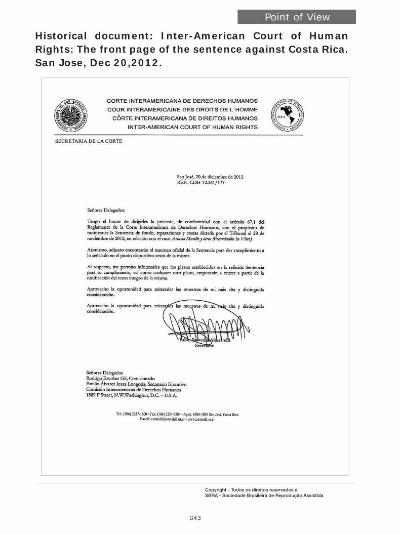
343
Point of View
Copyright - Todos os direitos reservados a SBRA - Sociedade Brasileira de Reprodução Assistida
Historical document: Inter-American Court of Human Rights: The front page of the sentence against Costa Rica. San Jose, Dec 20,2012.
JBRA6_150213.indb 343 15/02/2013 17:17:21

344
Point of View
Copyright - Todos os direitos reservados a SBRA - Sociedade Brasileira de Reprodução Assistida
COSTA RICA, again part of the Latin American Community of ART.
Maria do Carmo Borges de Souza¹ and Roberto Coco²
1 President of Redlara2 Vice President of Redlara
It was with great pleasure and enthusiasm that we received the result of the judgment of the Court of Human Rights with regard to the question that was in force in Costa Rica. In the judgment of the Court on November 28 last, won the reason, the desire to provide an offspring when not possible by natural means.We recognize the efforts of all who participated in this fight, in order to welcome the feelings of a population that suffered the limitation of both their human and reproductive rights.Carlos Valerio Monge and his fellow public defenders were valiant in Costa Rica and there has been an extraordinary effort in unity uttering opinions of experts, directly participating in public events in Costa Rica, producing opinions and subsidizing the Court with scientific data, giving us all one dimension of what we can do when together and united.Thanks Fernando Zegers, impeccable referee appointed by WHO and thank you Maria Teresa Urbina, Director of Redlara Region of Venezuela, Colombia and the Caribbean, for their tireless work. Thanks to Ian Cooke, Sheryl Vanderpoel, Santiago Munné, Klaus Wiemer, Vera Raposo and many others who have pledged and glowed, full of enthusiasm to cooperate.As points Fernando Zegers, the recent decision of the Court of Human Rights requires Costa Rica to re-establish the practice of assisted reproduction and make it enforceable to infertile people. Moreover, it forces the government to provide this service to individuals by a national health program, the latter on the basis of protecting the right to non-discrimination.Perhaps most relevant to this resounding failure is that explicitly states:1 - Reproductive rights are part of human rights and the right to found a family and the right to live autonomy privacy must be protected by the countries.2 - Recognize infertility as a disease of the reproductive system (WHO) that generates a disability. In this context, the prohibition to perform IVF constitute discrimination against persons with a disability3 - States that the embryo is not subject to the right to life as are the people. That protecting the embryo is exer-cised through the protection they are entitled to pregnant women (Article 4.1 of the Convention). Explicitly recog-nizes that conception begins at implantation of the embryo and not the moment of fertilization. The owner of the right to life is the pregnant woman and not the embryo.4 - Sets that the pre implantation embryos, ie generated by in vitro fertilization techniques, are not subject to current law as individuals. Moreover, explicitly recognizes that the acquisition of status of person, is established through a gradual process and not set immediately with fertilization.5 – Provides that the right of women to be protected by international right, cannot be affected by laws protecting embryos. That is, explicitly states the woman as head of law and not the embryo.The ruling also requires the government to pay the costs of the victims and to provide them with psychological support for four years. A complete disclosure in the media and in the Official Journal must be provided and it forces Costa Rica to train and educate the legal persons regarding reproductive rights.We are at a crucial moment in the history of the fight for reproductive rights, especially to women in the Americas. The Presidency and Board of Directors of REDLARA are very proud of our institution’s effective participation in this process as an active part of a moment in history.
JBRA6_150213.indb 344 15/02/2013 17:17:21
345
JBRA6_150213.indb 345 15/02/2013 17:17:23
346
Laboratórios Ferring - BrasilPça. São Marcos, 624 - 05455-050 - São Paulo - BrasilPABX - 55 11 3024.7500
MEN AN 112 Maio 2012
Menopur® (menotropina - LH 75 U.I. + FSH 75 U.I.).
USO ADULTO. INDICAÇÕES Mulheres: Esterilidade com insufi ciência ovariana hipo – ou normogonadotrófi ca: estimulação do crescimento folicular, incluindo mulheres com síndrome de ovário policístico que não responderam ao tratamento com citrato de clomifeno. Homens: Esterilidade com hipogonadismo hipo – ou normogonadotrófi co: em combinação com HCG, para estimular a espermatogênese. CONTRAINDICAÇÕES Em Mulheres: tumores na glândula pituitária ou no hipotálamo e no útero, ovários e mamas; gravidez e lactação; sangramento ginecológico de etiologia desconhecida; hipersensibilidade aos componentes da fórmula. aumento dos ovários ou cistos que não tenham sido causados por síndrome de ovário policístico; níveis altos de LH e FSH indicando uma insufi ciência ovariana primária. Menopur® não deve ser administrado nos casos de: falência primária ovariana; má-formação de órgãos sexuais incompatíveis com a gravidez; tumor fi broide do útero incompatível com a gravidez. Em Homens: câncer de próstata; tumores no testículo. As seguintes condições devem ser tratadas antes do início da terapia: disfunções da glândula tireoide e do córtex da glândula supra-renal; hiperprolactinemia; tumores na glândula pituitária ou no diencéfalo (hipotálamo). CUIDADOS E ADVERTÊNCIAS Não deve ser utilizado para induzir a ovulação em mulheres cujos ovários foram involuntariamente hiperestimulados. A terapia requer monitorização da resposta ovariana verifi cada apenas pela ultrassonografi a ou em combinação com a mensuração do nível de estradiol sanguíneo. Alguns pacientes podem apresentar baixa resposta. Pacientes que estão sob estímulo do crescimento folicular para técnicas de reprodução assistida podem apresentar aumento ovariano ou desenvolver hiperestimulação. Uso em idosos e crianças Menopur® é um medicamento de uso adulto. Gravidez e Lactação Menopur® não deve ser utilizado por mulheres grávidas ou lactentes. INTERAÇÕES MEDICAMENTOSAS O uso concomitante de Menopur® com o citrato de clomifeno pode aumentar a resposta folicular. Quando for utilizado um agonista de GnRH, uma dose mais alta de Menopur® deve ser necessária para atingir uma resposta folicular adequada. REAÇÔES ADVERSAS dores abdominais, náusea, aumento do abdômen, dor e reação no local da injeção, cefaleia, hiperestimulação dos ovários, dor pélvica, febre, ascite, hidrotórax, oligúria, hipotensão e fenômenos tromboembólicos. Em casos muito raros, pode causar a formação de anticorpos tornando o tratamento inefi caz. POSOLOGIA Para indução do crescimento folicular em procedimentos de baixa complexidade (coito programado e inseminação intrauterina) em mulheres normo ou hipogonadotrófi cas a dose depende da reação ovariana, a qual deve ser defi nida através de exames ultrassonográfi cos dos ovários e mensuração dos níveis de estradiol. Se a dosagem de Menopur® for muito alta, podem ocorrer crescimentos foliculares uni e bilaterais múltiplos. Em geral, a terapia é iniciada dentro dos 7 primeiros dias do ciclo menstrual com uma dosagem inicial diária correspondente a 75 - 150 U.I. de FSH. Se os ovários não respondem, a dosagem pode ser gradativamente aumentada até surgirem evidências de aumento da secreção de estradiol e de crescimento folicular. O ajuste na dose não deve ser feito em frequência maior que 7 dias. O tratamento com a mesma dosagem de Menopur® é continuado até atingir-se um nível sérico de estradiol pré-ovulatório. Se o nível aumentar muito rapidamente, a dosagem deve ser reduzida. A dose máxima diária não deve ultrapassar 225 U.I. Caso a paciente não responda após 4 semanas de tratamento, o tratamento deve ser reiniciado com uma dosagem inicial maior do que a do ciclo anterior. Para induzir a ovulação, após uma resposta ótima do tratamento com Menopur®, utiliza-se 5.000 ou 10.000 U.I. de HCG injetado por via intramuscular, 1 dia após a última administração de Menopur®. Importante: recomenda-se que a paciente tenha relação sexual no dia e no dia seguinte da administração do HCG. Como alternativa, a inseminação intrauterina pode ser realizada.OBSERVAÇÃO: Após a administração de uma dose muito alta de Menopur®, a administração subsequente de HCG pode causar uma hiperestimulação involuntária dos ovários. Neste caso, o tratamento deve ser interrompido tanto com Menopur® ou com HCG e a paciente deverá utilizar um método anticoncepcional ou evitar relações sexuais até o início da próxima menstruação. A dosagem de Menopur® em programas de fertilização assistida (FIV/ICSI) pode variar de pessoa para pessoa. De um modo geral recomendam-se os seguintes esquemas posológicos: Quando for utilizado agonista de GnRH em depósito, a terapia com Menopur® deve ser iniciada aproximadamente 2 semanas após o início do tratamento com o agonista. A dose inicial recomendada de Menopur® é 150 – 225 U.I. por, pelo menos, 5 dias iniciais do tratamento. Baseado na monitorização clínica (ultrassonografi a do ovário em combinação ou não com a mensuração do nível de estradiol sanguíneo) as doses subsequentes devem ser ajustadas de acordo com a resposta individual e não devem exceder a quantidade de 150 U.I. por ajuste. A dosagem máxima diária não deve exceder 450 U.I. por dia e na maioria dos casos não é recomendada a utilização acima de 20 dias. Caso não seja utilizado o agonista de GnRH, a terapia com Menopur® deve ser iniciada no 2° ou 3° dia do ciclo menstrual. É recomendada a utilização de esquemas de variação de doses acima citadas. Quando um número adequado de folículos atingir um tamanho apropriado, uma única injeção de 10.000 U.I. de HCG deve ser administrada para induzir a maturação folicular fi nal, na preparação da aspiração de oócitos. As pacientes devem ser cuidadosamente monitoradas por, pelo menos, 2 semanas após a administração de HCG. Caso seja obtida uma resposta em excesso de Menopur®, deve-se interromper o tratamento e descontinuar o uso de HCG e as pacientes devem utilizar um método contraceptivo de barreira (por exemplo: camisinha) ou evitar relações sexuais até iniciar-se a próxima menstruação. No Homem: Inicialmente, 1.000 – 3.000 U.I. de HCG são administrados 3 vezes por semana, até atingir-se um nível sérico de testosterona normal. Então, 75 - 150 U.I. de Menopur® são administradas 3 vezes por semana, por alguns meses e de acordo com o critério médico. Orientar a/o paciente quanto ao protocolo de uso, para que não haja comprometimento do sucesso terapêutico.Venda sob prescrição médica. Material de uso exclusivo à classe médica. A persistirem os sintomas, o médico deverá ser consultado. Reg. MS: 1.2876.0011 Farm. Resp.: Helena Satie Komatsu - CRF/SP: 19.714 - Laboratórios Ferring Ltda. Praça São Marcos, 624 - 05455-050 - São Paulo – SP / CNPJ: 74.232.034/0001-48 / SAC: 0800 772 4656.
Contraindicação: Para mulheres com tumores no útero, nos ovários e nas mamas. Interações medicamentosas: O uso concomitante com o citrato de clomifeno pode aumentar a resposta folicular.
Referências: 1 Ziebe, S. et al. Infl uence of ovarian stimulation with HP-hMG or recombinant FSH on embryo quality parameters in patient undergoing IVF. Human Reproduction, 22(9): 2404-2413, 2007. 2 Weghofer, A. the impact of LH-containing gonadotropin on diploidy rates in preimplantation embryos: long protocol stimulation. Human Reproduction, 23(3): 499-503, 2008. 3 Al-Inany HG, et al. Effi cacy and safety of human menopausal gonadotrophins versus recombinant FSH: a meta-analysis.Reprod Biomed OnLine.16:81-88, 2008. 4 Coomarasamy, A. Urinary hMG versus recombinant FSH for controlled ovarian hyperstimulation following an agonist long down-regulation protocol in IVf or ICSI treatment: a systematic review and meta-analysis. Human Reproduction, 23(2): 310-315, 2008. 5 Wely, V. et al. Recombinant versus urinary gonadotrophin for ovarian stimulation in assisted reproductive technology cycles. Cochrane Database Syst Rev. 16(2):CD005354. Review, 2011. 6 Al-Inany HG. et al. Highly purifi ed hMG achieves better pregnancy rates in IVF cycles but not ICSI cycles compared with recombinant FSH: a meta-analysis. Gynecol Endocrinol.25(6):372-8, 2009. 7 Platteau P. et al. Highly purifi ed HMG versus recombinant FSH for ovarian stimulation in IVF cycles. Reprod Biomed Online17(2):190-8, 2008. 8 Andersen, AN. et al. Clinical outcome following stimulation with highly purifi ed hMG or recombinant FSH in patients undergoing IVF: a randomized assessor-blind controlled trial. Human Reproduction, 21(12): 3217-3227, 2006. 9 Bosch, E. et al. Highly purifi ed hMG versus recombinant FSH in ovarian hyperstimulation with GnRH antagonists - a randomized study. Human Reproduction, 23(10): 2346-2351, 2008. 10 Jee BC. et al. Clinical effi cacy of highly purifi ed hMG versus recombinant FSH in IVF/ICSI cycles: a meta-analysis. Gynecol Obstet Invest., 70(2):132-7, 2010.
anuncio 17 05 a.indd 2 18/05/2012 14:49:15
EventosEventos2012
SETEMBRO14 a 17 de setembro 63rd Annual Meeting of the German Society of Urology (DGU)Hamburg – Germany Organisation:INTERPLAN E-mail:[email protected] Website:www.dgu-kongress.de/index.php?id=571
OUTUBRO7 a 12 de outubroXX FIGO WORLD CONGRESS OF GYNECOLOGYAND OBSTETRICS http://www.figo2012.org/fellowship/
10 a 14 de outubroXIV Simpósio Brasileiro de Patologia do Trato Genital Inferior e Colposcopiae V COLPOVIXLocal: Centro de Convençõesde Vitória – ESRealização: SOGOEShttp://www.sogoes.com.br
26 a 26 de outubro84th Congress of the SIU –Rome – ItalyE-mail:[email protected]:www.siu.it
25 a 27 de outubro19º Congresso Baianode Obstetrícia e GinecologiaLocal: Bahia Pestana HotelRealização: SOGIBAhttp://www.sogiba.com.br/
28 a 31 de outubroXVI Congresso Sul-Brasileiro de Ginecologia e Obstetrícia I Jornada Sul-Brasileira de MastologiaCostão do SantinhoResort - Florianópolis
ATENÇÃO:Informamos que a partir de janeiro de 2013, todo o conteúdo das futuras publicações do Jornal Brasileiro de Reprodução Assistida será em inglês.
JBRA - Assisted Reproduction
INDEXADO NAS SEGUINTES BASES DE DADOS – Indexed on the following databases:CompendexEMBASEExcepta MédicaGeobase
PERIODICA (México)Plataforma SCImago Journal & Country RankPORTAL DE PERIÓDICOS DA CAPESScopus (Holanda)
Jornalista Responsável: Heber Maia – MTb 31.660
Produção Editorial e Gráfica: AlamTec Ciência Médica Editorial LTDARua das Roseiras, 464CEP 03144-090 - São Paulo-SPTel/Fax: (11) 2341-8045e-mail: [email protected]
Endereço para Correspondência: Dra. Maria do Carmo Borges de Souza Av. das Américas, 4666 - Sl. 312 / 313Barra da Tijuca - RJ CEP 22649-900E-mail: [email protected]: (21) 2430-9060Fax: (21) 2430-9070
ÓRGÃO DE DIVULGAÇÃO DA SOCIEDADE BRASILEIRA DE REPRODUÇÃO ASSISTIDA E DA REDE LATINOAMERICANA DE REPRODUÇÃO ASSISTIDA
ISSN: 1517-5693 - V. 16 | nº1 | Jan-Feb / 2012
Você. Nós. Somos os pais da fertilidade
Veja a obra de arte que fizemosjuntos.
SAC Merck Serono: 0800.113320
Anúncio veiculado em Maio de 2011.
Merck Serono é uma divisão da Merck.
JBRA_01_2012_AlamTec_260712.indb 3 27/07/12 16:19
2013
MARÇODia 7 a 9 de Março de 2013Best of ESHRE & ASRMAtlantis, Paradise Island, BahamasWebsite: www.asrm.org/Atlantis2013/
JULHO7 a 10 de JulhoEshre Annual MeetingLondon, United KingdomWebsite: www.eshre2013.eu
JBRA6_150213.indb 346 15/02/2013 17:17:25
Laboratórios Ferring - BrasilPça. São Marcos, 624 - 05455-050 - São Paulo - BrasilPABX - 55 11 3024.7500
MEN AN 112 Maio 2012
Menopur® (menotropina - LH 75 U.I. + FSH 75 U.I.).
USO ADULTO. INDICAÇÕES Mulheres: Esterilidade com insufi ciência ovariana hipo – ou normogonadotrófi ca: estimulação do crescimento folicular, incluindo mulheres com síndrome de ovário policístico que não responderam ao tratamento com citrato de clomifeno. Homens: Esterilidade com hipogonadismo hipo – ou normogonadotrófi co: em combinação com HCG, para estimular a espermatogênese. CONTRAINDICAÇÕES Em Mulheres: tumores na glândula pituitária ou no hipotálamo e no útero, ovários e mamas; gravidez e lactação; sangramento ginecológico de etiologia desconhecida; hipersensibilidade aos componentes da fórmula. aumento dos ovários ou cistos que não tenham sido causados por síndrome de ovário policístico; níveis altos de LH e FSH indicando uma insufi ciência ovariana primária. Menopur® não deve ser administrado nos casos de: falência primária ovariana; má-formação de órgãos sexuais incompatíveis com a gravidez; tumor fi broide do útero incompatível com a gravidez. Em Homens: câncer de próstata; tumores no testículo. As seguintes condições devem ser tratadas antes do início da terapia: disfunções da glândula tireoide e do córtex da glândula supra-renal; hiperprolactinemia; tumores na glândula pituitária ou no diencéfalo (hipotálamo). CUIDADOS E ADVERTÊNCIAS Não deve ser utilizado para induzir a ovulação em mulheres cujos ovários foram involuntariamente hiperestimulados. A terapia requer monitorização da resposta ovariana verifi cada apenas pela ultrassonografi a ou em combinação com a mensuração do nível de estradiol sanguíneo. Alguns pacientes podem apresentar baixa resposta. Pacientes que estão sob estímulo do crescimento folicular para técnicas de reprodução assistida podem apresentar aumento ovariano ou desenvolver hiperestimulação. Uso em idosos e crianças Menopur® é um medicamento de uso adulto. Gravidez e Lactação Menopur® não deve ser utilizado por mulheres grávidas ou lactentes. INTERAÇÕES MEDICAMENTOSAS O uso concomitante de Menopur® com o citrato de clomifeno pode aumentar a resposta folicular. Quando for utilizado um agonista de GnRH, uma dose mais alta de Menopur® deve ser necessária para atingir uma resposta folicular adequada. REAÇÔES ADVERSAS dores abdominais, náusea, aumento do abdômen, dor e reação no local da injeção, cefaleia, hiperestimulação dos ovários, dor pélvica, febre, ascite, hidrotórax, oligúria, hipotensão e fenômenos tromboembólicos. Em casos muito raros, pode causar a formação de anticorpos tornando o tratamento inefi caz. POSOLOGIA Para indução do crescimento folicular em procedimentos de baixa complexidade (coito programado e inseminação intrauterina) em mulheres normo ou hipogonadotrófi cas a dose depende da reação ovariana, a qual deve ser defi nida através de exames ultrassonográfi cos dos ovários e mensuração dos níveis de estradiol. Se a dosagem de Menopur® for muito alta, podem ocorrer crescimentos foliculares uni e bilaterais múltiplos. Em geral, a terapia é iniciada dentro dos 7 primeiros dias do ciclo menstrual com uma dosagem inicial diária correspondente a 75 - 150 U.I. de FSH. Se os ovários não respondem, a dosagem pode ser gradativamente aumentada até surgirem evidências de aumento da secreção de estradiol e de crescimento folicular. O ajuste na dose não deve ser feito em frequência maior que 7 dias. O tratamento com a mesma dosagem de Menopur® é continuado até atingir-se um nível sérico de estradiol pré-ovulatório. Se o nível aumentar muito rapidamente, a dosagem deve ser reduzida. A dose máxima diária não deve ultrapassar 225 U.I. Caso a paciente não responda após 4 semanas de tratamento, o tratamento deve ser reiniciado com uma dosagem inicial maior do que a do ciclo anterior. Para induzir a ovulação, após uma resposta ótima do tratamento com Menopur®, utiliza-se 5.000 ou 10.000 U.I. de HCG injetado por via intramuscular, 1 dia após a última administração de Menopur®. Importante: recomenda-se que a paciente tenha relação sexual no dia e no dia seguinte da administração do HCG. Como alternativa, a inseminação intrauterina pode ser realizada.OBSERVAÇÃO: Após a administração de uma dose muito alta de Menopur®, a administração subsequente de HCG pode causar uma hiperestimulação involuntária dos ovários. Neste caso, o tratamento deve ser interrompido tanto com Menopur® ou com HCG e a paciente deverá utilizar um método anticoncepcional ou evitar relações sexuais até o início da próxima menstruação. A dosagem de Menopur® em programas de fertilização assistida (FIV/ICSI) pode variar de pessoa para pessoa. De um modo geral recomendam-se os seguintes esquemas posológicos: Quando for utilizado agonista de GnRH em depósito, a terapia com Menopur® deve ser iniciada aproximadamente 2 semanas após o início do tratamento com o agonista. A dose inicial recomendada de Menopur® é 150 – 225 U.I. por, pelo menos, 5 dias iniciais do tratamento. Baseado na monitorização clínica (ultrassonografi a do ovário em combinação ou não com a mensuração do nível de estradiol sanguíneo) as doses subsequentes devem ser ajustadas de acordo com a resposta individual e não devem exceder a quantidade de 150 U.I. por ajuste. A dosagem máxima diária não deve exceder 450 U.I. por dia e na maioria dos casos não é recomendada a utilização acima de 20 dias. Caso não seja utilizado o agonista de GnRH, a terapia com Menopur® deve ser iniciada no 2° ou 3° dia do ciclo menstrual. É recomendada a utilização de esquemas de variação de doses acima citadas. Quando um número adequado de folículos atingir um tamanho apropriado, uma única injeção de 10.000 U.I. de HCG deve ser administrada para induzir a maturação folicular fi nal, na preparação da aspiração de oócitos. As pacientes devem ser cuidadosamente monitoradas por, pelo menos, 2 semanas após a administração de HCG. Caso seja obtida uma resposta em excesso de Menopur®, deve-se interromper o tratamento e descontinuar o uso de HCG e as pacientes devem utilizar um método contraceptivo de barreira (por exemplo: camisinha) ou evitar relações sexuais até iniciar-se a próxima menstruação. No Homem: Inicialmente, 1.000 – 3.000 U.I. de HCG são administrados 3 vezes por semana, até atingir-se um nível sérico de testosterona normal. Então, 75 - 150 U.I. de Menopur® são administradas 3 vezes por semana, por alguns meses e de acordo com o critério médico. Orientar a/o paciente quanto ao protocolo de uso, para que não haja comprometimento do sucesso terapêutico.Venda sob prescrição médica. Material de uso exclusivo à classe médica. A persistirem os sintomas, o médico deverá ser consultado. Reg. MS: 1.2876.0011 Farm. Resp.: Helena Satie Komatsu - CRF/SP: 19.714 - Laboratórios Ferring Ltda. Praça São Marcos, 624 - 05455-050 - São Paulo – SP / CNPJ: 74.232.034/0001-48 / SAC: 0800 772 4656.
Contraindicação: Para mulheres com tumores no útero, nos ovários e nas mamas. Interações medicamentosas: O uso concomitante com o citrato de clomifeno pode aumentar a resposta folicular.
Referências: 1 Ziebe, S. et al. Infl uence of ovarian stimulation with HP-hMG or recombinant FSH on embryo quality parameters in patient undergoing IVF. Human Reproduction, 22(9): 2404-2413, 2007. 2 Weghofer, A. the impact of LH-containing gonadotropin on diploidy rates in preimplantation embryos: long protocol stimulation. Human Reproduction, 23(3): 499-503, 2008. 3 Al-Inany HG, et al. Effi cacy and safety of human menopausal gonadotrophins versus recombinant FSH: a meta-analysis.Reprod Biomed OnLine.16:81-88, 2008. 4 Coomarasamy, A. Urinary hMG versus recombinant FSH for controlled ovarian hyperstimulation following an agonist long down-regulation protocol in IVf or ICSI treatment: a systematic review and meta-analysis. Human Reproduction, 23(2): 310-315, 2008. 5 Wely, V. et al. Recombinant versus urinary gonadotrophin for ovarian stimulation in assisted reproductive technology cycles. Cochrane Database Syst Rev. 16(2):CD005354. Review, 2011. 6 Al-Inany HG. et al. Highly purifi ed hMG achieves better pregnancy rates in IVF cycles but not ICSI cycles compared with recombinant FSH: a meta-analysis. Gynecol Endocrinol.25(6):372-8, 2009. 7 Platteau P. et al. Highly purifi ed HMG versus recombinant FSH for ovarian stimulation in IVF cycles. Reprod Biomed Online17(2):190-8, 2008. 8 Andersen, AN. et al. Clinical outcome following stimulation with highly purifi ed hMG or recombinant FSH in patients undergoing IVF: a randomized assessor-blind controlled trial. Human Reproduction, 21(12): 3217-3227, 2006. 9 Bosch, E. et al. Highly purifi ed hMG versus recombinant FSH in ovarian hyperstimulation with GnRH antagonists - a randomized study. Human Reproduction, 23(10): 2346-2351, 2008. 10 Jee BC. et al. Clinical effi cacy of highly purifi ed hMG versus recombinant FSH in IVF/ICSI cycles: a meta-analysis. Gynecol Obstet Invest., 70(2):132-7, 2010.
anuncio 17 05 a.indd 2 18/05/2012 14:49:15JBRA6_150213.indb 347 15/02/2013 17:17:25
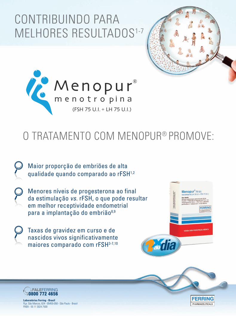
Laboratórios Ferring - BrasilPça. São Marcos, 624 - 05455-050 - São Paulo - BrasilPABX - 55 11 3024.7500
CONTRIBUINDO PARA MELHORES RESULTADOS1-7
O TRATAMENTO COM MENOPUR® PROMOVE:
Maior proporção de embriões de alta qualidade quando comparado ao rFSH1,2
Menores níveis de progesterona ao final da estimulação vs. rFSH, o que pode resultar em melhor receptividade endometrial para a implantação do embrião8,9
Taxas de gravidez em curso e de nascidos vivos significativamente maiores comparado com rFSH3-7,10
urofolitropina
anuncio 17 05 a.indd 1 18/05/2012 14:49:14JBRA6_150213.indb 348 15/02/2013 17:17:25