3d electron microscopy - thermo fisher scientific › tfs-assets › msd › ... · face and...
TRANSCRIPT
3D Electron Microscopy Exploring cells and tissue in their natural spatial context

Cover image: Rat brain, sample courtesy of Grahame Knott. This page: Neuron network from rat brain.
Imaging by Thermo Scientific Volumescope SEM. Data visualization by Thermo Scientific Amira Software.

About Thermo Fisher ScientificAs the world leader in serving science, we serve both academic and industrial life sciences researchers, providing an unmatched combination of complete workflow solutions ranging from cryo-EM structural determination of macromolecular complexes and protein sociology, in the native state, to reconstruction of 3D architecture of tissues and cells. Our solutions help researchers unlock the mysteries of underlying protein function and cellular process and bridge the gap between basic science and translational therapeutics.
Below: Mouse heart muscle, specimen courtesy of Madesh Muniswamy.
Imaging by Thermo Scientific Volumescope SEM. Data visualization by Thermo Scientific Amira Software.

Three technologies:
IntroductionGroundbreaking advances in large volume electron microscopy are enabling the three-dimensional visualization of specimens with unprecedented detail. This, in turn, is driving an increasing interest in the ultrastructural examination of cells and tissues in their natural spatial context.
Serial section techniques, previously the domain of specialists, are becoming increasingly automated with the development of solutions such as array tomography and serial block-face and focused ion beam scanning electron microscopes. These changes are rapidly broadening the scope of biomedical studies to
which 3D electron microscopy techniques can be applied. The acquisition of comprehensive functional information with ultrastructural details within a three-dimensional space will become routine. The stage is set for a challenging and exciting future.
DualBeam Focused Ion Beam +(AutoSlice&View)
• In situ ion beam milling
• Full automation
• Best data quality
• In situ sectioning
• Full automation
• Largest volumes
• Easiest navigation
• Largest imaging field
• Sample can be stored and re-imaged
Serial Block Face Imaging (VolumeScope 2)
Array Tomography module for Maps Software

SBFI Publications
The number of publications incorporating serial block face imaging (SBFI) has grown significantly over the last several years in a variety of specimen types of various research areas.
BORRETT, S. and HUGHES, L. (2016), Reporting methods for processing and analysis of data from serial block face scanning electron microscopy.
Journal of Microscopy. doi:10.1111/jmi.12377
Data from Jan. 21, 2019, based from Pubmed search: ((serial block face) OR SBFSEM OR SBEM OR SBF-EM OR 3View OR volumescope[all]) AND microscopy

1. A workflow for visualizing human cancer biopsies using large-format electron microscopyRiesterer JL, López CS, Stempinski ES, Williams M, Loftis K, Stoltz K, Thibault G, Lanicault C, Williams T and Gray JW.
bioRxiv 67537; doi:10.1101/67537
The protocols and mounting strategies described in this workflow are compatible with 2D large-format EM mapping, 3D focused ion beam SEM, and serial block-face SEM. The flexibility to use diverse imaging technologies available at most academic institutions makes this workflow useful and applicable for most life science samples.
2. 3D Microstructure of Tendon Collagen Fibrils using Serial Block-Face SEM and a Mechanical Model for Load Transfer.Safa BN, Peloquin JM, Natriello JR, Caplan JL, Elliott DM
bioRxiv 547281; doi: 10.1101/547281
In this study, the author used serial block-face scanning electron microscopy (SBF-SEM) to investigate the three-dimensional microstructure of fibrils in rat tail tendon. They found that tendon fibrils have a complex architecture with many helically wrapped fibrils. This study is significant in that it provides a three-dimensional view of the tendon microstructure and suggests friction between helically wrapped fibrils as a mechanism for load transfer, which is an important aspect of tendon biomechanics.
3. An automated workflow for segmenting single adult cardiac cells from large-volume serial block-face scanning electron microscopy data. Hussain, A., Ghosh, S., Kalkhoran, S.B., Hausenloy, D.J., Hanssen, E., Rajagopal, V.,
Journal of Structural Biology (2018), doi: 10.1016/j.jsb.2018.02.005
This paper presents a new algorithm to automatically segment the myofibrils, mitochondria and nuclei within single adult cardiac cells that are part of a large serial block-face scanning electron microscopy (SBF-SEM) dataset. The algorithm correctly classified pixels within the single cell with 97% accuracy when compared to manual segmentations.
4. Insights on the impact of mitochondrial organization on bioenergetics in high-resolution computational models of cardiac cell architecture.Ghosh S, Tran K, Delbridge LMD, Hickey AJR, Hanssen E, Crampin EJ, Rajagopal V.
PLoS Comput Biol. 2018 Dec 5;14(12):e1006640. doi: 10.1371/journal.pcbi.1006640. eCollection 2018 Dec.
How does the mitochondrial arrangement organized with several mitochondria aggregated into columns of varying sizes affect
the metabolite distributions within cardiomyocytes? And what is its impact on force dynamics? The authors address these questions by employing finite element modeling of cardiac bioenergetics on computational meshes derived from electron microscope images.
5. Mitochondrial Dysfunction Leads to Cortical Under-Connectivity and Cognitive Impairment.Fernandez A, Meechan DW, Karpinski BA, Paronett EM, Bryan CA, Rutz HL, Radin EA, Lubin N, Bonner ER, Popratiloff A, Rothblat LA, Maynard TM and LaMantia AS.
Neuron. 2019 May 2; doi: 10.1016/j.neuron.2019.04.013
The authors assessed cellular, molecular, and developmental origins of under-connectivity and its consequences for cognitive function. They also investigated that anti-oxidant restoration of mitochondrial integrity, cortical connectivity, and cognitive behavior defines oxidative stress as a therapeutic target in neurodevelopmental disorders.
6. Clearance by Microglia Depends on Packaging of Phagosomes into a Unique Cellular Compartment.Villani A, Benjaminsen J, Moritz C, Henke K, Hartmann J, Norlin N, Richter K, Schieber NL, Franke T, Schwab Y, Peri, F.
Developmental Cell Vol. 49(1), pp. 77-88.e7 (2019). doi: 10.1016/j.devcel.2019.02.014
Effective intracellular processing of ingested cells is likely to be crucial for microglial function, but the underlying cellular mechanisms are poorly understood. This work identifies a conserved crucial step in the phagocytic pathway of immune cells and provides a potential entry point for manipulating their behavior in development and disease.
Selected publications
Relationship between the microenvironment (red collagen fibers and blue fibroblasts) surrounding the center red and pink tumor cells is expected to be clinically relevant (Reisterer, 2019, bioRXiv 67537).
10 µm

7. Multimodal analysis of Plasmodium knowlesi-infected erythrocytes reveals large invaginations, swelling of the host cell, and rheological defects.Liu B, Blanch AJ, Namvar A, Carmo O, Tiash S, Andrew D, Hanssen E, Rajagopal V, Dixon MWA, Tilley L.
Cell Microbiol. 2019 Jan 11:e13005. doi: 10.1111/cmi.13005. [Epub ahead of print]
The simian parasite Plasmodium knowlesi causes severe and fatal malaria infections in humans, but the process of host cell remodeling is only poorly understood. The authors have used serial block-face scanning electron microscopy to explore the topography of P. knowlesi-infected red blood cells (RBCs) at different stages of asexual development.
8. Nitrogen mustard exposure perturbs oocyte mitochondrial physiology and alters reproductive outcomes. Brayboy LM, Clark H, Knapik LO, Schnirman RE, Wessel GM,
Reproductive Toxicology (2018), doi: 10.1016/j.reprotox.2018.10.002
The objective of this study is to investigate nitrogen mustard’s (NM) effects on oocyte mitochondria to understand risks facing female soldiers, cancer patients, and their children using mice models. Escalating doses of NM increased oxidative stress in parental and F1 generation oocytes, suggesting that mitochondrial damage by NM is enhanced by mitochondrial superoxide.
9. Retinal Cell Type DNA Methylation and Histone Modifications Predict Reprogramming Efficiency and Retinogenesis in 3D Organoid Cultures.Wang L, Hiler D, Xu B, AlDiri I, Chen X, Zhou X, Griffiths L, Valentine M, Shirinifard A, Sablauer A, Thiagarajan S, Barabas ME, Zhang J, Johnson D, Frase S, Dyer MA.
Cell Rep. 2018 Mar 6;22(10):2601-2614. doi: 10.1016/j.celrep.2018.01.075
Neurons are difficult to reprogram, and there has not been a systematic side-by-side characterization of reprogramming efficiency or epigenetic memory across different neuronal subtypes. Here, the authors compare reprogramming efficiency of five different retinal cell types at two different stages of development.
10. Bleb Expansion in Migrating Cells Depends on Supply of Membrane from Cell Surface Invaginations.Goudarzi M, Tarbashevich K, Mildner K, Begemann I, Garcia J, Paksa A, Reichman-Fried M, Mahabaleshwar H, Blaser H, Hartwig J, Zeuschner D, Galic M, Bagnat M, Betz T, Raz E.
Dev Cell. 2017 Dec 4;43(5):577-587.e5. doi: 10.1016/j.devcel.2017.10.030. Epub 2017 Nov 22
The authors report on the mechanisms allowing the inflation of the membrane during bleb formation. They show that the rapid expansion of the protrusion depends on membrane invaginations that are localized preferentially at the cell front.
11. Disrupting assembly of the inner membrane complex blocks Plasmodium falciparum sexual stage development.Parkyn Schneider M, Liu B, Glock P, Suttie A, McHugh E, Andrew D, Batinovic S, Williamson N, Hanssen E, McMillan P, Hliscs M, Tilley L, Dixon MWA.
PLoS Pathog. 2017 Oct 6;13(10):e1006659. doi: 10.1371/journal.ppat.1006659. eCollection 2017 Oct.
Using super-resolution optical and electron microscopies, the authors define the ultrastructure of the inner membrane complex at different stages of gametocyte development of the human pathogen, Plasmodium falciparum. They also characterized two new proteins of the gametocyte IMC, called PhIL1 and PIP1.
12. Advantages of Using a Variable Pressure Serial Block-Face Scanning Electron Microscope for 3D Volume Analyses.Lopez CS, Williams M, Bouchet-Marquis C.
Microsc. Microanal. 2017;23(Suppl 1): 1162-1163. doi: 10.1017/S143192761700647X.
The authors found that mounting flat embedded samples onto a 1% carbon-loaded resin block, by means of silver epoxy glue, mitigates charging, yielding the sample amenable for SBF-SEM mounting and imaging.
13. Heparan Sulfate Organizes Neuronal Synapses through Neurexin Partnerships.Zhang P, Lu H, Peixoto RT, Pines MK, Ge Y, Oku S, Siddiqui TJ, Xie Y, Wu W, Archer-Hartmann S, Yoshida K, Tanaka KF, Aricescu AR, Azadi P, Gordon MD, Sabatini BL, Wong ROL, Craig AM.
Cell. 2018 Sep 6;174(6):1450-1464.e23. doi: 10.1016/j.cell.2018.07.002. Epub 2018 Aug 9
The prototypical synapse-organizing complex neurexin-neuroligin mediates synapse development and function and is central to a shared genetic risk pathway in autism and schizophrenia. Neurexin’s role in synapse development is thought to be mediated purely by its protein domains, but the authors reveal a requirement for a rare glycan modification.
14. Evaluating seasonal changes of cone photoreceptor structure in the 13-lined ground squirrel.Sajdak BS, Salmon AE, Litts KM, Wells C, Allen KP, Dubra A, Merriman DK, Carroll J.
Vision Res. 2019 Mar 6;158:90-99. doi: 10.1016/j.visres.2019.02.009.
Despite the changes to cone structure during hibernation, cone density and packing remained unchanged throughout the seasonal cycle. Pairing non-invasive imaging with ultrastructural assessment may provide insight to the biological origins of cone photoreceptor signals.

For current certifications, visit thermofisher.com/certifications. © 2018 Thermo Fisher Scientific Inc. All rights reserved. All trademarks are the property of Thermo Fisher Scientific and its subsidiaries unless otherwise specified. BR0105-EN-10-2019
Find out more at thermofisher.com/EM-life-sciences
Large Volume Analysis Thermo Scientific Volumescope 2 is a novel serial block-face imaging solution that combines mechanical and optical sectioning using our proprietary multi-energy deconvolution technology to facilitate automated acquisition of large sample volumes at isotropic resolution.
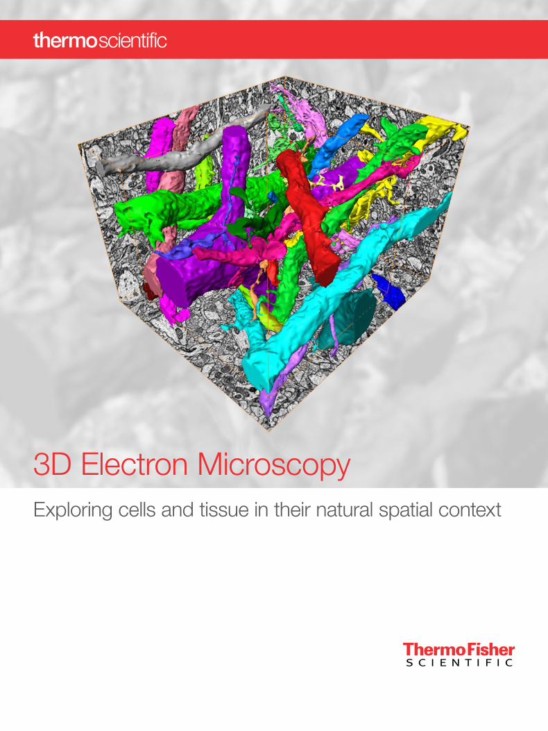
Volume reconstruction of neuronal processes in mouse brain acquired using serial block face SEM (Thermo Scientific™ Volumescope 2). Courtesy of Dr. P. Laserstein, Max Planck Institute for Brain Research, Germany.