3d imaging of biological specimens by electron microscopy · 3d imaging of biological specimens by...
TRANSCRIPT

3D Imaging of Biological Specimens by Electron
Microscopy
Focus on Microscopy
Hannover, May 2010
Elke Spiess, Wim Voorhout and Wim Busing
FEI Company
2
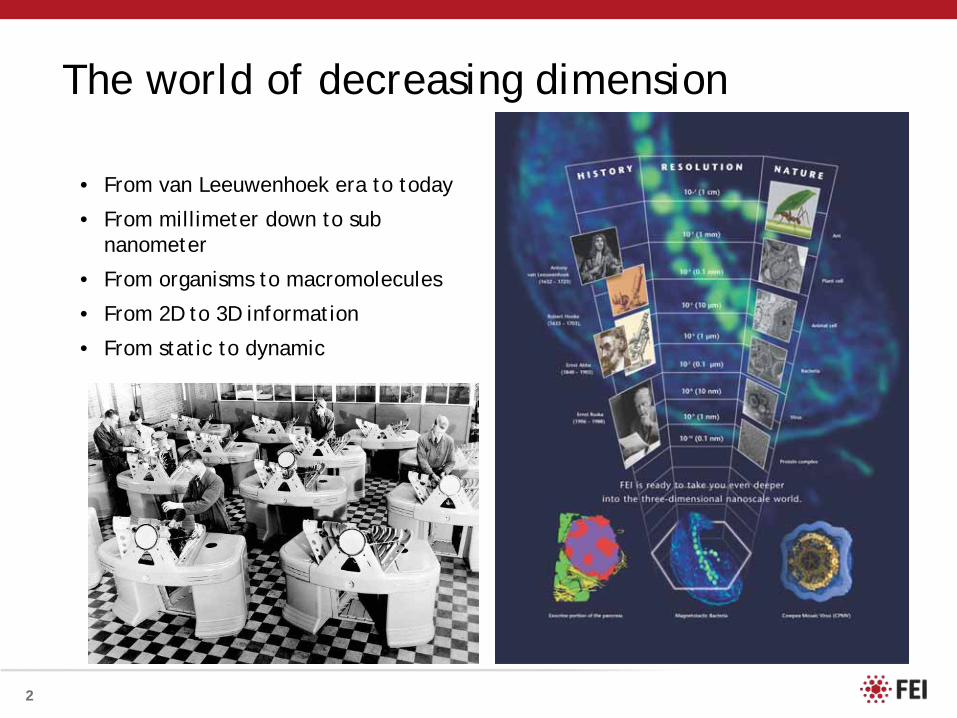
The world of decreasing dimension
• From van Leeuwenhoek era to today
• From millimeter down to sub nanometer
• From organisms to macromolecules
• From 2D to 3D information
• From static to dynamic

3
3D information
• Confocal Light Microscopy (CLSM)
• For looking at thick sections, whole cells or tissue using fluorescent probes
• Serial sectioning (TEM)
• build up a 3D image from consecutive sections
• SDB Slice & View
• removing thin layers and block face imaging
• Tomography (TEM)
• acquisition of a series of images at different tilts
• Single Particle reconstruction
• acquire many images of isolated structures

4
DNA/Genes Proteins Cells Organ/Systems Health
High resolutionSmall volume
Source: SDI Lab Analytical and Life Science Instrument Market ’06-’10
How to get there?
1. (Cryo) - Electron Tomography2. Single Particle Analysis
3. Template MatchingSegmentation
Low resolutionLarge volume

5
Acquisition
Alignment
Reconstruction
Visualization
Electron Tomography - Four Steps
Courtesy: Dr. Kobayashi, National Institute of Advanced Industrial Science and Technology, Osaka, Japan
1
2 4
3
Xplore3D

6 6
Volume rendering using ResolveRT
Rota Virus (negative stain) Tomography

7 7
3D ReconstructionAligned Data Set
HIV Infected Cells (epon fixed) Tomography
250 nm epon sections from HIV infected cells.Data collected on Tecnai Spirit BioTwinCourtesy: Robert-Koch Institute, Berlin

10
DNA/Genes Proteins Cells Organ/Systems Health
Source: SDI Lab Analytical and Life Science Instrument Market ’06-’10
How to get there?
1. (Cryo) - Electron Tomography2. Single Particle Analysis
3. Template MatchingSegmentation
The Structural Biology Playground
6,600x 60,000x660x
2D 3D
High resolutionSmall volume
Low resolutionLarge volume

11
Cryo EM – Imaging at cryo temperatures
6,600x
38,000x660x
2D 3D
To observe structures as close as possible to their natural state!

12
NOT AT ALL !!!
Cryo EM: Noisy and Ugly??
Herpes virus capsidDeng et al. J. Virol. 81:3640-3644 (2007)

13
3D reconstruction
Portal
50nmPenton
Hexons
Direct visualization of a bacteriophage-like portal in the Kaposi’s sarcoma-associated herpesvirus capsid by cryo-electron tomography
Deng et al. J. Virol. 81:3640-3644 (2007)
Examples: Cryo-Electron Tomography
14 14
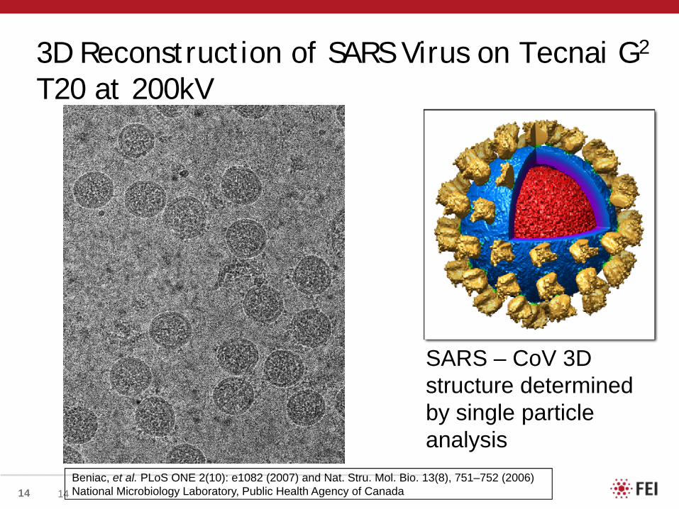
3D Reconstruction of SARS Virus on Tecnai G2
T20 at 200kV
Beniac, et al. PLoS ONE 2(10): e1082 (2007) and Nat. Stru. Mol. Bio. 13(8), 751–752 (2006)National Microbiology Laboratory, Public Health Agency of Canada
SARS – CoV 3D structure determined by single particle analysis

17
SPA – Tomography (Hybrid method)3D reconstruction of 80S ribosome fragment
Recording cryo tomogram of in-situ ribosome-rich area
254 particles of which tomograms have been taken are selected
Particle averaging on tomograms increases image information
Imaged at TU/e Titan Krios – Jason Pierson et al

19
Single Particle Resolution – Why?Secondary Structure Elements at different resolutions
At 4Å resolution, strands in the b-hairpin begin to separate, the pitch of the a-helix becomes visible and bulky side chains can start to be seen.
Segment extracted from the atomic model of HK97 capsid protein. An alpha-helix and a beta-hairpin joined together by a loop and filtered to different resolutions.
At 2Å resolution, the hole in each aromatic ring is resolved (red arrow).
Towards atomic resolution structural determination by single-particle cryo-electron microscopyCurrent Opinion in Structural Biology, Volume 18, Issue 2, April 2008, Pages 218-228 Z Hong Zhou
20
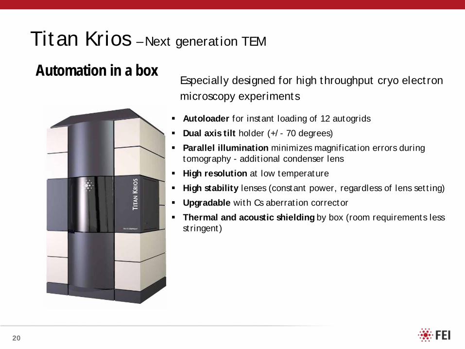
Titan Krios – Next generation TEM
Especially designed for high throughput cryo electron microscopy experiments
Autoloader for instant loading of 12 autogrids
Dual axis tilt holder (+/- 70 degrees)
Parallel illumination minimizes magnification errors during tomography - additional condenser lens
High resolution at low temperature
High stability lenses (constant power, regardless of lens setting)
Upgradable with Cs aberration corrector
Thermal and acoustic shielding by box (room requirements less stringent)
Automation in a box

22
Conclusions
• Single Particle
• Best resolution for macro molecular machines/viruses• Only works for identical structures
3.3A ° Cryo-EM Structure of a Non-enveloped Virus Reveals a Priming Mechanism for Cell Entry
Xing Zhang,1 Lei Jin,1 Qin Fang,2 Wong H. Hui,3 and Z. Hong Zhou1,3,*
1. Department of Microbiology, Immunology & Molecular Genetics, University of California, Los Angeles, Los Angeles, CA 90095-7364,USA 2. State Key Laboratory of Virology, Wuhan Institute of Virology, Chinese Academy of Sciences, Wuhan, Hubei 430071, China, 3. California Nano-Systems Institute, University of California, Los Angeles, Los Angeles, CA 90095-7364, USA
Cell 141, 1–11, April 30, 2010.