40 tracheal mass
TRANSCRIPT

40 Tracheal Mass/Narrowing
CLINICAL IMAGAGINGAN ATLAS OF DIFFERENTIAL DAIGNOSIS
EISENBERG
DR. Muhammad Bin Zulfiqar PGR-FCPS III SIMS/SHL
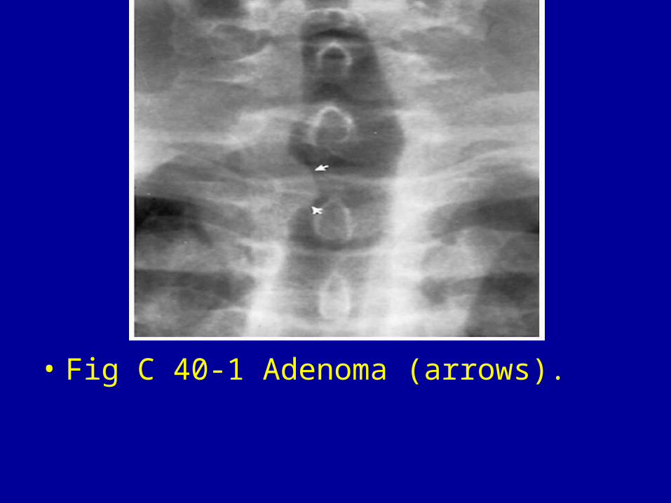
• Fig C 40-1 Adenoma (arrows).
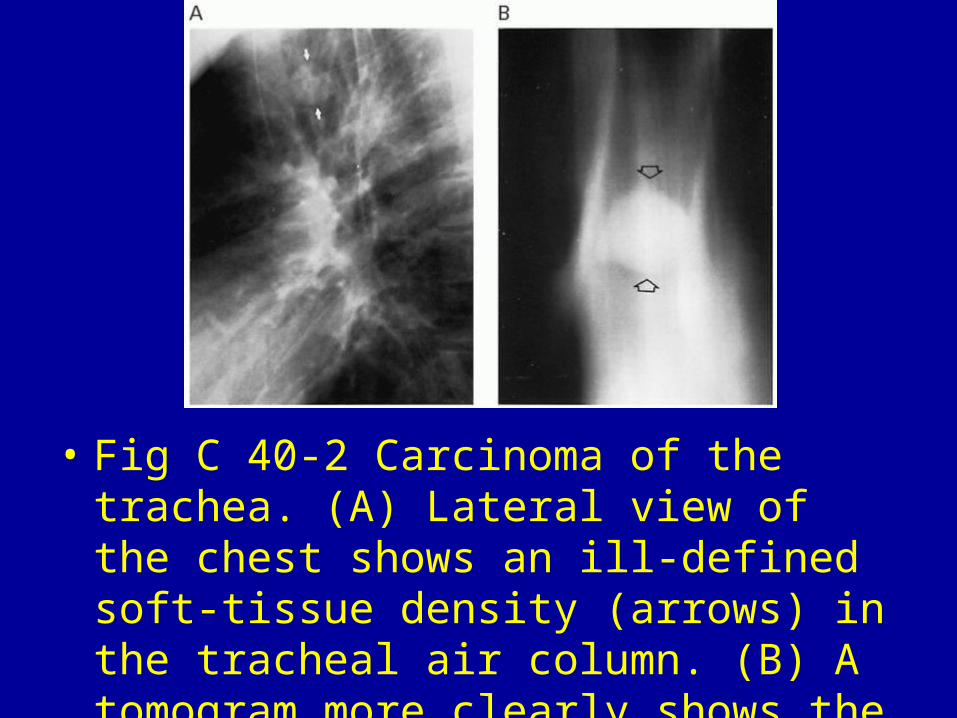
• Fig C 40-2 Carcinoma of the trachea. (A) Lateral view of the chest shows an ill-defined soft-tissue density (arrows) in the tracheal air column. (B) A tomogram more clearly shows the mass (arrows).

• Fig C 40-3 Extramedullary myeloma. Proliferation of plasma cells forms a mass (arrow) in the trachea.70
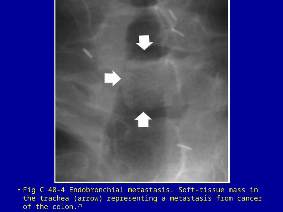
• Fig C 40-4 Endobronchial metastasis. Soft-tissue mass in the trachea (arrow) representing a metastasis from cancer of the colon.71
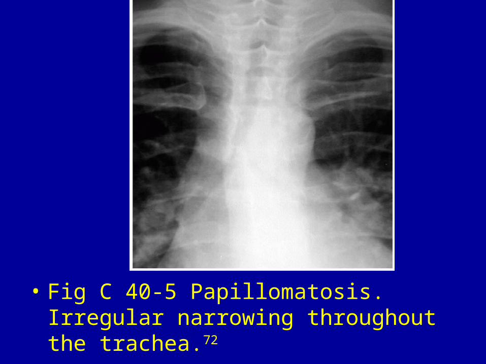
• Fig C 40-5 Papillomatosis. Irregular narrowing throughout the trachea.72
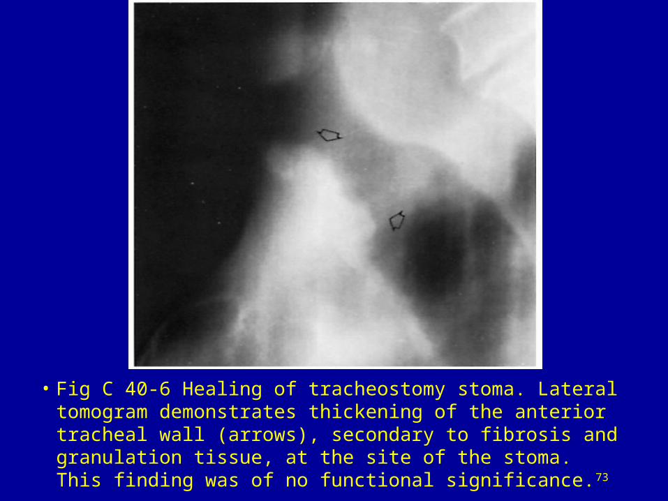
• Fig C 40-6 Healing of tracheostomy stoma. Lateral tomogram demonstrates thickening of the anterior tracheal wall (arrows), secondary to fibrosis and granulation tissue, at the site of the stoma. This finding was of no functional significance.73
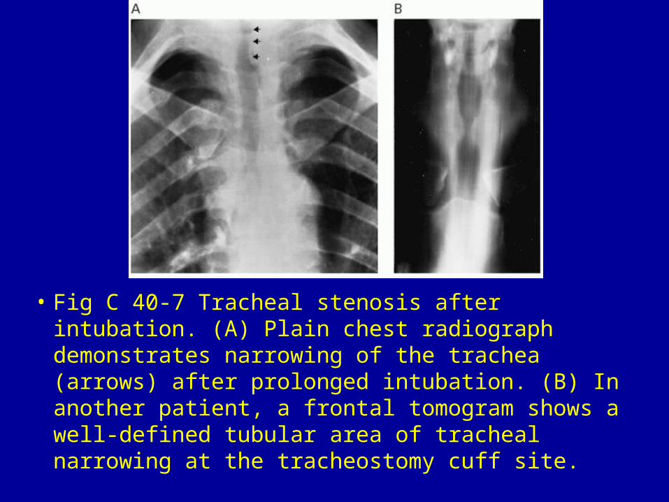
• Fig C 40-7 Tracheal stenosis after intubation. (A) Plain chest radiograph demonstrates narrowing of the trachea (arrows) after prolonged intubation. (B) In another patient, a frontal tomogram shows a well-defined tubular area of tracheal narrowing at the tracheostomy cuff site.
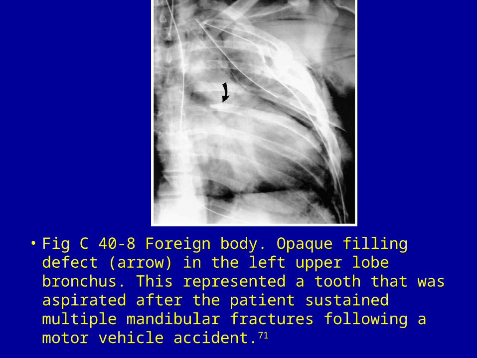
• Fig C 40-8 Foreign body. Opaque filling defect (arrow) in the left upper lobe bronchus. This represented a tooth that was aspirated after the patient sustained multiple mandibular fractures following a motor vehicle accident.71
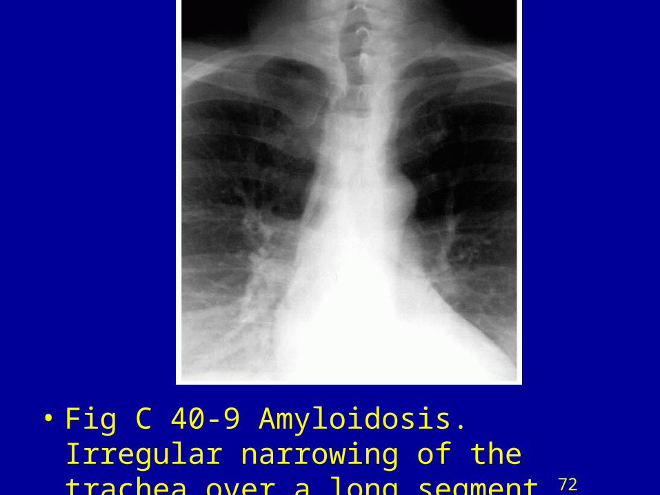
• Fig C 40-9 Amyloidosis. Irregular narrowing of the trachea over a long segment.72
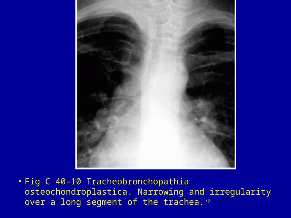
• Fig C 40-10 Tracheobronchopathia osteochondroplastica. Narrowing and irregularity over a long segment of the trachea.72
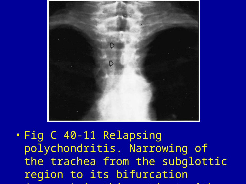
• Fig C 40-11 Relapsing polychondritis. Narrowing of the trachea from the subglottic region to its bifurcation (arrows) in this patient with long-standing disease.74
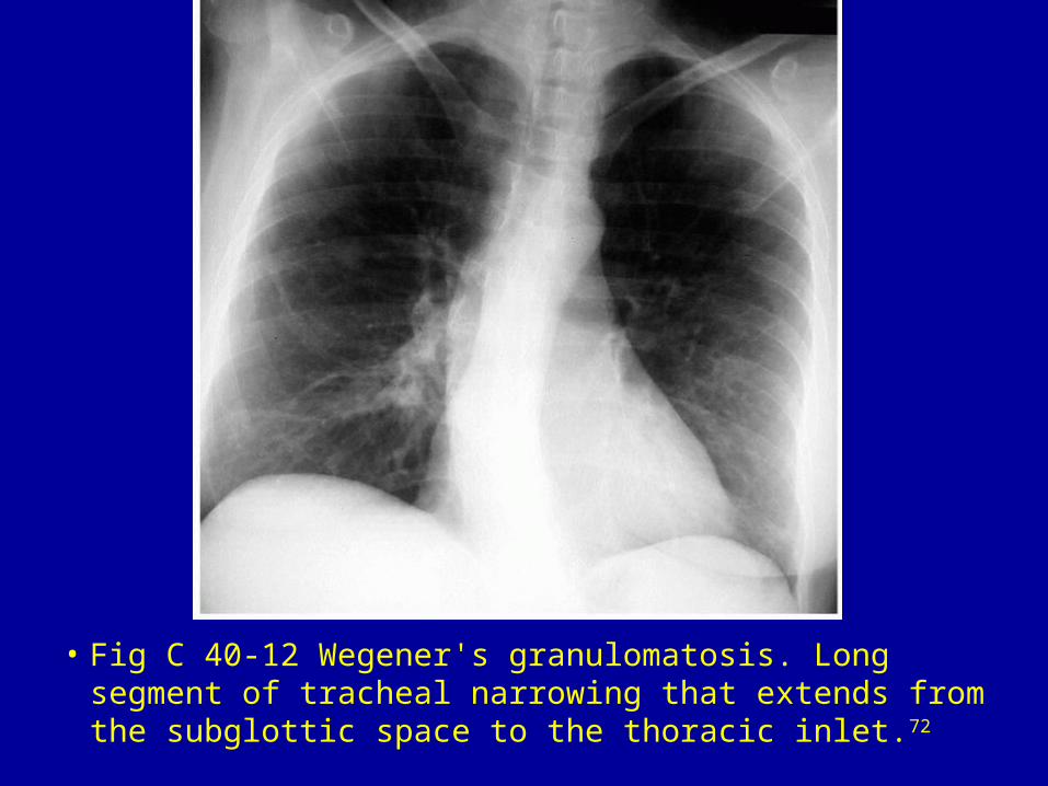
• Fig C 40-12 Wegener's granulomatosis. Long segment of tracheal narrowing that extends from the subglottic space to the thoracic inlet.72
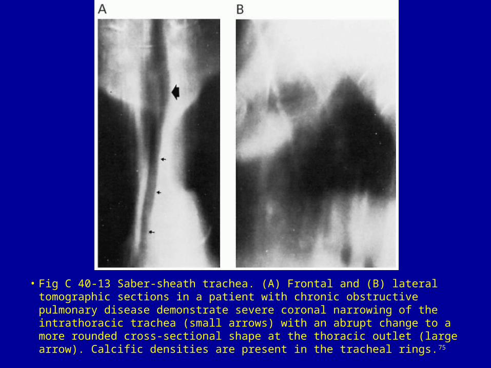
• Fig C 40-13 Saber-sheath trachea. (A) Frontal and (B) lateral tomographic sections in a patient with chronic obstructive pulmonary disease demonstrate severe coronal narrowing of the intrathoracic trachea (small arrows) with an abrupt change to a more rounded cross-sectional shape at the thoracic outlet (large arrow). Calcific densities are present in the tracheal rings.75
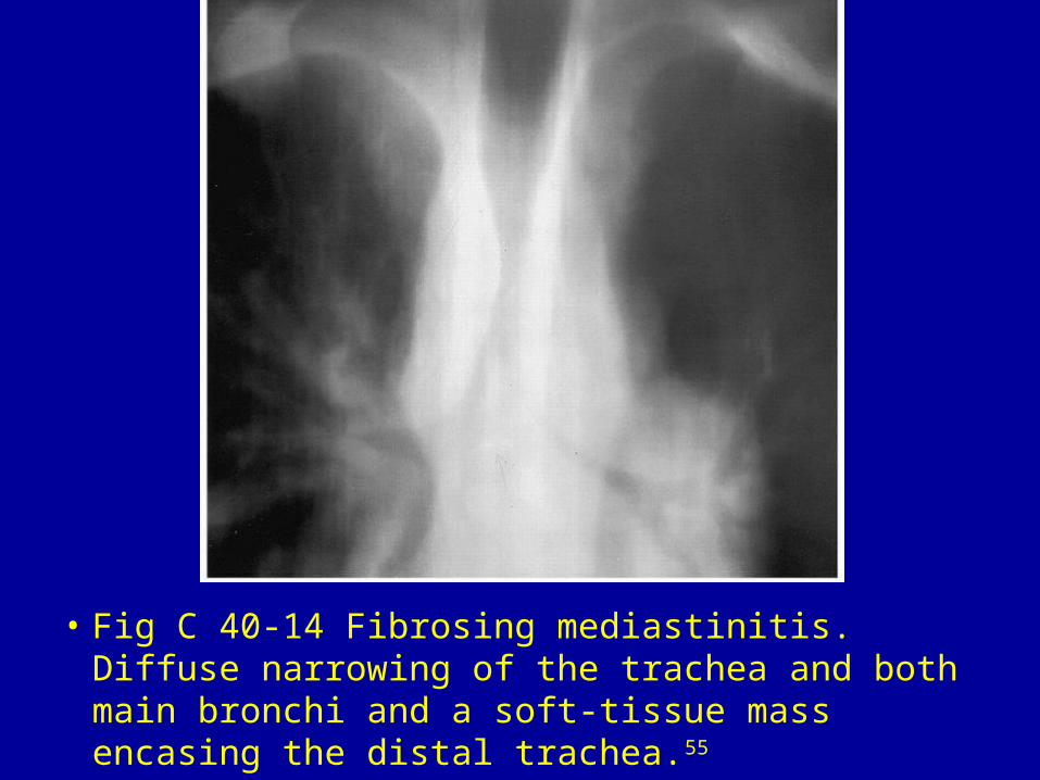
• Fig C 40-14 Fibrosing mediastinitis. Diffuse narrowing of the trachea and both main bronchi and a soft-tissue mass encasing the distal trachea.55


