4,400 117,000 130m · 2019-12-08 · denture stomatitis, loss of denture retention, fracture of the...
TRANSCRIPT

Selection of our books indexed in the Book Citation Index
in Web of Science™ Core Collection (BKCI)
Interested in publishing with us? Contact [email protected]
Numbers displayed above are based on latest data collected.
For more information visit www.intechopen.com
Open access books available
Countries delivered to Contributors from top 500 universities
International authors and editors
Our authors are among the
most cited scientists
Downloads
We are IntechOpen,the world’s leading publisher of
Open Access booksBuilt by scientists, for scientists
12.2%
117,000 130M
TOP 1%154
4,400
Chapter 9
Denture andOverdenture Complications
Elena Preoteasa, Cristina Teodora Preoteasa,Laura Iosif, Catalina Murariu Magureanu andMarina Imre
Additional information is available at the end of the chapter
http://dx.doi.org/10.5772/59250
1. Introduction
Dentures and overdentures, the most frequently used treatment options for the completeedentulism, can have local and systemic complications. For their prevention, treatment andreduction of their negative impact, it is necessary to understand their etiological context andto know their particularities of manifestation. Considering the relatively high rate of somecomplications of denture and overdenture treatment, knowing them is essential for ensuringa treatment that corresponds to the medical standards of care and patients’ needs andexpectations.
2. Context of denture and overdenture complications
All medical treatments should be approached with a holistic perspective in mind, due to thefact that there are numerous factors which, through interacting each other, have an impact onthe final medical outcome. Understanding the problem and its realistic possible approaches,but also considering its treatment limitations and performing an analysis that evaluates themedium and long-term prognosis ensures the highest premises for obtaining a good result.
The previous also applies to the treatment of edentulism using dentures or overdentures. Someof the key aspects that might help understand better the denture and overdenture complica‐tions, as they define the etiological context, are mentioned in Table 1.
© 2015 The Author(s). Licensee InTech. This chapter is distributed under the terms of the Creative CommonsAttribution License (http://creativecommons.org/licenses/by/3.0), which permits unrestricted use, distribution,and reproduction in any medium, provided the original work is properly cited.

Context
General medical and social factorsMedical and social perception of edentulismDemographics of edentulism
Denture and overdenture treatment factors
Treatment difficultyTreatment options overviewMaintenance therapyTechnical and biomechanical considerationsPrevious dental treatments
Edentulous patient factors
Oral health statusSystemic health status and medication useAgeHealth risk factorsPatient need and preferences
Table 1. Context of denture and overdenture complications – key factors
2.1. Medical and social perception of edentulism
Edentulism is defined as the loss of all permanent teeth. Tooth loss is an outcome of a complexinteraction between disease entities (e.g., caries and periodontal disease) and non-diseaseentities (e.g. economy, oral healthcare system, access to dental services, dental awareness,cultural tradition, education) [1]. Continuing exposure to risk factors after onset of edentulism(e.g., poor oral hygiene, smoking, deficient dental treatment) can have an etiological role inthe occurrence of complication.
Edentulism is a chronic, severe, irreversible medical condition and is described as the finalmarker of disease burden for oral health [2,3]. It is common for elderly people, but it is notregarded any more as an inevitable phenomenon that comes with age [4].
Edentulism has several deleterious consequences on oral health (e.g., residual ridge resorption,impaired masticatory function, trouble speaking), general health (deficient nutritional status,increased risk for certain systemic diseases), mental and social well-being (dissatisfaction withappearance, avoidance of social contacts) and on quality of life [1,2,4]. The previous haveimpact on prosthetic treatment to be performed.
Thus, the current perception on edentulism is as non-fatal sequelae of diseases and injuries,which still represents a tremendous global health care burden [5,6]. It can be considered aphysical impairment, because important body parts have been lost, a disability, because itassociates functional limitations or a handicap, as it sometimes limits or prevents normal lifeor work activities [1,2,7-9].
Considering the impact and demographics of the edentulism, the health care barriers that olderpeople face, the Active Ageing approach of the World Health Organization (keeping olderpeople socially engaged and productive), intensive measures and new regulations regardingcaring for the elderly population are needed. Consequently, implementation of gerodontology,as a new dental specialty, may be appropriate [10].
Emerging Trends in Oral Health Sciences and Dentistry194
2.2. Demographics of edentulism
According to the current reports and predictions, edentulism is and will continue to representa common disease for the elderly people segment.
There is a tendency for reduction of the edentulism prevalence, through the reduction of toothloss. Thus, in the United States in the period of 1999-2004, the prevalence of tooth retention inseniors (65 years and older) significantly increased from 17.9 teeth to 18.9 teeth and theprevalence of edentulism significantly decreased from approximately 34% to 27% [11]. Thisphenomenon can be justified through the progress made in the dental field, the emphasis onprevention measures, improved access to dental care services and mass education for ap‐proaching a healthy behavior [4]. But, despite these efforts, complete edentulism continues tohave a high prevalence, aspect associated mainly to the aging population phenomenonthrough growth of the life expectancy and thus the number of elderly people and the numberof edentulous patients [12,13].
Estimates show that edentulism is found in 2.3% of the world’s population regardless of age,respectively in 7-69% of adult populations internationally [5,14]. Considerably high disparitiesare noted between different countries, different regions, due to the important impact of thesocio-economical and behavioral factors.
The prognoses show that edentulism is decreasing, but most probably will continue to be acondition with a significant prevalence, especially in elderly’s people, which is estimated tobe a growing category in the global population [15]. Douglas estimates that in the United Statesthe population with one or two edentulous jaws will increase from 34 million in 1991 to 38million in 2020 [1,12]. Felton considers that most probably the necessity for complete denturetherapy will not disappear over the next 4 or 5 decades, and the economic conditions may evenlead to a growing need [1,6].
2.3. Treatment difficulty
Edentulism is generally regarded as a clinical condition with a high degree of treatmentdifficulty, often being hard to achieve optimal functional parameters. The complexity of theedentulous condition derives from the extensive oral changes, both anatomical and functional,that sometimes require preprosthetic surgical intervention in order to optimize the biome‐chanical conditions, which are superimposed on general alterations (related to age, systemicdisease, and psychosocial status). In order to support the differentiation of cases according totheir treatment difficulty degree, the ACP (American College of Prosthodontists) has puttogether the Prosthodontic Diagnostic Index (PDI) Classification System for the completeedentulism [16]. Higher complexity of edentulism condition increase the risk of treatmentcomplication (e.g., in cases with severe ridge resorption ill-fitting dentures are more frequentlynoticed), and complications can also contribute to increasing the degree of treatment difficulty(e.g., wearing unstable dentures accelerate the ridge resorption rate).
2.4. Treatment options overview
Complete denture used to be the only treatment option for the complete edentulism. Nowa‐days, this is still the most frequently used treatment option, but there can be seen a growing
Denture and Overdenture Complicationshttp://dx.doi.org/10.5772/59250
195

trend towards using implant prosthetic restorations fixed or removable. Each treatment optionhas the risk of specific complications, dependent on their manufacturing particularities andbio-mechanical features.
Dentures can have both local and systemic complications, such as gingival hyperplasia,denture stomatitis, loss of denture retention, fracture of the denture and functional impair‐ment, mastication deficiencies having a negative impact on the nutritional status. Somepatients cannot tolerate the dentures, aspect that can be connected to psychological factors, topatients’ needs and expectations, but also to age, oral conditions, denture deficiencies anddoctor-patient relationship.
Root supported overdentures, with or without attachment systems, have the advantage ofimproved retention and stability, with a positive impact on the oral functions and the accom‐modation with the future dentures. Their possible complications include the ones of theconventional dentures and, additionally, some modifications of the supporting teeth or theattachment system used.
Prosthetic implant restorations, either fixed or a removable, are alternatives that provide animproved functional integration and better treatment outcome, but are more complex andrequire preprosthetic interventions, with additional biological, financial and time costs. Usingthese treatment options involves the risk for additional complications, with regards to thehigher complexity of the treatment –e.g., treatment plan related, surgical complications,technical complications.
2.5. Maintenance therapy
Maintenance is very important for the longevity of the treatment, having a positive impact inreducing the frequency and severity of its complications. Both type of procedures, thoseperformed in the dental office, by the dentist and at home, by the patient, are relevant in thisrespect.
Periodical check-ups are essential, considering that there are some complications with a highprevalence rate both for dentures and implant overdentures (e.g., loss of denture stability dueto progressive ridge resorption, denture adjustments and relinings, clip activations) [17].Additionally, the edentulous patients are often elderly patients, and face access barriers todental care services, in relation to aspects like lack of finances or transportation difficulties[18,19]. Due to this, it is recommended to keep in mind the possible complications and to takethe appropriate preventive measures to limit them at the time the treatment is being plannedand performed.
Informing and instructing the patient on how to take proper care of the oral care andprosthetic restorations are important aspects, since complications can be tightly related tothis (e.g., the lack of appropriate cleaning of the denture, teeth or implants is associatedwith a higher risk for denture stomatitis, tooth or implant loss). Since we are frequentlydealing with elderly people, who have less manual dexterity, it is recommended to choosesimpler treatment option (e.g., if applicable, 2-implant overdentures are more appropriatethan 4-implant overdenture [20].
Emerging Trends in Oral Health Sciences and Dentistry196
2.6. Technical and biomechanical considerations
According to the current level of knowledge, treatment with dentures or implant/rootoverdentures must consider the risk for developing complications in relation to the technicaland biomechanical features (e.g., design, attachment components, materials).
There are different types of design for dentures and overdentures, with different possiblecomplications. Thus, using narrow-diameter implants associates a higher risk of implantfracture. Considering the occlusal scheme, there is evidence that patients prefer dentures withlingualized occlusion [21]. Metal or non-metal (glass and polyethylene fibers) inserts arerecommended for denture base reinforcement when there is a high risk of denture fracture orwhen there are more than 2 teeth or implants supporting the denture [22].
Material used for denture/overdenture fabrication associates the risk of developing compli‐cations in relation to their physico-chemical properties and their biocompatibility. Forexample, polymethylmethacrylate (PMMA), the material mostly used for manufacturing ofdentures or overdentures, through its features (porosity, increased wettability, low mechanicalstrength, monomer release after curing) facilitates the occurrence of complications such asmicrobial or contact denture stomatitis, fracture of the dentures, artificial teeth discolorationand wear [23].
2.7. Previous dental treatments
A key element in order to achieve a predictable outcome is the analysis of the previous dentaland prosthetic treatments, by connecting patient’s subjective complaints with prostheticrestoration’s objective deficiencies. This gives important information that could be used fordecision making in establishing the particularities of the future prosthetic treatment. Forexample, complete denture intolerance can be linked to personality traits, to objective patient’sfeatures that enhance the occurrence of functional deficiencies, or to some objective faults ofthe dentures. Differentiating between these three situations is the basis for selecting the optimaltreatment option, with the possibility to prevent the complications that occurred in the past.
2.8. Oral health status
The complete edentulism cannot be regarded simply as the loss of teeth. It is accompanied bymassive, progressive changes of the oral structures and functional alterations, which associatesa high degree of treatment difficulty and the occurrence of specific complications. Impact ofedentulism on oral health is mainly manifested in 3 directions: modifier of normal physiology;risk factor for impaired mastication; determinant of oral health [2]. Amongst the sequelae oftreatment with complete dentures, as the most commonly used treatment option, there can bementioned residual ridge resorption, mucosal reactions, burning mouth syndrome, denturestomatitis [24].
Considering the severe changes of the oral status in edentulous patients, the increasing elderlypopulation and the relatively frequent barriers to oral health care of older people (e.g., financialhardship, transportation difficulties), Petersen et al. makes a series of recommendations among
Denture and Overdenture Complicationshttp://dx.doi.org/10.5772/59250
197
which are the incorporation of age related oral health concerns into the promotion of generalhealth, that could ease the development of oral health care for older people [25].
2.9. Systemic health status and medication use
Between oral health and general health there are numerous interactions, that sometimesmaterializes as local or systemic complications.
The impact of complete edentulism on the general health status is manifested as an increasedrisk of conditions, such as nutritional deficiencies, inflammatory changes of the gastric mucosa,peptic or duodenal ulcers, obesity, noninsulin-dependent diabetes mellitus, hypertension,heart failure, ischemic heart disease, stroke, aortic valve sclerosis, chronic kidney disease,sleep-disordered breathing, including obstructive sleep apnea [2]. Additionally, functionallimitations, mental and social well-being alterations that negatively impact the quality of lifeare more common in edentulous patients.
The impact of general health status and the medication used on the oral health of the edentu‐lous patient is partially manifested through the occurrence of complications. Nutritionaldeficiencies increase the risk of occurrence of denture stomatitis, traumatic ulcer and burningmouth syndrome [25]. Patient’s personality and psychological well-being influences treatmentsatisfaction and tolerance [10]. Decreased manual dexterity has a negative impact on care andmaintenance of dentures/overdentures, which leads to negative effects on oral and systemichealth [14].
2.10. Age
Patient’s age is an important aspect to consider when planning the prosthetic treatment,being linked to particularities of oral and general health status, to specific needs andexpectation towards the prosthetic rehabilitation, to particular medical approaches in orderto ensure a good long-term prognosis. Prosthetic treatment of the edentulous patient shouldtake into account the current situation, but also the most probable evolution and, if present,the inherent complications (e.g., preventive measures to reduce alveolar ridge resorptionare recommended).
Young-elderly edentulous patients generally have more favorable clinical conditions forprosthetic rehabilitation, a better general health status, a faster adaptation to removableprosthesis if chosen and the ability to perform most accurately the necessary the maintenanceprocedures. They have higher expectations regarding the esthetics and functionality of theprosthetic rehabilitation and don’t easily accept the removable treatment options.
Old-elderly edentulous patients generally register an increased treatment difficulty, as aconsequence of numerous factors interacting. In previous ill-fitting complete denture wearersthere is a severe ridge resorption [26,27]. The prevalence of co-morbidities is increased, suchas physical or mental health problems that have a negative impact on oral health, systemichealth, functioning and behavior. Most of the times the elderly people are not regular users ofdental services since they overcome physical and psychological access treatment barriers (e.g.,
Emerging Trends in Oral Health Sciences and Dentistry198
the cost of dental care services, transportation problems, doctor’s attitudes-lack of responsive‐ness to patient’s concerns, the lack of perceived need for care, fear), which are more significantfor the functionally dependent elderly then for the independent elderly [28-30]. They havetreatment expectations that target first the rehabilitation of the masticatory function, andsecond the esthetics. They usually prefer more simple medical procedures, that include limitedsurgical interventions and that demand easy maintenance procedures. The older completelyedentulous patients show a more frequent rate of denture intolerance, probably due to lessadaptability to new situations.
Demographic changes, namely population ageing and decreasing prevalence of tooth loss,have impact on the edentulous patient profile. There is an increasing of the age when edentu‐lism occurs, aspects that associates an increased treatment difficulty. Considering the latter,additional measures are necessary to ensure adequate oral health for older edentulous patientse.g., access to and financing for dental services, an adequately trained workforce to providedental care and appropriate education to edentulous individuals [30].
2.11. Health risk factors
Health risk factors should be assessed since they can explain some of the case particularitiesand may have a negative impact on the treatment outcome. Among them, there can bementioned behavioral risk factors (e.g. tobacco and alcohol consumption, obesity related tophysical activity and diet), social risk factors (e.g., socio-economical status, social networksand social support, occupational factors, social inequalities), inadequate disease screeningpractices, exposure to increased stress [31]. Their role is proven both as a cause of completeedentulism and also as a factor that impacts the treatment outcome, being risk factors for somecomplications.
2.12. Patient need and preferences
Health care decisions require integrating the patient’s individual preferences and values,according to the ethical principle of respecting the patient’s autonomy [32]. A good relationand communication between doctor and patient offers the best premises for reaching aconsensus regarding the medical decision, with a positive effect on the treatment outcome.
Patient preferences are related to numerous variables, e.g., age, social status, personality type,education. Acknowledging them may be difficult, especially in elderly patients, sometimes inrelation to objective reasons, as physical changes that affect the communication (e.g., loss ofhearing or visual acuity). Additional efforts should be made in order to understand thepatient’s health needs and preferences, since they can have important consequences, such asrejection of the prosthetic treatment or even avoiding addressing for medical treatment.
3. Classification of denture and overdenture complications
The classification of denture and overdenture complications can enhance practitioner’sunderstanding of them, with a positive effect on their management and prognosis.
Denture and Overdenture Complicationshttp://dx.doi.org/10.5772/59250
199
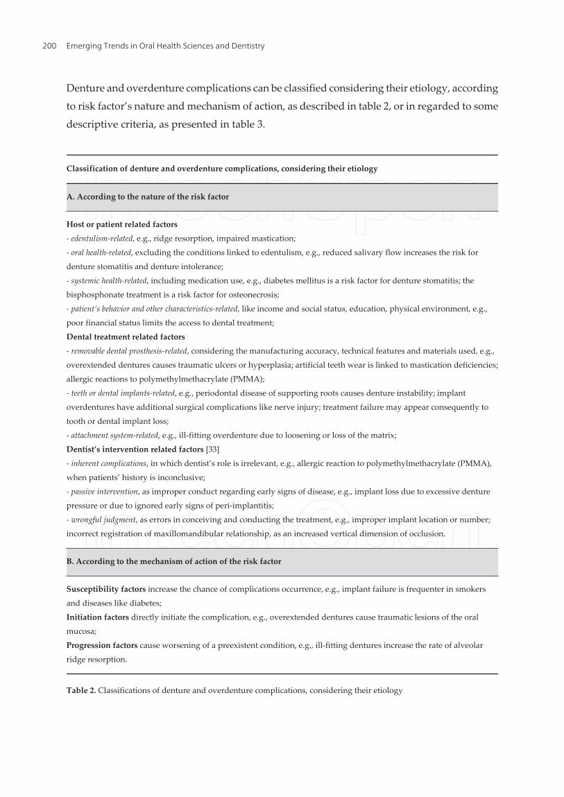
Denture and overdenture complications can be classified considering their etiology, accordingto risk factor’s nature and mechanism of action, as described in table 2, or in regarded to somedescriptive criteria, as presented in table 3.
Classification of denture and overdenture complications, considering their etiology
A. According to the nature of the risk factor
Host or patient related factors- edentulism-related, e.g., ridge resorption, impaired mastication;- oral health-related, excluding the conditions linked to edentulism, e.g., reduced salivary flow increases the risk fordenture stomatitis and denture intolerance;- systemic health-related, including medication use, e.g., diabetes mellitus is a risk factor for denture stomatitis; thebisphosphonate treatment is a risk factor for osteonecrosis;- patient’s behavior and other characteristics-related, like income and social status, education, physical environment, e.g.,poor financial status limits the access to dental treatment;Dental treatment related factors- removable dental prosthesis-related, considering the manufacturing accuracy, technical features and materials used, e.g.,overextended dentures causes traumatic ulcers or hyperplasia; artificial teeth wear is linked to mastication deficiencies;allergic reactions to polymethylmethacrylate (PMMA);- teeth or dental implants-related, e.g., periodontal disease of supporting roots causes denture instability; implantoverdentures have additional surgical complications like nerve injury; treatment failure may appear consequently totooth or dental implant loss;- attachment system-related, e.g., ill-fitting overdenture due to loosening or loss of the matrix;Dentist’s intervention related factors [33]- inherent complications, in which dentist’s role is irrelevant, e.g., allergic reaction to polymethylmethacrylate (PMMA),when patients’ history is inconclusive;- passive intervention, as improper conduct regarding early signs of disease, e.g., implant loss due to excessive denturepressure or due to ignored early signs of peri-implantitis;- wrongful judgment, as errors in conceiving and conducting the treatment, e.g., improper implant location or number;incorrect registration of maxillomandibular relationship, as an increased vertical dimension of occlusion.
B. According to the mechanism of action of the risk factor
Susceptibility factors increase the chance of complications occurrence, e.g., implant failure is frequenter in smokersand diseases like diabetes;Initiation factors directly initiate the complication, e.g., overextended dentures cause traumatic lesions of the oralmucosa;Progression factors cause worsening of a preexistent condition, e.g., ill-fitting dentures increase the rate of alveolarridge resorption.
Table 2. Classifications of denture and overdenture complications, considering their etiology
Emerging Trends in Oral Health Sciences and Dentistry200
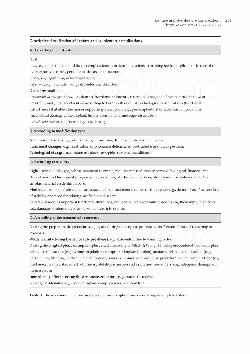
Descriptive classification of denture and overdenture complications
A. According to localization
Host- oral, e.g., oral soft and hard tissue complications, functional alterations; remaining teeth complications in case of rootoverdentures as caries, periodontal disease, root fracture;- facial, e.g., aged prognathic appearance;- systemic, e.g., malnutrition, gastro-intestinal disorders;Dental restoration- removable dental prosthesis, e.g., denture/overdenture fracture; retention loss; aging of the material, teeth wear- dental implants, that are classified according to Berglundh et al. [34] as biological complications (functionaldisturbances that affect the tissues supporting the implant, e.g., peri-implantitis) or technical complications(mechanical damage of the implant, implant components and suprastructures)- attachment system, e.g., loosening, loss, damage
B. According to modification type
Anatomical changes, e.g., alveolar ridge resorption; decrease of the muscular mass;Functional changes, e.g., mastication or phonation deficiencies, protruded mandibular position;Pathological changes, e.g., traumatic ulcers, atrophic stomatitis, candidiasis.
C. According to severity
Light – few clinical signs, whose treatment is simple, requires reduced costs in terms of biological, financial andclinical time and has a good prognosis, e.g., loosening of attachment system; ulcerations or irritations related tosurplus material on denture’s base;Moderate – functional alterations are associated and treatment requires medium costs, e.g., denture base fracture; lossof stability and need for relining; artificial tooth wear;Severe – associates important functional alterations, can lead to treatment failure, addressing them imply high costs,e.g., damage of inferior alveolar nerve, denture intolerance.
D. According to the moment of occurrence
During the preprosthetic procedures, e.g., pain during the surgical procedures for frenum plastia or reshaping ofexostosis;While manufacturing the removable prostheses, e.g., discomfort due to vomiting reflex;During the surgical phase of implant placement, according to Misch & Wang [35] being encountered treatment plan-related complications (e.g., wrong angulation or improper implant location), anatomy-related complications (e.g.,nerve injury, bleeding, cortical plate perforation, sinus membrane complication), procedure-related complications (e.g.,mechanical complications, lack of primary stability, ingestion and aspiration) and others (e.g., iatrogenic damage andhuman error);Immediately after inserting the denture/overdenture, e.g., traumatic ulcers;During maintenance, e.g., root or implant complications, retention loss
Table 3. Classifications of denture and overdenture complications, considering descriptive criteria
Denture and Overdenture Complicationshttp://dx.doi.org/10.5772/59250
201
4. Main complications of denture and overdenture
Some of the most common complications of the completely edentulous patient, treated bydentures or implant/root overdentures will be presented. Aspects related to their etiology,clinical features and management will be covered.
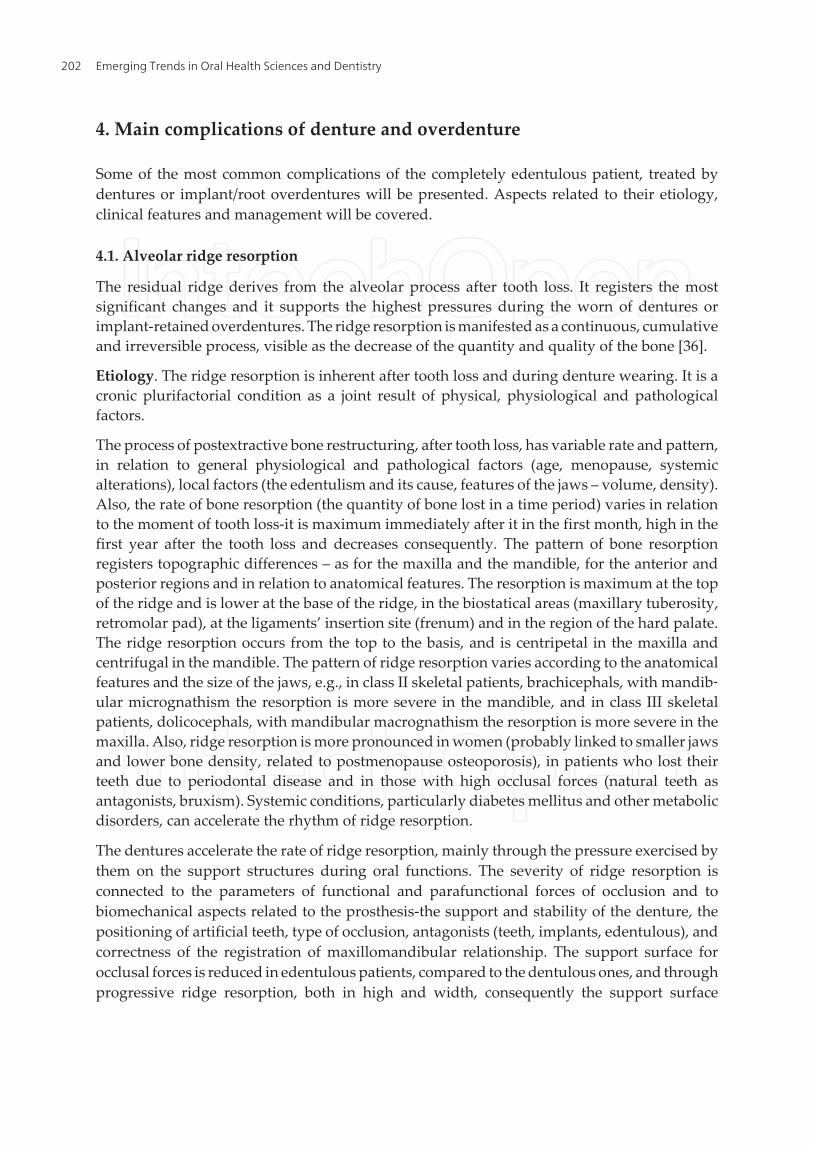
4.1. Alveolar ridge resorption
The residual ridge derives from the alveolar process after tooth loss. It registers the mostsignificant changes and it supports the highest pressures during the worn of dentures orimplant-retained overdentures. The ridge resorption is manifested as a continuous, cumulativeand irreversible process, visible as the decrease of the quantity and quality of the bone [36].
Etiology. The ridge resorption is inherent after tooth loss and during denture wearing. It is acronic plurifactorial condition as a joint result of physical, physiological and pathologicalfactors.
The process of postextractive bone restructuring, after tooth loss, has variable rate and pattern,in relation to general physiological and pathological factors (age, menopause, systemicalterations), local factors (the edentulism and its cause, features of the jaws – volume, density).Also, the rate of bone resorption (the quantity of bone lost in a time period) varies in relationto the moment of tooth loss-it is maximum immediately after it in the first month, high in thefirst year after the tooth loss and decreases consequently. The pattern of bone resorptionregisters topographic differences – as for the maxilla and the mandible, for the anterior andposterior regions and in relation to anatomical features. The resorption is maximum at the topof the ridge and is lower at the base of the ridge, in the biostatical areas (maxillary tuberosity,retromolar pad), at the ligaments’ insertion site (frenum) and in the region of the hard palate.The ridge resorption occurs from the top to the basis, and is centripetal in the maxilla andcentrifugal in the mandible. The pattern of ridge resorption varies according to the anatomicalfeatures and the size of the jaws, e.g., in class II skeletal patients, brachicephals, with mandib‐ular micrognathism the resorption is more severe in the mandible, and in class III skeletalpatients, dolicocephals, with mandibular macrognathism the resorption is more severe in themaxilla. Also, ridge resorption is more pronounced in women (probably linked to smaller jawsand lower bone density, related to postmenopause osteoporosis), in patients who lost theirteeth due to periodontal disease and in those with high occlusal forces (natural teeth asantagonists, bruxism). Systemic conditions, particularly diabetes mellitus and other metabolicdisorders, can accelerate the rhythm of ridge resorption.
The dentures accelerate the rate of ridge resorption, mainly through the pressure exercised bythem on the support structures during oral functions. The severity of ridge resorption isconnected to the parameters of functional and parafunctional forces of occlusion and tobiomechanical aspects related to the prosthesis-the support and stability of the denture, thepositioning of artificial teeth, type of occlusion, antagonists (teeth, implants, edentulous), andcorrectness of the registration of maxillomandibular relationship. The support surface forocclusal forces is reduced in edentulous patients, compared to the dentulous ones, and throughprogressive ridge resorption, both in high and width, consequently the support surface
Emerging Trends in Oral Health Sciences and Dentistry202
decreases even more. The magnitude of occlusal forces are generally lower in the edentulouspatients, but there are variations related to age, sex, parafunctions as bruxism, stress level, foodconsistency preferences, and also the correctness of prosthetic rehabilitation. Increasedduration of occlusion contacts, as a risk factor for ridge resorption, is related to bruxism, ill-fitting dentures, unstable occlusion and increased vertical dimension of occlusion. Comparedto maxillary edentulism, mandibular edentulism has greater risk of registering more severeridge resorption, due to the decreased denture support surface and related higher magnitudeof pressure beared. Also, denture wearing associates the risk of specific complications thatfavor the occurrence of an accelerated rate of ridge resorption, such as inflammatory lesionsof the oral mucosa (e.g., denture stomatitis). Due to these factors, it is considered that ridgeresorption is in tight relation with the period of wearing the dentures, but is also influencedby the quality of the treatment.
Clinical features. Ridge resorption is characterized by changes of the morphology of thealveolar ridges and of maxillomandibular relationship, with consequences on the prosthetictreatment and its outcome with time.
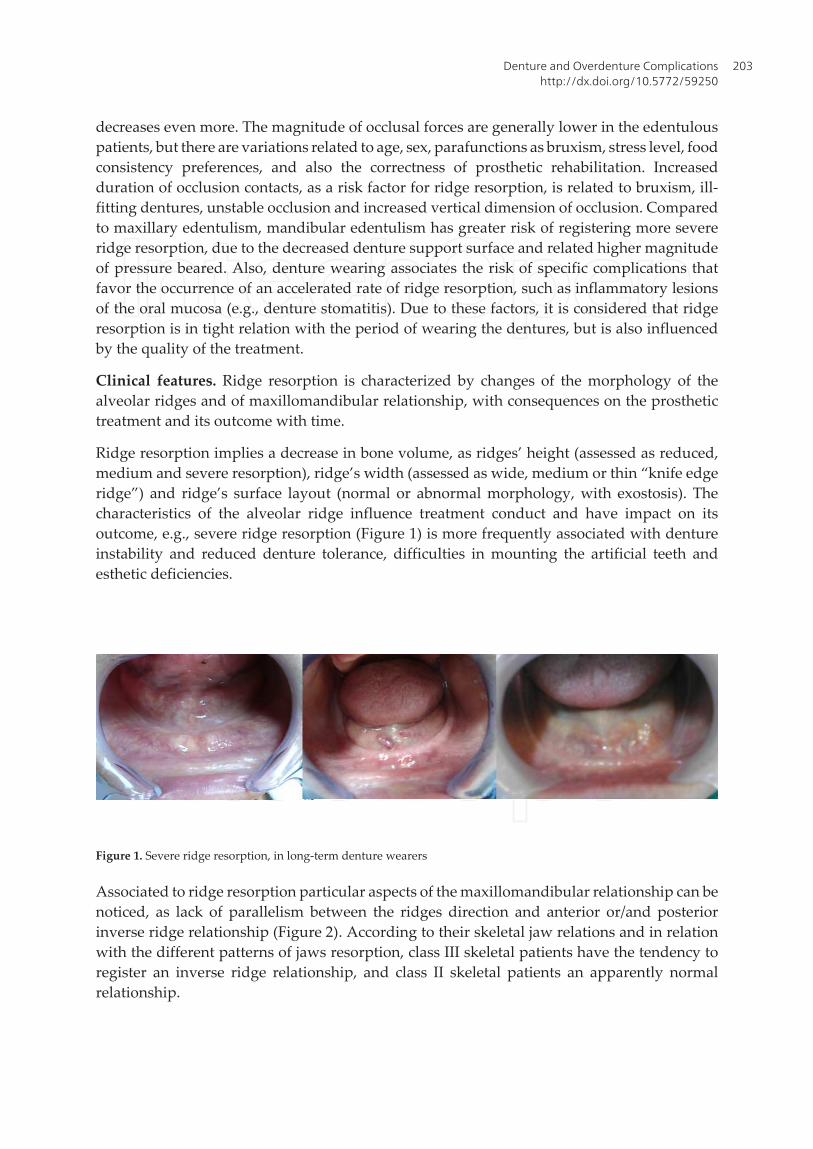
Ridge resorption implies a decrease in bone volume, as ridges’ height (assessed as reduced,medium and severe resorption), ridge’s width (assessed as wide, medium or thin “knife edgeridge”) and ridge’s surface layout (normal or abnormal morphology, with exostosis). Thecharacteristics of the alveolar ridge influence treatment conduct and have impact on itsoutcome, e.g., severe ridge resorption (Figure 1) is more frequently associated with dentureinstability and reduced denture tolerance, difficulties in mounting the artificial teeth andesthetic deficiencies.
Figure 1. Severe ridge resorption, in long-term denture wearers
Associated to ridge resorption particular aspects of the maxillomandibular relationship can benoticed, as lack of parallelism between the ridges direction and anterior or/and posteriorinverse ridge relationship (Figure 2). According to their skeletal jaw relations and in relationwith the different patterns of jaws resorption, class III skeletal patients have the tendency toregister an inverse ridge relationship, and class II skeletal patients an apparently normalrelationship.
Denture and Overdenture Complicationshttp://dx.doi.org/10.5772/59250
203
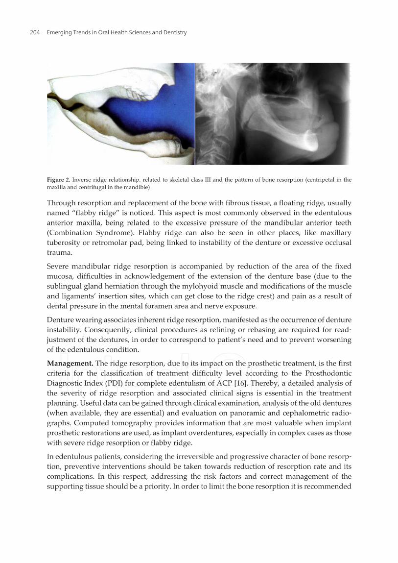
Figure 2. Inverse ridge relationship, related to skeletal class III and the pattern of bone resorption (centripetal in themaxilla and centrifugal in the mandible)
Through resorption and replacement of the bone with fibrous tissue, a floating ridge, usuallynamed “flabby ridge” is noticed. This aspect is most commonly observed in the edentulousanterior maxilla, being related to the excessive pressure of the mandibular anterior teeth(Combination Syndrome). Flabby ridge can also be seen in other places, like maxillarytuberosity or retromolar pad, being linked to instability of the denture or excessive occlusaltrauma.
Severe mandibular ridge resorption is accompanied by reduction of the area of the fixedmucosa, difficulties in acknowledgement of the extension of the denture base (due to thesublingual gland herniation through the mylohyoid muscle and modifications of the muscleand ligaments’ insertion sites, which can get close to the ridge crest) and pain as a result ofdental pressure in the mental foramen area and nerve exposure.
Denture wearing associates inherent ridge resorption, manifested as the occurrence of dentureinstability. Consequently, clinical procedures as relining or rebasing are required for read‐justment of the dentures, in order to correspond to patient’s need and to prevent worseningof the edentulous condition.
Management. The ridge resorption, due to its impact on the prosthetic treatment, is the firstcriteria for the classification of treatment difficulty level according to the ProsthodonticDiagnostic Index (PDI) for complete edentulism of ACP [16]. Thereby, a detailed analysis ofthe severity of ridge resorption and associated clinical signs is essential in the treatmentplanning. Useful data can be gained through clinical examination, analysis of the old dentures(when available, they are essential) and evaluation on panoramic and cephalometric radio‐graphs. Computed tomography provides information that are most valuable when implantprosthetic restorations are used, as implant overdentures, especially in complex cases as thosewith severe ridge resorption or flabby ridge.
In edentulous patients, considering the irreversible and progressive character of bone resorp‐tion, preventive interventions should be taken towards reduction of resorption rate and itscomplications. In this respect, addressing the risk factors and correct management of thesupporting tissue should be a priority. In order to limit the bone resorption it is recommended
Emerging Trends in Oral Health Sciences and Dentistry204
to preserve the tooth roots, to use dental implants, to realize immediate prosthetic rehabilita‐tion, especially in cases with tooth lost due to periodontal disease since this conduct favors amore reduced guided bone resorption. Correctness of dentures manufacturing is essential andit should rely on the principles of retention, stability and support, with proper maintenanceand on time replacement. Implant overdentures can be used both as a preventive solution, inorder to reduce the bone resorption, and as a curative solution, for solving the cases with severeridge resorption where conventional dentures did not succeed or were not tolerated.
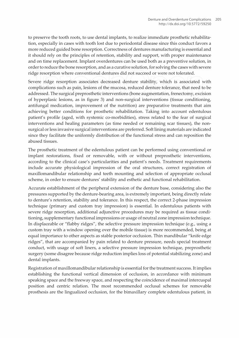
Severe ridge resorption associates decreased denture stability, which is associated withcomplications such as pain, lesions of the mucosa, reduced denture tolerance, that need to beaddressed. The surgical preprosthetic interventions (bone augmentation, frenectomy, excisionof hyperplasic lesions, as in figure 3) and non-surgical interventions (tissue conditioning,antifungal medication, improvement of the nutrition) are preparative treatments that aimachieving better conditions for prosthetic rehabilitation. Taking into account edentulouspatient’s profile (aged, with systemic co-morbidities), stress related to the fear of surgicalinterventions and healing parameters (as time needed or remaining scar tissues), the non-surgical or less invasive surgical interventions are preferred. Soft lining materials are indicatedsince they facilitate the uniformly distribution of the functional stress and can reposition theabused tissues.
The prosthetic treatment of the edentulous patient can be performed using conventional orimplant restorations, fixed or removable, with or without preprosthetic interventions,according to the clinical case’s particularities and patient’s needs. Treatment requirementsinclude accurate physiological impression of the oral structures, correct registration ofmaxillomandibular relationship and teeth mounting and selection of appropriate occlusalscheme, in order to ensure dentures’ stability and esthetic and functional rehabilitation.
Accurate establishment of the peripheral extension of the denture base, considering also thepressures supported by the denture-bearing area, is extremely important, being directly relateto denture’s retention, stability and tolerance. In this respect, the correct 2-phase impressiontechnique (primary and custom tray impression) is essential. In edentulous patients withsevere ridge resorption, additional adjunctive procedures may be required as tissue condi‐tioning, supplementary functional impressions or usage of neutral zone impression technique.In displaceable or “flabby ridges”, the selective pressure impression technique (e.g., using acustom tray with a window opening over the mobile tissue) is more recommended, being atequal importance to other aspects as stable posterior occlusion. Thin mandibular “knife edgeridges”, that are accompanied by pain related to denture pressure, needs special treatmentconduct, with usage of soft liners, a selective pressure impression technique, preprostheticsurgery (some disagree because ridge reduction implies loss of potential stabilizing zone) anddental implants.
Registration of maxillomandibular relationship is essential for the treatment success. It impliesestablishing the functional vertical dimension of occlusion, in accordance with minimumspeaking space and the freeway space, and respecting the coincidence of maximal intercuspalposition and centric relation. The most recommended occlusal schemes for removableprosthesis are the lingualized occlusion, for the bimaxillary complete edentulous patient, in
Denture and Overdenture Complicationshttp://dx.doi.org/10.5772/59250
205
skeletal class II patients or in severe mandibular ridge resorption or the linear occlusion, formandibular overdentures, in patients with combination syndrome or skeletal class III patternand severe maxillary ridge resorption.
Mandibular conventional dentures register frequently retention and stability deficiencies,mainly related to ridge resorption. These can be addressed through usage of implant prostheticrestorations, fixed or removable. There are multiple treatment options when considering usageof dental implants, as removable prosthesis (conventional or narrow dental implant overden‐ture, with different attachment systems as bars, ball, Locator) or fixed restorations (All an four,Fast & Fixed, conventional fixed implant restorations). Current perspective identifies 2 implantoverdentures as the minimum standard for mandibular edentulism taking into accountperformance, patient satisfaction, cost and clinical time [37]. Selecting between them requireacknowledgement of case futures and patient’s need and preferences. For example, fixedrestorations have better treatment outcome, but have limited usage due to aspects like costand higher complexity of the interventions required (e.g., sometimes surgical procedures asbone augmentation or sinus lift cannot be avoided).
4.2. Traumatic ulcers
Traumatic ulcers are small, painful mucosal lesions that most commonly develop in the firstdays after insertion of a new denture [38].
Etiology. Traumatic ulcers are caused by dentures with overextended margins, unbalancedocclusion, small excess of material or related to some conditions of the denture bearing area,like exostosis or tori. Ill-fitting dentures can lead to soft tissue irritation or ulceration due to
Figure 3. Preprosthetic surgical interventions for excision of hyperplastic lesions
Emerging Trends in Oral Health Sciences and Dentistry206
excessive movement of denture. Additional to the mechanical trauma, ulcers can appear dueto chemical or thermal insults.
Clinical features. The painful mucosal ulcerations are tender, have a yellowish floor and redmargins, with no hardening or thickening of mouth tissues. The irregularly shaped lesions areusually localized in the buccal and lingual sulcus, are covered by a grey necrotic membraneand surrounded by an inflammatory halo. It looks as a hyperemic area, covered or not withfibrin deposits..
Management. Traumatic ulcers usually heal fast, in about a week, after removal of the cause.Usually, denture base and occlusion adjustments are made. Additionally, benzamine hydro‐chloride 0.15% mouthwash or spray, to provide symptomatic relief, and chlorhexidinegluconate 0.2% mouthwash for oral rinses and soaking the dentures overnight, to prevent andtreat infection, can be recommended [39]. Traumatic ulcer decreased in frequency as the lengthof denture use increased and occurred more frequently during the first 5 years of denture use[40]. Traumatic ulcers must be differentiated from squamous carcinoma, bacterial, fungal andviral diseases, and other oral mucosal diseases, through their clinical aspect, evolution, lack ofresponse to treatment [39]. Patients with an ulcer of over three weeks' duration should bereferred for biopsy or other investigations to exclude malignancy or other serious conditionssuch as chronic infections.
4.3. Denture related hyperplasia
Denture related hyperplasia is an enlargement of the oral mucosa, appeared in relation to thedenture base. There are two main types of denture related hyperplasia, namely denture-relatedfibrous hyperplasia (epulis fissuratum) and inflammatory papillary hyperplasia.
Etiology. Denture-related fibrous hyperplasia occurs as a reaction to low-grade continuouschronic trauma induced by denture flanges, which have thin sharp edges. Other risk factorsare ill-fitting unstable dentures, increased vertical dimension of occlusion and parafunctionalhabits [41]. Inflammatory papillary hyperplasia occurs in relation to wearing the denturecontinuously, poor oral and denture hygiene, severe ridge resorption, unstable dentures,smoking, age-related changes and some systemic conditions [42]. Denture related hyperplasiais more common in elderly due to oral mucosa changes and their decreased immune responseto infection.
Clinical features. Denture-related fibrous hyperplasia appears as a reactive mucosal enlarge‐ment, corresponding to the denture flange, which is more common in the maxillary buccalsulcus. The pedunculated, sessile or nodular formations, single or multiple can be red,hyperemic or light pink, usually being asymptomatic. Microbial colonization can occur, mostcommon being Candida species.
In inflammatory papillary hyperplasia the hard palatal mucosa has an erythematous aspect,with a pebbly or papillary surface [42]. According to its severity, we can see forms with limitedlocalization or that cover the entire hard palatal mucosa. The previously described two typesof denture related hyperplasia can be observed in figure 4.
Denture and Overdenture Complicationshttp://dx.doi.org/10.5772/59250
207

Figure 4. Denture related hyperplasia
Management. Denture-related fibrous hyperplasia usually diminishes considerably, almostentirely, after removal of the cause, correcting the denture flanges. Sometimes minor surgeryis required.
The treatment of inflammatory papillary hyperplasia requires removal of the denture at night,improvement of the oral hygiene and denture hygiene. Antifungal therapy, surgical excisionof the hyperplastic tissues and renewal of the denture can be recommended in some cases [42].
4.4. Denture stomatitis
Denture stomatitis is a chronic infectious inflammatory disease of the oral mucosa that is indirect contact with the base of the removable prosthesis, either conventional or implant-supported.
Etiology. It has a multifactorial etiology, it is primary related to denture wearing, but thedominant etiological factor is the microbial one-frequently fungal infection with Candidaalbicans and other sub-strains, but also bacteria such as Staphylococcus and Streptococcusspecies being identified [43]. Additionally, there are local and systemic and behavioral riskfactors.
Acrylic dentures produce ecological changes that facilitate the accumulation of bacteria andyeasts and thus commensal organism may become pathogenic, denture stomatitis beingconsidered an opportunistic infection [44]. A higher prevalence is noticed in cases with poordenture hygiene with denture plaque accumulation, continuous wear of the dentures (includ‐ing at night) and in ill-fitting dentures. Other risk factors for denture stomatitis are related tothe material characteristics, as their changes in time that favor plaque accumulation andmicrobial colonization (soft linings materials through their fast deterioration and difficultiesof achieving proper hygiene; hard acrylic materials through their increased porosity thatoccurs in time) or as determining hypersensitivity reactions.
Host related risk factors for denture stomatitis include local factors (reduced salivary flow rate,low salivary pH, poor oral hygiene), general factors (physiological such as age, sex, nutritionalstatus and associated medication) which act towards decreasing the resistance and defensemechanisms of the oral mucosa. The prevalence of denture stomatitis is higher among elderlydenture users, women, smokers, alcohol consumers, vitamin A deficiency, diabetes and
Emerging Trends in Oral Health Sciences and Dentistry208
immune deficiency [44-47]. Changes in the salivary flow rate may be signs of a systemicdisease, as in Sjögren or Mikulicz syndromes, or associated to medication use, as diuretics,antihypertensive, antipsychotic, anxiolytic, analgesic, anti-inflammatory, antihistaminicdrugs. Also, incorrect antibiotic therapy, without fungal protection and broad spectrumantibiotics are seen as risk factors.
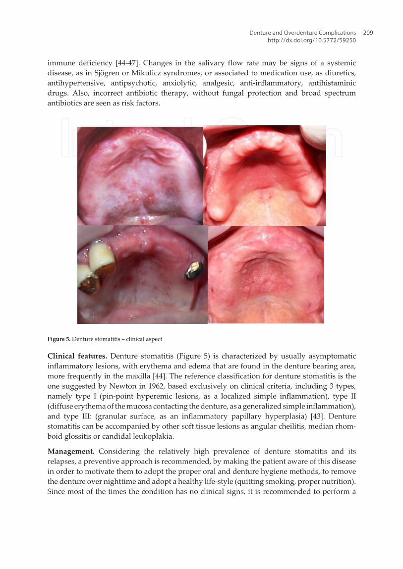
Figure 5. Denture stomatitis – clinical aspect
Clinical features. Denture stomatitis (Figure 5) is characterized by usually asymptomaticinflammatory lesions, with erythema and edema that are found in the denture bearing area,more frequently in the maxilla [44]. The reference classification for denture stomatitis is theone suggested by Newton in 1962, based exclusively on clinical criteria, including 3 types,namely type I (pin-point hyperemic lesions, as a localized simple inflammation), type II(diffuse erythema of the mucosa contacting the denture, as a generalized simple inflammation),and type III: (granular surface, as an inflammatory papillary hyperplasia) [43]. Denturestomatitis can be accompanied by other soft tissue lesions as angular cheilitis, median rhom‐boid glossitis or candidal leukoplakia.
Management. Considering the relatively high prevalence of denture stomatitis and itsrelapses, a preventive approach is recommended, by making the patient aware of this diseasein order to motivate them to adopt the proper oral and denture hygiene methods, to removethe denture over nighttime and adopt a healthy life-style (quitting smoking, proper nutrition).Since most of the times the condition has no clinical signs, it is recommended to perform a
Denture and Overdenture Complicationshttp://dx.doi.org/10.5772/59250
209
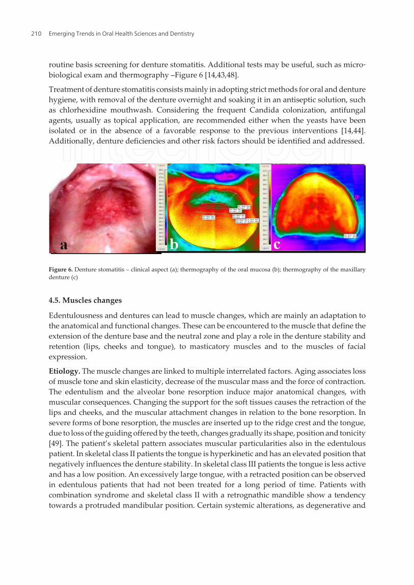
routine basis screening for denture stomatitis. Additional tests may be useful, such as micro‐biological exam and thermography –Figure 6 [14,43,48].
Treatment of denture stomatitis consists mainly in adopting strict methods for oral and denturehygiene, with removal of the denture overnight and soaking it in an antiseptic solution, suchas chlorhexidine mouthwash. Considering the frequent Candida colonization, antifungalagents, usually as topical application, are recommended either when the yeasts have beenisolated or in the absence of a favorable response to the previous interventions [14,44].Additionally, denture deficiencies and other risk factors should be identified and addressed.
Figure 6. Denture stomatitis – clinical aspect (a); thermography of the oral mucosa (b); thermography of the maxillarydenture (c)
4.5. Muscles changes
Edentulousness and dentures can lead to muscle changes, which are mainly an adaptation tothe anatomical and functional changes. These can be encountered to the muscle that define theextension of the denture base and the neutral zone and play a role in the denture stability andretention (lips, cheeks and tongue), to masticatory muscles and to the muscles of facialexpression.
Etiology. The muscle changes are linked to multiple interrelated factors. Aging associates lossof muscle tone and skin elasticity, decrease of the muscular mass and the force of contraction.The edentulism and the alveolar bone resorption induce major anatomical changes, withmuscular consequences. Changing the support for the soft tissues causes the retraction of thelips and cheeks, and the muscular attachment changes in relation to the bone resorption. Insevere forms of bone resorption, the muscles are inserted up to the ridge crest and the tongue,due to loss of the guiding offered by the teeth, changes gradually its shape, position and tonicity[49]. The patient’s skeletal pattern associates muscular particularities also in the edentulouspatient. In skeletal class II patients the tongue is hyperkinetic and has an elevated position thatnegatively influences the denture stability. In skeletal class III patients the tongue is less activeand has a low position. An excessively large tongue, with a retracted position can be observedin edentulous patients that had not been treated for a long period of time. Patients withcombination syndrome and skeletal class II with a retrognathic mandible show a tendencytowards a protruded mandibular position. Certain systemic alterations, as degenerative and
Emerging Trends in Oral Health Sciences and Dentistry210
autoimmune conditions, vascular accidents, paresis, burns, traumatisms, nutritional statusalterations as protein deficiencies, associate muscular changes.
Prosthetic treatment deficiencies favor abnormal muscular changes. Increased verticaldimension of occlusion and ill-fitting dentures cause muscle spasms, habitual and involuntarymovements. Oversized anterior buccal flange of the maxillary denture associates the overex‐tension of the upper lip, with possible anatomical and functional consequences. Associationof posterior artificial tooth wear with over jet or lack of coincidence of maximal intercuspalposition and centric relation leads to an abnormal protruded mandibular position, whichmakes difficult the registration of maxillomandibular relationship (centric relation).
Clinical features. Generally, the clinical aspects are the result of complex muscular changes,such as regarding the tone, volume and attachment of the muscle, combined with neuromus‐cular coordination and control deficiencies.
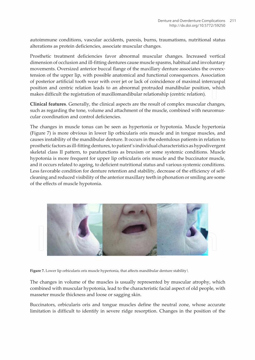
The changes in muscle tonus can be seen as hypertonia or hypotonia. Muscle hypertonia(Figure 7) is more obvious in lower lip orbicularis oris muscle and in tongue muscles, andcauses instability of the mandibular denture. It occurs in the edentulous patients in relation toprosthetic factors as ill-fitting dentures, to patient’s individual characteristics as hypodivergentskeletal class II pattern, to parafunctions as bruxism or some systemic conditions. Musclehypotonia is more frequent for upper lip orbicularis oris muscle and the buccinator muscle,and it occurs related to ageing, to deficient nutritional status and various systemic conditions.Less favorable condition for denture retention and stability, decrease of the efficiency of self-cleaning and reduced visibility of the anterior maxillary teeth in phonation or smiling are someof the effects of muscle hypotonia.
Figure 7. Lower lip orbicularis oris muscle hypertonia, that affects mandibular denture stability\
The changes in volume of the muscles is usually represented by muscular atrophy, whichcombined with muscular hypotonia, lead to the characteristic facial aspect of old people, withmasseter muscle thickness and loose or sagging skin.
Buccinators, orbicularis oris and tongue muscles define the neutral zone, whose accuratelimitation is difficult to identify in severe ridge resorption. Changes in the position of the
Denture and Overdenture Complicationshttp://dx.doi.org/10.5772/59250
211
muscle insertions occur, such as high muscle insertions, even on the ridge top (genioglossusand mentalis muscle), with detached oral mucosa. Considering that position of muscleattachments has a major impact to denture base stability and retention, through changes of thedenture bearing area, severe ridge resorption with consecutive muscles changes increase thetreatment difficulty degree, especially in the mandible.
Muscle force decreasing leads to decrease in the capacity of performing a voluntary act (suchas mastication). This occurs in relation to ageing, paresis, depression, denture instability orpain caused by the dentures. Alterations in jaw movements can occur in relation to deficienciesof the prosthetic restorations, as unstable occlusion, denture instability, increased verticaldimension of occlusion or in bruxism. Muscular spasms are encountered in particularsituations as in the jaw-closing muscles, related to an increased vertical dimension of occlusionor for jaw-opening muscles related to a decreased vertical dimension of occlusion.
Neuromuscular coordination and control deficiencies, which occur in relation to age andsystemic alterations, can increase treatment difficulty and negatively influence the accommo‐dation with the prosthesis. For example, in Parkinson disease a lack of neuromuscularcoordination occurs, which leads to difficulties in registration of maxillomandibular relation‐ship and in the insertion and removal of the denture or the overdenture. Abnormal, involun‐tary, patterned or stereotyped and purposeless orofacial movements (oral dyskinesia) canoccur linked to ill-fitting unstable dentures, oral discomfort, and lack of sensory contacts [2].Facial nerve paresis includes affected unilateral facial musculature movement with asymmetryof facial expression and functional disorders, taste alterations and salivary changes, all havingimpact on the prosthetic treatment – difficulties in impression taking and in registration ofmaxillomandibular relationship, reduced masticatory efficiency with unilateral mastication,increased risk of unstable dentures, aesthetic alterations and denture intolerance.
Management. Considering the importance of the muscle factor for the oral functioning, anaccurate evaluation should be performed. In some cases, besides the clinical evaluation,additional tests are recommended, such as electromyography or kinesiography, and some‐times special treatment conduct is required [50].
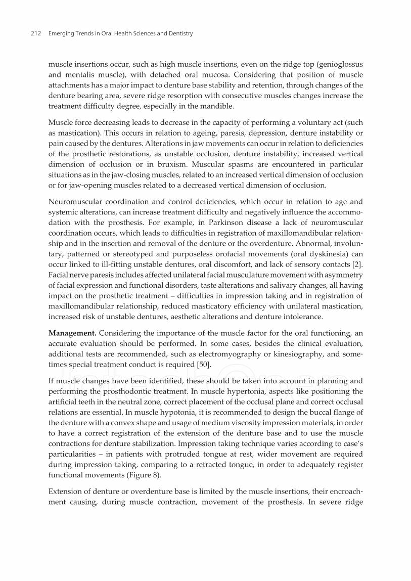
If muscle changes have been identified, these should be taken into account in planning andperforming the prosthodontic treatment. In muscle hypertonia, aspects like positioning theartificial teeth in the neutral zone, correct placement of the occlusal plane and correct occlusalrelations are essential. In muscle hypotonia, it is recommended to design the buccal flange ofthe denture with a convex shape and usage of medium viscosity impression materials, in orderto have a correct registration of the extension of the denture base and to use the musclecontractions for denture stabilization. Impression taking technique varies according to case’sparticularities – in patients with protruded tongue at rest, wider movement are requiredduring impression taking, comparing to a retracted tongue, in order to adequately registerfunctional movements (Figure 8).
Extension of denture or overdenture base is limited by the muscle insertions, their encroach‐ment causing, during muscle contraction, movement of the prosthesis. In severe ridge
Emerging Trends in Oral Health Sciences and Dentistry212
resorption cases, as for those with muscle insertions on the ridge top, preprosthetic surgeryfor repositioning of muscle and mucosal attachments is indicated [51].
In neuromuscular coordination and control deficiencies, considering the severe functionalalterations, conventional dentures usually don’t respond to patient’s need and implantoverdenture should be chosen instead. Compared to conventional dentures, implant over‐dentures provides better functional parameters – exertion of higher masticatory forcespromotes better nutrition through the ability to chew harder foods.
Last but not least, manufacturing of a new prosthesis requires an adjustment period for theestablishment of the new memory patterns for the masticatory muscles, of about 6 to 8 weeks,aspect that should be mentioned to the patient [52].
4.6. Facial alterations, including esthetic complications
The complete edentulism contributes greatly to the facial aspect known as the aged appear‐ance. Prosthetic treatment needs to adequately address this consequence of edentulism,considering the fact that patients’ complaints are frequently related to aesthetic reasons.
Etiology. The facial appearance of the edentulous patient is the result of factors related tocomplete edentulism and prosthetic treatment, combined with others such as ageing, local andgeneral particularities and medical conditions.
Edentulism associates significant anatomical and functional changes that impact the facialappearance. Lip and cheek support is severely altered by tooth loss and bone resorption. Atendency of increasing the facial concavity occurs in relation to the different pattern of boneresorption of the jaws (centripetal in the maxilla and centrifugal in the mandible). In associationwith the loss of the occlusal contacts, a counter-clockwise rotation of the mandible, with adecreasing height of the lower third of the face, and sometimes a tendency to a more advancedprotruded mandibular position occurs. Facial alterations that are directly linked to edentulismcan be considered worsening factors of the esthetic appearance, since there are also preexistentchanges in relation to other factors.
As a consequence of aging, there are changes related to the evolution of bones and soft tissues(muscles, fat and skin), in addition to noticeable effects of gravity, with effect on facial esthet‐ics [53]. Systemic health, medication use and behavior (e.g., alcohol and tobacco use) can
Figure 8. Tongue position at rest – anterior vs. posterior
Denture and Overdenture Complicationshttp://dx.doi.org/10.5772/59250
213
influence the facial appearance. For example, smoking causes changes particularly in the lowerand middle third of the face, like hyperpigmentation and accentuated wrinkles-deepernasolabial folds, upper lip wrinkles, lower lip vermillion wrinkles, lower lid hyperpigmenta‐tion [54]. Premature aged appearance occurs in some diseases like Cutis laxa or glomeruloneph‐ritis [55,56].
The prosthetic treatment of the edentulous patient addresses positively some of the previousmentioned facial alteration, but can also contribute to an aged appearance through its defi‐ciencies, as in cases with a decreased vertical dimension of occlusion, a reverse smile line ordarker, yellow artificial teeth.
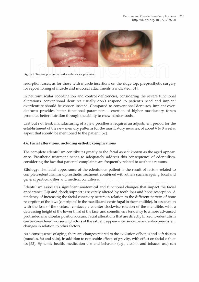
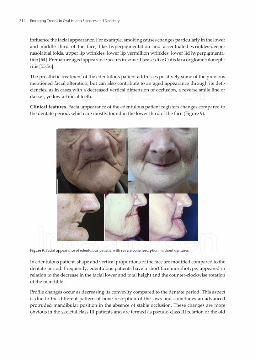
Clinical features. Facial appearance of the edentulous patient registers changes compared tothe dentate period, which are mostly found in the lower third of the face (Figure 9).
Figure 9. Facial appearance of edentulous patient, with severe bone resorption, without dentures
In edentulous patient, shape and vertical proportions of the face are modified compared to thedentate period. Frequently, edentulous patients have a short face morphotype, appeared inrelation to the decrease in the facial lower and total height and the counter-clockwise rotationof the mandible.
Profile changes occur as decreasing its convexity compared to the dentate period. This aspectis due to the different pattern of bone resorption of the jaws and sometimes an advancedprotruded mandibular position in the absence of stable occlusion. These changes are moreobvious in the skeletal class III patients and are termed as pseudo-class III relation or the old
Emerging Trends in Oral Health Sciences and Dentistry214
man's prognathism. Profile changes include also modification of nasolabial angle related tonose tip lowering and loss of upper lip support.
Lips register great changes, as reduction of vermilion height and their volume, color modifi‐cations, retraction due to support loss, elongation (upper lip) and shortening (lower lip),straight or reversed lip line and low smile line, and reduced lips dynamics that contribute toa decreased teeth exposure during speaking and smiling, which associated a reduction ofemotional display, as happiness or sadness [57].
Facial changes related to ageing mark the facial appearance. Lips and cheeks become lessprominent and there can be noticed marked folds and wrinkles, loose or sagging skin, changesin the skin texture and hyperpigmentation. These are mainly connected to muscle changes, ashypotonia, and skin changes, as loss of skin elastic recoil.
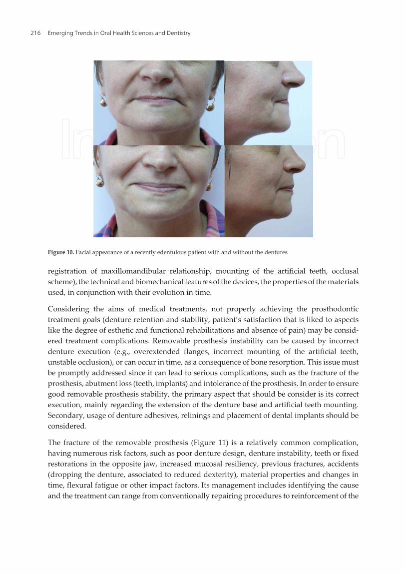
The prosthetic treatment has a positive impact on the facial esthetics (Figure 10). Generally, itprovides a support for the soft tissue, tries to compensate the tooth loss and bone resorption(through the artificial teeth and anterior buccal maxillary flange), ensures a functional verticaldimension of occlusion and give a natural look through exposure of the teeth during smilingor speaking. Some faulty prosthesis or some changes that occurs in time can have a negativeimpact on facial esthetic. Unpleasant facial appearance can be linked to errors in anteriorartificial tooth mounting (too forward, too backward), shade selection (chosen incorrectly, toolight, not matching the patient's age), to changes of the artificial teeth over time (through teethwear the smile line can become reversed, or through aging of the material discolorations canappear). A decreased vertical dimension of occlusion leads to an aged appearance, with deeperperioral folds, and an increased vertical dimension of occlusion associate an unnatural,tensioned look. An overextended buccal flange, encountered more often in the maxillarydentures, leads to an over-supported lip with a tensioned unnatural look. Unstable denturesnegatively influence facial appearance through movement while speaking and the facialchanges related to protruded mandibular position that many times is associated.
Management. Facial esthetic evaluation must consider changes’ severity and causes, in orderto properly address them and respond to patients’ need and expectations. In order to make anaccurate analysis, regular clinical examination (from frontal and lateral view, with and withoutdentures, in rest and in maximal intercuspal position, during speech and smiling) can besupplemented by radiological examination (cephalometric radiographs) and records from thedentate period, as photos, dental casts, radiographs. The prosthetic rehabilitation of thecompletely edentulous patient must consider, from an aesthetic point of view, beside thegeneral esthetic principles, also patient’s features that are relatively obvious in the dentateperiod and rather difficult to assess in the edentulous one. The previous should be related toother patient’s characteristics (e.g., age, sex, functional particularities, health status) and toprosthodontic biomechanical requirements in order to obtain a good treatment outcome.
4.7. Denture and overdenture biomechanical and technical complications
Removable dental prosthesis are described as having a series of complications in relation tothe correctness and accuracy of their planning and execution (extension of the denture base,
Denture and Overdenture Complicationshttp://dx.doi.org/10.5772/59250
215
registration of maxillomandibular relationship, mounting of the artificial teeth, occlusalscheme), the technical and biomechanical features of the devices, the properties of the materialsused, in conjunction with their evolution in time.
Considering the aims of medical treatments, not properly achieving the prosthodontictreatment goals (denture retention and stability, patient’s satisfaction that is liked to aspectslike the degree of esthetic and functional rehabilitations and absence of pain) may be consid‐ered treatment complications. Removable prosthesis instability can be caused by incorrectdenture execution (e.g., overextended flanges, incorrect mounting of the artificial teeth,unstable occlusion), or can occur in time, as a consequence of bone resorption. This issue mustbe promptly addressed since it can lead to serious complications, such as the fracture of theprosthesis, abutment loss (teeth, implants) and intolerance of the prosthesis. In order to ensuregood removable prosthesis stability, the primary aspect that should be consider is its correctexecution, mainly regarding the extension of the denture base and artificial teeth mounting.Secondary, usage of denture adhesives, relinings and placement of dental implants should beconsidered.
The fracture of the removable prosthesis (Figure 11) is a relatively common complication,having numerous risk factors, such as poor denture design, denture instability, teeth or fixedrestorations in the opposite jaw, increased mucosal resiliency, previous fractures, accidents(dropping the denture, associated to reduced dexterity), material properties and changes intime, flexural fatigue or other impact factors. Its management includes identifying the causeand the treatment can range from conventionally repairing procedures to reinforcement of the
Figure 10. Facial appearance of a recently edentulous patient with and without the dentures
Emerging Trends in Oral Health Sciences and Dentistry216

denture base with metal or non-metal products (as glass and polyethylene fibers or net), tochanging the previous denture or even the treatment option [58].
Figure 11. Overdenture fracture at the attachment site
The complications associated to the properties of the material used, mainly polymethylme‐thacrylate (PMMA), are linked to changes that appears during their evolution in time, asdiscolorations, artificial teeth wear, increased porosity and decrease flexural strength. Con‐sidering their functional and aesthetic impact, denture and overdenture treatment should berenewed at approximately every 5 years.
Additionally, signs of combination syndrome can appear when mandibular overdentures(supported or retained by roots or dental implants) are opposed by an edentulous maxilla. Inthis situation the masticatory field moves anteriorly, favoring the instability of the maxillarydenture and the increased bone resorption rate in the anterior maxilla. This iatrogenic effectcan be managed by using implants also in the maxilla, aiming to address or prevent thisfunctional consequence and the destructive process of the oral structures [59].
4.8. Teeth complications, with root overdentures
The root overdentures can have teeth related complications, mainly due to primary orrecurrent caries, periradicular lesions developed by vital teeth, endodontically lesionsdeveloped by endodontically treated teeth due to loss of the restoration sealing the root canal,periodontitis or root fracture [60]. Their management is dependent of the problem type, inmost severe forms tooth loss and recurrent failure of prosthodontic treatment occurring. It isimportant to preserve the roots as a prevention factor for bone resorption and due their positiveimpact on the oral functioning [61]. Patients’ awareness, instruction and motivation regardingmaintaining a proper oral hygiene are essential considering that is the main factor for perio‐dontal disease and caries control. When caries occur, it is important to identify them quicklyin order to have high a high success rate for the treatment. Topical fluoridation or coveragewith metallic caps can be performed preventively for patients with a high caries risk. For theperiodontal disease it is recommended to use Chlorhexidine 0.12% mouthwash twice daily.Also, the removal of the denture overnight and maintenance of proper denture hygiene are
Denture and Overdenture Complicationshttp://dx.doi.org/10.5772/59250
217
recommended. If tooth mobility appears, it can be addressed by reducing the tooth height,which leads to an increase in the crown to root ratio. The risk of root fracture is higher inendodontically treated teeth and when the magnitude of occlusal forces is higher, as in dentureinstability, bruxism, increased vertical dimension of occlusion, when teeth or fixed prosthesisin the opposite jaw. Preventively, thimble crowns can be used.
4.9. Implants complications, with implant overdentures
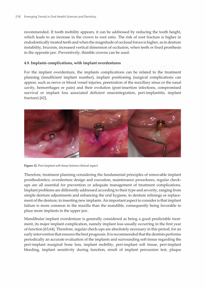
For the implant overdenture, the implants complications can be related to the treatmentplanning (insufficient implant number), implant positioning (surgical complications canappear, such as nerve or blood vessel injuries, penetration of the maxillary sinus or the nasalcavity, hemorrhages or pain) and their evolution (post-insertion infections, compromisedsurvival or implant loss associated deficient osseointegration, peri-implantitis, implantfracture) [62].
Figure 12. Peri-implant soft tissue lesions-clinical aspect
Therefore, treatment planning considering the fundamental principles of removable implantprosthodontics, overdenture design and execution, maintenance procedures, regular check-ups are all essential for prevention or adequate management of treatment complications.Implant problems are differently addressed according to their type and severity, ranging fromsimple denture adjustments and enhancing the oral hygiene, to denture relinings or replace‐ment of the denture, to inserting new implants. An important aspect to consider is that implantfailure is more common in the maxilla than the mandible, consequently being favorable toplace more implants in the upper jaw.
Mandibular implant overdenture is generally considered as being a good predictable treat‐ment, its major implant complication, namely implant loss usually occurring in the first yearof function [63,64]. Therefore, regular check-ups are absolutely necessary in this period, for anearly intervention that ensures the best prognosis. It is recommended that the dentists performsperiodically an accurate evaluation of the implants and surrounding soft tissue regarding theperi-implant marginal bone loss, implant mobility, peri-implant soft tissue, peri-implantbleeding, implant sensitivity during function, result of implant percussion test, plaque
Emerging Trends in Oral Health Sciences and Dentistry218
accumulation. The overdentures must be verified regarding the overdenture base that is indirect contact with the implant, as risk factor for peri-implant soft tissue complications,regarding the occlusion and maxillomandibular relationship whose faults may be related toexerting increased pressure on implants, as risk factor for implant failure, as its stability andhygiene. Other aspects, like the prosthetic treatment on the opposite jaw (an unstable dentureas antagonist can produce excessive forces on the implants) and parafunctions should bechecked.
4.10. Attachment system complication, with overdentures
Attachment system complications can occur as a consequence of an incorrect treatmentplanning, improper treatment conduct (e.g., errors during placement of the retentive housingin the overdenture base) or related to their changes that occur in time, during functioning (e.g.,loosening or damage). These vary according to the type of attachment system, e.g., bar, ball,Locator. Most frequent attachment system complications, with overdentures, are: decreasedprosthesis retention due to deactivation, detachment, damage or loss of the retentive housing;abutment screw loosening or fracture; fracture of the attachment system components (e.g., baror clip fracture); soft tissue lesions as hyperplasia under the bar or peri-implant mucositis.
The management of attachment system complications varies according to the attachmentsystem used and the complication type. Technical complications are more common for barthan ball attachments, and both of them are more common compared to locator system [65,66].Usually low severity complications occurs, such as loss of rubber ring and matrix deactivation,which need to be promptly addressed since they cause overdenture instability with possiblenegative impact on the dental implants. A more severe complication is bar fracture, thatrequires increased clinical time and expenses to be resolved, considering that usually theoverdenture must be replaced. In elderly edentulous patients simpler prosthetic reconstruc‐tions, with complications that require decreased time and money are preferred. Thus, if theoption of implant overdenture has been selected, the ball attachment system can be moreappropriate than the bar attachment system, due to the more simple maintenance proceduresand easier replacement of the implant if necessary.
4.11. Patient satisfaction and quality of life
The conventional dentures are the most common treatment option for the edentulous patients,and usually register good results in terms of patient’s satisfaction. Dissatisfaction reasons mostclaimed by patients are related to denture instability, improper mastication, esthetic deficiencyand phonation problems [67]. Denture intolerance is usually connected to subjective factors(the patient’s needs and expectations, psychological type, misconceptions) or objective factors(denture instability, pain, functional deficiencies).
The root or implant overdenture have improved retention that contributes to physical andpsychological comfort. According to the current evidence, mandibular implant overdenturesprovide a higher satisfaction and oral health related quality of life compared to conventionaldenture, but there is uncertainty about the true magnitude of difference between the two [68].
Denture and Overdenture Complicationshttp://dx.doi.org/10.5772/59250
219
5. Conclusions
Dentures and overdentures, the most frequently used treatment options for the completeedentulism, have complications that are related to patient and prostheses features. Patient’sgeneral and local conditions and behavior must be acknowledged as their manifestations,interactions and impact on the prosthetic treatment. Removable implant prosthodonticsprinciples should be well-known and respected during prosthesis execution. The previous,additional to regular check-ups, represent the basis of the prevention removable prosthesiscomplications.
Denture and overdenture complications are partially similar, differences being related todesign particularities, biomechanical aspects and execution procedures. Addressing themdepends on their nature and severity, requiring a specific medical conduct. Often simpleclinical interventions are needed, but sometimes complex procedures with increased clinical,biological and financial costs must be considered in order to achieve a medical result thatcorresponds to the current medical standards and patient needs and expectations.
Author details
Elena Preoteasa1*, Cristina Teodora Preoteasa2, Laura Iosif1, Catalina Murariu Magureanu1 andMarina Imre1
*Address all correspondence to: [email protected]
1 Department of Prosthodontics, Faculty of Dental Medicine, Carol Davila University ofMedicine and Pharmacy, Bucharest, Romania
2 Department of Oral Diagnosis, Ergonomics, Scientific Research Methodology, Faculty ofDental Medicine, Carol Davila University of Medicine and Pharmacy, Bucharest, Romania
References
[1] Carlsson GE, Omar R. The Future of Complete Dentures in Oral Rehabilitation. ACritical Review. Journal of Oral Rehabilitation 2010;37(2): 143-156.
[2] Emami E, de Souza RF, Kabawat M, Feine JS. The Impact of Edentulism on Oral andGeneral Health. International Journal of Dentistry 2013; 2013. DOI:10.1155/2013/498305 (accesed 1 September 2014).
[3] Cunha-Cruz J, Hujoel PP, Nadanovsky P. Secular Trends in Socio-Economic Dispari‐ties in Edentulism: USA, 1972–2001. Journal of Dental Research 2007;86(2): 131–13.
Emerging Trends in Oral Health Sciences and Dentistry220

[4] Beltrán-Aguilar ED, Barker LK, Canto MT, Dye BA, Gooch BF, Griffin SO, Hyman J,Jaramillo F, Kingman A, Nowjack-Raymer R, Selwitz RH, Wu T. Surveillance forDental Caries, Dental Sealants, Tooth Retention, Edentulism, and Enamel Fluorosis--United States, 1988-1994 and 1999-2002. Morbidity and Mortality Weekly Report.Surveillance Summaries 2005;54(3): 1-43.
[5] Vos T, Flaxman AD, Naghavi M, Lozano R, Michaud C, Ezzati M, Shibuya K, et al.Years Lived with Disability (YLDs) for 1160 Sequelae of 289 Diseases and Injuries1990-2010: A Systematic Analysis for the Global Burden of Disease Study 2010. Lan‐cet 2012; 380(9859): 2163-96. DOI: 10.1016/S0140-6736(12)61729-2 (accesed 1 Septem‐ber 2014).
[6] Felton DA. Edentulism and Comorbid Factors. Journal of Prosthodontics 2009;18(2):88–96.
[7] World Health Organization. International Classification of Functioning, Disabilityand Health. Geneva, Switzerland. World Health Organization; 2001.
[8] O′ Reilly E. Treatment Options for Our Edentulous Patients. http://www.burlington‐dentalclinic.ie/wp-content/uploads/2012/10/Dr-Eddie-OReilly-Edentulous-Pa‐tients.pdf (accesed 1 September 2014).
[9] Suarez-Sanchez OR. Evaluating Epidemiological Properties of the American Collegeof Prosthodontists (ACP) Prosthodontic Diagnostic Index for Complete Edentulism(PDI-CE)(PDI-Study). Harvard School of Dental Medicine; 2007.
[10] Papadaki E, Anastassiadou V. Elderly Complete Denture Wearers: A Social Ap‐proach to Tooth Loss. Gerodontology 2012; 29(2):e721-7. DOI: 10.1111/j.1741-2358.2011.00550.x. (accesed 14 September 2014).
[11] Dye BA, Tan S, Smith V, Lewis BG, Barker LK, Thornton-Evans G, Eke PI, Beltrán-Aguilar ED, Horowitz AM, Li CH. Trends in Oral Health Status: United States,1988-1994 and 1999-2004. Vital Health Stat 11. 2007;Apr. (248): 1-92.
[12] Douglass CW, Shih A, Ostry L. Will There Be a Need for Complete Dentures in theUnited States in 2020? Journal of Prosthetic Dentistry 2002;87(1): 5–8.
[13] Osterberg T, Carlsson GE. Dental State, Prosthodontic Treatment and Chewing Abili‐ty-a Study of Five cohorts of 70-Year-Old Subjects. Journal of Oral Rehabilitation2007;34(8): 553-9.
[14] Felton D, Cooper L, Duqum I, Minsley G, Guckes A, Haug S, Meredith P et al. Evi‐dence-Based Guidelines for the Care and Maintenance of Complete Dentures: a Pub‐lication of the American College of Prosthodontists. Journal of the American DentalAssociation 2011;142(Suppl 1): 1S-20S.
[15] Thompson GW, Kreisel PS. The Impact of the Demographics of Aging and the Eden‐tulous Condition on Dental Care Services. Journal of Prosthetic Dentistry 1998;79(1):56-9.
Denture and Overdenture Complicationshttp://dx.doi.org/10.5772/59250
221
[16] American College of Prosthodontists (ACP). The Prosthodontic Diagnostic Index forComplete Edentulism (PDI-CE). http://ww-w.gotoapro.org/assets/1/7/Complete_Edentulism_Checklist.pdf (accesed 1 September 2014).
[17] Al-Zubeidi MI, Payne AG. Mandibular Overdentures: A Review of Treatment Philos‐ophy and Prosthodontic Maintenance. New Zealand Dental Journal 2007;103(4):88-97.
[18] Kiyak HA, Reichmuth M. Barriers to and Enablers of Older Adults' Use of DentalServices. Journal of Dental Education 2005; 69(9): 975-6.
[19] [19]. Montini T, Tseng TY, Patel H, Shelley D. Barriers to Dental Services for OlderAdults. American Journal of Health Behaviour 2014;38(5): 781-8.
[20] Wismeijer D, Ten BC, Schulten EA. Notes Concerning an Overdenture on 2 Implantsas the Standard for Treating an Edentulous Mandible. Nederlands Tijdschrift VoorTandheelkunde 2011;118(12): 633-9.
[21] Sutton AF, Glenny AM, McCord JF. Interventions for Replacing Missing Teeth: Den‐ture Chewing Surface Designs in Edentulous People. The Cochrane Database of Sy‐sematic Reviews 2005;25(1). DOI: 10.1002/14651858.CD004941.pub2 (accesed 1September 2014).
[22] Preoteasa CT, Nabil Sultan A, Popa L, Ionescu E, Iosif, L, Ghica, MV, Preoteasa E.Wettability of Some Dental Materials. Optoelectronics and Advanced Materials –Rapid Communications 2011; 5(8): 874-8.
[23] Preoteasa E, Imre M, Preoteasa CT. A 3-Year Follow-up Study of Overdentures Re‐tained by Mini–Dental Implants. The International Journal of Oral & MaxillofacialImplants 2014; 29(5). 1034-41.
[24] Carlsson GE. Clinical Morbidity and Sequelae of Treatment with Complete Dentures.Journal of Prosthetic Dentistry 1998;79(1): 17-23.
[25] Petersen PE, Kandelman D, Arpin S, Ogawa H. Global Oral Health of Older People-Call for Public Health Action. Community Dental Health 2010;27(4 Suppl 2): 257-67.
[26] Hummel SK, Wilson MA, Marker VA, Nunn ME. Quality of Removable Partial Den‐tures Worn by the Adult U.S. Population. Journal of Prosthetic Dentistry 2002;88(1):37-43.
[27] Pietrokovski J, Harfin J, Levy F. The Influence of Age and Denture Wear on the Sizeof Edentulous Structures. Gerodontology 2003;20(2): 100-5.
[28] Fitzpatrick AL, Powe NR, Cooper LS, Ives DG, Robbins JA. Barriers to Health CareAccess Among the Elderly and Who Perceives Them. American Journal of PublicHealth 2004; 94(10):1788-94.
Emerging Trends in Oral Health Sciences and Dentistry222
[29] Dolan TA, Atchison KA. Implications of Access, Utilization and Need for OralHealth Care by the Non-Institutionalized and Institutionalized Elderly on the DentalDelivery System. Journal of Dental Education 1993;57(12): 876-87.
[30] Dolan TA, Atchison K, Huynh TN. Access to Dental Care Among Older Adults in theUnited States. Journal of dental Education 2005; 69(9): 961-74.
[31] Health and Behavior. The Interplay of Biological, Behavioral, and Societal Influences.Institute of Medicine (US) Committee on Health and Behavior: Research, Practice,and Policy. Washington DC. National Academies Press (US); 2001. http://www.ncbi.nlm.nih.gov/books/NBK43743/ (accesed 1 September 2014).
[32] Barratt A. Evidence Based Medicine and Shared Decision Making: The Challenge ofGetting Both Evidence and Preferences Into Health Care. Patient Education andCounseling 2008;73(3): 407-12.
[33] Graber T, Eliades T, Athanasiou AE. Risk Management in Orthodontics: Expers'Guide to Malpractice. Chicago: Quintessence Publishing Co; 2004.
[34] Berglundh T, Persson L, Klinge B. A Systematic Review of the Incidence of Biologicaland Technical Complications in Implant Dentistry Reported in Prospective Longitu‐dinal Studies of at least 5 Years. Journal of Clinical Periodontology 2002;29 (Suppl 3):197-212.
[35] Misch K, Wang H. Implant Surgery Complications: Etiology and Treatment. ImplantDentistry 2008; 17(2): 159-68.
[36] Atwood DA. Reduction of Residual Ridges: A Major Disease Entity. Journal of Pros‐thetic Dentistry 1971;26(3): 266-79.
[37] Thomason JM, Kelly SA, Bendkowski A, Ellis JS.Two implant retained overden‐tures--a review of the literature supporting the McGill and York consensus state‐ments. Journal of Dentistry 2012;40(1):22-34.
[38] Budtz-Jörgensen E. Oral Mucosal Lesions Associated with the Wearing of RemovableDentures. Journal of Oral Pathology 1981; 10(2): 65-80.
[39] Dunlap CL, Barker CF. A Guide to Common Oral Lesions. http://dentistry.umkc.edu/Practicing_Communities/asset/OralLesions (accessed 23 July 2014).
[40] Coelho CM, Sousa YT, Daré AM. Denture-Related Oral Mucosal Lesions in a Brazil‐ian School of Dentistry. Journal of Oral Rehabilitation 2004;31(2): 135-9.
[41] Sudarshan R, Sree Vijayabala G, Prem Kumar KS. Inflammatory Hyperplasia of theOral Cavity. Archives Medical Review Journal. 2012; 21(4): 299-307.
[42] Canger EM, Celenk P, Kayipmaz S. Denture-Related Hyperplasia: A Clinical Studyof a Turkish Population Group. Brazilian Dental Journal 2009; 20(3): 243-8.
Denture and Overdenture Complicationshttp://dx.doi.org/10.5772/59250
223
[43] European Association of Oral Medicine (EAOM). Denture Related Stomatitis. http://www.eaom.eu/empty_24.html (accesed 1 September 2014).
[44] Gendreau L, Loewy ZG. Epidemiology and Etiology of Denture Stomatitis. Journal ofProsthodontics 2011; 20(4): 251-60.
[45] Petersen PE, Yamamoto T. Improving the Oral Health of Older People: The Ap‐proach of the WHO Global Oral Health Programme. Community Dentistry and OralEpidemiology 2005;33(2): 81-92.
[46] Shulman JD, Rivera-Hidalgo F, Beach MM. Risk Factors Associated with DentureStomatitis in the United States. Journal of Oral Pathology and Medicine 2005 34(6):340-6.
[47] Lylajam S, Prasanath V. Denture Stomatitis-Etiological Factors and Management.Journal of Clinical Dentistry 2011;2(1): 9-11.
[48] Preoteasa E, Iosif L, Amza O, Preoteasa CT, Dumitrascu C. Thermography, an Imag‐istic Method in Investigation of the Oral Mucosa Status in Complete Denture Wear‐ers. Journal of Optoelectronics and Advanced Materials 2010;12(11): 2333–4.
[49] Pietrokovski J, Kaffe I, Arensburg B. Retromolar Ridge in Edentulous Patients: Clini‐cal Considerations. Journal of Prosthodontics 2007;16(6): 502-6.
[50] Melescanu Imre M, Preoteasa E, Tancu A, Preoteasa CT. Imaging Technique for theComplete Edentulous Patient Treated Conventionally or With Mini Implant Over‐denture. Jornal of Medicine and Life 2013;6(1): 86-92.
[51] Cawood JI, Stoelinga PJ. International Research Group on Reconstructive Prepros‐thetic Surgery. Consensus Report. International Journal of Oral Maxillofacial Surgery2000;29(3): 159-62.
[52] Goiato MC, Filho HG, Dos Santos DM, Barão VA, Júnior AC. Insertion and Follow-Up of Complete Dentures: A Literature Review. Gerodontology 2011;28(3): 197-204.
[53] Nkengne A, Bertin C. Aging and Facial Changes--Documenting Clinical Signs, Part 1:Clinical Changes of the Aging Face. Skinmed 2012;10(5): 284-289.
[54] Okada HC, Alleyne B, Varghai K, Kinder K, Guyuron B. Facial Changes Caused bySmoking: A Comparison Between Smoking and Nonsmoking Identical Twins. Plasticand Reconstructive Surgery 2013;132(5): 1085-92.
[55] Mataix J, Bañuls J, Lucas A, Pastor N, Betlloch I. Prematurely Aged Appearance.Clinical and Experimental Dermatology 2006;31(6): 833-4.
[56] Joss N, Boulton-Jones JM, More I. Premature Ageing and Glomerulonephritis. Neph‐rology Dialisis Transplantation 2001;16(3): 615-8.
Emerging Trends in Oral Health Sciences and Dentistry224
[57] Tupac R, et al. Parameters of Care for the Speciality of Prosthodontics. Jornal of pros‐thodontics. Implant, Esthetic, and Reconstructive Dentistry. Official Journal of TheAmerican College of Prosthodontists 2005;14(4)Suppl: 1-103.
[58] Preoteasa E, Murariu CM, Ionescu E, Preoteasa CT. Acrylic Resin ReinforcementWith Metallic and Nonmetallic Inserts. Revista Medico-Chirurgicala a Societatii deMedici si Naturalisti din Iasi 2007; 111(2): 487-93.
[59] Kreisler M, Behneke N, Behneke A, d´Hoedt B. Residual Ridge Resorption in theEdentulous Maxila in Patients With Implant-Supported Mandibular Overdentures:An 8-Years Retrospective Study. International Journal of Prosthodontics 2003;16(3):265-300.
[60] Ettinger RL, Qian F. Postprocedural Problems in An Overdenture Population: A Lon‐gitudinal Study. Journal of Endodontics 2004; 30(5): 310-4.
[61] Crum RJ, Rooney GE Jr. Alveolar Boneloss in Overdentures: A 5-Years Study. Jour‐nal of Prosthetic Dentistry 1978; 40(6): 610-3.
[62] Preoteasa E, Meleşcanu-Imre M, Preoteasa CT, Marin M, Lerner H. Aspects of OralMorphology as decision factors in mini-implant supported overdenture. RomanianJournal of Morphoogy and Embryology 2010;51(2): 309-14.
[63] Burns DR. Mandibular Implant Overdenture Treatment: Consensus and Controver‐sy. Journal of Prosthodontics 2000;9(1): 37-46.
[64] Eckert SE, Choi YG, Sánchez AR, Koka S. Comparison of Dental Implant Systems:Quality of Clinical Evidence and Prediction of 5-Year Survival. The InternationalJournal of Oral & Maxillofacial Implants 2005; 20(3):406-15.
[65] Gotfredsen K1, Holm B. Implant-Supported Mandibular Overdentures RetainedWith Ball Or Bar Attachments: A Randomized Prospective 5-Year Study. Internation‐al Journal of Prosthodontics 2000;13(2): 125-30.
[66] Cakarer S, Can T, Yaltirik M, Keskin C. Complications Associated with the Ball, BarAnd Locator Attachments for Implant-Supported Overdentures. Medicina Oral, Pa‐toogial Oral Y Cirurgia Bucal 2011;16(7): e953-9.
[67] Preoteasa E, Marin M, Imre M, Lerner H, Preoteasa CT. Patients' Satisfaction WithConventional Dentures and Mini Implant Anchored Overdentures. Revista Medico-Chirurgicala a Societatii de Medici si Naturisti din Iasi 2012;116(1): 310-16.
[68] Emami E, Heydecke G, Rompré PH, de Grandmont P, Feine JS. Impact of ImplantSupport for Mandibular Dentures on Satisfaction, Oral and General Health-RelatedQuality of Life: A Meta-Analysis of Randomized-Controlled Trials. Clinical Oral Im‐plants Research 2009; 20(6): 533-44.
Denture and Overdenture Complicationshttp://dx.doi.org/10.5772/59250
225