4892 4 supp 107509 lzpmwq supplementary figures supplementary figure s1. return loss (s11) of the...
TRANSCRIPT

1
Supplementary Figures
Supplementary Figure S1. Return loss (S11) of the wireless sensing element. The S11
parameter of various designs of the wireless sensing element was simulated using Ansoft HFSS
software. Inset shows the schematic of the LC design (left) and image of the design implemented
on a Si chip (right).

2
Supplementary Figure S2. Biotransfer of the sensor onto skin. (a) Optical image of the
graphene based wireless sensing element on a water soluble silk fibroin substrate. (b) Conformal
transfer of the sensing element onto human skin via the dissolution of the supporting silk
substrate. (c) Magnified optical image of the sensor after transfer. Scale bars are 7 mm.

3
Supplementary Figure S3. Optical microscopy images of graphene on surfaces. (a) Optical
microscope image of graphene film on Ni surface. (b) Optical microscope image of graphene
transferred onto a surface via the dissolution of the silk film.

4
Supplementary Figure S4. Stability of sensor in running water. Optical images of (a)
biotransferred sensor onto a human arm, (b) mild rinsing in running water, and (c) the sensor
following exposure to running water. Scale bars are 1 cm.

5
Supplementary Figure S5. Raman spectra of tooth enamel and Bombyx mori silk fibroin
film. Raman spectra of (a) bare tooth enamel surface (b) silk fibroin substrate. The amide band
at 1660 cm-1 indicates the presence of domains of silk I structure in the silk film61.

6
Supplementary Figure S6. Structural integrity testing of sensor biotransferred onto bovine
tooth enamel. Optical images of (a) sensor interfaced on tooth before testing, (b) immersion of
the tooth in mouthwash, (c) vortexing of the tooth sensor in mouthwash, (d) structurally intact
sensing element after vortexing, and (e) the tooth after removal from the solution. All scale bars
are 1 cm. (f) Raman spectrum of the graphene surface before vortexing. (g) Raman spectrum of
the graphene sensor after vortexing in mouthwash, showing higher edge-induced D band
intensity62.

7
Supplementary Figure S7. Structural integrity testing of sensor integrated onto IV bag.
Optical images of (a) IV bag sensor before testing, (b) Immersion of the IV bag sensor in water,
(c) Structurally stable sensor after recovery from water. Change in sheet resistance (d) and
transmittance (e) of graphene transducer after undergoing 3 cycles of harsh Q-tip rubbing. Scale
bars are 1 cm.

8
Supplementary Figure S8. Impedance spectrum of the reader coil. The impedance spectrum
of the reader coil antenna in the presence and absence of the sensing element.

9
Supplementary Figure S9. Impedance spectrum of the sensing element. The impedance
magnitude of the sensing element as a function of frequency, illustrating measurement of the
resonance bandwidth from the half-power points.

10
Supplementary Figure S10. Complex impedance spectrum of the sensing element. The
complex impedance of the sensing element as a function of frequency, showing impedance
magnitude, real part and imaginary part. The measurement of the characteristic frequencies of
resonance is illustrated.

11
Supplementary Figure S11. Electrical equivalent circuit of the graphene based wireless
sensing element-reader system. The graphene based wireless sensing element is modeled as an
LRC resonant circuit. The reader system consists of a coil antenna connected to a radio
frequency (RF) impedance analyzer63.
12
Supplementary Methods
The equivalent circuit of the graphene nanosensor reader device is illustrated in Supplementary
Figure S11. The sensing element is modeled as an LRC circuit where L is the inductance of the
meander line inductor, C is the capacitance of the graphene/interdigitated electrode capacitive
system and R is the resistance of the graphene transducing element. The reader system for the
wireless measurement consists of a coil antenna connected to a radio frequency impedance
analyzer (Hewlett-Packard 4191A). The reader antenna is used to inductively couple and power
the remote sensing element. The reader coil is excited by an AC voltage signal generated by the
regulated voltage source in the impedance analyzer, and the corresponding current response is
measured as the frequency is varied. The AC sinusoidal signal on the reader coil will result in a
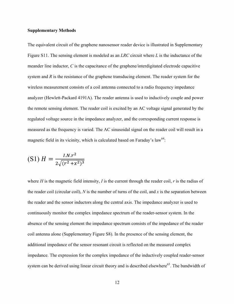
magnetic field in its vicinity, which is calculated based on Faraday’s law64:
(S1) . .
where H is the magnetic field intensity, I is the current through the reader coil, r is the radius of
the reader coil (circular coil), N is the number of turns of the coil, and x is the separation between
the reader and the sensor inductors along the central axis. The impedance analyzer is used to
continuously monitor the complex impedance spectrum of the reader-sensor system. In the
absence of the sensing element the impedance spectrum consists of the impedance of the reader
coil antenna alone (Supplementary Figure S8). In the presence of the sensing element, the
additional impedance of the sensor resonant circuit is reflected on the measured complex
impedance. The expression for the complex impedance of the inductively coupled reader-sensor
system can be derived using linear circuit theory and is described elsewhere65. The bandwidth of

13
resonance is measured between the half-power (-3dB) points (Supplementary Figure S9). The
characteristic frequencies of sensor resonance (resonance frequency and zero reactance
frequency) are monitored from the complex impedance spectrum (Supplementary Figure S10).
Supplementary References
61. Monti, P., Taddei, P., Freddi, G., Asakura, T. & Tsukada, M. Raman spectroscopic characterization of Bombyx mori silk fibroin: Raman spectrum of Silk I. J. Raman Spectrosc. 32, 103-107 (2001).
62. Kim, K. S. et al. Large-scale pattern growth of graphene films for stretchable transparent electrodes. Nature 457, 706-710 (2009).
63. Chen, P. J., Saati, S., Varma, R., Humayun, M. S. & Tai, Y. C. Wireless intraocular pressure sensing using microfabricated minimally invasive flexible-coiled LC sensor implant. J. Microelectromech. Syst. 19, 721-734 (2010).
64. Finkenzeller, K. RFID Hand Book, Fundamentals and Applications in Contactless Smart cards, Radio Frequency Identification and Near-Field Communication. (John Wiley & Sons, 2010).
65. Lorrain, P. & Corson, D. Electromagnetic Fields and Waves. (W.H. Freeman and Company, 1970).