4d ultrafast electron diffraction, crystallography, … · ultrafast electron diffraction (ued),...
TRANSCRIPT

1 Mar 2006 15:56 AR ANRV272-PC57-03.tex XMLPublishSM(2004/02/24) P1: IKH
10.1146/annurev.physchem.57.032905.104748
Annu. Rev. Phys. Chem. 2006. 57:65–103doi: 10.1146/annurev.physchem.57.032905.104748
Copyright c© 2006 by Annual Reviews. All rights reserved
4D ULTRAFAST ELECTRON DIFFRACTION,CRYSTALLOGRAPHY, AND MICROSCOPY
Ahmed H. ZewailLaboratory for Molecular Sciences and Physical Biology Center for Ultrafast Scienceand Technology, California Institute of Technology, Pasadena, California 91125;email: [email protected]
Key Words structural dynamics, ultrafast science, molecular imaging, biologicalimaging
■ Abstract In this review, we highlight the progress made in the development of 4Dultrafast electron diffraction (UED), crystallography (UEC), and microscopy (UEM)with a focus on concepts, methodologies, and prototypical applications. The jointatomic-scale resolutions in space and time, and sensitivity reached, make it possible todetermine complex transient structures and assemblies in different phases. These ap-plications include studies of isolated chemical reactions (molecular beams), interfaces,surfaces and nanocrystals, self-assembly, and 2D crystalline fatty-acid bilayers. In 4DUEM, we are now able, using timed, single-electron packets, to image nano-to-microscale structures of materials and biological cells. Future applications of these methodsare foreseen across areas of physics, chemistry, and biology.
INTRODUCTION
Chemical and biological structures transform on different timescales through in-termediate and transition states on complex energy landscapes, the surfaces offree energy (1). The global shape in nuclear-coordinate space reflects the possi-ble conformations (entropy) and multitude of interactions (enthalpy) that couldlead to the change. Over a century of developments (Figure 1), it has becomepossible in the past two decades to observe the atomic motions (femtochemistry)on their femtosecond timescale (2 and references therein), the scale of vibra-tional periods. Observed coherent nuclear motions on such timescales define afundamental transition, from ensemble-rate kinetics to single-molecule-trajectorydynamics.
However, when the systems (molecules or assemblies of them) are those withhundreds or thousands of atoms and the changes involve many possible conforma-tions, one must not only resolve the temporal behavior but also determine the 3Dmolecular structures during the change. Such combined atomic-scale resolutionsin space and time constitute the basis for a new field of study in what we referred to
0066-426X/06/0505-0065$20.00 65
Ann
u. R
ev. P
hys.
Che
m. 2
006.
57:6
5-10
3. D
ownl
oade
d fr
om a
rjou
rnal
s.an
nual
revi
ews.
org
by C
AL
IFO
RN
IA I
NST
ITU
TE
OF
TE
CH
NO
LO
GY
on
06/0
9/06
. For
per
sona
l use
onl
y.

1 Mar 2006 15:56 AR ANRV272-PC57-03.tex XMLPublishSM(2004/02/24) P1: IKH
66 ZEWAIL
Figure 1 Developments in studies of kinetics and dynamics, from seconds to the fem-
tosecond atomic scale as cited (date indicated) by Nobel prizes over a century.
earlier (see 3; 4 and references therein) as 4D ultrafast electron diffraction (UED),ultrafast electron crystallography (UEC), and ultrafast electron microscopy (UEM;vide infra).
Beginning with the discovery of X rays near the turn of the twentieth century(1895), diffraction techniques have allowed determination of equilibrium (time-averaged) 3D structures, from diatomic salts (NaCl) to DNA, to proteins, and tocomplex assemblies such as viruses (Figure 2) (5). Of equal importance, after thediscovery of the electron in 1897, was the development of electron diffractionfor gas-phase structural determination, surface structural analysis, and structuraldetermination of biological systems (Figure 2). In fact the first membrane pro-tein (water insoluble) crystal structure of bacteriorhodopsin (crystal thickness of∼10 nm) was determined using electron diffraction/microscopy (6); determinationof the structure of the photosynthetic reaction center, a membrane protein, by X-raydiffraction techniques was achieved in 1985 (7), and more recently the structure ofion channels was successfully completed (8). From the 3D structures determinedby electron or X-ray diffraction, one pictures the static spatial arrangements ofatoms, but the mechanism for the function cannot be directly unraveled withoutknowledge of structural dynamics.
Ann
u. R
ev. P
hys.
Che
m. 2
006.
57:6
5-10
3. D
ownl
oade
d fr
om a
rjou
rnal
s.an
nual
revi
ews.
org
by C
AL
IFO
RN
IA I
NST
ITU
TE
OF
TE
CH
NO
LO
GY
on
06/0
9/06
. For
per
sona
l use
onl
y.

1 Mar 2006 15:56 AR ANRV272-PC57-03.tex XMLPublishSM(2004/02/24) P1: IKH
4D ULTRAFAST IMAGING 67
Figure 2 Developments in structural determination, from sodium chloride crystals to DNA
and proteins, including the Nobel prizes awarded (date given is that of development).
In our laboratory at Caltech, the methods of choice for structural dynamics havebeen UED, UEC, and UEM, for the following reasons. First, the experiments are of“tabletop” scale and can be implemented with ultrafast (femtosecond and picosec-ond) laser sources. Second, the cross-section for electron scattering is about sixorders of magnitude larger than that of X-ray scattering. Third, electrons, becauseof their strong interaction with matter, can reveal transient structures of gases,surfaces, and (thin) crystals. Fourth, electrons are less damaging to specimens peruseful elastic scattering event (9). Fifth, electrons can be focused to obtain imagesin microscopy. And sixth, with properly timed sequences of electron pulses—framereferencing—we are able to “isolate” in time the evolving transient structure(s),as shown below.
In this review, we highlight recent developments of 4D UED, UEC, and UEMat Caltech. We present the methodology for the determination of transient, com-plex molecular structures (assemblies) with joint spatial (picometer) and temporal(picosecond and femtosecond) resolutions. Examples for applications in differ-ent phases are given, including studies of isolated chemical reactions (molecularbeams), interfaces, surfaces and nanocrystals, self-assembled monolayers, and 2Dcrystalline fatty-acid bilayers. We conclude by summarizing the progress made
Ann
u. R
ev. P
hys.
Che
m. 2
006.
57:6
5-10
3. D
ownl
oade
d fr
om a
rjou
rnal
s.an
nual
revi
ews.
org
by C
AL
IFO
RN
IA I
NST
ITU
TE
OF
TE
CH
NO
LO
GY
on
06/0
9/06
. For
per
sona
l use
onl
y.

1 Mar 2006 15:56 AR ANRV272-PC57-03.tex XMLPublishSM(2004/02/24) P1: IKH
68 ZEWAIL
so far in the direct imaging, in space and time, of nano-to-micro scale materi-als structures and of biological cells and the potential for exploration in areas ofphysics, chemistry, and biology. First, however, we will briefly attempt to illumi-nate the issue of complexity in structural changes and the need for 4D structuraldynamics.
COMPLEXITY: WHY 4D?
Even for chemical reactions involving tens of atoms, until recently the determi-nation of intermediate structures in 3D space was impossible because of theirfleeting nature on the timescale of a picosecond or less. These transient structuresare “dark” in that they undergo radiationless transitions into reactive or nonreactivechannels. This bifurcation obscures the mechanism, and only structural dynamicalstudies can resolve the complexity of pathways and the structures involved, asillustrated below.
Macromolecular structures, with hundreds to thousands of atoms, have theadditional complexity of changes involving numerous possible conformations,and with some that are “active” and others “inactive” in the biological function.Such nonequilibrium structures on a complex energy landscape, if determined, canprovide an understanding of the origin of reduced-coordinate space for the motionand the real enthalpic and entropic contribution to the free energy. Prime examplesof these energy landscapes are those describing protein folding and molecularrecognition. Remarkably, the nature and complexity of the transformation arecontrolled by the balance of weak forces, such as hydrogen bonding, electrostaticinteractions, dispersion, and hydrophobic forces, all having energy on the order ofa few kcal/mol.
It is perhaps useful to consider cases where the function and dynamics arestrongly correlated. In our laboratory, we have studied the dynamics of the ele-mentary processes involved in molecular recognition, protein hydration, and elec-tron transfer (Figure 3). The timescales were directly established, but the transientstructures at the critical stage of the function remained unknown. For example, therecognition of myoglobin to dioxygen, and similarly of a model picket fence todioxygen (Figure 3), were studied on the femtosecond and up to the millisecondtimescale. We were able to obtain the timescales for oxygen liberation, rebinding,and escape from the macromolecular structure. We also measured the diffusion-controlled rates (kon) and the dissociation rates (koff ) of the complexes. From theresults, we inferred the global nature of the landscape (10–12). But we still knowlittle about the active intermediate structure(s) critical to the different pathways inthe protein. These structures cannot be optically resolved.
Similarly, for RNA recognition of proteins (Figure 3), the dynamics of the com-plex was found to be interfacial and is controlled by a single residue, the interfacebetween tryptophan of polypeptides and bases of RNA (13, 14). The recognitionis critically dependent on conformational structures, stacked (active) or unstacked
Ann
u. R
ev. P
hys.
Che
m. 2
006.
57:6
5-10
3. D
ownl
oade
d fr
om a
rjou
rnal
s.an
nual
revi
ews.
org
by C
AL
IFO
RN
IA I
NST
ITU
TE
OF
TE
CH
NO
LO
GY
on
06/0
9/06
. For
per
sona
l use
onl
y.

1 Mar 2006 15:56 AR ANRV272-PC57-03.tex XMLPublishSM(2004/02/24) P1: IKH
4D ULTRAFAST IMAGING 69
Figure 3 Biological structures studied for their role in molecular recognition, charge
transport, and hydration. From top left: picket fence mimic of myglobin, myglobin, RNA-
polypeptide complex, DNA-drug complex, and protein (α-chymotrypsin) hydrated by a
dynamically ordered water layer (see text).
(inactive). The in vivo function (antitermination) experiments showed strong cor-relation with the femtosecond dynamics. The femtosecond timescale is uniqueto the active structures; the inactive structures have a nanosecond timescale. Wedo not know the precursor transient structures and the extent of conformationalflexibility.
The final examples given here are those of the DNA structures involved inelectron transport and protein structures involved in interfacial water hydration(Figure 3). The “mobility” of electron transport in DNA duplexes was found to beessentially controlled by the dynamics of stacked structures on the timescale of thetransport (15 and references therein; 16). Protein structures in water were foundto be dynamically hydrating (on the picosecond timescale) for them to reach thefolded state, and hydration plays a significant role in the function (17 and referencestherein; 18). Neither structures were determined during the act.
Ann
u. R
ev. P
hys.
Che
m. 2
006.
57:6
5-10
3. D
ownl
oade
d fr
om a
rjou
rnal
s.an
nual
revi
ews.
org
by C
AL
IFO
RN
IA I
NST
ITU
TE
OF
TE
CH
NO
LO
GY
on
06/0
9/06
. For
per
sona
l use
onl
y.
1 Mar 2006 15:56 AR ANRV272-PC57-03.tex XMLPublishSM(2004/02/24) P1: IKH
70 ZEWAIL
From these representative studies it is clear that for such complex structures,with complex free energy landscapes, an understanding of the function requiresan integration of the trilogy:
Structure—Dynamics—Function.
Though time-averaged molecular structural determination is important, the staticstructures do not elucidate the nonequilibrium functional structures, and 4Ddetermination in space and time can uncover the true nature of the landscape.
Before highlighting three related developments at Caltech, it is important torealize two concepts involved in structural dynamics—the time-energy uncertaintyand the relevant timescales. First, there should be no concern about the uncertaintyprinciple in limiting the information to be gained from femtosecond-to-picosecondtime resolution, as all structures are prepared coherently (2 and references therein;19, 20). Second, for any dynamical process, the change with time is continuous,and although some global events may occur at longer times—so-called “relevanttimescales”—these events are triggered by changes at early times. The primaryevents are an essential part of any complete description of the landscape and thedynamics. Thus the notion that “relevant” biological events occur only far beyondthe ultrafast time domain gives an incomplete picture and may prove, as in earlynotions of chemical reactions (2), to be inaccurate.
EVOLUTIONS AND REVOLUTIONS
As in the evolution (revolution) of any field, tools and concepts have been equallyimportant in the developments of UED, UEC, and UEM. For studies with elec-trons, Young’s interference (1801), J.J. Thomson’s corpuscle (1897), de Broglie’spostulate of the electron’s wave nature (1924; Doctoral Thesis), and Debye’s workon diffraction and structure of gases (1915, 1929) provided the foundation. Gasphase electron diffraction (GED) was observed first by Mark & Wierl in 1930(21), only three years after the discovery of electron diffraction by Davisson &Germer for a crystal of nickel (22) and by G.P. Thomson (the son of J.J.) & Reidfor thin films of aluminum, celluloid, and other materials (23). Methods and ap-plications have progressed over the following decades, culminating in studies of(time-averaged) structures that were—since Pauling’s days—and still are impor-tant for the understanding of the nature of the chemical bond (see, e.g., 24; 25and references therein). Thousands of static structures were obtained using GED.For solids, progress has been made in many areas of studies (26 and referencestherein), including those of low-energy electron diffraction (LEED) and reflectionhigh-energy electron diffraction (RHEED). For biological studies, as mentionedabove, microscopy and diffraction are powerful tools in structural determination.
Determining transient molecular structures on the ultrafast timescale demandsnot only the marriage of ultrafast probing techniques with those of conventional
Ann
u. R
ev. P
hys.
Che
m. 2
006.
57:6
5-10
3. D
ownl
oade
d fr
om a
rjou
rnal
s.an
nual
revi
ews.
org
by C
AL
IFO
RN
IA I
NST
ITU
TE
OF
TE
CH
NO
LO
GY
on
06/0
9/06
. For
per
sona
l use
onl
y.

1 Mar 2006 15:56 AR ANRV272-PC57-03.tex XMLPublishSM(2004/02/24) P1: IKH
4D ULTRAFAST IMAGING 71
diffraction, but also the development of new concepts for reaching simultaneouslythe temporal and spatial resolutions of atomic scale. Progress has been made inadvancing X-ray diffraction and absorption methods [e.g., see 27 (and referencestherein), 28–33 and below] but here we focus on the developments involvingultrafast electrons in UED, UEC, and UEM, for reasons discussed below.
Following the development of femtochemistry in the mid-1980s, we embarkedupon a new challenge, that of achieving time-resolved ultrafast electron diffractionfor structural determination. In 1991, we proposed that replacing the “spectro-scopic probes” of femtochemistry with ultrashort electron pulses would open up anew field for studying the nature of transient structures and their coherent atomicmotions (34, 35). A year later, we reported diffraction patterns with picosecondelectron pulses (∼10 ps), but without recording the temporal evolution of thereaction in the gas phase (36). Since those first images, the technical and theoret-ical machinery had to be developed, evolving from the first-generation apparatus(UED-1) (36) to the second (UED-2) (37), and culminating in the third-generationapparatus (UED-3). In UED-3, the spatial and temporal resolutions are 0.01 Aand 1 ps, respectively (38), for isolated molecular beams studies, and the sensitiv-ity of detecting a chemical change is as low as 1%. We then constructed UED-4for the sole purpose of UEC (39, 40) in reflection and transmission modes andreached in this apparatus the atomic-scale spatial (picometer) resolution and thefemtosecond time (300-fs) resolution, as discussed below; Figure 4 portrays theapparatus schematic for both UED and UEC. In 2004, we embarked on the de-velopment (3 and references therein; 41) of UEM (Figure 5), with capabilities forsingle-electron, femtosecond (≤100 fs) diffraction (UED-5), and microscopy.
For UED and UEC, the leap forward came from the integration of new 2D dig-ital processing with CCD cameras, the generation of ultrashort electron packetsusing femtosecond lasers and high extraction fields, and the in situ pulse sequenc-ing and clocking (4 and references therein)—all of which gave us unprecedentedlevels of sensitivity and spatiotemporal resolution to perform real experiments.Perhaps the most critical advance was the development of the frame-referencemethod; earlier we termed it the diffraction-difference method (4 and referencestherein; 42), but because of the development of UEC and UEM we find referringto it as the frame-reference method to be more general. When properly timedframe referencing is made, before and during the change, the evolving transientstructures can be determined. Armed with these developments, we have stud-ied a variety of complex molecular structures, resolved their temporal evolution,and studied phenomena both in the gas and condensed phases and on surfaces,as discussed below. For UEM, the paradigm shift, after many failed attempts toreach space-charge-free focusing and temporal broadening, was the developmentof timed, single-electron packets for imaging of materials structures and biologicalcells.
The earliest efforts at introducing time resolution into electron diffraction werenot developed for the ultrashort time domain. As in flash photolysis (2 and ref-erences therein), studies of radicals with electron diffraction were made using
Ann
u. R
ev. P
hys.
Che
m. 2
006.
57:6
5-10
3. D
ownl
oade
d fr
om a
rjou
rnal
s.an
nual
revi
ews.
org
by C
AL
IFO
RN
IA I
NST
ITU
TE
OF
TE
CH
NO
LO
GY
on
06/0
9/06
. For
per
sona
l use
onl
y.

1 Mar 2006 15:56 AR ANRV272-PC57-03.tex XMLPublishSM(2004/02/24) P1: IKH
72 ZEWAIL
Figure 4 Schematic of ultrafast electron diffraction and ultrafast electron crystallography
apparatus, together with the in situ measured electron pulse streaks and width (see text for
details).
Ann
u. R
ev. P
hys.
Che
m. 2
006.
57:6
5-10
3. D
ownl
oade
d fr
om a
rjou
rnal
s.an
nual
revi
ews.
org
by C
AL
IFO
RN
IA I
NST
ITU
TE
OF
TE
CH
NO
LO
GY
on
06/0
9/06
. For
per
sona
l use
onl
y.

1 Mar 2006 15:56 AR ANRV272-PC57-03.tex XMLPublishSM(2004/02/24) P1: IKH
4D ULTRAFAST IMAGING 73
Figure 5 Schematic of ultrafast electron microscopy apparatus, and a close-up of the
design for femtosecond pulse interface to the microscope.
Ann
u. R
ev. P
hys.
Che
m. 2
006.
57:6
5-10
3. D
ownl
oade
d fr
om a
rjou
rnal
s.an
nual
revi
ews.
org
by C
AL
IFO
RN
IA I
NST
ITU
TE
OF
TE
CH
NO
LO
GY
on
06/0
9/06
. For
per
sona
l use
onl
y.

1 Mar 2006 15:56 AR ANRV272-PC57-03.tex XMLPublishSM(2004/02/24) P1: IKH
74 ZEWAIL
electronic circuitry (“shutters”) on the milli-to-micro second timescale. The pat-terns, if properly deconvoluted from the contribution of the precursor, only givethe structure of the radical, not its molecular history or the reaction dynamics. Assuch, studies of this type provide valuable information on the structures of radicals(stable or metastable), but both the approach and time resolutions are inadequatefor obtaining direct information of the dynamics on the ultrashort timescale.
With submillisecond recording, Rood & Milledge (43) conducted studies of theradical ClO•, from ClO2, by combining flash photolysis and gas-phase electrondiffraction. A year earlier, using a different apparatus, Ischenko et al. (44) reportedon studies of infrared multiphoton excitation of CF3I, but the time resolution ofthe processes involved was not provided; for a critique, see References 43 and45. Bartell & Dibble (46) studied phase change in clusters produced in supersonicjets, with a time-of-flight resolution of ca. 1 μs. Ewbank et al. (47, 48) advancedthe temporal resolution to nanoseconds (and later into the subns) by combining anintense-laser initiated electron source with a linear diode array detector, operatingin the space-charge limit (∼1010 electrons per pulse). The groups of Schafer andEwbank made careful investigation of the photofragments of small molecules (e.g.,CS2) and their internal energies. Recently, Weber’s group (49) has succeeded inobtaining UED patterns, that of cyclohexadiene in the gas phase, a system we havestudied both theoretically and experimentally, as referenced below.
In 1982, Mourou & Williamson (50) introduced the methodology of a modifiedBradley-Sibbett streak camera to record diffraction from thin aluminum filmsin a transmission mode with 100-ps pulses; subsequently, 20-ps electron pulseswere produced to study the films before and after irradiation with a laser (51).Elsayed-Ali and colleagues succeeded in using 200-ps (and later shorter) electronpulses to investigate surface melting with RHEED (52, 53). More recently, thegroups of Cao (54) and Miller (55) have focused their efforts on the shortening ofthe pulses (vide infra) and demonstrations also on solid films of aluminum. Formicroscopy, the developments are more recent and will be covered in the section onUEM.
ULTRAFAST ELECTRON DIFFRACTION
The Frame-Reference Method and Clocking of Structures
The UED technique employs properly timed sequences of ultrafast pulses—a laserpulse to initiate the reaction and an electron pulse to probe the ensuing structuralchange in the molecular sample (Figure 6). The resulting electron diffraction pat-terns are then recorded on a CCD camera. This sequence of pulses is repeated,timing the electron pulse to arrive before or after the laser pulse; in effect, a series ofsnapshots of the evolving molecular structure are taken in a continuous recording.Each time-resolved diffraction pattern can then, in principle, be inverted to revealthe 3D molecular structure that gave rise to the pattern at that specific time delay.
Ann
u. R
ev. P
hys.
Che
m. 2
006.
57:6
5-10
3. D
ownl
oade
d fr
om a
rjou
rnal
s.an
nual
revi
ews.
org
by C
AL
IFO
RN
IA I
NST
ITU
TE
OF
TE
CH
NO
LO
GY
on
06/0
9/06
. For
per
sona
l use
onl
y.

1 Mar 2006 15:56 AR ANRV272-PC57-03.tex XMLPublishSM(2004/02/24) P1: IKH
4D ULTRAFAST IMAGING 75
Figure 6 (Top) Concept of ultrafast electron diffraction. (Bottom) Concept of ul-
trafast electron crystallography. Note the progressive frame referencing that can be
made at different times. The Laue zones are denoted by L in the reciprocal space.
Ann
u. R
ev. P
hys.
Che
m. 2
006.
57:6
5-10
3. D
ownl
oade
d fr
om a
rjou
rnal
s.an
nual
revi
ews.
org
by C
AL
IFO
RN
IA I
NST
ITU
TE
OF
TE
CH
NO
LO
GY
on
06/0
9/06
. For
per
sona
l use
onl
y.
1 Mar 2006 15:56 AR ANRV272-PC57-03.tex XMLPublishSM(2004/02/24) P1: IKH
76 ZEWAIL
However, in practice, a key challenge lies in recovering the molecular structuralinformation that is embedded in the as-acquired diffraction images.
One of the most powerful features of electron diffraction is that the electronsscatter off all atoms and atom-atom pairs in the molecular sample. Thus, unlikespectroscopy wherein the probe is tuned to specific transitions, the electron probe issensitive to all species in its path and can hence uncover structures that spectroscopymay be blind to. However, it is this strength of UED that also poses a tremendouschallenge in retrieving information on molecular structure change. The recordedelectron diffraction patterns contain contributions from incoherent atomic scatter-ing as well as the coherent molecular interferences arising from atom-atom pairs.Because there is no long-range order in gases to enhance coherent interferences,the incoherent atomic scattering from gases is orders of magnitude higher. Also,because the fraction of molecules undergoing change is small (typically 10% orless), the recorded diffraction patterns contain large contributions from unreactedmolecules.
A key advance in accessing this small population of changing structures embed-ded in the large background signal has been the application of the frame-referencemethod mentioned above. The method consists of timing the electron pulses so asto establish an in situ reference signal, usually the ground-state structure obtainedat negative time, or one of the evolving structures at positive times. At differentreference times (tref ) we can isolate selective changes, as shown in Figure 7. Thenature of our digital processing methodology then allows us to obtain the differ-ence of each time-resolved diffraction pattern from a particular reference signal.The methodology will be demonstrated below for a two-step chemical reaction.
The frame-reference method has several general advantages. First, the large(unwanted) background signal from atomic scattering is a common contributionto all images—regardless of the temporal delay and the nature of the reaction—andcan, therefore, be practically eliminated in the difference. Thus, whereas the totaldiffraction signal is dominated by the background intensity, the frame-referencecurve is dominated by the molecular scattering intensity. Second, any intrinsicsystematic error of the detection system will effectively be eliminated or greatlyreduced by the difference. Third, each frame-reference pattern reflects comparablecontributions from the reactant and transient structures—in contrast, in the orig-inal raw data, only a relatively small fraction of the signal comes from transientstructures, with the vast majority of the signal originating from the unreacted par-ent. Therefore, the significance of transient structure contribution is dramaticallyenhanced in the frame-reference patterns.
This development of the frame-reference methodology, combined with the highsensitivity achieved by the design of our CCD detection system, provides the im-petus for investigating diverse molecular phenomena with UED. However, severalother conceptual challenges had to be surmounted. First, there had not previouslybeen a way to determine in situ the zero-of-time (clocking) in UED experiments.Second, for an ultrafast electron pulse, electron-electron repulsion takes place.These space-charge effects broaden the pulse duration over time, leading to a
Ann
u. R
ev. P
hys.
Che
m. 2
006.
57:6
5-10
3. D
ownl
oade
d fr
om a
rjou
rnal
s.an
nual
revi
ews.
org
by C
AL
IFO
RN
IA I
NST
ITU
TE
OF
TE
CH
NO
LO
GY
on
06/0
9/06
. For
per
sona
l use
onl
y.

1 Mar 2006 15:56 AR ANRV272-PC57-03.tex XMLPublishSM(2004/02/24) P1: IKH
4D ULTRAFAST IMAGING 77
Figure 7 Frame-referencing methodology applied to the determination of structures in the
elimination reaction of C2F4I2. Note the absence of the peak at ∼5 A as tref was selected
at +5 ps (instead of negative time). The structure of the intermediate is determined to be
classical, as evidenced by the agreement between diffraction theory and experiment.
Ann
u. R
ev. P
hys.
Che
m. 2
006.
57:6
5-10
3. D
ownl
oade
d fr
om a
rjou
rnal
s.an
nual
revi
ews.
org
by C
AL
IFO
RN
IA I
NST
ITU
TE
OF
TE
CH
NO
LO
GY
on
06/0
9/06
. For
per
sona
l use
onl
y.

1 Mar 2006 15:56 AR ANRV272-PC57-03.tex XMLPublishSM(2004/02/24) P1: IKH
78 ZEWAIL
trade-off between temporal resolution and the electron pulse density. Third, theorders-of-magnitude lower density of gas-phase samples relative to solids andsurfaces results in much weaker scattering intensities. Last, the limited extent ofreaction requires unprecedented sensitivity in the number of molecules detected.These challenges in UED were major hurdles that had to be circumvented in orderto reach the current state-of-the-art.
To clock the change on the picosecond/femtosecond timescale, we havedeveloped an ion-induced “lensing method” (56). Basically, in the same diffrac-tion apparatus, we use a laser beam to induce gas ionization through femtosec-ond multiphoton processes. The generated photoelectrons escape this ionizedregion and leave behind positively charged ions whose transient electric field inthe formed plasma acts as an effective lens and “focuses” the electron beam,only when both the laser and electron pulses temporally overlap. In this way wecan identify the zero-of-time, or the threshold, for the purpose of clocking thechange.
Finally, to limit space-charge-induced broadening, the electron density in theultrashort electron packets is maintained low; 1 ps electron pulses typically contain1000 electrons in the UED apparatus (38). Consequently, the total scattering inten-sity is considerably lower when compared with conventional GED experiments.The orders-of-magnitude difference in the beam current of UED (pA) relative toconventional GED experiments (μA to mA) must be accounted for by the detectorsensitivity and length of the exposure. The introduction of a sensitive CCD cam-era system capable of single-electron detection was the key to overcoming thisproblem. For UEC and UEM, we also invoke the frame-reference method, but theincoherent diffraction is much attenuated. As shown below, it is possible to reachthe femtosecond timescale because of the increased sensitivity due to this long-range order in UEC and the single-electron imaging in UEM. We give examplesof studies in UED below.
Prototype Example: Chemical Reactions
The temporally and spatially resolved transient structures elucidated by UED havenow been studied in different reactions for excited states and for conformations atnonequilibrium geometries. A textbook case is that of the nonconcerted eliminationreaction of dihaloethanes. It demonstrates the UED methodology of using differentelectron-pulse sequences to isolate the reactant, intermediates-in-transition, andproduct structures. The specific reaction studied involves the elimination of twoiodine atoms from the reactant (ethanes) to give the product (ethylenes). Thestructures of all intermediates were unknown, and the challenge lay in determiningthe structural dynamics of the entire reaction. As detailed elsewhere (57, 58), thiswas achieved by referencing the diffraction to different time frames (–95 ps and+5 ps); see Figures 7 and 8. The temporal evolution of the two steps of the reactionwas also recorded, for the final step as a rise (25 ± 7 ps), and for the intermediateas a decay (26 ± 7 ps).
Ann
u. R
ev. P
hys.
Che
m. 2
006.
57:6
5-10
3. D
ownl
oade
d fr
om a
rjou
rnal
s.an
nual
revi
ews.
org
by C
AL
IFO
RN
IA I
NST
ITU
TE
OF
TE
CH
NO
LO
GY
on
06/0
9/06
. For
per
sona
l use
onl
y.
1 Mar 2006 15:56 AR ANRV272-PC57-03.tex XMLPublishSM(2004/02/24) P1: IKH
4D ULTRAFAST IMAGING 79
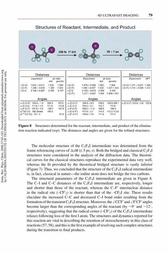
Figure 8 Structures determined for the reactant, intermediate, and product of the elimina-
tion reaction indicated (top). The distances and angles are given for the refined structures.
The molecular structure of the C2F4I intermediate was determined from theframe referencing curves of �sM (t; 5 ps; s). Both the bridged and classical C2F4Istructures were considered in the analysis of the diffraction data. The theoreti-cal curves for the classical structures reproduce the experimental data very well,whereas the fit provided by the theoretical bridged structure is vastly inferior(Figure 7). Thus, we concluded that the structure of the C2F4I radical intermediateis, in fact, classical in nature—the iodine atom does not bridge the two carbons.
The structural parameters of the C2F4I intermediate are given in Figure 8.The C–I and C–C distances of the C2F4I intermediate are, respectively, longerand shorter than those of the reactant, whereas the C–F′ internuclear distancein the radical site (–CF′
2) is shorter than that of the –CF2I site. These resultselucidate the increased C–C and decreased C–I bond order resulting from theformation of the transient C2F4I structure. Moreover, the ∠CCF′ and ∠F′CF′ anglesbecome larger than the corresponding angles of the reactant (by ∼9◦ and ∼12◦,respectively), suggesting that the radical center (–CF′
2) of the C2F4I intermediaterelaxes following loss of the first I atom. The structures and dynamics reported forthis reaction are vital in describing the retention of stereochemistry in this class ofreactions (57, 58), and this is the first example of resolving such complex structuresduring the transition to final products.
Ann
u. R
ev. P
hys.
Che
m. 2
006.
57:6
5-10
3. D
ownl
oade
d fr
om a
rjou
rnal
s.an
nual
revi
ews.
org
by C
AL
IFO
RN
IA I
NST
ITU
TE
OF
TE
CH
NO
LO
GY
on
06/0
9/06
. For
per
sona
l use
onl
y.

1 Mar 2006 15:56 AR ANRV272-PC57-03.tex XMLPublishSM(2004/02/24) P1: IKH
80 ZEWAIL
Dark Structures: Bifurcations in Radiationless Transitions
Another example in the applications of UED is that of excited-state structures,bifurcating to undergo radiationless transitions and chemical reactions. For exam-ple, in a recent publication (59), four prototypical hetero-aromatic (pyridine, 2-methylpyridine and 2,6-dimethylpyridine) and aromatic-carbonyl (benzaldehyde)organic molecules have been studied. For these molecules and others studied(Table 1), we determined the initial ground-state structure and followed uponexcitation the changes in the diffraction pattern with time (Figures 9 and 10). Thedepletion of old bonds and emergence of new bonds elucidate the structural ori-gin of dark transitions in the hetero-aromatics and the quinoid-like excited-statestructure of triplet benzaldehyde. Of significance is the understanding of the in-fluence of parent structure on the dynamical evolution of relaxation pathways andtheir relative timescales, and the possible bifurcation into physical and chemicalchannels on the energy landscape.
For the studies of the pyridine series, we focused on the so-called channelthree phenomenon, wherein, at a given internal energy threshold, the nonradia-tive decay rate increases abruptly with concomitant drop in the emission quan-tum yield. The channel three onset is much more pronounced for pyridine andpicoline (2-methlypyridine) than for lutidine (2,6-dimethlypyridine). Two funda-mental questions were unanswered: What is the origin of the abrupt change inthe radiationless behavior of the pyridines above a certain input energy threshold?Furthermore, how are such radiationless processes influenced by subtle changesin the molecular structure?
On the basis of the determined transient structures, we concluded that uponexcitation, pyridine and picoline undergo C–N bond scission to open the aromaticring and form a diradical structure. Lutidine does not undergo ring opening, but in-stead gives vibrationally hot ground/electronic state species. The refined structuresof ring-opened pyridine and picoline show alternating single- (near 1.45 A) anddouble-bond (near 1.35 A) character for the skeletal distances, indicating disrup-tion of the aromaticity of the parent ring structure. Moreover the farthest C · · · Ndistances (reflected as positive contributions in Figure 10) are >4 A, which areabsent in the f (r) of the parent ground-state structure. The refined hot lutidinestructure(s) shows the retention of aromatic distances in the product. The popu-lations of these transient structures gave the temporal growth with time constantsof 17 ± 1 ps, 28 ± 7 ps, and 16 ± 2 ps, for pyridine, picoline, and lutidine,respectively. The disruption of the ring, or lack thereof, was elucidated in recentcalculations of charge density maps by Shorokhov et al. in this laboratory and isfundamentally due to the nature of the nπ∗ versus ππ∗ excitation.
Can we determine excited-state structures and their possible bifurcation? Thestudy of the aromatic carbonyl benzaldehyde makes the answer affirmative. Uponlight absorption, benzaldehyde undergoes efficient nonradiative intersystem cross-ing to the triplet state as evidenced by its high phosphorescence yield. Previousinvestigations revealed photochemical dissociation into benzene and carbonmonoxide above an internal energy threshold (∼35,000 cm−1). We sought to
Ann
u. R
ev. P
hys.
Che
m. 2
006.
57:6
5-10
3. D
ownl
oade
d fr
om a
rjou
rnal
s.an
nual
revi
ews.
org
by C
AL
IFO
RN
IA I
NST
ITU
TE
OF
TE
CH
NO
LO
GY
on
06/0
9/06
. For
per
sona
l use
onl
y.

1 Mar 2006 15:56 AR ANRV272-PC57-03.tex XMLPublishSM(2004/02/24) P1: IKH
4D ULTRAFAST IMAGING 81
Figure 9 Diffraction images and radial distribution functions f(r), together with structural
parameters for the indicated molecules (see text for details). DMP, dimethylpyridine; COT3,
cycloocta-1,3,5-triene.
Ann
u. R
ev. P
hys.
Che
m. 2
006.
57:6
5-10
3. D
ownl
oade
d fr
om a
rjou
rnal
s.an
nual
revi
ews.
org
by C
AL
IFO
RN
IA I
NST
ITU
TE
OF
TE
CH
NO
LO
GY
on
06/0
9/06
. For
per
sona
l use
onl
y.

1 Mar 2006 15:56 AR ANRV272-PC57-03.tex XMLPublishSM(2004/02/24) P1: IKH
82 ZEWAIL
Figure 10 Structural dynamics for two reactive systems (top) and for the excited triplet state
(quinoid structure) that resulted from radiationless transition of benzaldehyde. The calculated
charge density map shows the quinoid structure in agreement with experiment (the calculated
image to scale is in Reference 60).
Ann
u. R
ev. P
hys.
Che
m. 2
006.
57:6
5-10
3. D
ownl
oade
d fr
om a
rjou
rnal
s.an
nual
revi
ews.
org
by C
AL
IFO
RN
IA I
NST
ITU
TE
OF
TE
CH
NO
LO
GY
on
06/0
9/06
. For
per
sona
l use
onl
y.
1 Mar 2006 15:56 AR ANRV272-PC57-03.tex XMLPublishSM(2004/02/24) P1: IKH
4D ULTRAFAST IMAGING 83
determine the structures and pathways—whether the photophysical and photo-chemical processes occur consecutively or else competitively as a result of bi-furcation. The diffraction data show the rupture of covalent C–C bonds (near 1.4A), loss of next-nearest neighbor distances (near 2.5 A), and depletion of longerdistances (>3.5 A)—a fragmentation and significant repositioning of the atomicnuclei must accompany the process.
We determined a quinoid structure, which is optically dark, for the tripletexcited-state (ππ∗) benzaldehyde formed as a result of intersystem crossing. Thisexcited-state structure exhibits well-defined single and double bonds, indicatingdisruption of aromaticity in the ring (Figure 10). On the other hand, benzeneformed in the dissociation pathway is in its ground electronic state, as indicatedby the refined C–C bond distance. The simultaneous emergence of the photophys-ical and photochemical products in the diffraction data indicates a bifurcation onthe excited singlet surface with apparent rise, through the intermediate, with timeconstants of 25 ± 4 ps and 38 ± 5 ps for excited triplet benzaldehyde and ben-zene, respectively. This observed bifurcation resolves long-standing issues. Thequantum chemical calculations of charge density maps by Shorokhov et al. (60)(Figure 10) support the conclusion regarding the electronic distribution of thestructures involved in the bifurcation.
Complex Landscapes and Light-Atom Structures
In order to extend these studies to systems of more complex energy landscapes andto structures with no heavy atoms, we studied organometallic structures, whichreact through multiple pathways and yield products of different spin multiplic-ity, and a series of hydrocarbons that undergo thermal/photochemical reactions.We determined the initial structures with high accuracy and those of the reactionintermediate(s). For the hydrocarbons, the sensitivity was still high even thoughthere were no heavy atoms to scatter from. The landscape involves multiple con-formations and we studied these nonequilibrium structures, both experimentallyand theoretically. More recently, we reported our study of the nature of the hydro-gen bond in acetylacetone and the structural dynamics of its elimination reactions.We also concerned ourselves with the theoretical treatment of diffraction on theultrashort timescale with particular focus on the role of coherence and the effectof orientational order on diffraction of reacting populations. We do not cover thesestudies in this review, but reference to these and other studies is made in Table 1.
ULTRAFAST ELECTRON CRYSTALLOGRAPHY
In UEC, the new features of the apparatus include three interconnected ultrahigh vacuum (UHV) chambers—the sample preparation, load-lock, and scatteringchambers. To this apparatus we interfaced a femtosecond laser system. The crys-tal is mounted on a computer-controlled goniometer for high-precision (0.005◦)angular rotation; it can be cooled to a temperature of 10 K and changed in five
Ann
u. R
ev. P
hys.
Che
m. 2
006.
57:6
5-10
3. D
ownl
oade
d fr
om a
rjou
rnal
s.an
nual
revi
ews.
org
by C
AL
IFO
RN
IA I
NST
ITU
TE
OF
TE
CH
NO
LO
GY
on
06/0
9/06
. For
per
sona
l use
onl
y.

1 Mar 2006 15:56 AR ANRV272-PC57-03.tex XMLPublishSM(2004/02/24) P1: IKH
84 ZEWAIL
directions, three translations, and two rotations. The preparation chamber has sput-tering and cleaning tools and is also equipped with LEED and Auger spectroscopyfor characterization of the crystal surface. Molecules can be studied on the surfaceeither as physisorbed or chemically functionalized entities, and thin crystals can bestudied in the reflection mode. We have also used the apparatus in the transmissionmode for studies of crystals and thin films.
Diffraction is recorded in the far field and analyzed using the observed Braggspots, streaks, and Laue zones of Ewald’s sphere (Figure 6). The electron andlight pulses used in UEC were temporally and spatially characterized. For the30 keV electron pulses, we used in situ streaking techniques and, for light, the nowstandard autocorrelation method. We have obtained the fastest streaking speed of140 ± 2 fs/pixel in UED-4, thus approaching the state of the art in streak cameras(Figure 4). For the extraction field of 10 kV/mm, the spreading time is ∼20 fs.From the measured streaking, a pulse width of 322 ± 128 fs was obtained. Usingthe same electron gun design, Cao et al. (54) obtained ∼300 fs pulses, albeit witha streaking speed of 250 fs/pixel. Miller’s group reported ∼600 fs resolution intransmission (55). The initiation laser pulse duration is typically 100 fs, and theoverall temporal resolution is determined by the geometry of the experiment andthe relative widths (shapes) and speeds of the two pulses. We have treated in detailelsewhere (61) conditions for velocity mismatch and its angular dependence foroptimum pulse width. In what follows we give examples of studies in UEC.
Surfaces and Crystals
A paradigm case study demonstrating the potential of UEC was reported byVigliotti et al. (62). Determination of surface structural dynamics, using framereferencing, was achieved for crystalline solids (GaAs), following the temperaturerise of the crystal. From the change of Bragg diffraction (shift, width, and inten-sity), we showed by direct inversion of the diffraction data that “compression”and “expansion” occur on the –0.01 A to +0.02 A scale, and that the “transienttemperature” reaches its maximum value (1565 K) in 7 ps (Figure 11). The onsetof structural change lags behind the rise in the temperature, demonstrating theevolution of nonequilibrium structures. These results were compared with thoseof nonthermal femtosecond optical probing reported by Mazur’s group (63), andthe agreement for the temperature response from the fluence dependence of thedielectric function is impressive, but we now have the dynamical structure.
The surface of GaAs was functionalized with a monolayer of chlorine atoms,chemically bonded. On the ultrashort timescale we observed, following the com-pression, the expansion that is due to the rise of phonon temperature. At longer time,the restructuring and the evolution toward the equilibrium state was clearly evidentin the diffraction (intensity and shift). Structural dynamics can be divided into threeregimes: potential driven change, which involves electronic redistribution with nomotion of nuclei (femtoseconds to a few picoseconds); coherent nonequilibriumlattice expansion (rise time of 7 ps); and restructuring and heat diffusion (50 ps tonanoseconds). The latter agrees well with the expansion reported in the literature
Ann
u. R
ev. P
hys.
Che
m. 2
006.
57:6
5-10
3. D
ownl
oade
d fr
om a
rjou
rnal
s.an
nual
revi
ews.
org
by C
AL
IFO
RN
IA I
NST
ITU
TE
OF
TE
CH
NO
LO
GY
on
06/0
9/06
. For
per
sona
l use
onl
y.

1 Mar 2006 15:56 AR ANRV272-PC57-03.tex XMLPublishSM(2004/02/24) P1: IKH
4D ULTRAFAST IMAGING 85
Figure 11 Ultrafast electron crystallography studies of GaAs single-crystal surfaces, ter-
minated with chlorine atoms. Shown here is only the change (from equilibrium) of the lattice
constant. The broadening and intensity of diffraction peaks were also monitored, and studies
of this system are continuing in this laboratory (see text).
Ann
u. R
ev. P
hys.
Che
m. 2
006.
57:6
5-10
3. D
ownl
oade
d fr
om a
rjou
rnal
s.an
nual
revi
ews.
org
by C
AL
IFO
RN
IA I
NST
ITU
TE
OF
TE
CH
NO
LO
GY
on
06/0
9/06
. For
per
sona
l use
onl
y.

1 Mar 2006 15:56 AR ANRV272-PC57-03.tex XMLPublishSM(2004/02/24) P1: IKH
86 ZEWAIL
for thermal heating (“infinite” time limit). We are continuing studies of this systembecause of, among other findings, the discovery of changes in signal phase whenspatially scanning the electron and heating pulses.
Similarly, we studied surface and bulk crystals of silicon, with and withoutadsorbates. Frame referencing to the ground-state structure shows the changes inthe structure caused by the initiating pulse, from ground-state pattern at negativetime to the observed change at positive time (Figure 12). The structural changeis evident in the shift with time of the in-phase Bragg peak of the rocking curve,whereas the increase in vibrational amplitude is reflected in the broadening. Theevolution takes place as a rise to a maximum shift and then a decay to the co-ordinates of the original structure. By gating on a Bragg spot, we followed thechanges with time. As with GaAs, we observed the motion of surface and bulkatoms and their timescales (39). Following the femtosecond rise in electron tem-perature, electron-phonon coupling results in the population of optical phonons,which then generate, after picoseconds delay, acoustic waves (lattice expansionand compression), and finally lattice heating. Such a two-temperature descriptionof heating is well known. Using UEC, we now can observe the ultrafast surfaceand bulk structural dynamics and follow the restructuring and diffusion at longertimes.
Because we can vary the fluence of the initiating pulse, we also studied thestructural changes involved in phase transitions when the temperature of the lat-tice is sufficiently high to cause large amplitude disorder. Initiating an ultrashorttemperature jump of the amorphous structure with the infrared femtosecond pulsegives new diffraction ring patterns, which we followed as a function of time byreferencing to the ground-state frame (Figure 12). The structural change is a phasetransition to the liquid-like state (39).
Interfacial Water: Hydrophilic and Hydrophobic Substrates
The directional molecular features of hydrogen bonding and the different structurespossible, from amorphous to crystalline, make the interfacial collective assemblyof water, on the mesoscopic scale, much less understood. Structurally, the natureof water on a substrate is determined by forces of orientation at the interface and bythe net charge density, which establishes the hydrophilic or hydrophobic characterof the substrate. However, the transformation from ordered to disordered structureand their coexistence critically depends on the timescales for the movements ofatoms locally and at long range. Therefore, it is essential to elucidate the nature ofthese structures and the timescales for their equilibration.
In a recent publication by Ruan et al. (64), we reported UEC determinationof the structural dynamics of interfacial water following substrate infrared tem-perature jump. Interfacial water was formed on a hydrophilic surface (silicon,chlorine-terminated) or hydrophobic surface (silicon, hydrogen-terminated) undercontrolled UHV conditions. We identified the interfacial and ordered (crystalline)
Ann
u. R
ev. P
hys.
Che
m. 2
006.
57:6
5-10
3. D
ownl
oade
d fr
om a
rjou
rnal
s.an
nual
revi
ews.
org
by C
AL
IFO
RN
IA I
NST
ITU
TE
OF
TE
CH
NO
LO
GY
on
06/0
9/06
. For
per
sona
l use
onl
y.

1 Mar 2006 15:56 AR ANRV272-PC57-03.tex XMLPublishSM(2004/02/24) P1: IKH
4D ULTRAFAST IMAGING 87
Figure 12 Ultrafast electron crystallography of single-crystal silicon surfaces and
nanometer crystals. The evolution of structures, using frame referencing, and the phase
transition at higher temperatures (diffraction rings) are displayed.
Ann
u. R
ev. P
hys.
Che
m. 2
006.
57:6
5-10
3. D
ownl
oade
d fr
om a
rjou
rnal
s.an
nual
revi
ews.
org
by C
AL
IFO
RN
IA I
NST
ITU
TE
OF
TE
CH
NO
LO
GY
on
06/0
9/06
. For
per
sona
l use
onl
y.

1 Mar 2006 15:56 AR ANRV272-PC57-03.tex XMLPublishSM(2004/02/24) P1: IKH
88 ZEWAIL
structure from the Bragg diffraction and the layered and disordered (polycrys-talline) structure from the Debye-Scherrer rings (Figures 13 and 14). The tempo-ral evolution of interfacial water and layered ice after the temperature jump wasstudied with monolayer sensitivity.
On the hydrophilic surface substrate the structure is cubic (Ic), not hexagonal(Ih), and structural dynamics are distinctive. The timescale for the breakage (large-amplitude motion) of long-range order of the interfacial layer (37 ps) is an orderof magnitude longer than that for breaking hydrogen bonds in bulk liquid water,and the local OH · · O and O · · · O bond distances from diffraction are directlyinvolved in the change but on different timescales. These results also indicate thatthe timescale for energy flow (in the 1 ps range) in the assembled water struc-ture is much shorter than that of energy localization for desorption of individualmolecules. Moreover, the restructuring time involving long-range order is longerthan the time for amorphization, a process in which the O · · · O correlation is lostbefore the OH · · O correlation, defining the vibrational motion involved. Finally,there is the relevance to biological water—perhaps it is not accidental that thetimescale for losing the hydrogen bond network (37 ps) is similar to that reportedfor interfacial water near hydrophilic protein surfaces (20 ps to 50 ps) and is verydifferent from that of bulk water (700 fs to 1.5 ps); see Figure 3 and Reference 17(and references therein).
On the hydrophobic surface, the structure is still cubic, but very different inorder. The structural dynamics is also different. The interface is dominated bypolycrystalline Ic, but coexisting in this phase are crystallite structures, not adja-cent to the surface of the substrate; see Figure 14 for rings and spots diffraction,and the comparison with theory. The change in the structure of Ic has differenttemporal behaviors reflecting the distinct difference in the transfer of energy topolycrystalline and crystallite Ic. We note that for the hydrophilic substrate studied,water adjacent to the surface is crystalline, or nearly so, whereas that away fromthe surface is polycrystalline. These studies were made recently in this laboratoryby D.-S. Yang, N. Gedik & S. Habershon, and a full account of these and the earlierstudy (64) will be published in a series of papers. The issues of interest are thecoexistence of these structures, their different dynamics, and the timescales forenergy transfer and disruption of the hydrogen bond network.
Bilayers of Crystalline 2D Fatty Acids: Molecular Assemblies
For the studies of membrane-type structures, we decided to first investigate abilayer of fatty acids deposited on a hydrophobic surface substrate, invoking thewell-known Langmuir-Blodgett (LB) technique. It allows for a controlled layer-by-layer deposition of ordered molecular films and has been used to create modelbiological membranes for studies under controlled conditions. Although biomem-branes are more complicated by the presence of intercalaters, the LB bilayersrepresent the building blocks of lipid bilayers, in our case with the molecularchains extending up to ∼50 A. LB films themselves are of considerable interest in
Ann
u. R
ev. P
hys.
Che
m. 2
006.
57:6
5-10
3. D
ownl
oade
d fr
om a
rjou
rnal
s.an
nual
revi
ews.
org
by C
AL
IFO
RN
IA I
NST
ITU
TE
OF
TE
CH
NO
LO
GY
on
06/0
9/06
. For
per
sona
l use
onl
y.

1 Mar 2006 15:56 AR ANRV272-PC57-03.tex XMLPublishSM(2004/02/24) P1: IKH
4D ULTRAFAST IMAGING 89
Figure 13 Ultrafast electron crystallography studies of interfacial water on a hydrophilic
substrate. The coexistence of crystalline and polycrystalline structures (Bragg spots and
Debye-Scherer rings) are clear in the patterns.
Ann
u. R
ev. P
hys.
Che
m. 2
006.
57:6
5-10
3. D
ownl
oade
d fr
om a
rjou
rnal
s.an
nual
revi
ews.
org
by C
AL
IFO
RN
IA I
NST
ITU
TE
OF
TE
CH
NO
LO
GY
on
06/0
9/06
. For
per
sona
l use
onl
y.

1 Mar 2006 15:56 AR ANRV272-PC57-03.tex XMLPublishSM(2004/02/24) P1: IKH
90 ZEWAIL
Figure 14 Ultrafast electron crystallography studies of interfacial water on a hy-
drophobic substrate. Shown also is the theoretical simulation of diffraction. (Bottom)
Frame referencing at positive time to show the depletion of “old” structures and for-
mation of “new” structures (dark and white regions).
Ann
u. R
ev. P
hys.
Che
m. 2
006.
57:6
5-10
3. D
ownl
oade
d fr
om a
rjou
rnal
s.an
nual
revi
ews.
org
by C
AL
IFO
RN
IA I
NST
ITU
TE
OF
TE
CH
NO
LO
GY
on
06/0
9/06
. For
per
sona
l use
onl
y.

1 Mar 2006 15:56 AR ANRV272-PC57-03.tex XMLPublishSM(2004/02/24) P1: IKH
4D ULTRAFAST IMAGING 91
various areas of research involving self-assembly, self-organization, and proteinimmobilization, and in technology developments such as molecular electronicsand nonlinear optics.
Previous static diffraction studies have provided patterns and analyses withfocus on the distances between the ions (X ray) and in thick films of many layers.In these investigations, however, the structures were not time-resolved and werenot directly reflective of the influence of the supporting surface. Femtosecond time-resolved studies (65) of biomembranes have examined the spectroscopy of localprobes, and time-resolved X-ray diffraction studies have shown the laser-induceddisorder above the damage threshold of the organic film (66). In the former studythe structure cannot be determined, and for the latter the film was 83 layers andit was not possible to solve for the structure of the fatty acid assembly. The laser-heating resulted in a destructive change above the damage threshold.
Recently, Chen et al. (67) reported on UEC of one bilayer (two chains ofC19H39COOH) of arachidic (eicosanoic) fatty acid. With UEC sensitivity andresolution, we determined the structure, thus establishing the orientation of thealiphatic chains and obtaining the subunit cell molecular –CH2–CH2– dimensions;Figure 15 displays the structure of the bilayer studied and the subunit cell in twodirections. In the same publication we reported the observation of the coherentand anisotropic dynamical expansion in the bilayer structure and the restructuringtoward equilibration at longer times. The timescales involved following the temper-ature jump were obtained from the diffraction frames taken every 1 ps (Figure 15).From these studies a structural dynamics picture emerges for the bilayer on thesubstrate and for its atomic motions.
All diffraction patterns are composed of spots (and/or streaks), showing thehigh quality of the 2D crystalline structure of the arachidic acid bilayer and theunderlying hydrogen terminated Si(111) surface. The diffraction patterns at neg-ative times and at low incidence angle of the electrons, parallel and perpendicularto the dipping direction, give the tilt angle of the chains (nearly zero) and providethe subunit cell parameters of the bilayer: a0 = 4.7 A, b0 = 8.0 A, and c0 = 2.54 A.The symmetry of the bilayer is an orthorhombic R(001) packing, with the (001)plane parallel to the Si(111) surface. At higher incidence angle, additional diffrac-tion Bragg spots appear, resulting from the single crystal silicon substrate, whichcan easily be indexed (see above).
These experimental values for the lattice parameters within the plane differfrom the predicted theoretical values of a0 = 4.96 A and b0 = 7.4 A (68).The difference can be explained by the fact that the theoretical values were cal-culated for infinite aliphatic chains and do not take into account the carboxylicend group of fatty acids. Moreover, the bilayer consists only of two monolay-ers, so the substrate and the deposition conditions (e.g., the dipping pressure orthe pHs of the solution) all have an important role in the order at the interface.The spacing c0 between CH2 planes agrees very well with the theoretical valueof 2.54 A, as it represents separation between strong C–C bonds. The rockingcurves, the changes of diffraction with incidence angle, were obtained and the
Ann
u. R
ev. P
hys.
Che
m. 2
006.
57:6
5-10
3. D
ownl
oade
d fr
om a
rjou
rnal
s.an
nual
revi
ews.
org
by C
AL
IFO
RN
IA I
NST
ITU
TE
OF
TE
CH
NO
LO
GY
on
06/0
9/06
. For
per
sona
l use
onl
y.

1 Mar 2006 15:56 AR ANRV272-PC57-03.tex XMLPublishSM(2004/02/24) P1: IKH
92 ZEWAIL
observations indicate a significant coherence length (nanometers) along the fattyacid chains.
With the frame-reference methodology invoked in these UEC studies, we ob-tained the structural dynamics selectively for the bilayer. As shown in Figure 15,immediately after the heating pulse (0 ps and 1 ps frames), an intensity loss of theBragg spots is observed, as dark (white) spots evolve in the different frames. The
Figure 15 Ultrafast electron crystallography of 2D fatty-acid bilayers. Shown are the sub-
unit cell structure determined and the dynamics from the frame referencing taken following
the heat pulse to the substrate. The frames at 0 and 1 ps show the onset of structural changes,
and the frames at longer times show the restructuring toward the equilibrium state.
Ann
u. R
ev. P
hys.
Che
m. 2
006.
57:6
5-10
3. D
ownl
oade
d fr
om a
rjou
rnal
s.an
nual
revi
ews.
org
by C
AL
IFO
RN
IA I
NST
ITU
TE
OF
TE
CH
NO
LO
GY
on
06/0
9/06
. For
per
sona
l use
onl
y.

1 Mar 2006 15:56 AR ANRV272-PC57-03.tex XMLPublishSM(2004/02/24) P1: IKH
4D ULTRAFAST IMAGING 93
change in the Bragg spots becomes more prominent over time (10–100 ps). Thelower part of the peaks becomes brighter while the upper part becomes darker,showing a downward shift of the Bragg spots. At longer times, the patterns getfainter again, indicating that the peaks are moving back to their original position.Remarkably, both the heating and electron pulses, because of their timed repeti-tion, ultrashort durations, and fluxes, do not damage the bilayer, as we repeatedthese experiments many times without observing damage.
In the vertical direction for all diffractions of the bilayer (c-component of themomentum transfer s-vector) we observe structural change. The behavior rep-resents an initial expansion (�c0 = 0.1 A) of the subcell in the bilayer afterimpulsive heating of the substrate followed by a subsequent contraction due tothe heat dissipation. The expansion takes place with a time constant of 25 ps,whereas the subsequent compression occurs with 55 ps time constant, and themuch longer time compression is a restructuring on the nanosecond timescale.Within our resolution, no significant change in lattice spacing is observed in theplane perpendicular to the molecular chains.
Structural dynamics of the bilayer can now be pictured. Because there is noabsorption resonance for the fatty acids at the wavelength of the heating pulse, thepulse is absorbed only by the substrate and the bilayer is practically transparent.The heating is through the phonon temperature described above in our studies ofSi and GaAs crystals (here substrate). Although the energy is directly transferredinto the bilayer from the substrate, the expansion in the bilayer is significant alongthe aliphatic chains of the molecules, indicating an efficient and “wire-like” energytransfer within the fatty acid assembly. The covalent bondings in the chains, asopposed to the weak interactions across chains, facilitate such directional motion,besides the 2D stacking interaction in the crystal-like structure. This apparentnonequilibrium behavior must be handled cautiously.
At the maximum extension of the fatty acids the effective temperature is abovethe melting point of the assembly (in the range of 100◦C), but the structure is farfrom equilibrium primarily in modes of the stretch and scissor (and twist) vibra-tions of the chains. Thus the motions along the chains should reflect the behaviorof coupled oscillators—the forming of wave motion by local change in velocityand amplitude (or diffusion). If true, the behavior is that of a soliton-type mo-tion. Molecular dynamics simulations (in collaboration with Professor T. Shoji’sgroup) and theoretical modeling (Dr. J. Tang, this group) of anharmonically cou-pled bonds are works in progress to address the timescales involved, the nature ofcoherent structural dynamics, and the incoherent energy dissipation in the restruc-turing at long times. An anisotropic expansion of the bilayer may prove significantin explaining mechanisms of motion and transport, and for this reason we are alsoexamining biological membranes. So far we have studied multiple-layered fattyacid systems and phospholipids and are continuing to explore the effect of layerthickness on diffraction changes, position, and intensity. There is another dimen-sion to this work, namely the origin of self-assembly (69) and crystallization, andUEC is our method of choice for the reasons given above.
Ann
u. R
ev. P
hys.
Che
m. 2
006.
57:6
5-10
3. D
ownl
oade
d fr
om a
rjou
rnal
s.an
nual
revi
ews.
org
by C
AL
IFO
RN
IA I
NST
ITU
TE
OF
TE
CH
NO
LO
GY
on
06/0
9/06
. For
per
sona
l use
onl
y.

1 Mar 2006 15:56 AR ANRV272-PC57-03.tex XMLPublishSM(2004/02/24) P1: IKH
94 ZEWAIL
ULTRAFAST ELECTRON MICROSCOPY
Transmission electron microscopy (TEM) with its wide-ranging arsenal of toolshas long been a powerful method in many areas of research, allowing for sub-nanometer spatial resolution but lacking the ultrashort time resolution. Optical mi-croscopy, using fluorescent probes, e.g., green fluorescent proteins, has providedthe means to visualize events occurring in vitro and within cells (41 and referencestherein). However, despite possessing the capability for temporal resolution, op-tical methods are limited in their spatial resolution to the wavelengths employed,typically 200–800 nm. The ultimate techniques would be those that have the spatialresolution of electron microscopy and the time resolution of optical methods. Asmentioned above, the atomic length scale can be studied with diffraction, but forbiological and nanoscopic materials with characteristic length scales ranging fromnanometers to micrometers, electron microscopy enjoys unique advantages.
Lobastov et al. (41), in a recent publication, reported the development of 4DUEM, which provides the ability to image complex structures with the spatialresolution of TEM, but as snapshots captured with ultrafast electron packets derivedfrom a train of femtosecond pulses. The images and diffraction patterns wereobtained at 120 keV for materials (single crystals of gold, amorphous carbon, andpolycrystalline aluminum) and for biological cells of rat intestines. The strobingpackets contain on average one electron per pulse and the specimen dose is a fewelectrons per A2, but the pulses are now fully controlled in space and time.
Conceptually, we built on the machinery of UED and UEC, but with a fundamen-tal difference, namely the realization of the significance of timed, single-electronpulses for imaging in UEM. The design of 4D UEM is shown in Figure 5, whichoutlines the interfacing of the femtosecond optical system with the transmissionelectron microscope. Space-charge broadening and defocusing of electron pulsesand stability of the electron flux during imaging are major issues that we had toresolve. To circumvent space-charge-induced broadening and the concomitant de-crease in the ability of the microscope optics to focus these electrons, we haveredesigned the transmission electron microscope by directly illuminating the pho-tocathode with extremely weak femtosecond pulses, but as a high-frequency trainof pulses separated by nanosecond or longer intervals. This crucial advance em-powered us to successfully record images and diffraction patterns with the requiredsensitivity and dosage, but with new dimensions.
The approach is entirely different from the one used in the important work ofDomer & Bostanjoglo (70 and references therein). They utilized a single giantpulse with ∼108 electrons, and the pulses are photoelectrically generated as inUED and UEC. Their microscope utilizes a ∼20 ns electron-pulse duration and acurrent of ∼2 mA. Such pulses, which were used to study laser-induced melting inmetals, pack within them the large number of electrons (∼108) that are detrimentalto achieving ultrashort pulse imaging. Moreover, because the time window forimaging in those experiments is nanoseconds, the uncertainty in spatial resolutiondue to noise statistics is on the order of micrometers, as pointed out by Bostanjoglo
Ann
u. R
ev. P
hys.
Che
m. 2
006.
57:6
5-10
3. D
ownl
oade
d fr
om a
rjou
rnal
s.an
nual
revi
ews.
org
by C
AL
IFO
RN
IA I
NST
ITU
TE
OF
TE
CH
NO
LO
GY
on
06/0
9/06
. For
per
sona
l use
onl
y.

1 Mar 2006 15:56 AR ANRV272-PC57-03.tex XMLPublishSM(2004/02/24) P1: IKH
4D ULTRAFAST IMAGING 95
(71). In fact our attempt to use amplified femtosecond light pulses with much higherpeak power density (on the order of 1012 W/cm2) resulted in electron bunches thatwere impervious to focusing by the microscope optics, and no doubt much broaderpulses due to electron-electron repulsion.
Using our 4D UEM we obtained images of materials, polycrystalline and sin-gle crystals (Figure 16). For calibration, we also obtained “images” when thefemtosecond pulses entering the microscope were blocked. The fact that no im-ages were observed when the light pulses were blocked indicates that the electrons
Figure 16 Ultrafast electron microscopy of biological cells from rat intestines at two
magnifications (top and bottom right). Shown also are a typical image of gold crystals (top,
left) and the diffraction pattern (bottom, left) (see text).
Ann
u. R
ev. P
hys.
Che
m. 2
006.
57:6
5-10
3. D
ownl
oade
d fr
om a
rjou
rnal
s.an
nual
revi
ews.
org
by C
AL
IFO
RN
IA I
NST
ITU
TE
OF
TE
CH
NO
LO
GY
on
06/0
9/06
. For
per
sona
l use
onl
y.

1 Mar 2006 15:56 AR ANRV272-PC57-03.tex XMLPublishSM(2004/02/24) P1: IKH
96 ZEWAIL
generated in the microscope were indeed those obtained optically and that thermalelectrons were negligible. The microscope can operate in UEM and TEM modes,and for atomic-scale investigations, we operated UEM in the diffraction mode byadjusting the intermediate lens to select the back focal plane of the objective lensas its object (Figure 16). The novel attributes of UEM encouraged us to test theviability of imaging biological cells. UEM images of rat intestinal cells were ob-tained in a few seconds and both the microvilli as well as the subcellular vesiclesof these epithelial cells were visualized in the UEM images. As in the case ofUED and UEC, UEM is now poised for measurements of structural dynamics.For such time-resolved investigations, the time axis is defined by distance steps inthe arrangement of Figure 5, noting that one micron of path difference correspondsto a frame separation of 3.3 fs. Applications are numerous.
CONCLUDING REMARKS
Because of limitations of space, it was not possible to cover all studies made usingUED, UEC, and UEM. In this review, we only highlighted the concepts, develop-ments, and, by way of prototypical examples, the myriad of possible applications.In order to reliably introduce the fourth dimension (time) into structural determi-nation and imaging in different phases we had to develop, through five generationsof table-top instruments, the conceptual framework and methodology needed toreach the atomic-scale spatiotemporal resolution and to cover both Fourier space(diffraction) and real space (microscopy). Our current resolutions are: spatial,picometer to nanometer (UED and UEC to UEM) and temporal, 1 picosecond(UED), 300 femtosecond (UEC, transmission), and less than or equal to 100 fem-tosecond (UEM, single electrons). Only with frame-referencing at different timescould we reach as low as 1% for the sensitivity of change and temporally isolate thetransient structures. For convenience, we list the subjects studied and publicationsfrom this laboratory in Table 1, covering nearly a decade of research.
In the same year (1991) we proposed UED as a method for structural dynamics,Thomas (72) wrote a “News and Views” piece entitled Femtosecond Diffraction.Toward the end of the piece he concluded with the following words: “If the ex-periment does indeed prove successful it will mark the dawn of an important newera. . . .” It took exactly a decade (2001), and the evolution from UED-1 to UED-3,to transform a dream into realization, from the exploration of the potential of theapproach to the explosion of the applications in real experimental determinationof isolated, transient molecular structures. The robustness of UED-3 made it pos-sible to study a variety of systems, and here we discussed two classes, prototypicalchemical reactions and excited-state dark structures that bifurcate through radia-tionless transitions. In this UED-3 laboratory, we are currently continuing studiesof structures in reactions and of excited states. Also, using a new apparatus weplan to study structural dynamics of macromolecular systems with the aid of newtheoretical formulation, including charge density maps (60), of diffraction fromisolated proteins. Oriented reactions (73, 74) are also part of the new effort in UED.
Ann
u. R
ev. P
hys.
Che
m. 2
006.
57:6
5-10
3. D
ownl
oade
d fr
om a
rjou
rnal
s.an
nual
revi
ews.
org
by C
AL
IFO
RN
IA I
NST
ITU
TE
OF
TE
CH
NO
LO
GY
on
06/0
9/06
. For
per
sona
l use
onl
y.

1 Mar 2006 15:56 AR ANRV272-PC57-03.tex XMLPublishSM(2004/02/24) P1: IKH
4D ULTRAFAST IMAGING 97
In retrospect, what is remarkable about Thomas’ piece was its broader visionfor the significance of determining structures in the act of change irrespectiveof the phase they are in. Our development of UEC, in both reflection and trans-mission modes, was for the sole purpose of extending UED to the condensedphase. Because of long-range order, it was possible to study structural dynam-ics of nanometer-scale crystals, surfaces, and molecular assembles on substrates.Here, we discussed prototypical cases of surface-atom motion, surface phase tran-sition, interfacial water on hydrophobic and hydrophilic substrates, and structuraldynamics of bilayers of fatty acids on hydrophobic substrates. In 2004, Thomasoverviewed (75) the significance of UEC, but equally important were his views onpotential applications in heterogeneous catalysis (76). Currently, our group is in-volved in extensions of UEC to studies of materials using transmission diffractionand biological assemblies using reflection diffraction. Transmission offers a bettertime resolution because of geometrical constraints in reflection, and for both weplan further improvement of the frame resolution in time by using composite-phaseshaped optical pulses (77).
From diffraction in Fourier space, both UED and UEC provide atomic-scalespatial resolution. An inversion to real space requires theoretical machinery and,for obtaining refined structures, dynamical theory may be invoked. So far we haveobtained structures using kinematic theory, principally because of the high electronenergy, the sample density, and the relatively low atomic number of atoms. Imagingin real space is the property of TEM. In 4D UEM, the sample sees timed single-electron packets, thus removing the detrimental space-charge effects of defocusingand broadening of pulses in imaging. Here, it was shown that such a microscopecan be applied to studies of materials and biological cells. In the same microscope,we obtained diffraction with atomic-scale spatial resolution, again using timed,single-electron packets.
Thomas, who has been following the trajectory of developments since its nais-sance, and who is a pioneer in the applications of microscopy to materials science(76, 78), has written a highlight (79) describing the revolutionary aspect of 4DUEM and the prospects for branched applications. Harris & Thomas (80) exploredsome applications in domains of biological macromolecules and solid state chem-istry. It did not escape our attention that 4D UEM should find applications in manyfields, in particular in biological imaging. The new microscope allows facile oper-ation in two modes—the UEM mode and the conventional TEM mode—offering auniversal methodology for structural studies in space and time. Moreover, becauseUEM has been designed using femtosecond pulse trains from the oscillator alone,without the need for an ultrafast pulse amplifier, it has the added technologicaladvantage of being beguilingly simple and easy to integrate. It may also proveunique (3) for limiting heat and blurring of images, and possibly radiation damage(9, 81), points under study in this laboratory. Currently, we have one completesystem operating at 120 keV, and shortly there will be three 4D UEMs, includingsingle-pulse recordings at a higher number of electrons but with picosecond res-olution. Applications involving higher spatial resolutions and microscopy-basedadvances (82–84) include studies of cryo and life cell imaging.
Ann
u. R
ev. P
hys.
Che
m. 2
006.
57:6
5-10
3. D
ownl
oade
d fr
om a
rjou
rnal
s.an
nual
revi
ews.
org
by C
AL
IFO
RN
IA I
NST
ITU
TE
OF
TE
CH
NO
LO
GY
on
06/0
9/06
. For
per
sona
l use
onl
y.

1 Mar 2006 15:56 AR ANRV272-PC57-03.tex XMLPublishSM(2004/02/24) P1: IKH
98 ZEWAIL
TABLE 1 Different generations of UED, UEC, and UEM: developments, subjects, and
references
Subject References
Concept Femtosecond transition-state dynamics 34
Structural femtochemistry: experimental methodology 35
UED-1 Ultrafast diffraction and molecular structure 36
UED: velocity mismatch and temporal resolution in
crossed-beam experiments
61
UED: molecular structures and coherent dynamics 73
UED: experimental time resolution and applications 56
UED-2 Clocking transient chemical changes by UED 37
UED: structures in dissociation dynamics of Fe(CO)5 42
UED: determination of radical structure with picosecond time
resolution
96
UED and direct observation of transient structures in a chemical
reaction
97
Ultrafast gas-phase electron diffraction 98
Conformations and barriers of haloethyl radicals
(CH2XCH2, X = F, Cl, Br, I)
99
UED of transient [Fe(CO)4]: determination of molecular
structure and reaction pathway
102
UED of transient cyclopentadienyl radical: a dynamic
pseudorotary structure
103
UED-3 Direct imaging of transient molecular structures with ultrafast
diffraction
38
CF2XCF2X and CF2XCF2 radicals (X = Cl, Br, I): ab initio and
DFT studies and comparison with experiments
58
Ultrafast diffraction of transient molecular structures in
radiationless transitions
100
Ultrafast diffraction and structural dynamics: the nature of
complex molecules far from equilibrium
101
UED and structural dynamics: transient intermediates in the
elimination reaction of C2F4I2
57
UED: complex landscapes of molecular structures in thermal
and light-mediated reactions
104
Direct determination of hydrogen-bonded structures in resonant
and tautomeric reactions using UED
105
UED: structural dynamics of the elimination reaction of
acetylacetone
106
Dark structures in molecular radiationless transitions determined
by ultrafast diffraction
59
UED: dynamical structures on complex energy landscapes 60
UED: oriented molecular structures in space and time 74
UEC UEC: transient structures of molecules, surfaces, and phase
transitions
39
(Continued)
Ann
u. R
ev. P
hys.
Che
m. 2
006.
57:6
5-10
3. D
ownl
oade
d fr
om a
rjou
rnal
s.an
nual
revi
ews.
org
by C
AL
IFO
RN
IA I
NST
ITU
TE
OF
TE
CH
NO
LO
GY
on
06/0
9/06
. For
per
sona
l use
onl
y.

1 Mar 2006 15:56 AR ANRV272-PC57-03.tex XMLPublishSM(2004/02/24) P1: IKH
4D ULTRAFAST IMAGING 99
TABLE 1 (Continued)
Subject References
UEC of interfacial water 64
UEC of surface structural dynamics with atomic-scale resolution 62
Structures and dynamics of self-assembled surface monolayers
observed by UEC
69
Atomic-scale dynamical structures of fatty acid bilayers
observed by UEC
67
UEM Four-dimensional UEM 41
Ultrafast photoelectron microscope 110
Reviews
and views
UED: a new development for the 4D determination of transient
molecular structures
4
Pulse, pump and probe 107
Structural chemistry, foreward 108
UED shapes up 109
Diffraction, crystallography and microscopy beyond three
dimensions: structural dynamics in space and time
3
UED: from the gas phase to the condensed phase with
picosecond and femtosecond resolution
40
Chemistry and biology in the new age 111
Femtosecond diffraction 72
UEC: the dawn of a new era 75
A revolution in electron microscopy 79
Time-resolved X-ray spectroscopy and diffraction are developing in many lab-oratories (27 and references therein; 28–33, 85–88). Ultrafast X-ray diffraction isstill advancing to achieve the reliability and sensitivity required for experimentalstudies of molecular-scale phenomena; demonstrations in melting and phonon’scoherence studies have been the prime examples. Pulsed X-ray sources can cur-rently be produced from a synchrotron facility (pulse duration 30–100 ps), laserplasma generation (several 100 fs), high-harmonic (XUV) generation (hundreds offemtoseconds to hundreds of attoseconds), or the proposed free-electron laser. Theissues involved are the brightness of the source, stability and detection sensitivity,and the wavelength to reach diffraction. Recently, multidimensional spectroscopy(89–92) was advanced to correlate frequencies of optical transitions with the tem-poral evolution, in analogy with NMR, thereby probing structural changes. Al-though structures are not resolved with atomic resolution it is possible with the aidof electronic structure calculations to elucidate ultrafast couplings and molecularchanges in optical space. Electron dynamical changes are now becoming feasibleto probe directly with spectroscopy on the subfemtosecond (attosecond) timescale(93 and references therein; 94, 95).
Four-dimensional UED, UEC, and UEM are at a turning point for exploitationin applications in physics, chemistry, and biology. The major hurdle in the use
Ann
u. R
ev. P
hys.
Che
m. 2
006.
57:6
5-10
3. D
ownl
oade
d fr
om a
rjou
rnal
s.an
nual
revi
ews.
org
by C
AL
IFO
RN
IA I
NST
ITU
TE
OF
TE
CH
NO
LO
GY
on
06/0
9/06
. For
per
sona
l use
onl
y.

1 Mar 2006 15:56 AR ANRV272-PC57-03.tex XMLPublishSM(2004/02/24) P1: IKH
100 ZEWAIL
of electron diffraction and imaging in microscopy was the space-charge problem,and with single-electron coherent packets, this problem no longer exists. More-over, local structures of less than 105 unit cells, as opposed to an average over1015 unit cells, can be explored and their constituents imaged in real space. Inour laboratory, the applications span both the physical and biological sciences. In1937, Davisson & Thomson received the Nobel Prize in physics “for their exper-imental discovery of the diffraction of electrons by crystals.” That discovery wasfor “static” diffraction. Only 70 years later could the fourth dimension of time beintroduced to diffraction and microscopy, integrating the physics of space and timein structural dynamics.
ACKNOWLEDGMENTS
This paper is based on a series of lectures (Cavendish’s Scott Lectures) I gave inCambridge, UK, in the spring of 2005. We gratefully acknowledge the generoussupport by the National Science Foundation for building the new generations ofUED and UEC, and Caltech for some of the initial help in building UEM. Partialsupport was provided by the Air Force Office of Scientific Research for studiesin UED. I thank all members of the “diffraction group,” both present and past,and whose efforts, expressed in the publications cited, made the story told herepossible; some parts are adapted from the original references. Originally, the visionfor this new field met very noticeable resistance, but it was their dedication andhard work that made us sail through. For this review, I especially thank SpencerBaskin and Dmitry Shorokhov for their thorough reading of the manuscript andhelpful comments, Jerry (Ding-Shyue) Yang for his help in the preparation of thefigures, and De Ann Lewis for typing the manuscript uninterruptedly and late afterwork hours. Finally, I am grateful for the timely support granted by the Gordonand Betty Moore Foundation for the UST Center devoted to the new horizons ofultrafast microscopy in physics, chemistry, and biology.
The Annual Review of Physical Chemistry is online athttp://physchem.annualreviews.org
LITERATURE CITED
1. McMillan PF, Clary DC, eds. 2005. Phi-los. Trans. R. Soc. A (Special Issue) 363:
309
2. Zewail AH. 2000. In Les Prix Nobel: TheNobel Prizes 1999, ed. T Frangsmyr, p.
103. Stockholm: Almqvist, Wiksell
3. Zewail AH. 2005. Philos. Trans. R. Soc.A 364:315
4. Srinivasan R, Lobastov VA, Ruan C-Y,
Zewail AH. 2003. Helv. Chim. Acta 86:
1763
5. Zewail AH, ed. 1992. The ChemicalBond: Structure and Dynamics. Boston,
MA: Academic
6. Henderson R, Unwin PNT. 1975. Nature257:28
7. Deisenhofer J, Epp O, Miki K, Huber R,
Michel H. 1985. Nature 318:618
Ann
u. R
ev. P
hys.
Che
m. 2
006.
57:6
5-10
3. D
ownl
oade
d fr
om a
rjou
rnal
s.an
nual
revi
ews.
org
by C
AL
IFO
RN
IA I
NST
ITU
TE
OF
TE
CH
NO
LO
GY
on
06/0
9/06
. For
per
sona
l use
onl
y.

1 Mar 2006 15:56 AR ANRV272-PC57-03.tex XMLPublishSM(2004/02/24) P1: IKH
4D ULTRAFAST IMAGING 101
8. Doyle DA, Cabral JM, Pfuetzner RA,
Kuo AL, Gulbis JM, et al. 1998. Science280:69
8a. Bass RB, Strop P, Barclay M, Rees DC.
2002. Science 298:1582
9. Henderson R. 1995. Q. Rev. Biophys.28:171
10. Steiger B, Baskin JS, Anson FC, Zewail
AH. 2000. Angew. Chem. Int. Ed. 39:257
11. Zou S, Baskin JS, Zewail AH. 2002. Proc.Natl. Acad. Sci. USA 99:9625
12. Wang Y, Baskin JS, Xia T, Zewail AH.
2004. Proc. Natl. Acad. Sci. USA 101:
18000
13. Xia T, Becker H-C, Wan C, Frankel A,
Roberts RW, Zewail AH. 2003. Proc.Natl. Acad. Sci. USA 100:8119
14. Xia T, Wan C, Roberts R, Zewail
AH. 2005. Proc. Natl. Acad. Sci. USA.102:13013
15. Wan C, Fiebig T, Kelley SO, Treadway
CR, Barton JK, Zewail AH. 1999. Proc.Natl. Acad. Sci. USA 96:6014
16. O’Neill MA, Becker H-C, Wan C, Barton
JK, Zewail AH. 2003. Angew. Chem. Int.Ed. 42:5896
17. Pal SK, Zewail AH. 2004. Chem. Rev.104:2099
18. Kamal JKA, Xia T, Pal SK, Zhao L, Ze-
wail AH. 2004. Chem. Phys. Lett. 387:209
19. Zewail AH. 2001. Nature 412:279
20. Zewail AH. 2001. Angew. Chem. Int. Ed.40:4371
21. Mark H, Wierl R. 1930. Naturwis-senschaften 18:205
22. Davisson C, Germer LH. 1927. Phys. Rev.30:705
23. Thomson GP, Reid A. 1927. Nature 119:
890
24. Pauling L, Brockway LO. 1935. J. Am.Chem. Soc. 57:2684
24a. Brockway LO. 1936. Rev. Mod. Phys. 8:
231
25. Hargittai I, Hargittai M, eds. 1988. Stere-ochemical Applications of Gas-PhaseElectron Diffraction. New York: VCH
26. Goodman P, ed. 1981. Fifty Years of Elec-tron Diffraction. Dordrecht: Reidel
27. Bressler C, Chergui M. 2004. Chem. Rev.104:1781
28. Rousse A, Rischel C, Gauthier J-C. 2001.
Rev. Mod. Phys. 73:17
29. Chin AH, Schoenlein RW, Glover TE,
Balling P, Leemans WP, Shank CV. 1999.
Phys. Rev. Lett. 83:336
30. Lindenberg AM, Kang I, Johnson SL,
Missala T, Heimann PA, et al. 2000. Phys.Rev. Lett. 84:111
31. von der Linde D. 2003. Science 302:1345
31a. Sokolowski-Tinten K, Blome C, Blums J,
Cavalleri A, Dietrich C, et al. 2003. Na-ture 422:287
32. Oulianov DA, Tomov IV, Dvornikov AS,
Rentzepis PM. 2002. Proc. Natl. Acad.Sci. USA 99:12556
33. Raksi F, Wilson KR, Jiang Z, Ikhlef A,
Cote CY, Kieffer J-C. 1996. J. Chem.Phys. 104:6066
34. Zewail AH. 1991. Faraday Discuss.Chem. Soc. 91:207
35. Williamson JC, Zewail AH. 1991. Proc.Natl. Acad. Sci. USA 88:5021
36. Williamson JC, Dantus M, Kim SB, Ze-
wail AH. 1992. Chem. Phys. Lett. 196:529
37. Williamson JC, Cao J, Ihee H, Frey H,
Zewail AH. 1997. Nature 386:159
38. Ihee H, Lobastov VA, Gomez UM, Good-
son BM, Srinivasan R, et al. 2001. Science291:458
39. Ruan C-Y, Vigliotti F, Lobastov VA, Chen
S, Zewail AH. 2004. Proc. Natl. Acad. Sci.USA 101:1123
40. Lobastov VA, Srinivasan R, Vigliotti F,
Ruan C-Y, Feenstra JS, et al. 2004. In
Ultrafast Optics IV, Springer Series inOptical Sciences, ed. F Krausz, G Korn,
P Corkum, IA Walmsley, 95:419. New
York: Springer; work was presented at the
conference in Vienna (2003)
41. Lobastov VA, Srinivasan R, Zewail AH.
2005. Proc. Natl. Acad. Sci. USA 102:
7069
42. Ihee H, Cao J, Zewail AH. 1997. Chem.Phys. Lett. 281:10
43. Rood AP, Milledge J. 1984. J. Chem. Soc.Faraday Trans. 2 80:1145
Ann
u. R
ev. P
hys.
Che
m. 2
006.
57:6
5-10
3. D
ownl
oade
d fr
om a
rjou
rnal
s.an
nual
revi
ews.
org
by C
AL
IFO
RN
IA I
NST
ITU
TE
OF
TE
CH
NO
LO
GY
on
06/0
9/06
. For
per
sona
l use
onl
y.

1 Mar 2006 15:56 AR ANRV272-PC57-03.tex XMLPublishSM(2004/02/24) P1: IKH
102 ZEWAIL
44. Ischenko AA, Golubkov VV, Spiridonov
VP, Zgurskii AV, Akhmanov AS, et al.
1983. Appl. Phys. B 32:161
45. Mastryukov VS. 1991. Moscow Univ.Chem. Bull. 46:30; Transl. Vestn. Mosk.Univ. Ser. 2 Khimiya 32:249 (In Russian)
46. Bartell LS, Dibble TS. 1990. J. Am. Chem.Soc. 112:890
47. Ewbank JD, Faust WL, Luo JY, English
JT, Monts DL, et al. 1992. Rev. Sci. In-strum. 63:3352
47a. Lobastov VA, Ewbank JD, Schafer L,
Ischenko AA. 1998. Rev. Sci. Instrum.69:2633
48. Ischenko AA, Schafer L, Luo JY, Ewbank
JD. 1994. J. Phys. Chem. 98:8673
49. Dudek RC, Weber PM. 2001. J. Phys.Chem. A 105:4167
50. Mourou G, Williamson S. 1982. Appl.Phys. Lett. 41:44
51. Williamson S, Mourou G, Li JCM. 1984.
Phys. Rev. Lett. 52:2364
52. Elsayed-Ali HE, Mourou GA. 1988. Appl.Phys. Lett. 52:103
53. Aeschlimann M, Hull E, Cao J, Schmut-
tenmaer CA, Jahn LG, et al. 1995. Rev.Sci. Instrum. 66:1000
54. Cao J, Hao Z, Park H, Tao C, Kau D,
Blaszczyk L. 2003. Appl. Phys. Lett. 83:
1044
55. Siwick BJ, Dwyer JR, Jordan RE, Miller
RJD. 2003. Science 302:1382
56. Dantus M, Kim SB, Williamson JC,
Zewail AH. 1994. J. Phys. Chem.98:2782
57. Ihee H, Goodson BM, Srinivasan R,
Lobastov VA, Zewail AH. 2002. J. Phys.Chem. A 106:4087; for the initial report,
see Reference 38
58. Ihee H, Kua J, Goddard WA III, Zewail
AH. 2001. J. Phys. Chem. A 105:3623
59. Srinivasan R, Feenstra JS, Park ST, Xu S,
Zewail AH. 2005. Science 307:558
60. Shorokhov D, Park ST, Zewail AH. 2005.
Chem. Phys. Chem. 6:2228
61. Williamson JC, Zewail AH. 1993. Chem.Phys. Lett. 209:10
62. Vigliotti F, Chen S, Ruan C-Y, Lobastov
VA, Zewail AH. 2004. Angew. Chem. Int.Ed. 43:2705
63. Sundaram SK, Mazur E. 2002. Nat. Mater.1:217
64. Ruan C-Y, Lobastov VA, Vigliotti F,
Chen S, Zewail AH. 2004. Science304:80
65. Bursing H, Ouw D, Kundu S, Vohringer
P. 2001. Phys. Chem. Chem. Phys. 3:2378
66. Rousse A, Rischel C, Uschmann I, Forster
E, Albouy PA, et al. 1999. J. Appl. Cryst.32:977
67. Chen S, Seidel MT, Zewail AH. 2005.
Proc. Natl. Acad. Sci. USA 102:8854
68. Kitaigorodskii AI. 1961. Organic Chemi-cal Crystallography. New York: Consult.
Bur.
69. Ruan C-Y, Yang D-S, Zewail AH. 2004.
J. Am. Chem. Soc. 126:12797
70. Domer H, Bostanjoglo O. 2003. Rev. Sci.Instrum. 74:4369
71. Bostanjoglo O. 2002. Adv. Imaging Elec-tron Phys. 121:1
72. Thomas JM. 1991. Nature 351:694
73. Williamson JC, Zewail AH. 1994. J. Phys.Chem. 98:2766
74. Baskin JS, Zewail AH. 2005. Chem. Phys.Chem. 6:2261
75. Thomas JM. 2004. Angew. Chem. Int. Ed.43:2606
76. Thomas JM, Thomas JW. 1997. Prin-ciples and Practice of HeterogeneousCatalysis. New York: VCH
77. Warren WS, Zewail AH. 1983. J. Chem.Phys. 78:2279, 2298
78. Thomas JM, Midgley PA. 2004. Chem.Commun. 2004:1253
79. Thomas JM. 2005. Angew. Chem. Int. Ed.44:5563
80. Harris KDM, Thomas JM. 2005. Crys-tal Growth and Design (Special Issue).
5:2124
81. Glaeser RM. 1999. J. Struct. Biol. 128:3
82. Spence JCH. 1999. Mater. Sci. Eng. Rep.26:1
83. Thomas JM, Terasaki O, Gai PL, Zhou
WZ, Gonzalez-Calbet J. 2001. Acc.Chem. Res. 34:583
Ann
u. R
ev. P
hys.
Che
m. 2
006.
57:6
5-10
3. D
ownl
oade
d fr
om a
rjou
rnal
s.an
nual
revi
ews.
org
by C
AL
IFO
RN
IA I
NST
ITU
TE
OF
TE
CH
NO
LO
GY
on
06/0
9/06
. For
per
sona
l use
onl
y.
1 Mar 2006 15:56 AR ANRV272-PC57-03.tex XMLPublishSM(2004/02/24) P1: IKH
4D ULTRAFAST IMAGING 103
84. Howie A. 2004. Microsc. Microanal. 10:
685
85. Chen LX. 2005. Annu. Rev. Phys. Chem.56:221
86. Moffatt K. 2003. Faraday Discuss. 122:
65
87. Plech A, Wulff M, Bratos S, Mirloup F,
Vuilleumier R, et al. 2004. Phys. Rev. Lett.92:125505
88. Nugent-Glandorf L, Scheer M, Samuels
DA, Bierbaum V, Leone SR. 2002. Rev.Sci. Instrum. 73:1875
89. Mukamel S. 1995. Principles of Nonlin-ear Optical Spectroscopy. New York: Ox-
ford Univ. Press
90. Zanni MT, Hochstrasser RM. 2001. Curr.Opin. Struct. Biol. 11:516
91. Jonas DM. 2003. Annu. Rev. Phys. Chem.54:425
92. Brixner T, Stenger J, Vaswani HM, Cho
M, Blankenship RE, Fleming GR. 2005.
Nature 434:625
93. Drescher M, Krausz F. 2005. J. Phys. B38:S727
94. Drescher M, Hentschel M, Kienberger R,
Tempea G, Spielmann C, et al. 2001. Sci-ence 291:1923
95. Fohlisch A, Feulner P, Hennies F, Fink
A, Menzel D, et al. 2005. Nature 436:
373
96. Cao J, Ihee H, Zewail AH. 1998. Chem.Phys. Lett. 290:1
97. Cao J, Ihee H, Zewail AH. 1999. Proc.Natl. Acad. Sci. USA 96:338
98. Williamson JC. 1998. Ultrafast gas-phaseelectron diffraction. PhD thesis. Calif.
Inst. Technol., Pasadena, CA
99. Ihee H, Zewail AH, Goddard WA III.
1999. J. Phys. Chem. A 103:6638
100. Lobastov VA, Srinivasan R, Goodson BM,
Ruan C-Y, Feenstra JS, Zewail AH. 2001.
J. Phys. Chem. A 105:11159
101. Ruan C-Y, Lobastov VA, Srinivasan R,
Goodson BM, Ihee H, Zewail AH. 2001.
Proc. Natl. Acad. Sci. USA 98:7117
102. Ihee H, Cao J, Zewail AH. 2001. Angew.Chem. Int. Ed. 40:1532
103. Ihee H, Feenstra JS, Cao J, Zewail AH.
2002. Chem. Phys. Lett. 353:325
104. Goodson BM, Ruan C-Y, Lobastov VA,
Srinivasan R, Zewail AH. 2003. Chem.Phys. Lett. 374:417
105. Srinivasan R, Feenstra JS, Park ST, Xu
SJ, Zewail AH. 2004. J. Am. Chem. Soc.126:2266
106. Xu SJ, Park ST, Feenstra JS, Srinivasan
R, Zewail AH. 2004. J. Phys. Chem. A108:6650
107. Hoffmann R. 1999. Am. Sci. 87:308
108. Karle J. 2000. Struct. Chem. 11:91
109. Helliwell JR. 2001. Phys. World 14:25
110. Lobastov VA, Zewail AH. 2004. U.S.
Patent (pending)
111. Hall N. 2002. Chem. Commun. 2002:2185
112. Feenstra JS, Park ST, Zewail AH. 2005. J.Chem. Phys. 123:221104–1
113. Habershon S, Zewail AH. 2006. Develop-
ment of genetic algorithm for determining
molecular structures and conformations.
Chem. Phys. Chem. In press
114. Lin MM, Shorokhov D, Zewail AH. 2006.
Helicity resonance in UED of helix-to-
coil transitions in proteins. Chem. Phys.Lett. In press
NOTE ADDED IN PROOF
Since the submission of this review, several relevant contributions have been made.These include References 112–114.
Ann
u. R
ev. P
hys.
Che
m. 2
006.
57:6
5-10
3. D
ownl
oade
d fr
om a
rjou
rnal
s.an
nual
revi
ews.
org
by C
AL
IFO
RN
IA I
NST
ITU
TE
OF
TE
CH
NO
LO
GY
on
06/0
9/06
. For
per
sona
l use
onl
y.

1 Mar 2006 15:56 AR ANRV272-PC57-03.tex XMLPublishSM(2004/02/24) P1: IKH
104
Ann
u. R
ev. P
hys.
Che
m. 2
006.
57:6
5-10
3. D
ownl
oade
d fr
om a
rjou
rnal
s.an
nual
revi
ews.
org
by C
AL
IFO
RN
IA I
NST
ITU
TE
OF
TE
CH
NO
LO
GY
on
06/0
9/06
. For
per
sona
l use
onl
y.

P1: JRX/KUV
February 23, 2006 1:40 Annual Reviews AR272-FM
Annual Review of Physical ChemistryVolume 57, 2006
CONTENTS
REFLECTIONS ON PHYSICAL CHEMISTRY: SCIENCE AND SCIENTISTS,
Joshua Jortner 1
ON A RESEARCH ROLLERCOASTER WITH FRIENDS,
Robin M. Hochstrasser 37
4D ULTRAFAST ELECTRON DIFFRACTION, CRYSTALLOGRAPHY,
AND MICROSCOPY, Ahmed H. Zewail 65
HETEROGENEOUS CHEMISTRY OF CARBON AEROSOLS,
Amanda M. Nienow and Jeffrey T. Roberts 105
PROGRESS IN THE THEORY OF MIXED QUANTUM-CLASSICAL
DYNAMICS, Raymond Kapral 129
STARK DECELERATION AND TRAPPING OF OH RADICALS,
Sebastiaan Y.T. van de Meerakker, Nicolas Vanhaecke,and Gerard Meijer 159
ATMOSPHERIC FIELD MEASUREMENTS OF THE HYDROXYL RADICAL
USING LASER-INDUCED FLUORESCENCE SPECTROSCOPY,
Dwayne E. Heard 191
EXCITONS IN CONJUGATED OLIGOMER AGGREGATES, FILMS,
AND CRYSTALS, Frank C. Spano 217
LASER PROBING OF SINGLE-AEROSOL DROPLET DYNAMICS,
Jonathan P. Reid and Laura Mitchem 245
CONNECTING CHEMICAL DYNAMICS IN GASES AND LIQUIDS,
Christopher G. Elles and F. Fleming Crim 273
NEAR-FIELD OPTICAL MICROSCOPY AND SPECTROSCOPY WITH
POINTED PROBES, Lukas Novotny and Stephan J. Stranick 303
ON THE NATURE OF IONS AT THE LIQUID WATER SURFACE,
Poul B. Petersen and Richard J. Saykally 333
CORRELATED ELECTRONIC STRUCTURE NONLINEAR RESPONSE
METHODS FOR STRUCTURED ENVIRONMENTS, Kurt V. Mikkelsen 365
COHERENT EXCITATION OF VIBRATIONAL MODES IN METALLIC
NANOPARTICLES, Gregory V. Hartland 403
ix
Ann
u. R
ev. P
hys.
Che
m. 2
006.
57:6
5-10
3. D
ownl
oade
d fr
om a
rjou
rnal
s.an
nual
revi
ews.
org
by C
AL
IFO
RN
IA I
NST
ITU
TE
OF
TE
CH
NO
LO
GY
on
06/0
9/06
. For
per
sona
l use
onl
y.

P1: JRX/KUV
February 23, 2006 1:40 Annual Reviews AR272-FM
x CONTENTS
ION PAIR DISSOCIATION: SPECTROSCOPY AND DYNAMICS,
Arthur G. Suits and John W. Hepburn 431
REACTIVITY OF THE GERMANIUM SURFACE: CHEMICAL PASSIVATION
AND FUNCTIONALIZATION, Paul W. Loscutoff and Stacey F. Bent 467
SCANNING TUNNELING MICROSCOPY MANIPULATION OF COMPLEX
ORGANIC MOLECULES ON SOLID SURFACES, Roberto Otero,Federico Rosei, and Flemming Besenbacher 497
RAMAN CRYSTALLOGRAPHY AND OTHER BIOCHEMICAL
APPLICATIONS OF RAMAN MICROSCOPY, Paul R. Carey 527
FEMTOSECOND TIME-RESOLVED PHOTOELECTRON IMAGING,
Toshinori Suzuki 555
SINGLE-MOLECULE ELECTRICAL JUNCTIONS, Yoram Selzerand David L. Allara 593
DYNAMICAL STUDIES OF THE OZONE ISOTOPE EFFECT: A STATUS
REPORT, R. Schinke, S. Yu. Grebenshchikov, M.V. Ivanov,and P. Fleurat-Lessard 625
INDEXES
Subject Index 663
Cumulative Index of Contributing Authors, Volumes 53–57 683
Cumulative Index of Chapter Titles, Volumes 53–57 685
ERRATA
An online log of corrections to Annual Review of Physical Chemistrychapters may be found at http://physchem.annualreviews.org/errata.shtml
Ann
u. R
ev. P
hys.
Che
m. 2
006.
57:6
5-10
3. D
ownl
oade
d fr
om a
rjou
rnal
s.an
nual
revi
ews.
org
by C
AL
IFO
RN
IA I
NST
ITU
TE
OF
TE
CH
NO
LO
GY
on
06/0
9/06
. For
per
sona
l use
onl
y.