532 case report - ageb
TRANSCRIPT
A man with abdominal pain and eosinophilia : tissue is the issue
L. Crapé1,2, B. Strubbe1, E. Cesmeli1, F. de Clerck1
(1) Department of Gastro-enterology AZ Sint Lucas, Ghent, Belgium ; (2) Groene Briel, Ghent, Belgium.
Abstract
A 24-year-old male presented with abdominal pain, postprandial vomiting and weight loss. Lab results showed an elevated serum eosinophil count and CT-scan demonstrated a thickened antral, duodenal and jejunal wall. Repetitive endoscopic mucosal biopsies were normal. Work-up of eosinophilia-associated gastro-intestinal disorders excluded secondary causes. Bone marrow showed an elevated eosinophil count without arguments for a primary hypereosinophilic syndrome. Endoscopic ultrasound-guided fine needle biopsy detected a strongly elevated number of eosinophils in the muscularis layer of the duodenum. The diagnosis of muscularis-predominant eosinophilic gastroenteritis together with a secondary hypereosinophilic syndrome was made. The patient was started on steroids and all symptoms vanished within a few days. (Acta gastroenterol. belg., 2019, 82, 532-535).
Keywords: eosinophilic gastroenteritis, EUS, endoscopic ultrasound, FNB.
Introduction
Diagnosis of eosinophilic gastroenteritis can be difficult, given the patchy nature of the disease on one hand, and due to the fact that not all wall layers are (equally) involved on the other hand. Thus, taking multiple biopsies is necessary (1). When the eosinophilic infiltration happens mainly in the muscular or serosal layer false negative biopsies are frequent (1). In literature, taking surgical full thickness biopsies is suggested when endoscopic biopsies are negative (2). However, this is an invasive technique. We present a case where the diagnosis of eosinophilic gastroenteritis is made by endoscopic ultrasound (EUS)-guided fine needle biopsy (FNB).
Case report
A 24-year-old Caucasian male presented at the outpatient clinic with continuous and progressive epigastric and pain in the left hypochondriac region since 1 week. The pain worsened postprandially and there was postprandial vomiting and constipation. There was no fever or recent travelling. He had lost a few kilograms in weight. Further systemic anamnesis was negative. Medical history included a peptic ulcer, for which he took pantoprazole as chronic medication. On clinical examination, he had a slightly distended abdomen with tenderness in the epigastric and left hypochondriac region, with slight rebound tenderness. The patient was admitted to the hospital.
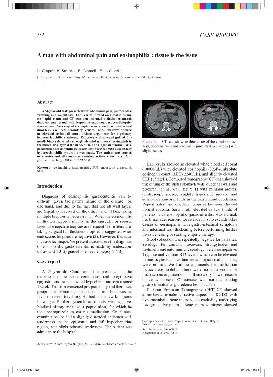
Lab results showed an elevated white blood cell count (10000/µL) with elevated eosinophils (22,4%, absolute eosinophil count (AEC) 2240/µL), and slightly elevated CRP (15mg/L). Computed tomography (CT) scan showed thickening of the distal stomach wall, duodenal wall and proximal jejunal wall (figure 1) with minimal ascites. Gastroscopy showed slightly hyperemic mucosa and edematous mucosal folds in the antrum and duodenum. Repeat antral and duodenal biopsies however showed normal mucosa. Serum IgE, elevated in two thirds of patients with eosinophilic gastroenteritis, was normal. For these latter reasons, we intended first to exclude other causes of eosinophilia with gastro-intestinal symptoms and intestinal wall thickening before performing further invasive testing or starting empiric therapy.
Stool collection was repeatedly negative for parasites. Serology for anisakis, toxocara, strongyloides and trichinella and auto-immune serology were also negative. Tryptase and vitamin B12 levels, which can be elevated in mastocytosis and certain hematological malignancies, were normal. We had no arguments for medication induced eosinophilia. There were no macroscopic or microscopic arguments for inflammatory bowel disease or celiac disease. C1-esterase was normal, making gastro-intestinal angio-edema less plausible.
Positron Emission Tomography (PET)-CT showed a moderate metabolic active aspect of D2-D3 with hypermetabolic bone marrow, not excluding underlying low grade lymphoma. Bone marrow biopsy showed
Correspondence to : Lara Crapé, Groene Briel 1, Ghent, Belgium.E-mail : [email protected]
Submission date : 04/10/2018Acceptance date : 06/01/2019
Acta Gastro-Enterologica Belgica, Vol. LXXXII, October-December 2019
532 CASE REPORT
Figure 1. — CT-scan showing thickening of the distal stomach wall, duodenal wall and proximal jejunal wall (red arrows) with slight ascites.
11-Crapé.indd 532 30/10/19 11:34

A man with abdominal pain and eosinophilia : tissue is the issue 533
Acta Gastro-Enterologica Belgica, Vol. LXXXII, October-December 2019
The diagnosis of muscularis-predominant eosino-philic gastroenteritis together with a secondary hyper-eosinophilic syndrome with single organ involvement was made.
Therapy was initiated with methylprednisolone 32mg per day, in combination with montelukast 10mg per day. Calcium substitution was associated. There was a quick resolution of symptoms within 10 days. Methylprednisolone was tapered to 16 mg over three weeks and the patient was then initiated on budesonide 9mg in off-label use (solubilized budesonide), tapered over 3 months. After 8 months our patient presented with recurrent symptoms. He was restarted on methylprednisolone with fast resolution of symptoms and a fast tapering. He is now on budesonide 9mg, slower tapering over 6 months is planned.
DiscussionEosinophilic gastroenteritis (EGE) is a rare inflam-
matory disorder first described by Kaijser in 1937 (3). Prevalence is estimated between 8,4 and 28 per 100 000 and is higher in children. Mostly, adults are diagnosed between their third and fifth decade of life (1). Concomi-tant allergic disorders, including asthma, rhinitis, eczema and drug or food allergies, are present in 45% to 63% of the reported EGE cases (1,4). Association with other autoimmune conditions such as celiac disease, ulcerative colitis and systemic lupus erythematosus (1,5) has been described.
EGE can affect the entire gastrointestinal tract, antrum and duodenum being most common (1). Symptoms can vary depending on the affected segment as well as on the affected layer of the gastrointestinal wall. Klein et al described three main categories based on the involved gut layers namely mucosal, muscular and serosal (6) .Mucosal infiltration (> 70%) causes abdominal pain, diarrhea, vomiting, protein-losing enteropathy and mal-absorption (1). Muscular disease leads to thickening of the gastrointestinal wall causing gastric outlet or intestinal obstruction, or even biliary obstruction and pancreatitis when affecting the peripapillary region (1). Serosal disease causes signs of ascites and peritonitis. Muscular and serosal types are often associated with mucosal infiltration, supporting the hypothesis of centrifugal disease progression (1).
Pathogenesis is not fully understood, but given the high correlation with other atopic conditions, an allergen-mediated hypersensitivity response is strongly suspected (1). It is postulated that exposure of the gastrointestinal mucosa to allergens promotes a Th-2 mediated immune response. These Th-2 cells produce interleukin (IL)-4, IL-5 and IL-13, promoting the production of eosinophils as well as IgE (7). Risk factors are higher socioeconomic status, Caucasian race and obesity (1).
Pittfalls in diagnosis.
Diagnosis is made based on three criteria: gastro-intestinal symptoms, eosinophilic infiltration of the
extensive central eosinophilia (24%) without elevated blasts or atypical eosinophils and without argument for lymphoma invasion. Additional fluorescent in situ hybridization (FISH) techniques showed no chromosomal abnormalities such a PDGFRB or FIP1L-PDGFRA gene reformation which are expected in primary HES with clonal eosinophil expansion.
After four days, eosinophil count rose to 53% or 6790/µL AEC and our patient further lost 4 kilograms.
Starting the patient on steroids could have been a possible diagnostic option but since endoscopic mucosal biopsies and serology were all negative for eosinophilic gastroenteritis we opted for a final evaluation with endoscopic ultrasound including fine-needle biopsy. We didn’t perform laparoscopy due to the risk of post-procedural leakage after surgical full-thickness biopsy in the setting of an extremely edematous duodenal wall.
EUS examination showed a circumferential wall thickening of the mucosal, submucosal and muscularis propria layers of the antrum, pylorus, bulbus and D2, with a still intact multilayer aspect suggestive for an inflammatory process rather than malignancy (figure 2). We performed an FNB of the bulboduodenal wall with a 22ga Acquire needle from Boston with 2 passes. The pathologic evaluation of the material showed a transmural infiltrate with a very high level of eosinophils, especially in the muscularis propria (figure 3).
Figure 2. — EUS showing thickening of the mucosal (red arrow), submucosal (yellow arrow) and muscularis propria (green arrow) layers of the antrum, pylorus, bulbus and D2, with guarded multiple layer aspect suggestive for an inflammatory process rather than a malignancy. Measurement of the antral wall showed a wall thickness of 8,1mm (normally 2-3mm).
Figure 3. — Fine needle biopsy showing muscularis propria layer with infiltration of numerous eosinophils (> 100/ HPF).
11-Crapé.indd 533 30/10/19 11:34

534 L. Crapé et al.
Acta Gastro-Enterologica Belgica, Vol. LXXXII, October-December 2019
diffuse hypoechoic thickening is seen with, in contrast to inflammatory disease, fusion of the layers (2).
Histopathologic diagnosis should be made by an experienced pathologist. The gastrointestinal tract, except for the esophagus, contains different amounts of eosinophils in basal circumstances, with the caecal and appendiceal region having the highest concentrations (7). In literature consisting of case reports and case series, an absolute eosinophil count of > 20 eosinophils/hpf in the lamina propria of the duodenum is suggested as cut-off for diagnosis (1). Other findings such as epithelial infiltration, eosinophilic cryptitis and degranulation of mast cells can increase the reliability of the histopathologic diagnosis (7).
Finally, other causes of gastrointestinal eosinophilia should be excluded. These include parasitic infections (i.e., Strongyloides, Ascaris, Ancylostoma, Anisakis, Capillaria, Toxicara, Trichiura and Trichinella), drugs (such as azathioprine, gemfibrozil, enalapril, and carba-mazepine (7)), vasculitis (i.e., Churg-Strauss syn-drome, polyarteritis nodosa), connective tissue diseases, inflammatory bowel diseases, celiac disease, lymphoma, leukemia, mastocytosis and primary hypereosinophilic syndrome (1).
Management
In up to 40% spontaneous remission is described. However, in most cases medication is needed, and eosinophilic gastroenteritis is treated by a short course of prednisone 0,5mg/kg during 2 weeks, then tapered over a period of 6-8 weeks. Response to prednisone is to be expected in up to 90 % of the cases. Pineton de Chambrun et al (11) described three long-term patterns: non-relapsing disease (42%), commonly seen in patients with the serosal type, relapsing-remitting disease (37%), occurring primarily in patients with the muscular type and chronic disease (21%), predominantly observed in patients with the mucosal type. Alternative treatments can be used in steroid dependent or refractory cases. Budesonide, elemental or empirical diet, leukotriene inhibitors, azathioprine, anti-histamines and mast-cell stabilizers have all been used (1). All of these treatment approaches have been described in small case series, but no randomized controlled or comparative trials are available (4). Budesonide has been used as induction and maintenance therapy in about ten case reports. In case of proximal disease (antrum, duodenum, jejunum), off-label use of Entocort (solubilized budesonide) is reported (12,13,14). Allergy testing is not recommended since food allergy testing by specific IgE and skin prick tests lack both sensitivity and specificity. Allergy-guided diets have not been proven effective in eosinophilic gastrointestinal disorders (1,2).
Conclusion
EGE is a disease with a broad differential diagnosis, often mimicking other gastrointestinal disorders.
gastrointestinal wall and exclusion of secondary causes of eosinophilic infiltration (1).
Clinical diagnosis can be difficult given the wide array of nonspecific symptoms, low incidence and the absence of pathognomonic findings. Abnormal laboratory tests can raise suspicion, but none are specific for EGE. In two-thirds of the cases peripheral eosinophilia and elevated IgE are seen (1). Imaging studies may be normal in up to 90% of the cases (7). Computed tomography (CT) scan can detect diffuse thickening of the intestinal wall, mucosal folds, ascites and obstruction, but also the “halo sign” and the “araneid-limb-like sign”, both of which can aid in differentiating between an inflammatory and a neoplastic lesion (1,4).
Endoscopic findings can vary from normal to ery-thema, pseudopolyps or ulcerative disease. In mucosal disease, diagnosis can be made by endoscopy with biopsies. At least 6 biopsies are recommended from both normal and abnormal appearing mucosa due to the patchy nature of the disease. In this way, diagnosis is made in 80% of patients with mucosal disease. When mucosal involvement is suspected, a repeat endoscopy is suggested in case of negative biopsies (1).
For diagnosis of submucosal or muscular infiltration literature generally advises surgical full-thickness biop-sy. However this is an invasive surgical procedure with risk of post-operative complications (2).
Only a handful of case reports have described endo-scopic ultrasound findings in muscular and serosal dis-ease (2,8,9) and only one made a diagnosis using EUS-guided FNA (10). Alnaser et al. was the first to describe an abnormal EUS image of EGE, with a significant thick-ening of the antral and duodenal mucosal and submuco-sal layers correlating well with the microscopic pathol-ogy of the resected surgical specimen (8). Akishisa et al. described a transmural circumferential thickening of the antrum of 1,2 centimeters predominantly involving the muscularis propria with homogeneous hypoechoic inter-nal areas and preservation of the five-layered structure. Here, mucosal biopsies could confirm the diagnosis (2). Andriulli et al described an asymmetrical thickening of the muscular layer of the antral wall (9).
Thickening of the gastric wall is not a specific sign of eosinophilic gastroenteritis. It is also observed in Menetrier’s disease, anisakiasis, acute gastric mucosal lesion (AGML), and infiltrating neoplasms (lymphoma and scirrhous carcinoma). However, Menetrier’s disease shows a hyperechoic thickening of the mucosal layer alone with preservation of the layers. Anisakiasis shows a thickening of the submucosal layer alone. AGML are divided into submucosal type and mucosal type with heterogeneous hypoechoic internal areas. Thus, the locations of these diseases are quite different from eosinophilic gastroenteritis with predominant muscular layer involvement. When EUS demonstrates a thickened muscularis propria, malignancy should be strongly suspected. However, in case of lymphoma, a
11-Crapé.indd 534 30/10/19 11:34

A man with abdominal pain and eosinophilia : tissue is the issue 535
Acta Gastro-Enterologica Belgica, Vol. LXXXII, October-December 2019
3. NAIK R.P., JOSHIPURA V.P., PATEL N.R., PATWARI S.I., BHAVSAR M.S. A rare case of predominantly muscular infiltrative eosinophilic gastroenteritis with ascites: a case report and review of the literature. Trop Gastroenterol., 2009, 30: 225-226.
4. SHIH H. M, BAIR M. J., CHEN H. L., LIN I. T. Eosinophilic gastroenteritis: Brief Review. Acta Gastroenterol Belg., 2016, 79 : 239-244.
5. BUTTERFIELD J.H., MURRAY J.A. Eosinophilic gastroenteritis and gluten-sensitive enteropathy in the same patient. J Clin Gastroenterol., 2002, 34 : 552-553.
6. KLEIN N.C., HARGROVE R.L., SLEISENGER M.H., JEFFRIES G.H. Eosinophilic gastroenteritis. Medicine (Baltimore), 1970, 49 : 299-319.
7. SHIFFLET A., FOROUHAR F., WU G.Y. Eosinophilic Digestive Diseases: Eosinophilic Esophagitis, Gastroenteritis, and Colitis. J Formos Med Assoc., 2009, 108 : 834-843.
8. ALNASER S., ALJEBREEN A.M. Endoscopic ultrasound and hisopathologic correlates in eosinophilic gastroenteritis. Gastroenterol, 2007, 13 : 91-94.
9. ANDRIULLI A., RECCHIA S., VALENTE G., PERA A., VERME G. Endoscopic ultrasonography in eosinophilic infiltration of gastric wall. Ital J Gastroenterol, 1990, 22 : 129-132.
10. MÜLLER M., KELLER K., STALLMANN S., ECKARDT A.J. Clinicopathologic Findings in Eosinophilic Gastroenteritis: A German Case Series. J Genet Syndr Gene Ther., 2014, 5 : 230.
11. PINETON DE CHAMBRUN G., GONZALEZ F., CANVA J. Y., GONZALEZ S., HOUSSIN L., DESREUMAUX P. et al. Natural history of eosinophilic gastroenteritis. Clin Gastroenterol Hepatol., 2011, 9 : 950-956.
12. TAN A.C., KRUIMEL J.W., NABER T.H. Eosinophilic gastroenteritis treated with non-enteric-coated budesonide tablets. Eur J Gastroenterol Hepatol., 2001, 13 : 425.
13. SIEWERT E., LAMMERT F., KOPPITZ P., SCHMIDT T., MATERN S. Eosinophilic gastroenteritis with severe protein-losing enteropathy: successful treatment with budesonide. Dig Liver Dis., 2006, 38 : 55.
14. ELSING C., PLACKE J., GROSS-WEEGE W. Budesonide for the treatment of obstructive eosinophilic jejunitis. Z Gastroenterol., 2007, 45 : 187.
Diagnosis requires a combination of clinical and patho-logic criteria and can be challenging especially in the muscular and serosal form since invasive surgical biopsies are needed. We presented a case in which the diagnosis of a muscular predominant for of EGE was made by using EUS-guided FNB. Mostly, EGE is treated with prednisone. However, there are many therapeutic options, all of which have been reported in case series and have shown variable efficacy. A maintenance regimen is often needed, preferably based upon a safe steroid-sparing drug.
Conflict of interest
There is no conflict of interest.
References
1. ANTOINE A.R., WEAM E.H. Eosinophilic gastroenteritis: Approach to diagnosis and management. World J Gastrointest Pharmacol Ther., 2016, 7 : 513-523.
2. FUKUDA A., KAJIYAMA T., KISHIMOTO H., SOMEDA H., SAKAI M., SENO H., et al. Eosinophilic gastroenteritis with predominant muscular layer disease: feature of endoscopic ultrasonography. Dig Endosc, 2004, 16 : 368-371.
11-Crapé.indd 535 30/10/19 11:34