55 yo male presented with - department of surgery at suny
TRANSCRIPT


55 yo male presented with› Left popliteal fossa and calf pain for 4 days› Difficulty walking› No history of trauma› No claudication

PMH/Social› HTN› Right adrenal
adenoma with hyperaldosteronism
› DM› Hypercholesterole-
mia› Smoker (2 P/D)› Social drinking
PSH› Varicocelectomy› Knee arthroscopy› Heel spur

Physical examPertinent for-Left pulsating popliteal mass, 3X3cm,
warm foot w/o evidence of acute ischemic changes and good capillary refill bilaterally
DP/PT pulses non palpable/biphasic dopler signals bilaterally

LABS- no pertinent findings; WNL
LE Duplex- Left popliteal artery aneurysm partially thrombosed 4.5x4.8cm
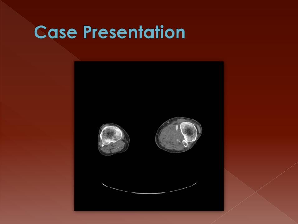
CTA of Aorta with Distal Runoff


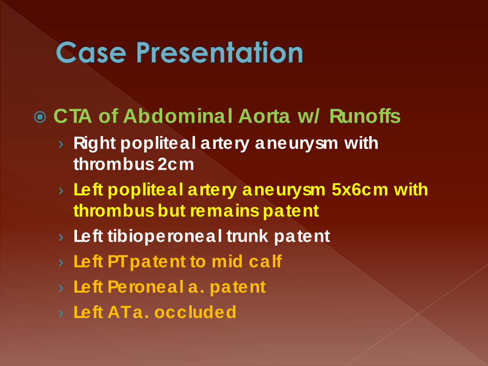
CTA of Abdominal Aorta w/ Runoffs› Right popliteal artery aneurysm with
thrombus 2cm› Left popliteal artery aneurysm 5x6cm with
thrombus but remains patent› Left tibioperoneal trunk patent› Left PT patent to mid calf› Left Peroneal a. patent› Left AT a. occluded

Initial plan-admission for anticoagulationVein mapping Semi-elective interventionAngiogram
However,Increase in symptoms during night
OR in morning

PROCEDURE- Left Common Femoral to Below Knee Popliteal Bypass w/ Ringed PTFE Graft and Popliteal Aneurysm Ligation
FINDINGS-› Severe inf. reaction, perianeurysmal› Venous hypertension› Heavily calcified popliteal artery› Periadventitial adventia (a. & v.)› Dense venous collateral

POD1-8› Improved swelling › Decreased pain› Ambulation/PT› Anticoagulation- Lovenox/coumadin› Palpable pulses› No neurologic findings or complaints

POD9C/O pain and numbness of the left leg
and footWeakness of the foot/drop footWarm extremityNon palpable pulses/biphasic
doplerable signals

Working diagnosisgraft occlusion v/s expansion of PAA
and nerve compression
Heparin drip
CTA of lower extremity


CTA LEWidely patent graft
9 cm PAA with intra-aneurysmal flow
Patent trifurcation

PROCEDURE- Left PAA exclusion, revision of bypass graft and intraoperative angiogram› Proximal control of the aneurysm above the
knee› Angiogram through graft showed retrograde
filling of aneurysm› Control/ligation of aneurysm below knee› Improvement of distal pulses and flow through
graft› Completion angio- no filling of aneurysm, patent
Peroneal a. reconstituting the DPA

POD1-3 (POD10-13)Extubated POD1Mild improvement in symptoms (3/5
improvement in dorsiflexion and eversion/inversion of foot) However, persistent weakness and numbness
Maintained anticoagulationPT/RehabAntibiotic coverage
POD4- OR for thrombus evacuation

PROCEDURE- Exploration of left poplitealfossa and PA aneurysmectomy and debridement via a posterior approach
FINDINGS-› Stretched tibial nerve over the sac› PAA controlled and excised- gelatinous
material that shelled out easily; little thrombus
› Obliteration of sac› JP drain in fossa

PATHOLOGYAll specimens- organizing thrombus,
part of vessel wallCoccoid bacteriaCultures- Staph epidermidis
MYCOTIC ANEURYSM

Postoperative courseSlowly improving neuro symptomsRehab and physical therapyLong term antibioticsLong term anticoagulation and antiPt
Discharge on POD15 (POD19/POD29)

Normal PA diameter-- 0.5-1.1cm- Varies with gender- Varies with segment of PA
Aneurysm = 1.5x normal diameter of adjacent PA segment

95-100% male Second most common aneurysms, 70%
of aneurysms of LE However rare- 1-7.4/100000 Bilateral 50-54% PAA+ AAA 40% Bilateral PAA + AAA up to 70% AAA + PAA up to 8% Risk of other aneurysms over 10 yrs- 50%

Asymptomatic mass- 38-40%will develop symptoms at a rate of
14%/yr (2) Intermittent claudication (chronic
ischemia)- 25%-40% (1,2) Pressure related (pain, DVT, swelling)- 5-
10% (2) Rupture- 0-7% (1) Acute ischemia- 21-35%; thrombosis or
embolisation (1)

Physical Exam- Unreliable Duplex US- more reliable; determines
size, presence of thrombus and patency of outflow vessels
Anatomical studies- needed when decision to operate is made› Angiography› CTA› MRA

SIZEAsymptomatic 2cm or lessSymptomatic 3cm or more ( 3 for
ischemia and 3.45 for compression)Rate of growth 3mm/y (2,3)
DISTORTIONAsymptomatic 0°Acute thrombosis 60°Compression 45° (2,3)

SIZE/DISTORTIONSize + distortion = best PPV and NPV
for complicationsSize > 3cm + Distortion > 45 ° (3)
RUNOFFsAbsent distal pulses = higher rate of
complications (1,2,3)

Elective intervention may have higher patency and limb salvage rates but difference is not significant
Observation may be a viable option in PAA< 3cm with little or no distortion and no intramural thrombus (2,3)

INDICATIONSThrombosisSymptomaticSize greater than 3 cm (2,3)

Medial approachsmall fusiform aneurysmsconventional bypass and ligation
Posterior approachlarge saccular aneurysmssymptoms of compressioninterposition grafting

Saphenous vein exposure Exposure of above knee and below
knee pop a. through same incision Large aneurysm → difficult exposure Tunnel from above-knee to below-knee
pop space between heads of gastro-cnemius
Bypass- end to side, reversed/non reversed, in-situ if CFA to distal a.


Aneurysm ligation- proximal and distal ligation as close to the aneurysm as possible to decrease the risks of expansion
Aneurysm should be open and back-bleeding branches ligated- difficult with medial approach

Prone position Lazy S incision across
the popliteal fossa Can be extended
over lesser saphenous vein if adequate for bypass

Exposure- between semimembranosusand semitendinosusand biceps muscles
Attention!! tibial and peroneal nerves
Open aneurysm sac Ligate backbleeders Bypass or
interposition graft

Acute Ischemia
Threatened Limb?No Yes
Heparin dripAngiogram
AngioSurgery
Occluded aneurysm
Patent DistalsYes No
Bypass Thrombolysis
Patent distalsYes No
Bypass Thromboembolectomy+/-
intraopthrombolysisEmergency Surgical Treatment

25-45% of acutely ischemic patients will have thrombosis of the PAA with either no runoffs or compromised runoffs unsuitable to bypass
Catheter-directed intra-arterial thrombolysis with TPA or urokinase
Mechanical thrombectomy may be used

Re-image at 6-24 hours If improvement → prepare for surgery Total clearance is not necessary for
bypass If no improvement beyond 24h → high
risk of amputation Do not continue beyond 24h

Improves outflow before bypass in 29-45% of acute ischemia patients
Reduces amputation rate from 96 to 69%

Galland- increased number of complications other than bleeding-Acute deterioration of the limb during thrombolysis
Destabilization and embolization of a large thrombus
Intraoperative thrombolysis (1,2)

ApproachPosterior and medial approaches
seem to have equally good results

Bisdas et al. Angiology 2010 61:248-252

Kropman HJR et al. J Vasc Surg 2007; 46:24-30

Saphenous vein vs graftPatency of saphenous vein superior to
graft overall- 94% vs 63%Regardless of approachMost significant in patients with acute
ischemia

Pulli et al. (6)159 PAA between 1984 and 200442% asymptomatic1/3rd 3 runoffs, 1/3rd 2 runoffs 1/3rd 1 or
0 runoffsfollow up- clinical + sono at 1,6 and 12
months then yearly thereafter

VARIABLE 30-day amputation rate P Value
Asymptomatic 1/67 (1.4%) .05
Symptomatic 6/92 (6.5%)
30 day Amputation Rate

VARIABLE Symptomatic Asymptomatic P Value
Survival 87% 81.4% NS
Limb salvage 80.4% 93.4% 0.03
Primary Patency 51.6% 86.5% 0.001
Secondary patency 79.6% 89.4% NS
Estimated 60 Months Outcomes

60 m
onth
s lim
b sa
lvag
e ra
te
in s
ympt
omat
ic li
mbs
(c
laud
ican
tvs
acut
e isc
hem
ia)

Factors associated with outcome on multivariate analysis (6)
Clinical presentation
Runoff Status (5 yr patency 72% w/ 2 or more runoffs vs 47.4% w/ worse runoffs)
Site of distal anastomosis (popliteal vstibial a.)
Mehta et al.26 PAAs between 1995 and 2001Mean F/U 38 monthsDuplex, CTA, MRA to document
aneurysm size (7)

Results (7)38%- persistent flow from geniculate
arteries23%- increased max sac diameter12%- rupture w/ 4% limb lossAll PAAs had near systemic pressure
Pathophysiology similar to type II endoleaks› Geniculate a. transmit syst. pressure to the PAA
causing its growth (7)

Conclusions (7)Routine use of posterior approach
whenever possible with ligation of collateral vessels and endoaneurysmor-raphy esp. for large saccular aneurysms with compressive symptoms
Medial approach should be reserved for extensive fusiform PAAs and still the collateral vessels should be ligated

Long term results unknown Short term results
patency rates lower than open repairhigher rates of reintervention
Reserved for patients at high surgical risks and appropriate anatomy

Contraindications› Access a. should accommodate 9-11Fr
introducers› Thrombosed aneurysms› Occluded SFA› Distal embolization (may be worsened by
deployment)› Very diffuse aneurysms extending above the
adductor hiatus or involving the entire below knee popliteal a. or SFA should be treated surgically


Tipsgrafts should be oversized by 10-20%if more than 1 stent is required a 3 cm
overlap at least should be done
Follow upDuplex at 3 months intervals the first
year then Q6 months thereafter

ResultsWeighted average for primary and
secondary patency rates at 1.5 yr- 65% and 76%
Decreased length of stay and recovery
Increased rates of thrombosis and repeat interventions
Antonello et al. (8)Prospective randomized trial30 PAAs between 1999 and 200315 patients OR/15 patients ETHemobahn graft- self expanding
nitinol stent w/ internal linining of PTFEsize 2cm or more

Technical details (8)Embolization of geniculate branches
when presentIntraoperative study in flexion- if
stenosis >50% → conversion Follow-up (8)
› Duplex and ABI with flexion upon discharge› Same at 1and 3 months and Q6 months after› Forced leg flexion CTA at 6 and 12 mo then
Qyear

Group A (OR) Group B (ET) PGraft/endograftocclusion 0% 6.7% NS
Primary Patency 100% 93.3% NS
Assisted Patency 100% NS
Limb Salvage Rate 100% 100% NS
Endoleaks 0% NS
Mean Operative Time 195.3mn 75.4mn <.01
Mean Hospital Stay 7.7 days 4.3 days <.01

GROUP A (OR) GROUP B (ET) P
12 months primary patency 100% 86.7% NS
48 months primary patency 81.8% 80% NS

There was no endoleaks No evidence of kinking of the graft
during the follow up period (45 months) as evidenced by CTA with forced flexions and ABIs with flexion and at rest
Conclusion› ET seems to be safe and as good as open
repair› Study limitation and no long term follow up

1- Cronenwett: Rutherford’s Vascular surgery, 71th ed. Saunders 2010
2- Galland RB. Popliteal aneurysms: controversies in their management. Am J Surg 190 (2005): 314-318
3- Galland RB. Popliteal aneurysms: from John Hunter to the 21st century. Ann R Coll Surg Engl 2007; 89: 466-471
4- Bisdas T, Parskevas KI, Pichlmaier M et al. Dorsal versus medial approach for the surgical repair of popliteal artery aneurysms. Angiology 2010; 61(3): 248-252
5- Kropman HJR, van Santvoort HC, Teijink J et al. The medial versus the posterior approach in the repair of popliteal artery aneurysm: a multicenter case-matched study. J Vasc Surg 2007; 46: 24-30
6- Pulli R, Dorigo W, Troisi N et al. Surgical management of popliteal artery aneurysms: which factors affect outcomes? J Vasc Surg 2006; 43: 481-487
7- Mehta M, Champagne B, Darling C et al. Outcome of popliteal artery aneurysms after exclusion and bypass: significance of residual patent branches mimicking type II endoleak

8- Antonello M, Frigatti P, Battocchio p et al. Open versus endovascular treatment for asymptomatic popliteal artery aneurysm: results of a prospective randomized study. J Vasc Surg 2005; 42: 185-193
9- Moore RD, Hill AB. Open versus endovascular repair of popliteal artery aneurysms. J Vasc Surg 2010; 51: 271-276