document
TRANSCRIPT
© 1999 Macmillan Magazines Ltd
letters to nature
NATURE | VOL 397 | 18 FEBRUARY 1999 | www.nature.com 625
NaCl, 10 mM EDTA, 5 mM MgCl2, 1mM DTT, 1 mM GTP-gS, pH 7.5, for 90
min at room temperature under rotation. Afterwards, buffer 1 was removed
and the GTP-gS form of GST±Rab5 was stabilized with buffer (buffer 2)
containing 20 mM HEPES, 100 mM NaCl, 5 mM MgCl2, 1mM DTT, pH 7.5, in
the presence of 1 mM GTP-gS for 20 min at room temperature under rotation.
Beads were then incubated for 120 min at 4 8C with bovine brain cytosol
obtained as follows: 14 bovine brains were homogenized in a blender with
buffer 2 and the homogenate was centrifuged at 4,200g at 4 8C for 50 min. The
resulting postnuclear supernatant was then centrifuged at 100,000g at 4 8C for
60 min. The high-speed supernatant was dialyzed against buffer 2 (without
nucleotide) before incubation with the af®nity column. After incubation with
cytosol, beads were washed with ten column volumes of buffer 2 containing 10
mM GTP-gS, ten column volumes of buffer 2 containing 250 mM NaCl ®nal
concentration and 10 mM GTP-gS, and one column volume of 20 mM HEPES,
250 mM NaCl, 1 mM DTT, pH 7.5.
Bound proteins were eluted with 1.5 column volumes of a buffer containing
20 mM HEPES, 1.5 M NaCl, 20 mM EDTA, 1mM DTT, 5 mM GDP, pH 7.5,
incubated with the beads for 20 min at room temperature under rotation.
EDTA was used at this step to remove Mg2+ from Rab5 and release the effectors
from the column. As EEA1 is a Zn2+-binding protein and EDTA chelates Zn2+
(refs 23, 30), our subsequent assays were done in the presence of 1 mM ZnCl2®ltered (0.22 mM) before its use. The GDP form of GST±Rab5 (1 ml) was made
as described above for the GTP-gS form of GST±Rab5 with the following
changes: all buffers contained GDP instead of GTP-gS, except for the elution
buffer which contained GTP-gS (1 mM) instead of GDP. In the case of GST
column (100 ml), there was no nucleotide in the buffers.
Fractionation of Rab5 effectors. The eluate (30 ml) from the 20-ml af®nity
column containing the mixture of Rab5-interacting proteins was ®rst treated
twice for 1 h at 4 8C with 1.5 ml glutathione Sepharose beads to remove GST±
Rab5 which leaked from the af®nity column during the elution step. The
sample was then desalted using PD10 columns from Pharmacia in a buffer
containing 20 mM HEPES, 150 mM NaCl, 1mM DTT, pH 7.5, and diluted
three times with 20 mM HEPES, 1 mM DTT, pH 7.5, resulting in a ®nal volume
of 135 ml. The diluted sample was loaded on a 1-ml MQ FPLC column
(Pharmacia) and bound proteins were step-eluted with 20 mM HEPES, 1 M
NaCl, 1mM DTT, pH 7.5, in a total volume of 1 ml (concentration step). This
eluate was fractionated on a 24-ml Superose 6 FPLC gel ®ltration column
(Pharmacia). Fractions of 0.4 ml were collected, aliquoted and frozen at -80 8C.
Endosome docking assay. HeLa cells grown in suspension were harvested,
washed with PBS and incubated for 5 min with 20 mg ml-1 rhodamine-labelled
transferrin at 37 8C for 5 min. Then endosomes were isolated15 and incubated in
a 10 ml reaction for 25 min at 37 8C with reagents mentioned in Fig. 4. At the
end of the reaction, samples were put on ice and visualized with a Zeiss
Axiophot ¯uorescence microscope. Images were taken using a Cohu camera.
Quantitation of docking was done by taking random pictures, followed by
counting of the occupied squares (an indication of organelle area) on a
reference grid.
Other preparations. GST±Rab5 and GST proteins were expressed in
Escherichia coli using the P-GEX vector (Pharmacia). his-Rab GDI16, his-a-
SNAP and his-a-SNAP(L294A)25 were produced as described. The preparation
of recombinant Rabaptin-5/Rabex-5 complex will be described elsewhere
(R. Lippe and M. Zerial, manuscript in preparation). Early endosome fusion
assay was done as described16.
Received 9 October 1998; accepted 8 January 1999.
1. Rothman, J. E. Mechanisms of intracellular protein transport. Nature 372, 55±63 (1994).2. Weber, T. et al. SNAREpins: Minimal machinery for membrane fusion. Cell 92, 759±772 (1998).
3. Robinson, L. M. & Martin, T. F. J. Docking and fusion in neurosecretion. Curr. Opin. Cell Biol. 10,
483±492 (1998).
4. von Mollard, G. F., Nothwehr, S. F. & Stevens T. H. The yeast v-SNARE Vti1p mediates two vesicle
transport pathways through interactions with the t-SNAREs Sed5p and Pep12p. J. Cell Biol. 137,1511±1524 (1997).
5. Holthuis, J. C., Nichols, B. J., Dhruvakumar, S. & Pelham, H. R. Two syntaxin homologues in the
TGN/endosomal system of yeast. EMBO J. 17, 113±126 (1998).
6. Hay, J. C. et al. Localization, dynamics, and protein interactions reveal distinct roles for ER and Golgi
SNAREs. J. Cell Biol. 141, 1489±1502 (1998).7. Sapperstein, S. K., Lupashin, V. V., Schmitt, H. D. & Waters, M. G. Assembly of the ER to Golgi SNARE
complex requires Uso1p. J. Cell Biol. 132, 755±767 (1996).
8. Cao, X., Ballew, N. & Barlowe, C. Initial docking of ER-derived vesicles requires Uso1p and Ypt1p but
is independent of SNARE proteins. EMBO J. 17, 2156±2165 (1998).
9. VanRheenen, S. M., Cao, X., Lupashin, V. V., Barlowe, C. & Waters, G. M. Sec35p, a novel peripheralmembrane protein, is required for ER to Golgi vesicle docking. J. Cell Biol. 141, 1107±1119 (1998).
10. Novick, P. & Zerial. M. The diversity of Rab proteins in vesicle transport. Curr. Opin. Cell Biol. 9, 496±
504 (1997).11. Sùgaard, M. et al. A Rab protein is required for the assembly of SNARE complexes in the docking of
transport vesicles. Cell 78, 937±948 (1994).
12. Lian, J. P., Stone, S., Jiang, Y., Lyons, P. & Ferro-Novick, S. Ypt1p implicated in v-SNARE activation.
Nature 372, 698±701 (1994).
13. Mayer, A. & Wickner, W. Docking of yeast vacuoles is catalyzed by the Ras-like GTPase Ypt7p aftersymmetric priming by Sec18p (NSF). J. Cell Biol. 136, 307±317 (1997).
14. Lupashin, V. V. & Waters, M. G. t-SNARE activation through transient interaction with a rab-like
guanosine triphosphatase. Science 276, 1255±1258 (1997).
15. Gorvel, J.-P., Chavrier, P., Zerial, M. & Gruenberg, J. Rab5 controls early endosome fusion in vitro. Cell
64, 915±925 (1991).16. Horiuchi, H. et al. A novel Rab5 GDP/GTP exchange factor complexed to Rabaptin-5 links nucleotide
exchange to effector recruitment and function. Cell 90, 1149±1159 (1997).
17. Gournier, H., Stenmark, H., Rybin, V., Lippe, R. & Zerial, M. Two distinct effectors of the small
GTPase Rab5 cooperate in endocytic membrane fusion. EMBO J. 17, 1930±1940 (1998).
18. Simonsen, A. et al. EEA1 links phosphatidylinositol 3-kinase function to Rab5 regulation ofendosome fusion. Nature 394, 494±498 (1998).
19. Emans, N. et al. Annexin II is a major component of fusogenic endosomal vesicles. J. Cell Biol. 120,
1357±1369 (1993).
20. Colombo, M. I., Beron, W. & Stahl, P. D. Calmodulin regulates endosome fusion. J. Biol. Chem. 272,7707±7712 (1997).
21. Rodriguez, L., Stirling, C. J. & Woodman, P. G. Multiple N-ethylmaleimide-sensitive components are
required for endosomal vesicle fusion. Mol. Biol. Cell 5, 773±783 (1994).
22. Colombo, M.I., Taddese, M., Whiteheart, S. W. & Stahl, P. D. A possible predocking attachment site
for N-ethylmaleimide-sensitive fusion protein. Insights from in vitro endosome fusion. J. Biol. Chem.271, 18810±18816 (1996).
23. Wiedemann, C. & Cockcroft, S. Vesicular transport: sticky ®ngers grab a lipid. Nature 394, 426±427
(1998).
24. Patki, V. et al. Identi®cation of an early endosomal protein regulated by phosphatidylinositol 3-kinase.
Proc. Natl Acad. Sci. USA 94, 7326±7330 (1997).25. Barnard, R. J. O., Morgan, A. & Burgoyne, R. D. Stimulation of NSF ATPase activity by alpha-SNAP is
required for SNARE complex disassembly and exocytosis. J. Cell Biol. 139, 875±883 (1997).
26. Mayer, A., Wickner, W. & Haas, A. Sec18p (NSF)-driven release of Sec17p (a-SNAP) can precede
docking and fusion of yeast vacuoles. Cell 85, 83±94 (1996).
27. Sonnichsen, B. et al. A role for giantin in docking COPI vesicles to Golgi membranes. J. Cell Biol. 140,1013±1021 (1998).
28. Barlowe, C. Coupled ER to golgi transport reconstituted with puri®ed cytosolic proteins. J. Cell Biol.
139, 1097±1108 (1997).
29. Mu, F. et al. EEA1, an early endosome-associated protein. J. Biol. Chem. 270, 13503±13511 (1995).
30. Stenmark, H., Aasland, R., Toh, B.H. & D'Arringo, A. Endosomal localization of the autoantigenEEA1 is mediated by zinc-binding FYVE ®nger. J. Biol. Chem. 271, 24048±24054 (1996).
Acknowledgements. We thank R. Lippe for the supply of recombinant Rabaptin-5/Rabex-5 complex;H. Stenmark for the GST±Rab5 cDNA construct; J. Rothman for pQE9 a-SNAP; A. Giner for technicalassistance; V. Rybin for helpful discussions on technical points; E. Nielsen and B. Sonnichsen for thesupply of rhodamine-labelled early endosomes; P. Scheiffele, B. Sonnichsen and members of thelaboratory for discussions and critical reading of the manuscript; and K. Ashman and M. Wilm formproviding mass spectroscopy data. S.C. is supported by an EU TMR fellowship. H.M.M. is recipient of anAlexander vonHumboldt Stiftung. This work was supported by the Max Planck Gesellschaft, and bygrants from the Human Frontier Science Program, EU TMR and Biomed (to M.Z.). This work isdedicated to the memory of Thomas Kreis.
Correspondence and requests for materials should be addressed to M.Z. (e-mail: [email protected]).
Photosynthetic control ofchloroplast geneexpressionThomas Pfannschmidt*, Anders Nilsson & John F. Allen
Plant Cell Biology, Lund University, Box 7007, S-220 07 Lund, Sweden. . . . . . . . . . . . . . . . . . . . . . . . . . . . . . . . . . . . . . . . . . . . . . . . . . . . . . . . . . . . . . . . . . . . . . . . . . . . . . . . . . . . . . . . . . . . . . . . . . . . . . . . . . . . . . . . . . . . . . . . .
Redox chemistryÐthe transfer of electrons or hydrogen atomsÐis central to energy conversion in respiration and photosynthesis.In photosynthesis in chloroplasts, two separate, light-drivenreactions, termed photosystem I and photosystem II, are con-nected in series by a chain of electron carriers1±3. The redox stateof one connecting electron carrier, plastoquinone, governs thedistribution of absorbed light energy between photosystems I andII by controlling the phosphorylation of a mobile, light-harvesting,pigment±protein complex4,5. Here we show that the redox state ofplastoquinone also controls the rate of transcription of genesencoding reaction-centre apoproteins of photosystem I andphotosystem II. As a result of this control, the stoichiometrybetween the two photosystems changes in a way that counteractsthe inef®ciency produced when either photosystem limits the rateof the other. In eukaryotes, these reaction-centre proteins areencoded universally within the chloroplast. Photosynthetic control
* Present address: Plant Cell Physiology and Molecular Biology, University of Bochum, Universitaets-
strasse 150, 44780 Bochum, Germany.
© 1999 Macmillan Magazines Ltd
letters to nature
626 NATURE | VOL 397 | 18 FEBRUARY 1999 | www.nature.com
of chloroplast gene expression indicates an evolutionary explana-tion for this rule: the redox signal-transduction pathway can beshort, the response rapid, and the control direct.
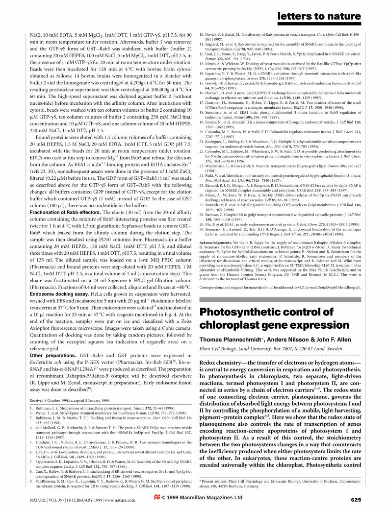
Photosynthesis and plant growth can be supported by light with awavelength composition that favours either photosystem II (`light2') or photosystem I (`light 1')2,3. Figure 1 shows the effects ofsuch photosystem-speci®c light on chloroplast composition andchloroplast gene expression. Figure 1a, b shows the relativetranscriptional rates of speci®c chloroplast genes in 7-day-oldmustard plants grown in light 1, in light 2, and after changesbetween the two regimes. Plants described as `light 2 ! 1' weregrown for 5 days under photosystem-II light, before being trans-ferred to photosystem-I light for 2 days; `light 1' plants were grownunder light 1 for 7 days; white-light-grown plants were used as thecontrol; `light 2' plants were grown under light 2 for 7 days; and`light 1 ! 2' plants were grown for 5 days under photosystem-Ilight, and then transferred to photosystem-II light for 2 days.Cotyledons of seedlings were collected, and chloroplasts wereisolated and used in chloroplast run-on transcription experiments.This method leads to radioactive pulse-labelling of messenger RNA
from a gene that was actually being transcribed at the time ofchloroplast isolation. The transcriptional rate of the genes for thetwo major reaction-centre subunits of photosystem I, psaAB, showsan incremental increase from left to right (Fig. 1a), as photosystem Ichanges from being light-saturated to being light-limited. Thetranscriptional rate for the psbA gene, which encodes the D1
reaction-centre protein of photosystem II, shows broadly the reversepattern (Fig. 1b): the transcriptional rate of psbA is highest whenphotosystem II is rate-limiting (`light 2 ! 1'). The RNA quantitiesof psaAB (Fig. 1c) and, to a lesser extent, psbA (Fig. 1d), followchanges in their respective transcriptional rates (Fig. 1a, b).
Figure 1e, f shows that the quantity of functional reaction centresfollows the same pattern of response to illumination as the rate oftranscription (Fig. 1a, b) and as transcript pool size (Fig. 1c, d). P700
is the specialized chlorophyll a that is bound to the psaAB geneproducts. It undergoes primary charge separation in photosystem I(ref. 6). P700 (Fig. 1e) is least abundant when the light suppliedfavours its activity (light 2 ! 1), and most abundant when it doesnot (light 1 ! 2). QA is the species of plastoquinone that is boundto the psbA gene product. It acts as a secondary electron acceptor inphotosystem II (ref. 7). QA (Fig. 1f) is most abundant whereillumination favours photosystem I (light 2 ! 1), and least abun-dant where it favours photosystem II (light 2 and light 1 ! 2). Thechlorophyll a/b ratio (Fig. 1g) follows the same pattern as P700 and asthe rate of transcription of psaAB. The ratio of photosystem II to I,measured as the molar ratio of QA to P700 (Fig. 1h), follows the samepattern as the absolute quantity of QA and as the rate of transcrip-tion of psbA. These results indicate that when either photosystembecomes rate-limiting to photosynthesis, transcription of genes forits reaction-centre proteins is induced. Genes for reaction-centreproteins of the other photosystem, which has surplus photochemi-cal capacity, are simultaneously repressed.
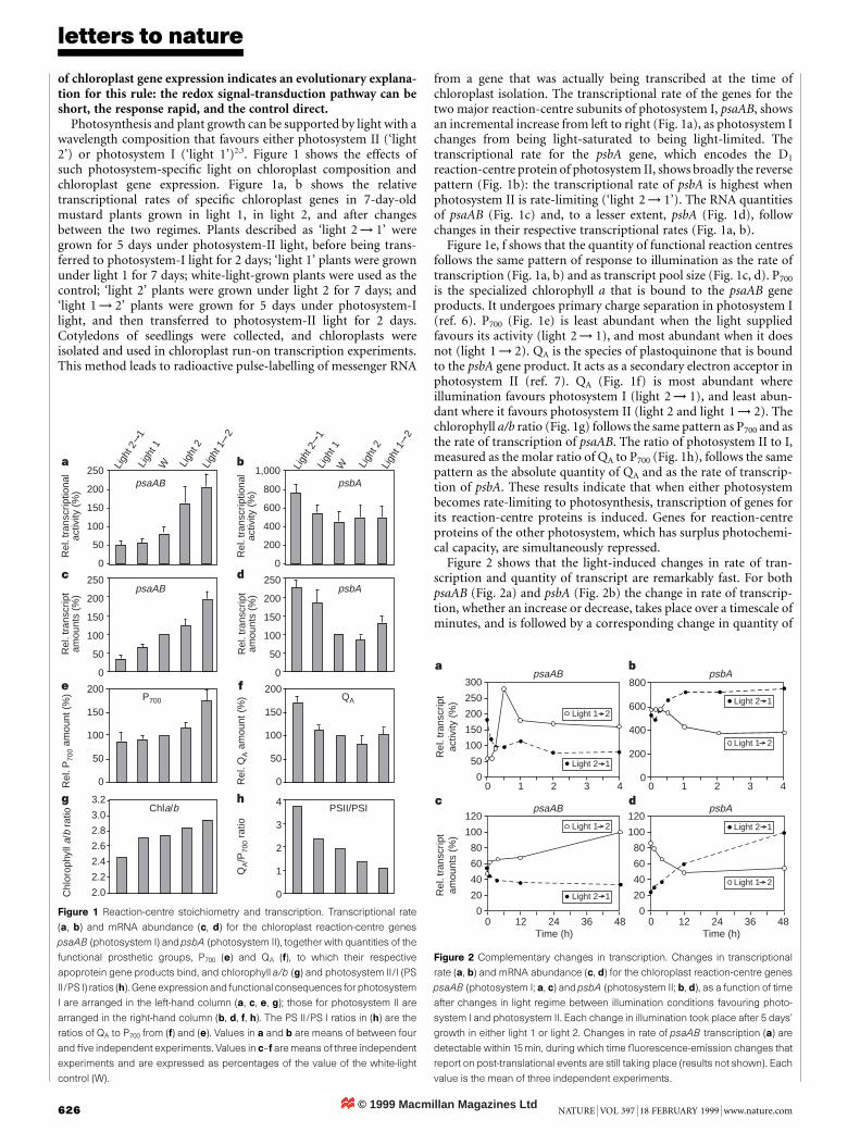
Figure 2 shows that the light-induced changes in rate of tran-scription and quantity of transcript are remarkably fast. For bothpsaAB (Fig. 2a) and psbA (Fig. 2b) the change in rate of transcrip-tion, whether an increase or decrease, takes place over a timescale ofminutes, and is followed by a corresponding change in quantity of
1,000
800
600
400
200
0
250
200
150
100
50
0
250
200
150
100
50
0
3.2
3.0
2.8
2.6
2.4
2.2
2.0
200
150
100
50
0
200
150
100
50
0
4
3
2
1
0
250
200
150
100
50
0
Chla/b PSII/PSI
P700 QA
psaAB psbA
psaAB psbA
Ligh
t 21
Ligh
t 1W Li
ght 2
Ligh
t 12
Ligh
t 21
Ligh
t 1W Li
ght 2
Ligh
t 12
Rel
. tra
nscr
iptio
nal
activ
ity (
%)
Rel
. tra
nscr
iptio
nal
activ
ity (
%)
Rel
. tra
nscr
ipt
amou
nts
(%)
Rel
. tra
nscr
ipt
amou
nts
(%)
Rel
. P70
0 am
ount
(%
)
Rel
. QA a
mou
nt (
%)
QA/P
700
ratio
Chl
orop
hyll
a/b
ratio
a b
c d
e f
g h
Figure 1 Reaction-centre stoichiometry and transcription. Transcriptional rate
(a, b) and mRNA abundance (c, d) for the chloroplast reaction-centre genes
psaAB (photosystem I) and psbA (photosystem II), together with quantities of the
functional prosthetic groups, P700 (e) and QA (f), to which their respective
apoprotein gene products bind, and chlorophyll a/b (g) and photosystem II/I (PS
II/PS I) ratios (h). Gene expression and functional consequences for photosystem
I are arranged in the left-hand column (a, c, e, g); those for photosystem II are
arranged in the right-hand column (b, d, f, h). The PS II/PS I ratios in (h) are the
ratios of QA to P700 from (f) and (e). Values in a and b are means of between four
and ®ve independent experiments. Values in c±f are means of three independent
experiments and are expressed as percentages of the value of the white-light
control (W).
300
250
200
150
100
50
0
120
100
80
60
40
20
0
120
100
80
60
40
20
0
800
600
400
200
00 1 2 3 4 0 1 2 3 4
0 12 24 36 48 0 12 24 36 48
Rel
. tra
nscr
ipt
amou
nts
(%)
Rel
. tra
nscr
ipt
activ
ity (
%)
psaAB psbA
psaAB psbA
Time (h) Time (h)
Light 1 2Light 2 1
Light 1 2
Light 2 1
Light 1 2
Light 2 1
Light 1 2
Light 2 1
a b
c d
Figure 2 Complementary changes in transcription. Changes in transcriptional
rate (a, b) and mRNA abundance (c, d) for the chloroplast reaction-centre genes
psaAB (photosystem I; a, c) and psbA (photosystem II; b, d), as a function of time
after changes in light regime between illumination conditions favouring photo-
system I and photosystem II. Each change in illumination took place after 5 days'
growth in either light 1 or light 2. Changes in rate of psaAB transcription (a) are
detectable within 15 min, during which time ¯uorescence-emission changes that
report on post-translational events are still taking place (results not shown). Each
value is the mean of three independent experiments.
© 1999 Macmillan Magazines Ltd
letters to nature
NATURE | VOL 397 | 18 FEBRUARY 1999 | www.nature.com 627
mRNA (Fig. 2c, d). We conclude that changes in the rate oftranscription (Fig. 2a, b) arise from light-induced perturbation ofthe redox state of the plastoquinone pool. The altered rates oftranscription result, in turn, in altered mRNA quantities (Fig. 2c, d)and, ultimately, in the altered stoichiometries of the two photo-systems of the chloroplast thylakoid membrane (Fig. 1e±h).
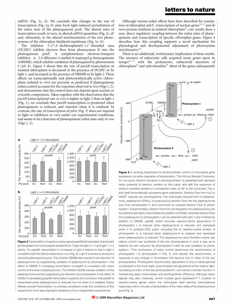
The inhibitor 3-(39,49-dichlorophenyl)-1,19-dimethyl urea(DCMU) inhibits electron ¯ow from photosystem II into theplastoquinone pool8. A complementary electron-transportinhibitor is 2,5-dibromo-3-methyl-6-isopropyl-p-benzoquinone(DBMIB), which inhibits oxidation of plastoquinol by photosystemI (ref. 8). Figure 3 shows that the rate of psaAB transcription inisolated chloroplasts is decreased in the presence of DCMU or bylight 1, and increased in the presence of DBMIB or by light 2. Theseeffects on transcriptionally and photosynthetically active chloro-plasts isolated in vitro are precisely as predicted if plastoquinoneredox control accounts for the responses observed in vivo (Figs 1, 2),and demonstrate that the control does not depend upon nuclear orcytosolic components. Taken together with the observation that thepsaAB transcriptional rate in vivo is higher in light 2 than in light 1(Fig. 1), we conclude that psaAB transcription is promoted whenplastoquinone is reduced, and retarded when it is oxidized. Incontrast, the rate of transcription of psbA (Fig. 3) does not respondto light or inhibitors in vitro under our experimental conditions,and seems to be a function of plastoquinone redox state only in vivo(Figs 1, 2).
Although various redox effects have been described for transla-tion of chloroplast psbA9, transcription of nuclear genes10±12 and denovo protein synthesis in isolated chloroplasts13, our results show anew, direct regulatory coupling between the redox state of plasto-quinone and transcription of speci®c chloroplast genes. Figure 4describes how this coupling supports a novel mechanism forphysiological and developmental adjustment of photosystemstoichiometry14.
There is an additional, evolutionary implication of these results.The ancestor of eukaryotic cells acquired many genes upon itsmerger15±17 with the prokaryotic, eubacterial ancestors ofchloroplasts18 and mitochondria19. Most of the genes subsequently
300
250
200
150
100
50
0
500
400
300
200
100
0
1,400
1,200
1,000
800
600
400
200
0
1,200
1,000
800
600
400
200
0
Ox Red Ox Red Ox Red Ox Red
Ox Red Ox Red Ox Red Ox Red
Rel
. tra
nscr
iptio
nal a
ctiv
ity (
%)
Rel
. tra
nscr
iptio
nal a
ctiv
ity (
%)
Ligh
t 1
Ligh
t 2Li
ght 1
+ D
CM
ULi
ght 1
+ D
BMIB
Ligh
t 1+
DBM
IB
Ligh
t 1
Ligh
t 2Li
ght 1
+ D
CM
U
Ligh
t 1
Ligh
t 2Li
ght 2
+ D
CM
ULi
ght 2
+ D
BMIB
Ligh
t 2+
DBM
IB
Ligh
t 1
Ligh
t 2Li
ght 2
+ D
CM
U
PQ-pool
PQ-pool
psaAB psbA
psaAB psbA
Light 2 1 chloroplasts
Light 1 2 chloroplasts
a
b
Figure 3 Transcription of reaction-centre genes psaAB (photosystem I) and psbA
(photosystem II) in chloroplasts isolated from 7-day-old light 2 ! 1 and light 1 ! 2
plants. For psaAB, transcription is increased in light 2 relative to that in light 1,
consistent with the effects observed in vivo (Fig. 2). Light 2 is known to produce a
reduced plastoquinone pool. The inhibitor DBMIB also results in net reduction of
plastoquinone, by suppressing oxidation of plastoquinol by photosystem I: the
effect of DBMIB in increasing transcription of psaAB is consistent with redox
control at the level of plastoquinone. The inhibitor DCMU causes oxidation of the
plastoquinone pool by suppressing its reduction by photosystem II: the effect of
DCMU in decreasing psaAB transcription supports the conclusion that psaAB is
transcribed when plastoquinone is reduced, but not when it is oxidized. Redox
effects on psbA transcription, in contrast, are absent under the conditions of this
experiment. Error bars represent variations of four independent experiments.
-400
0
+400
+800 psbA
psaAB
protein RNA DNA
H2O
Fd NADP
PQH2
PQ
P700*
P680*
P680
P700
protein DNA
Photosystem I
e–
e–e–
e–
+
+
light 2
light 1
Photosystem II
DCMU DBMIB
RNA
Sta
ndar
d re
dox
pote
ntia
l (m
V)
Figure 4 A working hypothesis for photosynthetic control of chloroplast gene
expression by redox regulation of transcription. The Hill and Bendall `Z-scheme'
for non-cyclic electron transport in photosynthesis1 is presented with standard
redox potential of electron carriers on the y-axis, and with the sequence of
electron transfers plotted on a projected z-axis, at 308 to the horizontal. The x-
axis itself schematically represents gene expression. Electron ¯ow from H2O to
NADP+ requires two photosystems. The chemically reduced form of plastoqui-
none, plastoquinol (PQH2), is produced by electron ¯ow into the plastoquinone
pool from photosystem II, and consumed by outward electron ¯ow to photo-
system I. Experimentally, electron ¯ow from photosystem II to plastoquinone can
be selected with light 2 and inhibited by addition of DCMU, whereas electron ¯ow
from plastoquinol to photosystem I can be selected with light 1 and inhibited by
addition of DBMIB. psaAB, which encodes reaction-centre apoproteins of
photosystem I, is induced when plastoquinone is reduced, and repressed
when it is oxidized (PQ). psbA, encoding the DI reaction-centre protein of
photosystem II, is induced when plastoquinone is oxidized and repressed
when plastoquinone is reduced. The plastoquinone pool therefore exerts reg-
ulatory control over synthesis of the two photosystems in such a way as to
balance its own reduction by photosystem II with its own oxidation by photo-
system I. This mechanism of redox control determines the stoichiometry of
photosystem I to photosystem II (Fig. 1), and adjusts this stoichiometry in
response to any change in illumination that favours one or other of the two
photosystems. Photosystem stoichiometry adjustment is thus a transcriptional
counterpart to the more rapid, post-translational adjustment of the relative light-
harvesting function of the two photosystems4,5, and serves a similar function in
maintaining redox homeostasis and photosynthetic ef®ciency. Although redox
signals may also, indirectly, control nuclear gene expression10±12, location of
reaction-centre genes within the chloroplast itself permits transcriptional
responses within minutes of perturbation of the redox state of the plastoquinone
pool (Fig. 2).

© 1999 Macmillan Magazines Ltd
letters to nature
628 NATURE | VOL 397 | 18 FEBRUARY 1999 | www.nature.com
retained have now been removed to the cell nucleus, but a small andrelatively constant sub-set of genes has remained in situ, within theorganelle. So why do chloroplasts and mitochondria retain anygenes at all? One proposal is that chloroplast and mitochondrialgenetic systems are required to permit direct redox regulation ofgene expression20,21. Our results are consistent with this hypothesis,and identify plastoquinone or a near neighbour as the site of a rapidand direct redox control of chloroplast transcription. The transcrip-tional responses shown here are speci®c and are many times morerapid (Fig. 2a) than comparable effects on nuclear genes10±12. Thesekinetics are reminiscent of transcriptional control in prokaryoticsystems. Such rapid and direct regulatory coupling may dependupon the genes concerned being present in the same intracellularcompartment as the electron-transport chain that regulates theirexpression, and upon the persistence there of prokaryoticallyderived, redox signal-transduction pathways to provide the meansof control. M. . . . . . . . . . . . . . . . . . . . . . . . . . . . . . . . . . . . . . . . . . . . . . . . . . . . . . . . . . . . . . . . . . . . . . . . . . . . . . . . . . . . . . . . . . . . . . . . . . . . . . . . . . . . . . . . . . . . . . . . .
Methods
Plant material and illumination conditions. Mustard seedlings (Sinapis alba
L.) were grown on vermiculite at 22 8C under light 1, light 2, white light (7 days)
or light 1/light 2, light2/light 1 (5/2 days) in a 16:8 h light±dark cycle at 30±
35 mE m-2 s-1. Light 1 was provided by 40-W incandescent light bulbs and
®ltered through a red ®lter giving 50% transmittance at 650 nm (Lee Filters, 027
Medium Red); light 2 was provided by 30-W cool-white ¯uorescent strip lamps
and ®ltered through an orange-yellow ®lter giving 50% transmittance at
560 nm (Strand Lightning Filters, 405 Orange). For white light, a combination
of incandescent bulbs and cool-white ¯uorescent strips was used without
additional spectral ®ltering. Measurements of the modulated chlorophyll-
¯uorescence emission con®rmed the selective effects of light 1 and light 2 on
photosystems I and II, respectively22: light 1 oxidized plastoquinol and induced
the transition to light-state 1, whereas light 2 reduced plastoquinone and
induced the transition to light-state 2 (refs 3±5).
Chloroplast isolation and spectroscopy. Chloroplasts and thylakoid
membranes were prepared by a method based on ref. 23. Concentrations of
QA and P700 were determined spectroscopically according to ref. 24 using the
extinction coef®cients of 11 mM-1 cm-1 for QA (ref. 7) and 64 mM-1 cm-1 for
P700 (ref. 6). Chlorophyll concentrations of plant cell subfractions and of total
plant material were determined spectroscopically after extraction in 80% (v/v)
buffered acetone using the extinction coef®cients of ref. 25.
Chloroplast run-on transcription. Run-on assays were performed essentially
as described26. Between 2:5 3 107 and 3 3 107 plastids were used in the
standard assay. DNA probes representing mustard chloroplast genes for
detection of labelled transcripts in run-on transcription experiments were
psaAB (pSA224-EBH1.9)27, psbA (pSA452a)28 and rrn16 (pBSH895)29. Gene-
speci®c expression rates were quanti®ed in phosphorimaging analyses. Values
were normalized to the value for rrn16, which was found not to be in¯uenced
by the light sources, and are means from between four and ®ve independent
experiments. In inhibitor and light-effect experiments, plastids were pre-
incubated with 0.5 mM DCMU or 0.2 mM DBMIB in the presence of 20 mM
Na4P2O7 and 10 mM NaHCO3 and exposed for 30 min to light 1 or light 2. They
were then subjected to the standard procedure for run-on transcriptional assay,
as described above.
Northern analysis. Isolation of total RNA and northern analyses were done
using standard protocols30. Hybridization probes for psaAB and psbA gene
transcripts were generated by in vitro transcription with T3 and T7 RNA
polymerases30 of plasmids pSA224-EBH1.9 (ref. 27) and pSA452a (ref. 28).
Received 12 November 1998; accepted 7 January 1999.
1. Hill, R. & Bendall, F. Function of the two cytochrome components in chloroplasts, a workinghypothesis. Nature 186, 136±137 (1960).
2. Duysens, L. N. M. & Amesz, J. Function and identi®cation of two photochemical systems in
photosynthesis. Biochim. Biophys. Acta 64. 243±260 (1962).
3. Myers, J. Enhancement studies in photosynthesis. Annu. Rev. Plant Physiol. 22, 289±312 (1971).
4. Allen, J. F., Bennett, J., Steinback, K. E. & Arntzen, C. J. Chloroplast protein phosphorylation couplesplastoquinone redox state to distribution of excitation energy between photosystems. Nature 291, 25±
29 (1981).
5. Allen, J. F. Protein phosphorylation in regulation of photosynthesis. Biochim. Biophys. Acta 1098,
275±335 (1992).
6. Hiyama, T. & Ke, B. Difference spectra and extinction coef®cients of P700. Biochem. Biophys. Acta 267,160±171 (1972).
7. van Gorkom, H. J. Identi®cation of the reduced primary electron acceptor of photosystem II as a
bound semiquinone anion. Biochim. Biophys. Acta 347, 439±442 (1974).
8. Trebst, A. Inhibitors in electron ¯ow: tools for the functional and structural localization of carriers
and energy conservation sites. Methods Enzymol. 69, 675±715 (1980).9. Danon, A. & May®eld, S. P. Light-regulated translation of chloroplast messenger RNAs through redox
potential. Science 266, 1717±1719 (1994).
10. Escoubas, J.-M., Loumas, M., LaRoche, J. & Falkowski, P. G. Light intensity regulation of cab gene
transcription is signaled by the redox state of the plastoquinone pool. Proc. Natl Acad. Sci. USA 92,10237±10241 (1995).
11. Maxwell, D. P., Laudenbach, D. E. & Huner, N. P. A. Redox regulation of light-harvesting complex II
and cab mRNA abundance in Dunaliella salina. Plant Physiol. 109, 787±795 (1995).
12. Karpinski, S., Escobar, C., Karpinski, B., Creissen, G. & Mullineaux, P. M. Photosynthetic electron
transport regulates the expression of cytosolic ascorbate peroxidase genes in Arabidopsis during excesslight stress. Plant Cell 9, 627±640 (1997).
13. Allen, C. A., HaÊkansson, G. & Allen, J. F. Redox conditions specify the proteins synthesised by isolated
chloroplasts and mitochondria. Redox Report 1, 119±123 (1995).
14. Fujita, Y., Murakami, A. & Ohki, K. Regulation of photosystem composition in the cyanobacterial
photosynthetic system: the regulation occurs in response to the redox state of the electron pool locatedbetween the two photosystems. Plant Cell Physiol. 28, 283±292 (1987).
15. Whatley, J. M., John, P. & Whatley, F. R. From extracellular to intracellular: the establishment of
mitochondria and chloroplasts. Proc. R. Soc. Lond. B 204, 165±187 (1979).
16. Cavalier-Smith, T. The simultaneous symbiotic origin of mitochondria, chloroplasts, and micro-
bodies. Ann. NY Acad. Sci. 503, 55±71 (1987).17. Martin, W. & MuÈller, M. The hydrogen hypothesis for the ®rst eukaryote. Nature 392, 37±41 (1998).
18. Ellis, R. J. The nuclear domination of chloroplast development. Sci. Prog. Oxf. 69, 129±142 (1984).
19. Attardi, G. & Schatz, G. Biogenesis of mitochondria. Annu. Rev. Cell Biol. 4, 289±333 (1988).
20. Allen, J. F. Control of gene expression by redox potential and the requirement for chloroplast and
mitochondrial genomes. J. Theor. Biol. 165, 609±631 (1993).21. Allen, J. F. & Raven, J. A. Free-radical-induced mutation vs redox regulation: costs and bene®ts of
genes in organelles. J. Mol. Evol. 42, 482±492 (1996).
22. Allen, J. F., Mullineaux, C. W., Sanders, C. E. & Melis, A. State transitions, photosystem stoichiometry
adjustment and non-photochemical quenching in cyanobacterial cells acclimated to light absorbed by
photosystem I or photosystem II. Photosynth. Res. 22, 157±166 (1989).23. Walker, D. A. Chloroplast (and grana): aqueous (including high carbon ®xation ability). Methods
Enzymol. 23, 211±220 (1971).
24. Melis, A. & Brown, J. S. Stoichiometry of system I and system II reaction centres and of plastoquinone
in different photosynthetic membranes. Proc. Natl Acad. Sci. USA 77, 4712±4716 (1980).
25. Porra, R. J., Thompson, W. A. & Kriedemann, P. E. Determination of accurate extinction coef®cientsand simultaneous equations for assaying chlorophylls a and b extracted with four different solvents:
veri®cation of the concentration of chlorophyll standards by atomic absorption spectroscopy.
Biochim. Biophys. Acta 975, 384±394 (1989).
26. Mullet, J. E. & Klein, R. R. Transcription and RNA stability are important determinants of higher plant
chloroplast RNA levels. EMBO J. 6, 1571±1579 (1987).27. Dietrich, G., Detschey, S., Neuhaus, H. & Link, G. Temporal and light control of plastid transcript
levels for proteins involved in photosynthesis during mustard (Sinapis alba L.) seedling development.
Planta 172, 393±399 (1987).
28. Link, G. Cloning and mapping of the chloroplast DNA sequences for two messenger RNAs from
mustard (Sinapis alba L.). Nucleic Acids Res. 9, 3681±3694 (1981).29. Pfannschmidt, T. & Link, G. The A and B forms of plastid DNA-dependent RNA polymerase from
mustard (Sinapis alba L.) transcribe the same genes in a different developmental context. Mol. Gen.
Genet. 257, 35±44 (1997).
30. Sambrook, J., Fritsch, E. F. & Maniatis, T. Molecular Cloning: A Laboratory Manual (Cold SpringHarbor Laboratory Press, 1989).
Acknowledgements. We thank G. Link for discussions and for providing mustard seeds and DNA probes,and A. Tullberg for assistance with the experiments on isolated chloroplasts. This work was supported bythe Swedish Natural Sciences Research Council and the Swedish Council for Co-ordination and Planningof Research. T.P. was the recipient of a postdoctoral fellowship of the Deutsche Forschungsgemeinschaft.
Correspondence and requests for materials should be addressed to J.F.A. (e-mail: [email protected]).