document
TRANSCRIPT
AG
AA
bst
ract
s
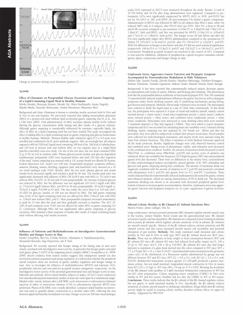
Change in pressures during cecal distention (pattern 2)
Tu1998
Effect of Glutamate on Postprandial Glucose Excursion and Gastric Emptyingof a Lipid-Containing Liquid Meal in Healthy HumansHiroko Hosaka, Motoyasu Kusano, Hiroaki Zai, Shiko Kuribayashi, Atsuto Nagoshi,Masaki Maeda, Yasuyuki Shimoyama, Osamu Kawamura, Masatomo Mori
Background and Aims: Glutamate is known to stimulate insulin secretion both In Vitro andIn Vivo in rats and humans. We previously reported that adding monosodium glutamate(MSG) to a protein-rich meal without lipid accelerated gastric emptying (Zai H, et al., AmJ Clin Nutr 2009). Oral administration of MSG increases plasma insulin levels withoutaltering the glucose concentration while fasting and after intake of a glucose solution.Although gastric emptying is considerably influenced by nutrients, especially lipids, theeffect of MSG on a lipid-containing meal has not been studied.This study investigated theeffect of adding MSG to a lipid-containing meal on gastric emptying and glucose homeostasisin healthy humans. Methods: Thirteen healthy male volunteers aged 25.5 ± 5.0 years wereenrolled and confirmed to be H. pylori antibody negative. After an overnight fast, all subjectsingested a 400 ml (520 kcal) liquid meal containing 100 kcal of fat, 300 kcal of carbohydrate,and 120 kcal of protein with and without MSG on two separate days in a single-blindplacebo-controlled cross-over study. On one of the two days, the test meal contained MSG(2 g, 0.5% wt:vol) in random order. Plasma glucose,serum insulin and glucose-dependentinsulinotropic polypeptide (GIP) were measured before and until 120 min after ingestionof the meal. Gastric emptying was assessed with a 13C acetate breath test (Breath ID System,Exalenz Bioscience Ltd., Israel) over 240 min. Postprandial dyspeptic symptoms (fullness,satiation, bloating, nausea, belching, epigastric pain, and heartburn) were assessed on a 10cm visual analogue scale (VAS). Results: After the test meal, plasma glucose and seruminsulin levels increased rapidly and reached a peak by 60 min. The insulin peak time wassignificantly shortened with addition of MSG (39.2±18.9 min with MSG vs. 53.1±26.4 minwithout MSG, P<0.05). At 30 min and 45 min postprandially, the increase of plasma glucosewas reduced by MSG, but the difference was not significant (65.7±16.2 mg/dl with MSGvs. 75.4±16.0 mg/dl without MSG, p=0.055 at 30 min postprandially, 59.1±29.0 mg/dl vs.76.0±25.2 mg/dl, P=0.094 at 45 min). The area under the curve from 0 to 120 min (AUC(0-120)) for glucose, insulin, or GIP was not altered by MSG. The half gastric emptyingtime (t1/2) of the lipid-containing meal was not affected by MSG (136±25 min with MSGvs. 124±16 min without MSG; p>0.1). Most postprandial symptoms increased immediatelyto peak by 15 min after the meal and then gradually returned to baseline. The AUC (0-120) of each symptom score (VAS) was not affected by either MSG or gastric emptying (t1/2). Conclusion: Although MSG did not significantly affect gastric emptying or glucoseexcursion, MSG initiated a faster response of insulin after intake of a lipid-containing liquidmeal without affecting total insulin secretion.
Tu1999
Influence of Naloxone and Methylnaltrexone on Interdigestive GastrointestinalMotility and Hunger Scores in ManEmidio Scarpellini, Rita Vos, Eveline Deloose, Athanasios A. Papathanasopoulos,Alessandra Rotondo, Inge Depoortere, Jan F. Tack
Background: We recently reported that hunger ratings in the fasting state in man wereclosely correlated with interdigestive motor activity, in particular that hunger peaks coincidedwith gastric phase 3 (GF3) of the migrating motor complex (MMC) (Scarpellini et al., DDW2009). Recent evidence from animal studies suggests that endogenous opioids are alsoinvolved in nutrient acquisition and energy regulation. It is still unclear whether the peripheralopioid receptors alone are involved in gastric motility regulation and hunger ratings inman. Aim: to investigate the influence of methylnaltrexone (MNTX) and naloxone (NA),peripherally selective and non-selective μ-opioid receptor antagonists, respectively, on theinterdigestive motor activity of the proximal gastrointestinal tract and hunger scores in man.Materials and methods: Eleven fasted healthy subjects (2 males; 29.5±3.5 years) underwentfour antroduodenojejunal manometry studies at least one week apart in a randomized single-blinded order: twenty minutes after a full MMC cycle intravenous or subcutaneous infusion/injection of saline or intravenous infusion of NA or subcutaneous injection MNTX wereperformed. Phases of the MMC were visually identified. Computer-aided baseline reconstruc-tion was used to quantify phasic contractions as a motility index (MI), reflecting the areabetween signal and baseline normalized over time. Hunger scores (on 100mm visual analogue
S-868AGA Abstracts
scales (VAS expressed as AUC)) were measured throughout the study. Results: A total of25 F3s before and 16 F3s after drug administration were registered. Compared to pre-treatment, GF3s were significantly suppressed by MNTX (6/12 vs. 0/10, p=0.0152) butnot by NA (6/13 vs. 0/6, p=0.1093); all post-treatment F3s lacked a gastric component.Administration of MNTX was followed by SBF3 in all subjects after 80±11 mins, while NAinduced SBF3 only in 6 subjects, after 83±14 mins (p< 0.05). After NA, a drop in SB andantral MI occurred compared to pre-treatment (3.43±0.37 vs 2.96±0.64 and 1.96±1.20 vs1.06±0.47, both p=0.0001), and this was prevented by MNTX (3.26±1.03 vs 3.05±0.62and 1.72±1.61 vs 1.58±0.91, both p=NS). The hunger scores 20 min before and after theF3s were significantly higher after MNTX administration compared to the spontaneous F3(before F3 147±18.3 vs 81.6±23.9, p=0.02; after F3 156.4±14.9 vs 122±18.7, p=0.05).With NA differences in hunger scores before and after F3 did not reach statistical significance(respectively 148.4±15.9 vs 111.8±21.9, p=0.07 and 118.2±27.3 vs 144.9±13.1, p=0.47).Conclusions: Peripheral μ-opioid receptors are involved in the control of GF3. Comparedto non-selective inhibition, inhibition of peripheral μ—opioid receptors stimulates interdi-gestive phasic contractions and hunger ratings in man.
Tu2000
Unpleasant Stress Aggravates Gastric Function and Dyspeptic SymptomAccompanied by Neuroendocrine Modulation in Male VolunteersAkihito Iida, Yasushi Funaki, Hiroshi Kaneko, Masahiro Matsunaga, Toshihiro Konagaya,Kentaro Tokudome, Naotaka Ogasawara, Makoto Sasaki, Masashi Yoneda, Kunio Kasugai
Background: It has been reported that experimentally induced anxiety decreases gastricaccommodation and results in satiety, fullness, and bloating after drinking. This phenomenalook like the postprandial distress syndrome in functional dyspepsia (FD). Aim:We examined1) experimentally induced unpleasantness influences the gastric function as well as dyspepticsymptoms under slowly drinking nutrient, and 2) underlying mechanisms among feeling,gut function and symptom. Methods: Eleven male volunteers were recruited. The participantswere ordered to drink the liquid meal at a rate of 15mL /min continuously. In this drinktest, the participants were exposed to 4 types of visual and auditory stimuli, namely control(neutral picture + classic music), visual stress (unpleasant picture + classic music), auditorystress (neutral picture + white noise), and combined stress (unpleasant picture + whitenoise) conditions. Participants were instructed to cease drinking when their score reached5 (maximal satiation) or they had ingested 1,500mL. They were asked to report dyspepticsymptoms and ECG was recorded for calculating heart rate variability every 5 minutes duringdrinking. Gastric emptying was also analyzed by 13C breath test . Before and after theprocedure, they were asked to subjectively evaluate their present mood states, blood sampleswere collected for endocrinological assessment, and anxiety levels were assessed using theState-Trait Anxiety Inventory. The Ethics Committee of Aichi Medical University approvedall the study protocols. Results: Significant changes were only observed between controland combined stress. Rating scores of pleasantness, vitality, and relaxation were decreasedin the combined stress condition. Further, the amount of maximum drink decreased (control582±57mL vs. combined stress 498±44mL; p<0.05) and severity score of satiation wasenhanced. Heart rate variability low-frequency (LF)/ high-frequency (HF) ratio and peripheralgastrin level also decreased. There were no differences in the anxiety level, concentrationsof other endocrinological makers (acyl-ghrelin, desacyl-ghrelin, CCK, NPY, adrenaline andglucose) and gastric emptying among four conditions. The amount of maximum drink wasnegatively correlated with the severity of satiation (r=-0.7; p<0.05) and positively correlatedwith pleasantness (r=0.3; p<0.05) and gastrin level (r= 0.3; p<0.05). Conclusion: Theseresults indicated that the experimentally induced unpleasantness decreased the gastric volumeand enhanced satiation, which are among characteristics of FD in drink test. The decreasedmaximum drinking was shown to be associated with decreased peripheral gastrin level.Gastrin is known to increase gastric accommodation, therefore, unpleasant stress may aggrav-ate gastric function and dyspepsia symptom via, in a part, suppression of gastrin secretion.
Tu2001
Altered Colonic Motility in Bk Channel β1 Subunit Knockout MiceMarion France, James Galligan, Hui Xu
Large-conductance calcium-activated potassium (BK) channels regulate smooth muscle tonein the trachea, urinary bladder, blood vessels and the gastrointestinal tract. BK channelactivation is partly calcium-dependent. BK channels are composed of pore forming α subunitsand accessory β subunits which regulate calcium sensitivity of the α subunit. β1 subunitsare smooth muscle specific. Absence of the β1 subunit reduces calcium sensitivity and BKchannel activity and this causes increased smooth muscle cell excitability and potentialdisruption of gut motility. Methods. This study examined small intestinal and colonicmotility In Vivo and In Vitro in wild type (WT) and β1 subunit knock-out (KO) mice.Results. There was no difference in body weight or food consumption between (WT) andβ1 subunit KO mice. β1 subunit KO mice had reduced fecal pellet output (n=13, 140 ±12 g) vs. WT mice (n=13, 230 ± 20 g, P<0.001). β1 subunit KO mice also had longerlatencies to expulsion of a glass bead inserted into the colon compared to WT mice (WT =7.7 ± 0.4 min; KO = 12 ± 1 min; P <0.01). Gastrointestinal transit measured as the geometriccenter (GC) of the distribution of a fluorescent marker administered by gavage was notdifferent between WT and KO mice (WT GC = 4.0 ± 0.2, n=8; KO GC = 3.4 ± 0.4, n=8,P>0.05). Bethanechol (muscarinic receptor agonist, 0.1-100 μM) produced a greater max-imum colonic, but not small intestinal, longitudinal muscle contraction In Vitro in tissuesfrom KO (135 ± 13 mg) vs. WT mice (89 ± 14 mg, n= 9, P < 0.05). Blocking the α subunitof the BK channel with paxilline (0.3 μM) increased bethanechol contractions in WT butnot KO colon preparations. Colonic migrating motor complexes (CMMC) In Vitro weresimilar in WT and KO tissues. Paxilline did not alter the CMMC in WT or KO tissues.Conclusions. These results indicate that the β1 subunit of the BK channel regulates colonicbut not gastric or small intestinal motility In Vivo. Specifically, the β1 subunit reducessensitivity of colonic smooth muscle to cholinergic stimulation. Drugs which alter β1 subunitactivity might be useful in treating colonic motility disorders without effects on upper GImotility. Supported by DK57039