6 pemphigus (1)
TRANSCRIPT

Pemphigus
Pemphigus is a rare group of blistering autoimmune diseases that
affect the skin and mucous membranes.

external resources

Types
• There are three types of pemphigus which vary in severity: pemphigus vulgaris, pemphigus foliaceus, and paraneoplastic pemphigus.
• The most common form of the disorder is pemphigus vulgaris (PV - ICD-10 L10.0). It occurs when antibodies attack Desmoglein 3. Sores often originate in the mouth, making eating difficult and uncomfortable. Although pemphigus vulgaris may occur at any age, it is most common among people between the ages of 40 and 60. It is more frequent among Ashkenazi Jews. Rarely, it is associated with myasthenia gravis. Nail disease may be the only finding and has prognostic value in management.

Pemphigus foliaceus
• Pemphigus foliaceus (PF) is the least severe of the three varieties. Desmoglein 1, the protein that is destroyed by the autoantibody, is only found in the top dry layer of the skin. PF is characterized by crusty sores that often begin on the scalp, and may move to the chest, back, and face. Mouth sores do not occur. It is not as painful as pemphigus vulgaris, and is often mis-diagnosed as dermatitis or eczema

Paraneoplastic pemphigus
• The least common and most severe type of pemphigus is paraneoplastic pemphigus (PNP). This disorder is a complication of cancer, usually lymphoma and Castleman's disease. It may precede the diagnosis of the tumor. Painful sores appear on the mouth, lips, and the esophagus. In this variety of pemphigus, the disease process often involves the lungs, causing bronchiolitis obliterans (constrictive bronchiolitis). Complete removal and/or cure of the tumor may improve the skin disease, but lung damage is generally irreversible .
Classification
• Pemphigus is a group of autoimmune blistering diseases that may be classified into the following types

Classification
– Pemphigus vulgaris, of which there several forms: » Pemphigus vegetans » Pemphigus vegetans of Hallopeau » Pemphigus vegetans of Neumann
– Pemphigus foliaceus, of which there several forms: » Pemphigus erythematosus » Endemic pemphigus
– Paraneoplastic pemphigus – IgA pemphigus, of which there several forms:
» Subcorneal pustular dermatosis » Intraepidermal neutrophilic IgA dermatosis

Diagnosis
• Pemphigus is recognized by a dermatologist from the appearance and distribution of the skin lesions. It is also commonly diagnosed by periodontists, oral and maxillofacial surgeons (specialists qualified in both medicine and dentistry)and ophthalmologists (eye doctors) as lesions can affect the eyes and mucous membrane of the oral cavity.

Diagnosis
• Definitive diagnosis requires examination of a skin or mucous membrane biopsy by a dermatopathologist or oral pathologist. The skin biopsy is taken from the edge of a blister, prepared for histopathology and examined with a microscope. The pathologist looks for an intraepidermal vesicle caused by the breaking apart of epidermal cells (acantholysis). Thus, the superficial (upper) portion of the epidermis sloughs off, leaving the bottom layer of cells on the "floor" of the blister. This bottom layer of cells is said to have a "tombstone appearance".

Treatment
• If not treated, pemphigus can be fatal due to overwhelming infection of the sores. The most common treatment is the administration of oral steroids, especially prednisone, and often in high doses. The side effects of cortico-steroids may require the use of so-called steroid-sparing or adjuvant drugs. The immuno-suppressant CellCept (Mycophenolic acid) is among those being used

Mucosal lichen planus
• Mucosal lichen planus, or oral lichen planus (OLP), is an inflammatory auto-immune[citation needed] disease that affects oral mucosa, with or without the involvement of the skin and other mucous membranes.
Epidemiology
• OLP affects women more than men (at a ratio of 3:2), and occurs most often in middle-aged adults. OLP in children is rare.

Lichen planus affecting the lower lip.

Cause
• The cause of lichen planus is not known. It is not contagious[1] and does not involve any known pathogen.OLP has been reported as a complication of chronic hepatitis C virus infection and can be a sign of chronic graft-versus-host disease of the mucous membrane (and skin).It has been suggested that OLP may respond to stress, where lesions may present on the mucosa (or skin) during times of stress in those with the disease

Clinical presentation
• The reticular form is the most common presentation and manifests as white lacy streaks on the mucosa (known as Wickham's striae) or as smaller papules (small raised area). The lesions tend to be bilateral and are asymptomatic. The lacy streaks may also be seen on other parts of the mouth, including the gingiva (gums), the tongue, palate and lips.
• The bullous form presents as fluid-filled vesicles which project from the surface.
• The erosive form presents with erythematous (red) areas that are ulcerated and uncomfortable. The erosion of the thin epithelium may occur in multiple areas of the mouth, or in one area, such as the gums, where they resemble desquamative gingivitis. Wickham's striae may also be seen near these ulcerated areas. This form may undergo malignant transformation.

Histo-pathological appearance
• The microscopic appearance of lichen planus is pathognomonic for the condition
• Hyperparakeratosis with thickening of the granular cell layer
• Development of a "saw-tooth" appearance of the rete pegs
• Degeneration of the basal cell layer • Infiltration of inflammatory cells into the
subepithelial layer of connective tissue

Micrograph of lichen planus. H&E stain

Differential Diagnosis
• The clinical presentation of OLP may also resemble other conditions, including:
• Lichenoid drug reaction. This entity is identical to OLP both clinically and histologically. However, lichenoid lesions may be single (in comparison to the usual bilateral appearance of OLP) with proximity to amalgam (metal alloys) dental restoration.
• Other oral vesiculo-ulcerative conditions such as Pemphigus vulgaris and Benign mucous membrane pemphigoid
• Discoid lupus erythematosus • Chronic ulcerative stomatitis • Frictional keratosis and Morsicatio buccarum (chronic cheek biting) • Oral leukoplakia • Chronic graft-versus-host-disease may manifest as lichenoid reaction. This
type of lichenoid lesions have a higher risk of malignant transformation to oral squamous cell carcinoma in comparison to the classical oral lichen planus. Graft-versus-host-disease-associated oral cancer may have more aggressive behavior with poorer prognosis, when compared to oral cancer in non-hematopoietic stem cell transplantation patients.

Treatment
• Medicines used to treat lichen planus include:• Oral and topical steroids. • Oral retinoids • immunosuppressant medications • hydroxychloroquine • tacrolimus • dapsone • Aloe vera[3] • Pusley Portulaca oleracea

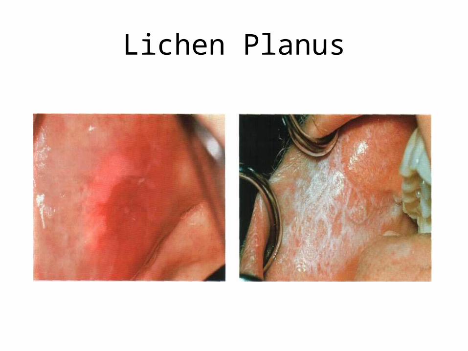
Lichen planus
• Lichen planus is a chronic mucocutaneous disease that affects the skin, tongue, and oral mucosa. The disease presents itself in the form of papules, lesions, or rashes. Lichen planus does not involve lichens, the fungus/algae symbionts that often grow on tree trunks; the name refers to the dry and undulating, "lichen-like" appearance of affected skin. It is sometimes associated with certain medications and diseases, but is basically of unknown cause

Lichen Planus

Classification
• Configuration Annular lichen planus
Linear lichen planus Morphology of lesion
Hypertrophic lichen planus Atrophic lichen planus Vesiculobullous lichen planus Ulcerative lichen planus Follicular lichen planus Actinic lichen planus Lichen planus pigmentosus

Classification
• Site of involvement – Lichen planus of the palms and soles (Palmoplantar lichen
planus) – Mucosal lichen planus – Lichen planus of the nails – Lichen planus of the scalp ( leading to cicatricial alopecia) – Inverse lichen planus
• Special forms – Drug-induced lichen planus – Lupus erythematosus-lichen planus overlap syndrome – Lichen planus pemphigoides – Keratosis lichenoides chronica – Lichenoid reaction of graft-versus-host disease – Lichenoid keratosis – Lichenoid dermatitis

Signs and symptoms
• The typical rash of lichen planus is well-described by the "5 Ps": well-defined pruritic, planar, purple, polygonal papules. The commonly affected sites are near the wrist and the ankle. The rash tends to heal with prominent blue-black or brownish discoloration that persists for a long time. Besides the typical lesions, many morphological varieties of the rash may occur. The presence of cutaneous lesions is not constant and may wax and wane over time. Oral lesions tend to last far longer than cutaneous lichen planus lesions.
Lichen Planus
Oral lichen planus (OLP) may present in one of three forms.
• The reticular form is the most common presentation and manifests as white lacy streaks on the mucosa (known as Wickham's striae) or as smaller papules (small raised area). The lesions tend to be bilateral and are asymptomatic. The lacy streaks may also be seen on other parts of the mouth, including the gingiva (gums), the tongue, palate and lips.
• The bullous form presents as fluid-filled vesicles which project from the surface.

Thirth form
• The erosive forms (Atrophic LP & Ulcerative LP) present with erythematous (red) areas that are ulcerated and uncomfortable. The erosion of the thin epithelium may occur in multiple areas of the mouth (more prominent on the posterior buccal mucosa), or in one area, such as the gums, where they resemble desquamative gingivitis. Wickham's striae may also be seen near these ulcerated areas. This form may undergo malignant transformation, although this is controversial. The malignant transformation rate is thought to be less than 1%, however it has been reported to be as high as 5%.[3] For any persistent oral lesion of erosive lichen planus that does not respond to topical corticosteroids, a biopsy is recommended to rule out precancerous (premalignant) change or malignant transformation.

Cause
• The cause of lichen planus is not known. It is not contagious and does not involve any known pathogen. Some lichen planus-type rashes (known as lichenoid reactions) occur as allergic reactions to medications for high blood pressure, heart disease and arthritis, in such cases termed drug-induced lichenoid reactions. These lichenoid reactions are referred to as lichenoid mucositis (of the mucosa) or dermatitis (of the skin).

Treatment
• Medicines used to treat lichen planus include:• Oral and topical steroids. • Oral retinoids • immunosuppressant medications • hydroxychloroquine • tacrolimus • dapsone • Non-drug treatments:• UVB NarrowBand Phototherapy• Aloe vera • Purslane

Lupus erythematosus
• Lupus erythematosus is a category for a collection of diseases with similar underlying problems with immunity autoimmune disease

Symptoms
• Symptoms of these diseases can affect many different body systems, including joints, skin, kidneys, blood cells, heart, and lungs

Four main types of lupus exist
• systemic lupus erythematosus, discoid lupus erythematosus, drug-induced lupus erythematosus, and neonatal lupus erythematosus.

Systemic lupus erythematosus
• Systemic lupus erythematosus often abbreviated to SLE or lupus, is a systemic autoimmune disease (or autoimmune connective tissue disease) that can affect any part of the body. As occurs in other autoimmune diseases, the immune system attacks the body's cells and tissue, resulting in inflammation and tissue damage.It is a Type III hypersensitivity reaction caused by antibody-immune complex formation.

Systemic lupus erythematosus

Signs and symptoms
• SLE is one of several diseases known as "the great imitators" because it often mimics or is mistaken for other illnesses. SLE is a classical item in differential diagnosis, because SLE symptoms vary widely and come and go unpredictably. Diagnosis can thus be elusive, with some people suffering unexplained symptoms of untreated SLE for years.

Lupus erythematosus

Dermatological manifestations
• As many as 30% of sufferers have some dermatological symptoms (and 65% suffer such symptoms at some point), with 30% to 50% suffering from the classic malar rash (or butterfly rash) associated with the disease. Some may exhibit thick, red scaly patches on the skin (referred to as discoid lupus).

Musculoskeletal
• The most commonly sought medical attention is for joint pain, with the small joints of the hand and wrist usually affected, although all joints are at risk. The Lupus Foundation of America estimates more than 90 percent of those affected will experience joint and/or muscle pain at some time during the course of their illness

Hematological
• Anemia may develop in up to 50% of cases. Low platelet and white blood cell counts may be due to the disease or a side effect of pharmacological treatment. People with SLE may have an association with antiphospholipid antibody syndrome (a thrombotic disorder), wherein autoantibodies to phospholipids are present in their serum. Abnormalities associated with antiphospholipid antibody syndrome include a paradoxical prolonged partial thromboplastin time (which usually occurs in hemorrhagic disorders) and a positive test for antiphospholipid antibodies; the combination of such findings have earned the term "lupus anticoagulant-positive". Another autoantibody finding in SLE is the anticardiolipin antibody, which can cause a false positive test for syphilis
Cardiac
• A person with SLE may have inflammation of various parts of the heart, such as pericarditis, myocarditis, and endocarditis. The endocarditis of SLE is characteristically noninfective (Libman-Sacks endocarditis), and involves either the mitral valve or the tricuspid valve. Atherosclerosis also tends to occur more often and advances more rapidly than in the general population
Pulmonary
• Lung and pleura inflammation can cause pleuritis, pleural effusion, lupus pneumonitis, chronic diffuse interstitial lung disease, pulmonary hypertension, pulmonary emboli, pulmonary hemorrhage, and shrinking lung syndrome

Renal
• Painless hematuria or proteinuria may often be the only presenting renal symptom. Acute or chronic renal impairment may develop with lupus nephritis, leading to acute or end-stage renal failure. Because of early recognition and management of SLE, end-stage renal failure occurs in less than 5% of cases.

Laboratory tests
• Antinuclear antibody (ANA) testing and anti-extractable nuclear antigen (anti-ENA) form the mainstay of serologic testing for SLE. Several techniques are used to detect ANAs. Clinically the most widely used method is indirect immunofluorescence. The pattern of fluorescence suggests the type of antibody present in the patient's serum
Diagnostic criteria
• Some physicians make a diagnosis on the basis of the American College of Rheumatology (ACR) classification criteria. The criteria, however, were established mainly for use in scientific research including use in randomized controlled trials which require higher confidence levels, so some people with SLE may not pass the full criteria

Treatment
• The treatment of SLE involves preventing flares and reducing their severity and duration when they occur.
• Treatment can include corticosteroids and anti-malarial drugs. Certain types of lupus nephritis such as diffuse proliferative glomerulonephritis require bouts of cytotoxic drugs. These drugs include cyclophosphamide and mycophenolate.
• Hydroxychloroquine (HCQ) was the last medication approved by the FDA for lupus in 1955.[61] Some drugs approved for other diseases are used for SLE 'off-label'. In November 2010, an FDA advisory panel recommended approving Benlysta (belimumab) as a treatment for the pain and flare-ups common in lupus. The drug was approved by the FDA in March 2011

Discoid lupus erythematosus

DLE
• Discoid lupus erythematosus (DLE) is a chronic skin condition of sores with inflammation and scarring favoring the face, ears, and scalp and at times on other body areas. These lesions develop as a red, inflamed patch with a scaling and crusty appearance. The center areas may appear lighter in color with a rim darker than the normal skin.

Localized
• Localized discoid lupus erythematosus typically presents with skin lesions localized above the neck, with favored sites being the scalp, bridge of nose, cheeks, lower lip, and ears

Lupus erythematosus
• Lupus erythematosus, also called Lupus, is a disease. It is chronic, which means it does not go away. The Immune system is made up of white blood cells in your body that fight off disease.

A facial rash, as is common for Lupus erythematosus
Mucosal lichen planus
• Mucosal lichen planus, or oral lichen planus (OLP), is an inflammatory auto-immune[citation needed] disease that affects oral mucosa, with or without the involvement of the skin and other mucous membranes.

Epidemiology
• OLP affects women more than men and occurs most often in middle-aged adults. OLP in children is rare.

Cause
• The cause of lichen planus is not known. It is not contagious and does not involve any known pathogen.OLP has been reported as a complication of chronic hepatitis C virus infection and can be a sign of chronic graft-versus-host disease of the mucous membrane (and skin).It has been suggested that OLP may respond to stress, where lesions may present on the mucosa (or skin) during times of stress in those with the disease.

Clinical presentation
• OLP may present in one of three forms.• The reticular form is the most common presentation and manifests
as white lacy streaks on the mucosa (known as Wickham's striae) or as smaller papules (small raised area). The lesions tend to be bilateral and are asymptomatic. The lacy streaks may also be seen on other parts of the mouth, including the gingiva (gums), the tongue, palate and lips.
• The bullous form presents as fluid-filled vesicles which project from the surface.
• The erosive form presents with erythematous (red) areas that are ulcerated and uncomfortable. The erosion of the thin epithelium may occur in multiple areas of the mouth, or in one area, such as the gums, where they resemble desquamative gingivitis. Wickham's striae may also be seen near these ulcerated areas. This form may undergo malignant transformation.

Differential Diagnosis
• Lichenoid drug reaction. This entity is identical to OLP both clinically and histologically. However, lichenoid lesions may be single (in comparison to the usual bilateral appearance of OLP) with proximity to amalgam (metal alloys) dental restoration.
• Other oral vesiculo-ulcerative conditions such as Pemphigus vulgaris and Benign mucous membrane pemphigoid
• Discoid lupus erythematosus • Chronic ulcerative stomatitis • Frictional keratosis and Morsicatio buccarum (chronic cheek biting) • Oral leukoplakia • Chronic graft-versus-host-disease may manifest as lichenoid reaction. This
type of lichenoid lesions have a higher risk of malignant transformation to oral squamous cell carcinoma in comparison to the classical oral lichen planus. Graft-versus-host-disease-associated oral cancer may have more aggressive behavior with poorer prognosis, when compared to oral cancer in non-hematopoietic stem cell transplantation patients
Treatment
• Care of OLP is within the scope of Oral medicine speciality. It is generally accepted that OLP (as well as other mucous membrane lichen planus, such as genital) is more difficult to manage than skin lichen planus.
• Currently there is no cure for lichen planus but there are certain types of medicines used to reduce the effects of the inflammation. Lichen planus may go into a dormant state after treatment. There are also reports that lichen planus can flare up years after it is considered cured.

Medicines used to treat lichen planus include:
• Oral and topical steroids. • Oral retinoids • immunosuppressant medications • hydroxychloroquine • tacrolimus • dapsone • Aloe vera[3] • Pusley Portulaca oleracea