69451 weinheim, germany spectra: am-250 or 400 avance bruker; chemical shifts (δ) in ppm related to...
TRANSCRIPT

Supporting Information
© Wiley-VCH 2008
69451 Weinheim, Germany
A Caged Retinoic Acid for Use with One- andTwo-Photon Excitation in Zebrafish Embryos
Pierre Neveu,[a],[b] Isabelle Aujard,[b] Chouaha Benbrahim,[b] Thomas Le Saux,[b]
Jean-François Allemand,[a] Sophie Vriz,[c],[d] David Bensimon,[a] Ludovic Jullien[b],∗
[a] Dr. P. Neveu, Dr. J. F. Allemand, Dr. D. Bensimon,
Ecole Normale Supérieure,
Laboratoire de Physique Statistique and Département de Biologie
UMR 8550, 24 rue Lhomond, F-75231 Paris, France
[b] Dr. P. Neveu, Dr. I. Aujard, C. Benbrahim, Dr. T. Le Saux, Prof. Dr. L. Jullien,
Ecole Normale Supérieure,
Département de Chimie,
UMR CNRS-ENS-UPMC Paris 06 8640 PASTEUR,
24 rue Lhomond, F-75231 Paris, France
[c] Prof. Dr. S. Vriz,
Inserm U770, Hémostase et Dynamique Cellulaire Vasculaire,
80 rue du Général Leclerc, 94276 Le Kremlin-Bicêtre, France
[d] Prof. Dr. S. Vriz,
Université Paris 7 Denis Diderot, 75005 Paris, France
Supporting Information
The Supporting Information reports on:
• The Experimental Methods.
• The one-photon photoconversion kinetics of cRA upon UV illumination.
• The comparison of the phenotypes from RA and two cis-retinoic acids.
• The teratogenic effects induced by UV illumination of cRA in injected embryos.
• The two-photon uncaging kinetics of cRA in vivo.
1

Experimental Methods
Syntheses of the caged compounds obtained from all-trans retinoicacid RA
The 7-dimethylamino-coumarin-4-ylmethyl ester cRACoum was prepared by condensa-
tion between RA and the 7-dimethylamino-coumarin-4-yl methanol HOCoum [1] in the
presence of DCC (scheme 1Sa).
DCC, DMAP+
HOCoum
OH
O
RA
O
O
OCH2Cl2OO
HO
NMe2
cRACoum
O
NMe2
Scheme 1Sa. Synthesis of cRACoum.
The three 2-nitrobenzyl esters: cRA, cRABr, and cRACN were obtained under sim-
ilar conditions by condensation between RA and the corresponding 2-nitrobenzyl alcohols
HOP, HOPBr, and HOPCN [2] (Scheme 1Sb).
OMe
O2N OMe
DCC, DMAPHO+
R
HOP
HOPBr
HOPCN
cRA
cRABr
cRACN
OH
O
RA
O
O NO2
OMeOMe
R
R
H
Br
CN
CH2Cl2
Scheme 1Sb. Syntheses of the 2-nitrobenzyl esters of cRA, cRABr, and cRACN.
General
The commercially available chemicals were used without further purification. Anhydrous
solvents were freshly distilled before use. Column chromatography (CC): silica gel 60
(0.040-0.063 mm) Merck. Analytical and thin layer chromatography (TLC): Merck silica
gel 60 F254 precoated plates; detection by UV (254 nm). Melting point: Büchi 510. 1H-
NMR Spectra: AM-250 or 400 AVANCE Bruker; chemical shifts (δ) in ppm related to
protonated solvent as internal reference (1H: CHCl3 in CDCl3, 7.26 ppm; CHD2SOCD3 in
CD3SOCD3, 2.49 ppm. 13C: 13CDCl3 in CDCl3, 77.0 ppm; 13CD3SOCD3 in CD3SOCD3,
39.6 ppm); Coupling constants J in Hz. Mass spectrometry (chemical ionization and high
resolution with NH3 or CH4) was performed at the Service de Spectrométrie de masse
2
de l’ENS. Microanalyses were obtained from the Service de Microanalyses de l’Université
Pierre et Marie Curie, Paris.
Experimental Procedures
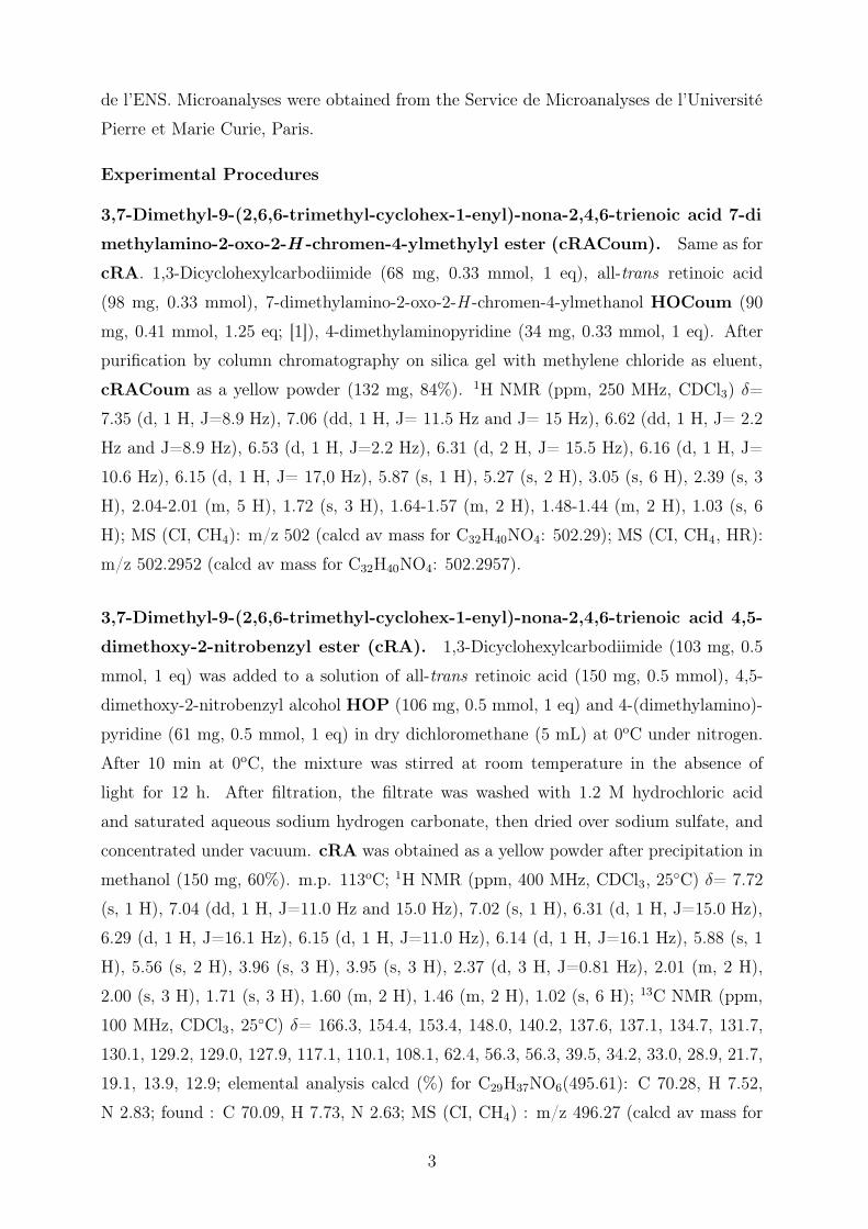
3,7-Dimethyl-9-(2,6,6-trimethyl-cyclohex-1-enyl)-nona-2,4,6-trienoic acid 7-di
methylamino-2-oxo-2-H -chromen-4-ylmethylyl ester (cRACoum). Same as for
cRA. 1,3-Dicyclohexylcarbodiimide (68 mg, 0.33 mmol, 1 eq), all-trans retinoic acid
(98 mg, 0.33 mmol), 7-dimethylamino-2-oxo-2-H -chromen-4-ylmethanol HOCoum (90
mg, 0.41 mmol, 1.25 eq; [1]), 4-dimethylaminopyridine (34 mg, 0.33 mmol, 1 eq). After
purification by column chromatography on silica gel with methylene chloride as eluent,
cRACoum as a yellow powder (132 mg, 84%). 1H NMR (ppm, 250 MHz, CDCl3) δ=
7.35 (d, 1 H, J=8.9 Hz), 7.06 (dd, 1 H, J= 11.5 Hz and J= 15 Hz), 6.62 (dd, 1 H, J= 2.2
Hz and J=8.9 Hz), 6.53 (d, 1 H, J=2.2 Hz), 6.31 (d, 2 H, J= 15.5 Hz), 6.16 (d, 1 H, J=
10.6 Hz), 6.15 (d, 1 H, J= 17,0 Hz), 5.87 (s, 1 H), 5.27 (s, 2 H), 3.05 (s, 6 H), 2.39 (s, 3
H), 2.04-2.01 (m, 5 H), 1.72 (s, 3 H), 1.64-1.57 (m, 2 H), 1.48-1.44 (m, 2 H), 1.03 (s, 6
H); MS (CI, CH4): m/z 502 (calcd av mass for C32H40NO4: 502.29); MS (CI, CH4, HR):
m/z 502.2952 (calcd av mass for C32H40NO4: 502.2957).
3,7-Dimethyl-9-(2,6,6-trimethyl-cyclohex-1-enyl)-nona-2,4,6-trienoic acid 4,5-
dimethoxy-2-nitrobenzyl ester (cRA). 1,3-Dicyclohexylcarbodiimide (103 mg, 0.5
mmol, 1 eq) was added to a solution of all-trans retinoic acid (150 mg, 0.5 mmol), 4,5-
dimethoxy-2-nitrobenzyl alcohol HOP (106 mg, 0.5 mmol, 1 eq) and 4-(dimethylamino)-
pyridine (61 mg, 0.5 mmol, 1 eq) in dry dichloromethane (5 mL) at 0oC under nitrogen.
After 10 min at 0oC, the mixture was stirred at room temperature in the absence of
light for 12 h. After filtration, the filtrate was washed with 1.2 M hydrochloric acid
and saturated aqueous sodium hydrogen carbonate, then dried over sodium sulfate, and
concentrated under vacuum. cRA was obtained as a yellow powder after precipitation in
methanol (150 mg, 60%). m.p. 113oC; 1H NMR (ppm, 400 MHz, CDCl3, 25◦C) δ= 7.72
(s, 1 H), 7.04 (dd, 1 H, J=11.0 Hz and 15.0 Hz), 7.02 (s, 1 H), 6.31 (d, 1 H, J=15.0 Hz),
6.29 (d, 1 H, J=16.1 Hz), 6.15 (d, 1 H, J=11.0 Hz), 6.14 (d, 1 H, J=16.1 Hz), 5.88 (s, 1
H), 5.56 (s, 2 H), 3.96 (s, 3 H), 3.95 (s, 3 H), 2.37 (d, 3 H, J=0.81 Hz), 2.01 (m, 2 H),
2.00 (s, 3 H), 1.71 (s, 3 H), 1.60 (m, 2 H), 1.46 (m, 2 H), 1.02 (s, 6 H); 13C NMR (ppm,
100 MHz, CDCl3, 25◦C) δ= 166.3, 154.4, 153.4, 148.0, 140.2, 137.6, 137.1, 134.7, 131.7,
130.1, 129.2, 129.0, 127.9, 117.1, 110.1, 108.1, 62.4, 56.3, 56.3, 39.5, 34.2, 33.0, 28.9, 21.7,
19.1, 13.9, 12.9; elemental analysis calcd (%) for C29H37NO6(495.61): C 70.28, H 7.52,
N 2.83; found : C 70.09, H 7.73, N 2.63; MS (CI, CH4) : m/z 496.27 (calcd av mass for
3

C29H37NO6 : 495.61); MS (CI, CH4, HR) : m/z 496.2693 (calcd av mass for C29H38NO6 :
496.2699).
3,7-Dimethyl-9-(2,6,6-trimethyl-cyclohex-1-enyl)-nona-2,4,6-trienoic acid 2,2,2-
tribromo-1-(4,5-dimethoxy-2-nitro-phenyl)-ethyl ester (cRABr). Same as for
cRA. 1,3-Dicyclohexylcarbodiimide (52 mg, 0.25 mmol, 1 eq), all-trans retinoic acid (75
mg, 0.25 mmol), 2,2,2-tribromo-1-(4,5-dimethoxy-2-nitrophenyl)ethanol HOPBr [2] (145
mg, 0.31 mmol, 1.25 eq), 4-dimethylaminopyridine (30 mg, 0.25 mmol, 1 eq). After pu-
rification by column chromatography on silica gel with cyclohexane/ethyl acetate: 9/1 as
eluent, cRABr as a yellow oil (165 mg, 88%). 1H NMR (ppm, 250 MHz, CDCl3, 25◦C)
δ= 7.79 (s, 1 H), 7.57 (s, 1 H), 7.41 (s, 1 H), 7.01 (dd, 1 H, J=15.0 Hz, J=11.4 Hz), 6.31
(d, 1 H, J=15.0 Hz), 6.25 (d, 1 H, J=15.6 Hz), 6.09 (d, 1 H, J=13.6 Hz), 6.08 (d, 1 H,
J=15.6 Hz), 5.90 (s, 1 H), 3.92 (s, 3 H), 3.90 (s, 3 H), 2.32 (s, 3 H), 1.94 (s, 3 H), 1.85
(m, 2 H), 1.64 (s, 3 H), 1.54 (m, 2 H), 1.38 (m, 2 H), 0.95 (s, 6 H); 13C NMR (ppm,
100 MHz, CDCl3, 25◦C) δ= 163.9, 163.1, 156.9, 155.1, 152.1, 149.3, 142.9, 140.9, 137.6,
137.2, 134.3, 132.6, 130.3, 129.4, 129.1, 123.4, 115.6, 111.4, 108.2, 73.3, 56.5, 56.4, 39.5,
33.1, 28.9, 21.7, 19.1, 14.2, 12.9; MS (CI, NH3): m/z 763.0, 745.9 (calcd av mass for
C30H40N2O6Br3: 763.0, calcd av mass for C30H36NO6Br3 : 746.32 ); MS (CI, NH3, HR):
m/z 763.0406 and 765.0383 (calcd av mass for C30H40N2O6Br3: 763.0418 and 765.0401).
3,7-Dimethyl-9-(2,6,6-trimethyl-cyclohex-1-enyl)-nona-2,4,6-trienoic acid cya
no-(4,5-dimethoxy-2-nitro-phenyl)-methyl ester (cRACN). Same as for cRA.
1,3-Dicyclohexylcarbodiimide (103 mg, 0.5 mmol, 1 eq), all-trans retinoic acid (150 mg,
0.5 mmol), hydroxy-(4,5-dimethoxy-2-nitrophenyl)acetonitrile HOPCN [2] (150 mg, 0.62
mmol, 1.25 eq), 4-dimethylaminopyridine (61 mg, 0.5 mmol, 1 eq). cRACN as a yellow
powder (270 mg, 48%). m.p. 145oC; 1H NMR (ppm, 400 MHz, CDCl3, 25◦C) δ= 7.74 (s,
1 H), 7.18 (s, 1 H), 7.10 (dd, 1 H, J=12.3 Hz and 15.1 Hz), 6.33 (d, 1 H, J=15.1 Hz), 6.30
(d, 1 H, J=14.6 Hz), 6.15 (d, 1 H, J=12.3 Hz), 6.14 (d, 1 H, J=14.6 Hz), 6,17 (s, 1 H),
5.82 (s, 1 H), 4.03 (s, 3 H), 3.98 (s, 3 H), 2.38 (s, 3 H), 2.02 (s, 3 H), 2.02 (m, 2 H), 1.71
(s, 3 H), 1.61 (m, 2 H), 1.46 (m, 2 H), 1.02 (s, 6 H); 13C NMR (ppm, 100 MHz, CDCl3,
25◦C) δ= 163.9, 157.7, 153.8, 149.7, 141.3, 139.8, 137.6, 137.0, 134.0, 133.1, 130.4, 129.6,
129.1, 122.3, 115.8, 114.4, 110.0, 108.4, 58.8, 56.7, 56.5, 39.5, 34.2, 33.1, 28.9, 21.7, 19.1,
14.2, 12.9; MS (CI, NH3): m/z 521 (calcd av mass for C30H36N2O6 : 520.62 ); MS (CI,
NH3, HR): m/z 521.2657 (calcd av mass for C30H37N2O6: 521.2652).
4

Experiments with one-photon excitation
UV illumination
All one-photon excitation experiments were performed at 20oC by putting a bench top
UV lamp (365 nm;1 6 W; Fisher Bioblock) above a glass Petri dish (diameter: 5.5 cm)
containing V = 20 mL of solution. We used the uncaging experiment performed with the
model caged compound PheP to derive an order of magnitude of the photon flux I0 of the
incident beam in the wavelength range leading to uncaging. By taking Q(1)u = ε
(1)u Φ
(1)u = 48
M−1cm−1,[2] V = 20 mL, and l = 0.9 cm, I0 ≈ 4 10−5 Einstein min−1 was obtained from
k2.[2]
We checked that, when illuminated for up to 4 minutes under the present conditions,
control embryos developed normally (data not shown).[3] We also verified that the side
product resulting from uncaging was not responsible of the observed phenotypes (data
not shown).
Capillary electrophoresis
Electrophoretic measurements were performed with a PACE/MDQ (Beckman Coulter)
capillary electrophoresis system. Migrations were performed at 20 kV in bare fused silica
capillaries (Polymicro, Phoenix, AZ), 50 µm I.D. × 50 cm filled with running buffer (15
mM Na2B4O7 and 20 mM αCD containing 10% (v/v) acetonitrile; [4]) at 25oC. The
analytes were detected by UV absorbance at 350 nm. In view of the similarity of the
absorption spectra of the three analyzed retinoic acids at that wavelength, we assimilated
the corrected areas of the peaks to the amounts of the corresponding retinoic acids.
Preliminary screening of the caged retinoic acids
Zebrafish embryos were maintained at 28oC.[5] The compounds were assayed by injecting
zebrafish embryos (number of embryos injected: 50-100 range) at the 32-cell stage with 5
nl of 0.1 mM of the investigated substrate. Embryos were illuminated for various durations
with the 365 nm UV lamp placed on top of the dish. They were checked for developmental
abnormalities beyond 30 hours post fertilization (hpf) and compared with those incubated
with (or without) known concentrations of RA.
Teratogenicity assay
Zebrafish embryos were maintained at 28oC. The teratogenicity of the various compounds
was assayed by incubating pronase dechorionated zebrafish embryos (number of embryos1The lamp spectrum provided by the lamp purchaser is essentially gaussian around 350 nm with a 40
nm width at half height.
5

treated: 50-100 range) at the 128-cell stage for 90 min in embryo medium2 [5] supple-
mented with 10 µM of the investigated substrate. After washing with plain embryo
medium, the embryos were put in the Petri dish introduced above and illuminated for
various durations with the 365 nm UV lamp placed on top of the dish. They were checked
for developmental abnormalities beyond 30 hpf and compared with those incubated with
(or without) known concentrations of RA.
In vivo RA degradation
Embryos were injected at 32 cell stage with 5 nl of 0.1 mM cRA (final intra-cellular
concentration 10 µM, number of embryos injected: 50-100 range). Embryos were put in
the Petri dish introduced above and illuminated for various duration with the 365 nm
UV lamp placed on top of the dish. They were checked for developmental abnormalities
beyond 30 hpf.
Experiments with two-photon excitation
Instrumental setup
For the in vivo uncaging calibration studies, a home-built microscope [6] equipped with
an Olympus UPlanApo 60× 1.2 NA water immersion objective was used to image the
embryos on a JAI-40 CCD camera and locate the focal spot of the two-photon excitation.
Illumination (200 fs, 76 kHz, 750 nm) was provided by a mode-locked Ti-Sapphire laser
(Mira, Coherent). The incident power at the sample (≤ 4.5 mW) was measured with
a Lasermate powermeter (Coherent). The fluorescence photons were collected through
filters (AHF Analysentechnik, emission filter: 480/60 nm, excitation wavelengths: 750
nm for the experiments with cF and 720 nm with cRA) and optical fibers (FG200LCR
multimode fiber, Thorlabs) and detected with avalanche photodiodes (SPCM-AQR-14,
Perkin Elmer) coupled to an ALV-6000 correlator (ALV GmbH). The emission filters were
chosen to match the emission characteristics of RA when bound to CRABP and CRBP.[7,
8] The intensity of the collected fluorescence emission and its temporal correlation function
were recorded. The geometrical characteristics of the focal point were determined by
Fluorescence Correlation Spectroscopy (FCS) measurements [9] on a fluorescein solution
of known concentration (50 nM in 0.1 M NaOH). All the series of experiments reported in
the present work have been performed in a regime of laser powers in which the examined
fluorophores exhibit a quadratic dependence of the intensity of fluorescence emission on
the illumination power.2Embryo medium is composed of 0.80 g NaCl, 0.04 g KCl, 3.58 mg Na2HPO4, 6 mg KH2PO4, 0.144 g
CaCl2, 0.246 g MgSO4,7H2O, 0.35 g NaHCO3 per liter of dd H2O balanced with 1 M NaOH to pH 7.2.
6

Zebrafish embryo conditioning
Embryos were manually dechorionated at 1-3 somite stage. They were subsequently in-
cubated in embryo medium supplemented with 1 µM cF or 10 µM cRA for 90 min.
We located the two-photon focal spot in the dorsal part of the retina and continuously
monitored the fluorescence intensity. Experiments were conducted on 12 embryos in each
case. As a control, the same experiment was done on non-treated embryos.
Evaluation of possible photoinduced damages with two-photon excitation
Two-photon excitation requires a high flux of photons which may induce physical processes
that could damage the embryos.
Light absorption which leads to energy dissipation as heat could be detrimental to
the cell. Two types of light absorption processes can be here considered: water and
endogenous chromophores. Schönle and Hell [10] have computed the temperature increase
due to absorption by water molecules: it is in the 0.1 K range at 100 mW excitation power
at 850 nm for an illumination duration of 1 s. It is negligible, at the laser intensities used
here (less than 5 mW at the sample). Endogenous chromophores are present in a much
smaller concentration than water but they can exhibit large molar absorption coefficients
with one-photon absorption in the near IR. We can estimate the temperature increase due
to absorption of a chromophore present at 1 mM concentration with a molar absorption
coefficient of 5000 M−1cm−1 (a value similar to the one of hemoglobin [11]). Taking profit
of the calculation by Schönle and Hell, we estimate this increase to be at most ∼ 5 K at
10 mW laser power. Such an increase is not detrimental to the cell and we may therefore
neglect the temperature increase resulting from absorption phenomena.
Another source of photoinduced physical damage is the possibility to ionize the cel-
lular medium due to the electric field [11]: a plasma is formed at the focal point. This
phenomenon is widely used in laser surgery to perform tissue ablation. This plasma cre-
ates a cavitation bubble made of the evaporated compounds. This bubble may emit shock
wave that may modify the membrane permeability with a direct effect on cellular viability
without affecting the cellular morphology [11]. In our experimental conditions (surfacic
laser power: 1016 W.m−2), the threshold for cavitation can be estimated to be at a laser
power of ∼ 11 mW (see Noack et al., [12], Vogel et al. [13] and Sacchi [14]). Indeed when
we applied powers larger than 10 mW, we observed the formation of a cavitation bubble
and a shock wave which did not affect tissue morphology. However up to 5 mW average
power, we never observed the formation of any cavitation bubble even when the embryo
was illuminated for as long as one hour, and it eventually developed normally.
7
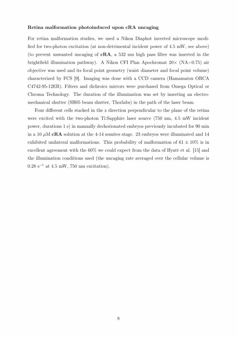
Retina malformation photoinduced upon cRA uncaging
For retina malformation studies, we used a Nikon Diaphot inverted microscope modi-
fied for two-photon excitation (at non-detrimental incident power of 4.5 mW, see above)
(to prevent unwanted uncaging of cRA, a 532 nm high pass filter was inserted in the
brightfield illumination pathway). A Nikon CFI Plan Apochromat 20× (NA=0.75) air
objective was used and its focal point geometry (waist diameter and focal point volume)
characterized by FCS [9]. Imaging was done with a CCD camera (Hamamatsu ORCA
C4742-95-12ER). Filters and dichroics mirrors were purchased from Omega Optical or
Chroma Technology. The duration of the illumination was set by inserting an electro-
mechanical shutter (SH05 beam shutter, Thorlabs) in the path of the laser beam.
Four different cells stacked in the z direction perpendicular to the plane of the retina
were excited with the two-photon Ti:Sapphire laser source (750 nm, 4.5 mW incident
power, durations 1 s) in manually dechorionated embryos previously incubated for 90 min
in a 10 µM cRA solution at the 4-14 somites stage. 23 embryos were illuminated and 14
exhibited unilateral malformations. This probability of malformation of 61 ± 10% is in
excellent agreement with the 60% we could expect from the data of Hyatt et al. [15] and
the illumination conditions used (the uncaging rate averaged over the cellular volume is
0.28 s−1 at 4.5 mW, 750 nm excitation).
8

The photoconversion kinetics of cRA upon UV illumi-nation.
Kinetic model
In view of the photochemical behavior of all-trans retinoic acid,[16, 17] we considered
three different processes to occur under UV illumination of a cRA solution: uncaging,
photoisomerization and photodegradation. Scheme 2S displays the simple kinetic model
which we adopted to analyze our data. The rate constants k1, k−1 relate to trans/cis
photoisomerization, k2 to uncaging, and k3 to photodegradation. For a sake of simplicity,
we here assume that i) the photoisomerization kinetics is similar for the caged ester and
for the retinoic acid; ii) the uncaging rate as well as the photodegradation kinetics do not
depend on the stereochemistry of the conjugation path.
cTk1
k-1
k2
cC
Tk1
k-1C
k2
k3 k3
k3 k3
D D
D D
Scheme 2S. Kinetic model that satisfactorily accounts for the photochemical events occurringduring uncaging of the 4,5-dimethoxy-2-nitrobenzyl ester of all-trans retinoic acid cRA. cT, cC,T, C, D designate caged all-trans retinoic acid (cRA), caged cis retinoic acids, all-trans retinoicacid, cis retinoic acids and degradation products respectively.
Scheme 2S is associated to the system of differential equations Eqs.(1–4) linking the
concentrations in cT, cC, T, C, and D:
dcT
dt= −(k1 + k2 + k3)cT + k−1cC (1)
dcC
dt= k1cT − (k−1 + k2 + k3)cC (2)
dT
dt= k2cT − (k1 + k3)T + k−1C (3)
dC
dt= k2cC + k1T − (k−1 + k3)C (4)
9

The four nontrivial eigenvalues of its determinant3 are λ1 = −(k1 + k−1 + k3), λ2 =
−(k2 + k3), λ3 = −k3, and λ4 = −(k1 + k−1 + k2 + k3). In principle, the photoactivation
kinetics is characterized by four time scales: τi = − 1λi
. However if the photoisomerization
kinetics is the fastest (as it indeed is; see Main Text and vide infra), the first and last
time scales are similar τ1 ≈ τ4. Under such conditions, only three characteristic times
τ1, τ2 and τ3 associated to photoisomerization, uncaging and photodegradation should be
observed for cRA under UV illumination. The temporal evolution of the concentrations
in cT, cC, T, C, D should be correspondingly triexponential with τ1, τ2 and τ3 decay
times: A1e−t/τ1 + A2e
−t/τ2 + A3e−t/τ3 . Much beyond τ1 as in Figure 1c, this fitting law
reduces to A2e−t/τ2 + A3e
−t/τ3 .
Complementary experiments
Due to the large number of fitting parameters involved in the model displayed in Scheme
2S, we tried to evaluate separately the kinetics of the different pathways: photoisomer-
ization, uncaging, and photodegradation, in the 365 nm UV illumination conditions used
to test the cRA teratogenic effect on a whole zebrafish embryo. We used capillary elec-
trophoresis and UV-Vis absorption to analyze the photoisomerization and photodegrada-
tion of all-trans retinoic acid and thus to derive orders of magnitude for k1 and k3 by
relying on the similarities of the electronic demands in cRA and RA. To support the
extracted k2 value, we independently measured the uncaging rate constant of a substrate
that did not photoisomerize nor photodegrade, since it was suggested that the uncaging
kinetics did not markedly depend on the caged substrate, but on the photolabile moiety.[2]
Kinetics of photoisomerization and photodegradation of all-trans retinoic acidRA
Investigation by capillary electrophoresis In view of preceding work,[16, 17] we
first used capillary electrophoresis (CE) to investigate the course of the photochemical
reactions exhibited by all-trans retinoic acid RA under the UV illumination conditions
used in our experiments on zebrafish embryos.
Three different peaks were observed in the CE electropherograms in the course of the
UV illumination. They were attributed by comparison with reference samples of all-trans3 ∣∣∣∣∣∣∣∣
λ + (k1 + k2 + k3) −k−1 0 0−k1 λ + (k−1 + k2 + k3) 0 0−k2 0 λ + (k1 + k3) −k−1
0 −k2 −k1 λ + (k−1 + k3)
∣∣∣∣∣∣∣∣
10

retinoic acid RA and two cis retinoic acids involved in its photoisomerization: the major
13-cis retinoic acid C13 and the minor 9-cis retinoic acid C9 (Chart 1S).
13
OHO
9
O OHC13C9
Chart 1S. Formula of the two cis retinoic acids identified from RA photoisomerization: 9-cisretinoic acid, C9, and 13-cis retinoic acid, C13.
As shown in Figure 1Sa, the relative concentration in RA drops as the relative con-
centrations of C13 and C9 increase.
a
0
0.2
0.4
0.6
0.8
1
0 50 100 150 200 250 300
Re
lative
pro
po
rtio
n
t (s)
b
0
0.2
0.4
0.6
0.8
1
0 500 1000 1500 2000
Corr
ecte
d a
rea (
a.u
.)
t (s)
Figure 1S. One-photon irradiation of a 25 µM RA solution in acetonitrile/embryo medium1/1 (v/v) at 293 K. a: Photoconversion extent as a function of time extracted from the CE elec-tropherograms of the irradiated solution (circles: RA, uppointing triangles: C13, downpointingtriangles: C9, crosses: C = C13 + C9; lines: exponential fits; b: Temporal evolution of thetotal amount RA + C13 + C9 as extracted from CE electropherograms (squares: experimentalpoints; solid line: exponential fit).
Clumping both cis-isomers into one species C = C13 + C9, one deduces on the short
time scale where photoisomerization dominates:
T (t)
T (0)=
k−1
k1 + k−1+
k1
k1 + k−1e−(k1+k−1)t (5)
C(T )
T (0)=
k1
k1 + k−1− k1
k1 + k−1e−(k1+k−1)t (6)
Both curves can be fitted with a single exponential curve to yield a similar estimate
for k1 + k−1 = 6 ± 0.5 10−2 s−1 providing τ1 ≈ 20 s as an estimate of the relaxation time
associated to photoisomerization.
The concentrations in the intermediates RA, C13, and C9 subsequently remains sta-
tionary between 100 and 1000 s. In this intermediate kinetic regime, the retinoic acids
11

RA, C13, and C9 are submitted to a photoinduced dynamic exchange and the steady-state
concentration in RA is equal to 20 % of the initial concentration of cRA.
On a longer time scale, one observes a continuous decrease of the total concentration
of the various interconverting isomers of retinoic acid (RA + C13 + C9) (Figure 1Sb).
An exponential fit to the data yields an estimate of the rate of retinoic acid degradation
in our experimental conditions: k3 = 5 ± 2 10−4s−1 (associated time scale τ3 ≈ 2000 s).
Investigation by UV-Vis absorption Figure 2Sa displays the time evolution of the
UV-Vis absorption of a RA solution that was irradiated under the UV illumination con-
ditions used in our experiments on zebrafish embryos. As anticipated from the preceding
CE investigation, the behavior is singular at the τ1 ≈ 20 s time scale: Up to 60 s, one ob-
serves wavelength shifts and absorption changes which are in line with the perturbation of
the conjugation path associated to the photoisomerization process. The absorbance sub-
sequently smoothly evolves as a function of time: The strong absorption band associated
to the polyene motif drops whereas a much weaker absorption band in agreement with
the less conjugated pattern expected for degradation products grows. The exponential fit
of the absorbance associated to retinoic acids provides a value k3 = 6 ± 2 10−4s−1 as an
estimate of the rate of retinoic acid degradation rate which is in satisfactory agreement
with the value derived from CE experiments (Figure 2Sb).
a)
0
0.2
0.4
0.6
0.8
1
1.2
250 300 350 400 450
Ab
so
rba
nce
(nm)
b)
0
0.2
0.4
0.6
0.8
1
1.2
0 500 1000 1500 2000
Ab
so
rba
nce
t (s)
Figure 2S. One-photon irradiation of a 25 µM RA solution in acetonitrile/embryo medium1/1 (v/v) at 293 K under the same illumination conditions as in Figures 1S. a: Evolution of theUV-Vis absorption spectra of the solution as a function of time (t(s)=0, 20, 60, 100, 160, 280,580, 1180, 1780); b: Evolution of the absorbance at 343 nm (disks) and at 364 nm (circles) as afunction of time (markers: experimental points; lines: monoexponential fit.
12

Uncaging kinetics of a model compound upon UV illumination
To support the extracted value of the cRA uncaging rate k2, we examined the simpler
4,5-dimethoxy-2-nitrobenzyl caged substrate PheP which does not isomerize and yields
upon UV illumination a strongly colored photoproduct that facilitates the study of its
uncaging kinetics (Scheme 3S).[2]
OH
NO2
O
NO2
OMeOMe
NO2
PheP
HCONO
OMeOMe
Phe
hν+
Scheme 3S. Uncaging of PheP releases the weakly acidic substrate Phe that dissociates toyield a strongly absorbing anion.
Figure 3Sa displays the evolution of the absorbance of a 25 µM PheP solution as a
function of time in the UV illumination conditions used in our experiments on zebrafish
embryos. The PheP absorption band continuously drops whereas an absorption band
near 400 nm corresponding to the released 4-nitrophenate anion increases with a typical
rate: k2 = 3.5 ± 0.5 10−3 s−1 associated to the time scale τ2 ≈ 300 s (Figure 3Sb).
a
0
0.1
0.2
0.3
0.4
0.5
250 300 350 400 450 500
Ab
so
rba
nce
(nm)
b
0
0.2
0.4
0.6
0.8
1
0 100 200 300 400 500 600
u
t (s)
Figure 3S. One-photon irradiation of a 25 µM solution of PheP in acetonitrile/20 mM TrispH=9 buffer 1/1 (v/v) at 293 K. a: Evolution of the UV-Vis absorption spectra of the solutionas a function of time (t(s)=0, 20, 60, 100, 160, 280); b: Uncaging extent ξu as a function oftime as extracted from the evolution of the total absorbance at 400 nm, Atot(400, t): ξu(t) =Atot(400,t)−Atot(400,0)
Atot(400,∞)−Atot(400,0) = 1 − exp(−k2t). Dots: experimental points; solid line: exponential fit.
Kinetics of cRA Uncaging/Photoisomerization/Photodegradation
The conclusion from the preceding complementary investigations is that the photoac-
tivation kinetics of cRA should be characterized by three separate time scales: a fast
13

photoisomerization time: τ1 ∼ 20 s followed by uncaging on a time scale τ2 ∼ 300 s before
photodegradation with characteristic time: τ3 ∼ 2000 s.
The UV-Vis observations reported in the Main Text are in fair agreement with the
preceding estimates. We derived: τ1 ∼ 20 s, τ2 ∼ 400 s, and τ3 ∼ 2000 s. They are also
in reasonable agreement with the capillary electrophoresis observation of 1± 0.5 µM RA
concentration after 280 s of illumination of 25 µM cRA.4 Indeed, at 280 s, we evaluate
the uncaging extent to exp(-2.5 10−3 × 280)≈ 50 %. In addition, RA would represent
about 20 % of the photoreleased retinoic acids (vide supra). Thus one would expect to
observe about 2.5 µM RA.
4cRA did not exit from the EC column under the required conditions to separate the retinoic acids;[4]cRA photoactivation kinetics was correspondingly derived from the observation on the intermediates RA,C13, C9 (the threshold for EC quantification was in the 1 µM range during the present series of EC).
14

Comparison of the phenotypes from trans and cis-retinoicacids
As uncaging is preceded by photoisomerizations (vide supra), we compared the significance
of the concentration in the different identified retinoic acids on the phenotypes retained
in that study.
We first investigated the teratogenicity of the retinoic acids. Embryos have been
incubated at 128 cell stage in various concentrations of RA, C9, and C13 for 90 min.
Embryos were then washed and checked for malformations in the anterio-posterior axis at
24 hpf. We observed that both cis retinoic acids produce phenotypes similar to the ones
observed with comparable concentrations in all-trans retinoic acid.
We similarly performed control experiments related to retina malformation with RA,
C9, and C13. 15 hpf embryos were incubated for 90 min in embryo medium supplemented
with 1 µM retinoic acid. Whereas RA yields a 38% malformation rate, the C9-treated
embryos yield a 19% malformation rate (17/88) and the C13-ones a low 7% malformation
rate (8/118) (they also generated developmental defects in the tail).
15

Teratogenic effects induced by UV illumination of cRAin injected embryos
In addition to the experiments reported in the Main Text, we also investigated the pho-
toinduced teratogenic effects of cRA in injected embryos. Figure 4S displays the results.
a)
b)
c)
d)
Figure 4S. Dependence of the cRA photoinduced phenotype on the duration of UV illumina-tion in zebrafish embryos after injection of 5 nl 0.1 mM cRA in 32 cell embryos and subsequentone photon 365 nm illumination for 40 (a), 160 (b) or 240 s (c); (d) control (injection of 5 nl1% DMSO).
For the longest illumination durations, the zebrafish embryos develop normally which
suggests that, at that stage of development (32 cell embryo), the various retinoids are
efficiently photodegraded in vivo. This is in accordance with the in vitro experiments
which show that RA photorelease upon cRA uncaging occurs only over a defined window
of illumination duration.
16

The two-photon uncaging kinetics of cRA in vivo.
General kinetic model
Incubation of the embryo in a solution of caged compound cA (either the caged fluo-
rophore cF or cRA) is assumed to lead to an initially uniform distribution of cA through-
out the embryo (initial concentration cA0). Introducing A and G as the active substrate
and the caging group respectively, the uncaging reaction:
cA 2hν→ A + G (7)
is considered to take place in the illuminated volume Vexc resulting from laser focusing
with a rate kunc which has been derived by Kiskin et al.:[18]
kunc = 0.737δuT
τP
(λ
πhcω2xy
)2
P 2 (8)
δu = 22 mGM (1 GM = 10−50 cm4.s/photon) is the uncaging cross section of the 4,5-
dimethoxy-2-nitrobenzyl caging group for two-photon absorption at the excitation wave-
length λ = 750 nm,[2] T = 13.6 ns is the period of the laser pulses of incident power
P , duration τP = 200 fs and waist at the focal point ωxy (0.3 µm for a 60× objective
of NA=1.2; measured by Fluorescence Correlation Spectroscopy with two-photon excita-
tion).
Neglecting diffusion of the reactant and products of the reaction (7) in and out of the
cells within the uncaging timescales, each embryo cell can be assumed as a closed system,
of volume V . Noticing that the effective uncaging time τ2 = 1/k2 = V/(kuncVexc) ∼ 1 s
is larger than the diffusion time in the volume V of the cell (τV � 20 ms), the reactant
and products can be considered to be homogeneously distributed within the cell. Then
the concentrations in cA and A obey:dcA
dt= −dA
dt= −k2cA (9)
which solution is:
A(t) = cA0
(1 − e−k2t
)(10)
Preliminary experiments with a caged fluorophore cF
We designed a favorable caged model fluorophore for cRA: cF [2] is a non-fluorescent
caged molecule sharing with cRA its photolabile protecting group and a similar hydropho-
bicity/hydrophilicity balance (Scheme 4S). In contrast to cRA, it releases a strongly flu-
orescent coumarin F upon uncaging which makes it the most appropriate for i) evaluating
the preceding kinetic model; ii) measuring the uncaging rate at a given laser power; iii)
characterizing the targeted retina cells.
17

cF
2 hν+
HCONO
OMeOMe
F
O OEt2N
O
O NO2
OMeOMe O OEt2N
OH
O
Scheme 4S. Uncaging of the model caged compound cF releasing the strongly fluorescentcoumarin F upon two-photon excitation.
Figure 5Sb displays the increase in fluorescence intensity δI(t) arising from the pho-
torelease of the fluorescent substrate F in a single cell of the developing retina of a zebrafish
embryo, previously incubated in a cF solution. Figure 5Sb also shows that the intensity
exhibits an exponential growth as predicted by the expression (Eq.10) derived from the
kinetic model.
b) c)
+ cF
a)
fluorescence
continuous
collectionillumination
V1/3 / µmt / s
I / kHz P
Figure 5S. Two photon cF uncaging in vivo. a: Principle of the experiment. b: Increase influorescence observed upon illuminating at 1.8 mW laser power a single retina cell of a manuallydechorionated 4-14 somite embryo previously incubated for 90 min in 1 µM cF. The rise of the flu-orescence intensity I(t) results from uncaging the fluorescent coumarin F from its non-fluorescentprecursor cF. Continuous line: fit to I(t) = Imax(1− e−k2t) with rate constant k2 = 0.04 s−1; c:Probability distribution of cell sizes deduced from the distribution of the values of k2 measuredin different cells as in b. Error bars represent s.e.m.
Due to variation in cellular volume, the extracted values of k2 vary from cell to cell. The
average value deduced from fits with Eq.(10) from data obtained in different cells yields
< τ2 >= 25 s−1 at an excitation power of 1.8 mW. We measured Vexc by Fluorescence
Correlation Spectroscopy with two-photon excitation of a fluorescein solution of known
concentration and calculated kunc from Eq. 8 to extract the volume V of the illuminated
cell from the relaxation time τ2. Figure 5Sc shows the resulting distribution of cell size
typically evaluated as V 1/3. The mean value < V 1/3 >= 13.5 ± 6 µm (error is standard
deviation) agrees with accepted values for the size of the targeted cells.
Altogether, this series of experiments:
• shows that two-photon cF uncaging obeys Eq. (10) and leads to the release of the
F substrate in a single cell that can be considered as impermeable on the timescale
18

of the experiment (a few minutes5);
• calibrates the uncaging rate of the 4,5-dimethoxy-2-nitrobenzyl caging moiety. We
found < k2 >= 0.04 s−1 at an excitation power of 1.8 mW and we checked that k2
was quadratically depending on the laser power P (thus one should expect < k2 >=
0.012 s−1 at an excitation power of 1 mW; see Main Text);
• provides the typical size distribution of the targeted retina cells.
5The structurally close but more hydrophobic retinol has been reported to diffuse out of cells witha 2.5 min timescale [19]. We thus expect the timescale for RA diffusion to be much larger, this beingconsistent with the absence of diffusion of RA out of the illuminated cell over several minutes.
19

References
[1] V. R. Shembekar, Y. Chen, B. K. Carpenter, G. P. Hess, Biochemistry 2005, 44,
7107–7114.
[2] I. Aujard, C. Benbrahim, M. Gouget, O. Ruel, J.-B. Baudin, P. Neveu, L. Jullien,
Chem. Eur. J. 2006, 12, 6865–6879.
[3] A recent report also underlines the innocuousness of 365 nm UV light in the zebrafish
system. See I. A. Shestopalov, S. Sinha, J. K. Chen, Nat. Chem. Biol. 2007 3, 650–
651.
[4] D. K. Bempong, I. L. Honigberg, N. M. Meltzero, J. Pharm. Biomed. Anal. 1993,
11, 829–833.
[5] The Zebrafish Book; A guide for the laboratory use of zebrafish (Danio rerio), M.
Westerfield, University of Oregon Press, Eugene, 2000.
[6] N. Gagey, P. Neveu, L. Jullien, Angew. Chem. Int. Ed. Engl. 2007, 46, 2467–2469.
[7] J . S. Bailey, C. H. Siu, J. Biol. Chem. 1988, 263, 9326-9332.
[8] P. N. MacDonald, D. E. Ong, J. Biol. Chem. 1987, 262, 10550–10556.
[9] O. Krichevsky, G. Bonnet, Rep. Prog. Phys. 2002, 65, 251–297.
[10] A. Schönle, S. W. Hell, Opt. Lett. 1998, 23, 325–327.
[11] A. Vogel, V. Venugopalan, Chem. Rev. 2003, 103, 577–644.
[12] J. Noack, D. X. Hammer, G. D. Noojin, B. A. Rockwell, A. Vogel, J. App. Phys.
1998 83, 7488–7495.
[13] A. Vogel, J. Noack, G. Hüttman, G. Paltauf, App. Phys. B 2005, 85, 1015–1047.
[14] C. A. Sacchi, J. Opt. Soc. Am. B 1991, 8, 337–345.
[15] G. A. Hyatt, E. A. Schmitt, N. Marsh-Armstrong, P. McCaffery, U. C. Drager, J. E.
Dowling, Development 1996, 122, 195–204.
[16] R. W. Curley Jr, J. W. Fowble, Photochem. Photobiol. 1988, 47, 831–835.
[17] A. Murayama, T. Suzuki, M. Matsui, J. Nutr. Sci. Vitaminol. 1997, 43, 167–176.
20
[18] N. Kiskin, R. Chillingworth, J. A. McCray, D. Piston, D. Ogden, Eur. Biophys. J.
2002, 30, 588–604.
[19] C. C. Chen, J. Heller, J. Biol. Chem. 1977, 252, 5216–5221.
21