7 moth eaten or punched-out osteolytic destructive lesions of
TRANSCRIPT
7 Moth-Eaten or Punched-Out Osteolytic Destructive Lesions of
Bone

CLINICAL IMAGAGINGAN ATLAS OF DIFFERENTIAL DAIGNOSIS
EISENBERG
DR. Muhammad Bin Zulfiqar PGR-FCPS III SIMS/SHL
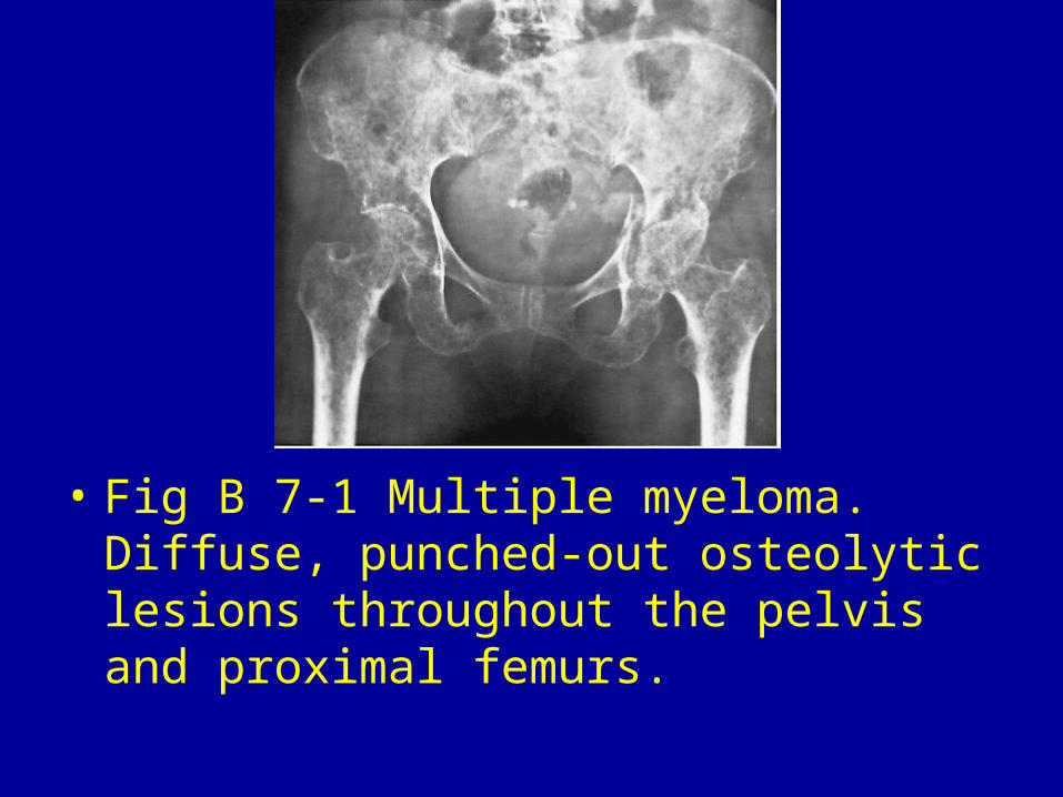
• Fig B 7-1 Multiple myeloma. Diffuse, punched-out osteolytic lesions throughout the pelvis and proximal femurs.
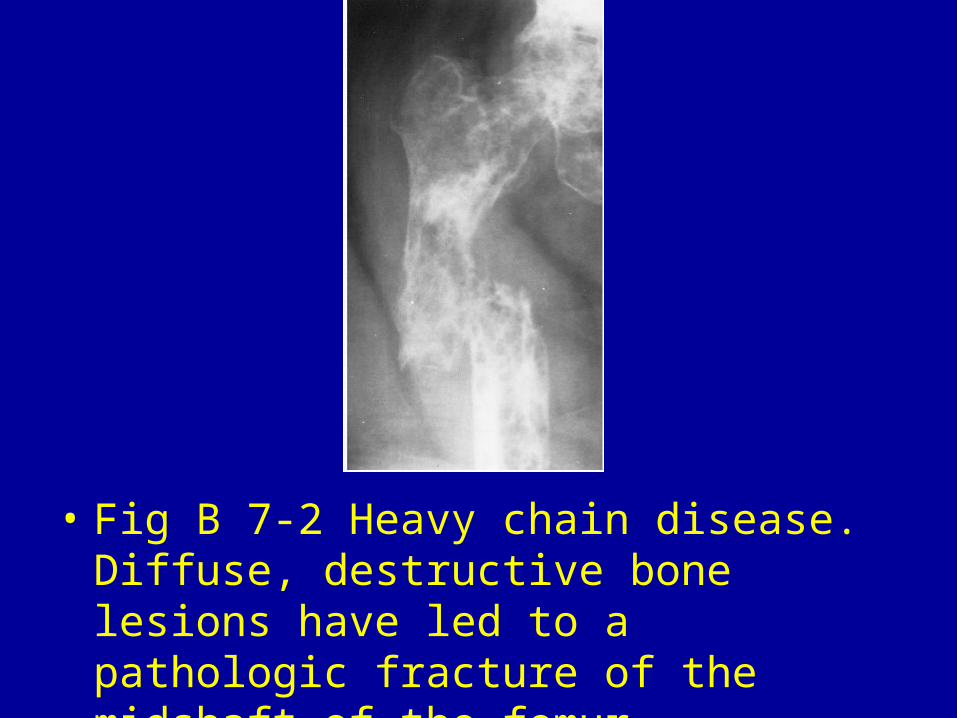
• Fig B 7-2 Heavy chain disease. Diffuse, destructive bone lesions have led to a pathologic fracture of the midshaft of the femur.
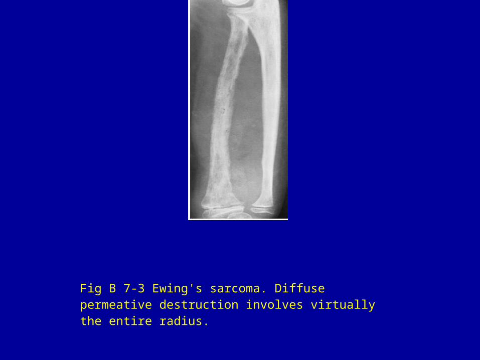
Fig B 7-3 Ewing's sarcoma. Diffuse permeative destruction involves virtually the entire radius.
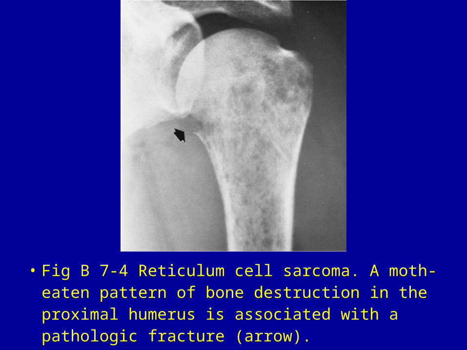
• Fig B 7-4 Reticulum cell sarcoma. A moth-eaten pattern of bone destruction in the proximal humerus is associated with a pathologic fracture (arrow).
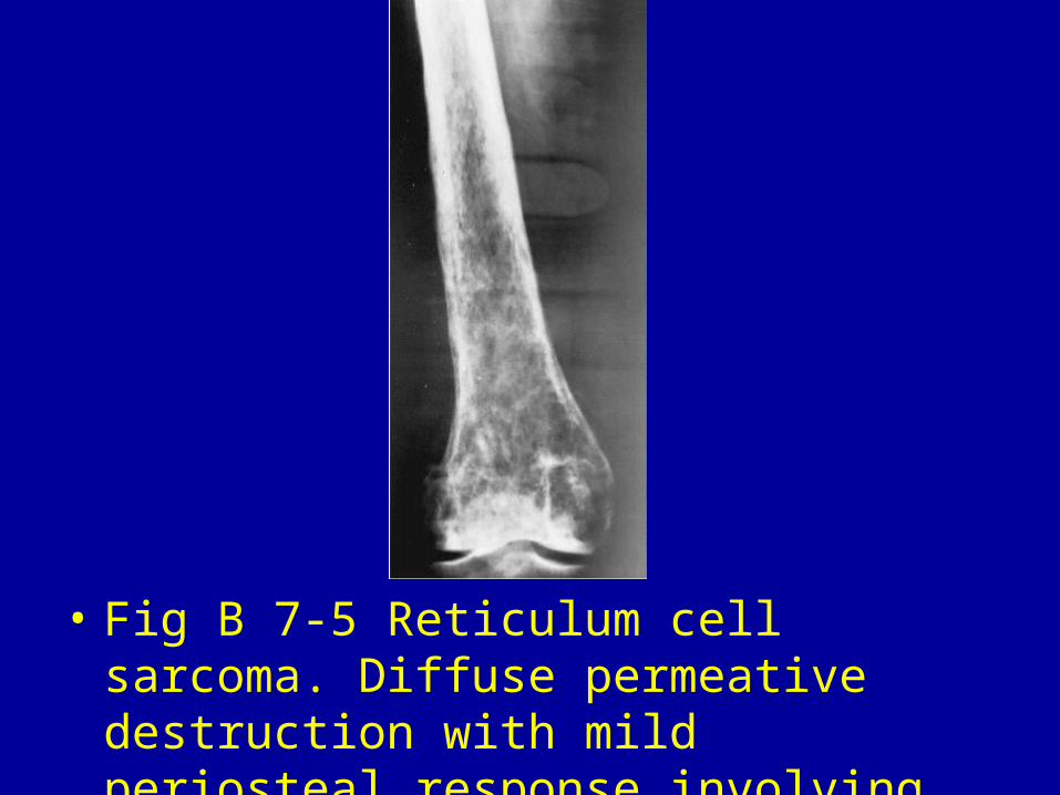
• Fig B 7-5 Reticulum cell sarcoma. Diffuse permeative destruction with mild periosteal response involving the distal half of the femur.
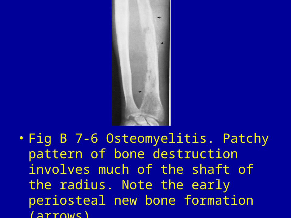
• Fig B 7-6 Osteomyelitis. Patchy pattern of bone destruction involves much of the shaft of the radius. Note the early periosteal new bone formation (arrows).
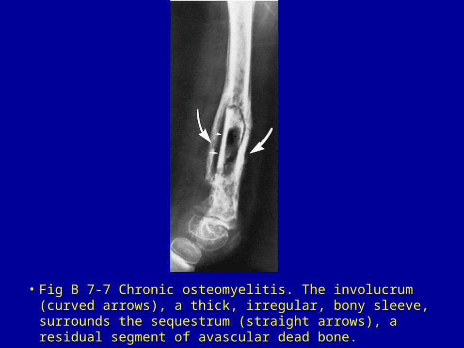
• Fig B 7-7 Chronic osteomyelitis. The involucrum (curved arrows), a thick, irregular, bony sleeve, surrounds the sequestrum (straight arrows), a residual segment of avascular dead bone.
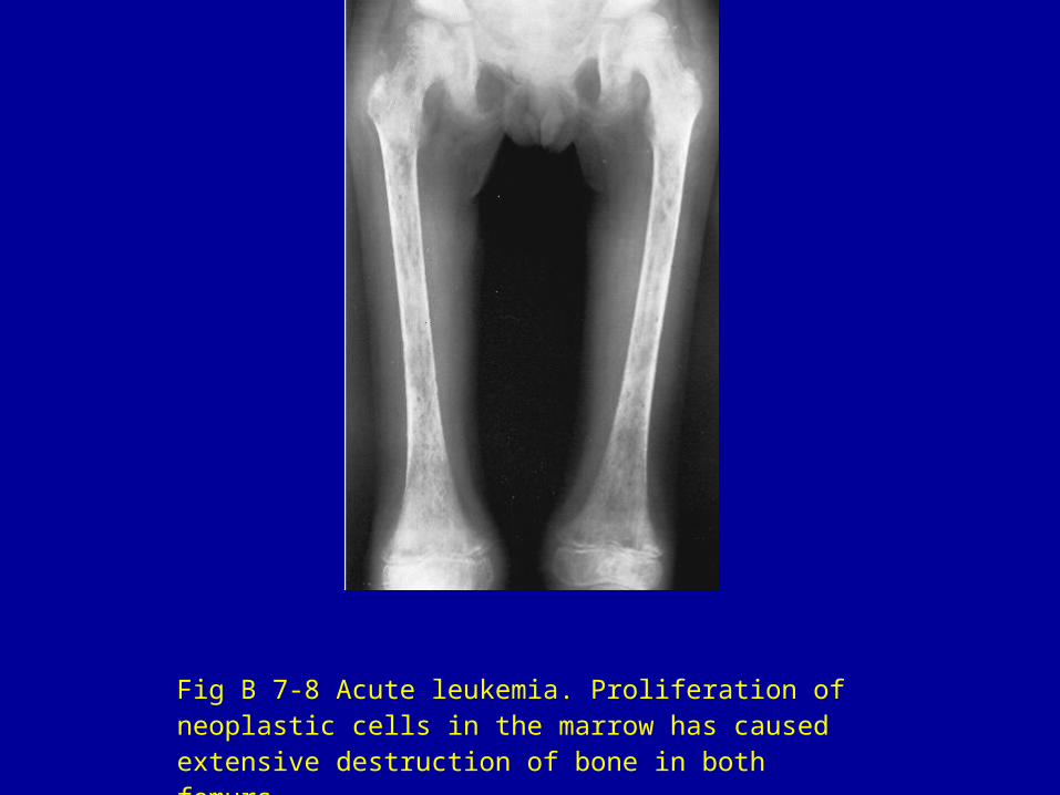
Fig B 7-8 Acute leukemia. Proliferation of neoplastic cells in the marrow has caused extensive destruction of bone in both femurs.
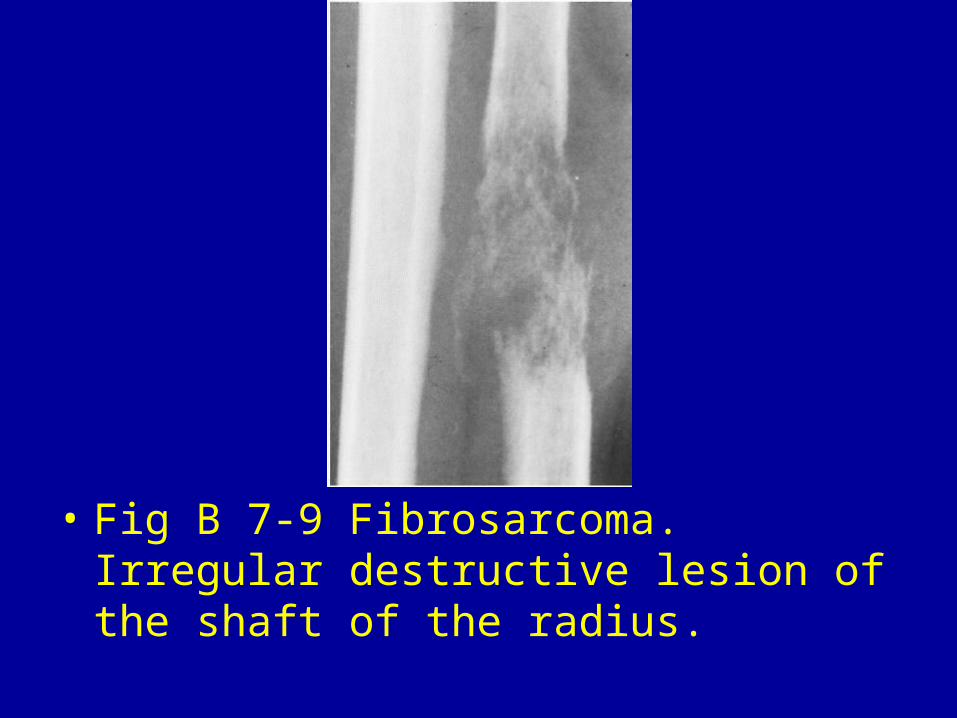
• Fig B 7-9 Fibrosarcoma. Irregular destructive lesion of the shaft of the radius.
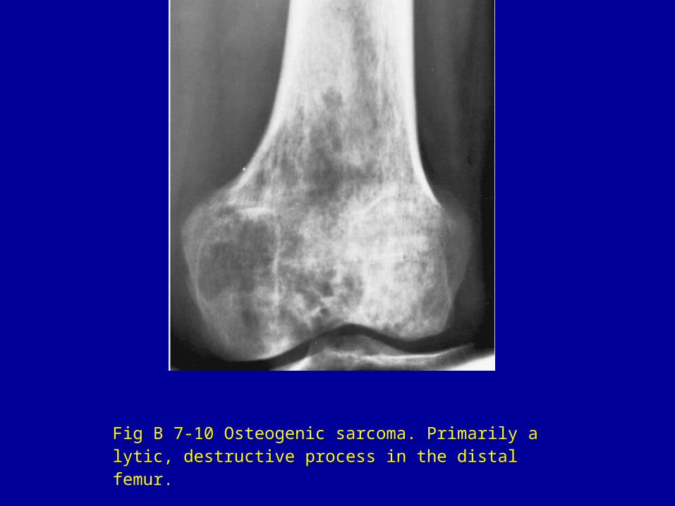
Fig B 7-10 Osteogenic sarcoma. Primarily a lytic, destructive process in the distal femur.
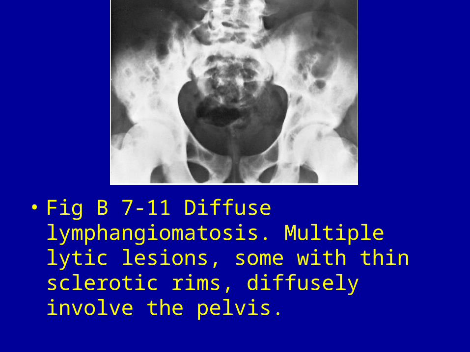
• Fig B 7-11 Diffuse lymphangiomatosis. Multiple lytic lesions, some with thin sclerotic rims, diffusely involve the pelvis.

