8 platelets and hemostasis
TRANSCRIPT
Platelet or Thrombocyte Platelet or Thrombocyte PhysiologyPhysiology
Platelet or Thrombocyte Platelet or Thrombocyte PhysiologyPhysiology
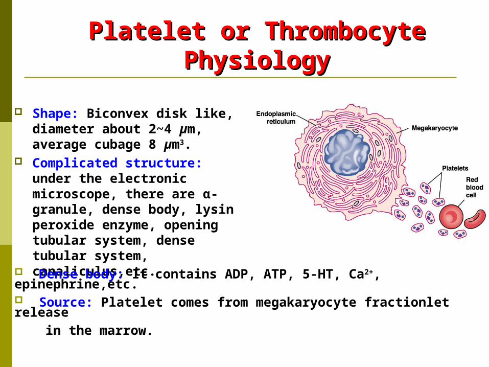
Shape: Biconvex disk like, diameter about 2~4 µm, average cubage 8 µm3.
Complicated structure: under the electronic microscope, there are α-granule, dense body, lysin peroxide enzyme, opening tubular system, dense tubular system, canaliculus,etc.
Dense body: It contains ADP, ATP, 5-HT, Ca2+, epinephrine,etc. Source: Platelet comes from megakaryocyte fractionlet release in the marrow.
Normal Value and Function of Normal Value and Function of PlateletPlatelet
Normal value: 100×109 ~ 300×109, range from 6%~10% Normal changes: more number in the afternoon than in
the morning, more in winter than in spring, more in the venous blood than capillary, after sport↑, pregnacy↑.
*Functions: 1. It maintains capillary endothelial cells smooth and integrated (repairing endothelium and providing nutrition). 2. It is involved in physiological hemostasis. Platelet and clinic relation: decrease of platelet, abnormal immune reaction, will
results in hemorrhage or bleeding, purpuric symptom.
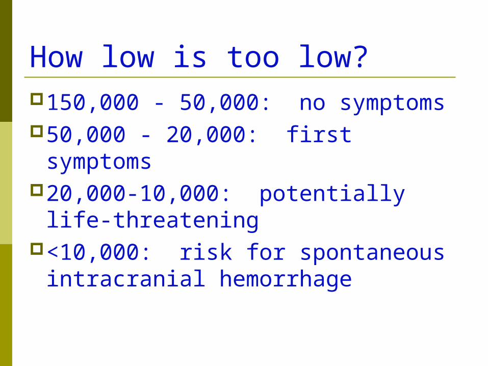
How low is too low?150,000 - 50,000: no symptoms50,000 - 20,000: first symptoms20,000-10,000: potentially life-
threatening<10,000: risk for spontaneous
intracranial hemorrhage
Platelet Hypofunction- Symptoms mucocutaneous bleeding, ie “Oozing
and bruising” epistaxis gum bleeding bruising heavy menses Petechiae Post-surgical bleeding
Platelet Forming and Platelet Forming and RegulationRegulation
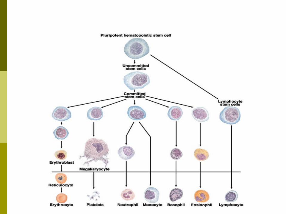
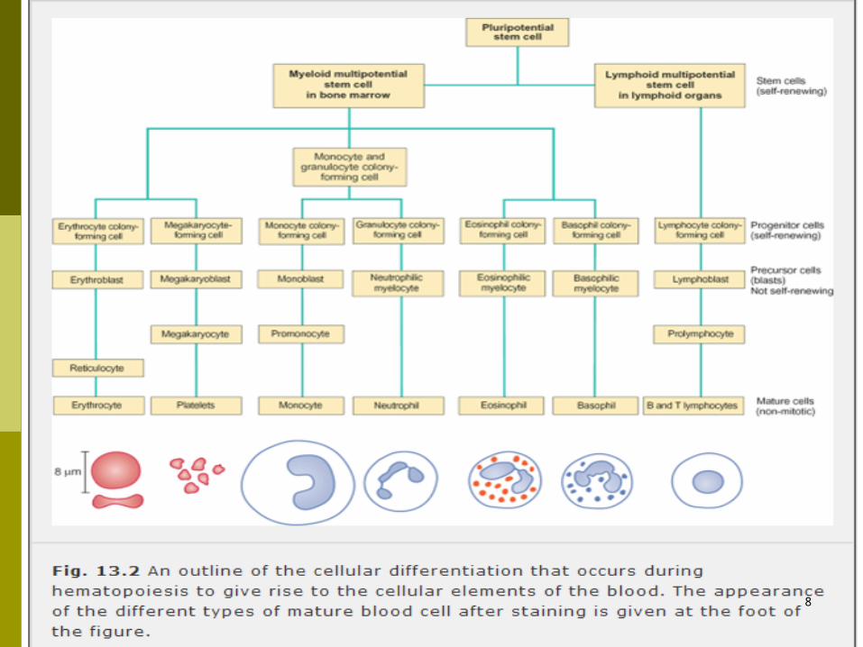
Platelet forming: Birth place is bone marrow, originating from hemopoietic
stem cells, and differentiating into burst forming unit- megakaryocyte, BFU-MK, then continuously into CFU-MK, and into megakaryocyte , into fractionlet release to the blood requiring 8~10 days.
>(one megakaryocyte can produce 200~7700 platelet).
8
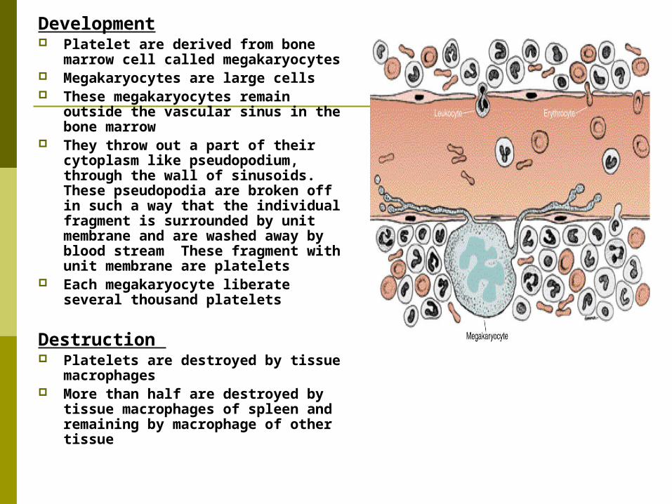
Development Platelet are derived from bone
marrow cell called megakaryocytes Megakaryocytes are large cells These megakaryocytes remain
outside the vascular sinus in the bone marrow
They throw out a part of their cytoplasm like pseudopodium, through the wall of sinusoids. These pseudopodia are broken off in such a way that the individual fragment is surrounded by unit membrane and are washed away by blood stream These fragment with unit membrane are platelets
Each megakaryocyte liberate several thousand platelets
Destruction Platelets are destroyed by tissue
macrophages More than half are destroyed by
tissue macrophages of spleen and remaining by macrophage of other tissue
Platelet Production Platelet production is regulated by
thrombopoietin (TPO). TPO synthesis is static Thrombopoietin binds to the surface of
megakaryocytes and platelets-- only free TPO stimulates platelet production Therefore, platelet mass negatively regulates
free TPO and further platelet production
Platelet Forming and Platelet Forming and RegulationRegulation
Regulation: Protein, Mpl, expressed by c-mpl (oncogene) exists
in CD34+ located at hemopoietic stem cells/
>committed progenitors, megakaryocyte and platelet, found by Methin in 1993, and its ligand named thrombopoietin, TPO was discovered in 1994 which promoted hemopoietic stem cells differentiating into megakaryocyte as hemopoietic stem cells positive regulating factor.
Life- Span and Breakage of Life- Span and Breakage of PlateletPlatelet
Life-span:Life-span: Averagely, 7~14 days in the blood. It can Averagely, 7~14 days in the blood. It can
be consumed when it displays physiological be consumed when it displays physiological
functions.functions.
Breakage:Breakage: Aged platelet can be processed by Aged platelet can be processed by
phagocytosis in liver, spleen and lymphatic node. phagocytosis in liver, spleen and lymphatic node.
IV. Physiological IV. Physiological HemostasisHemostasis
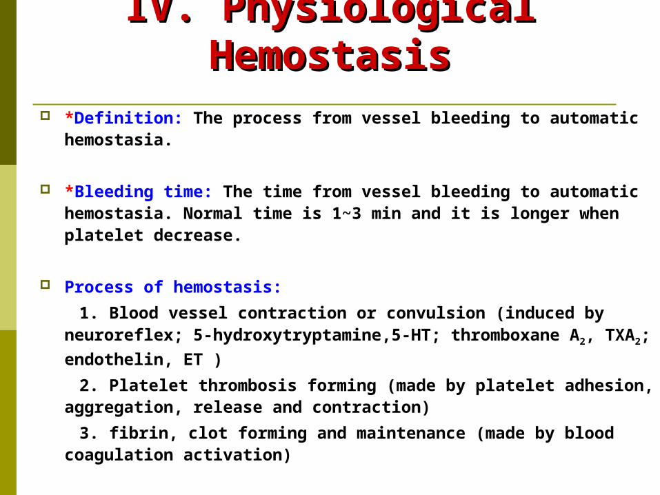
*Definition: The process from vessel bleeding to automatic hemostasia.
*Bleeding time: The time from vessel bleeding to automatic hemostasia. Normal time is 1~3 min and it is longer when platelet decrease.
Process of hemostasis:
1. Blood vessel contraction or convulsion (induced by neuroreflex; 5-hydroxytryptamine,5-HT; thromboxane A2,
TXA2; endothelin, ET )
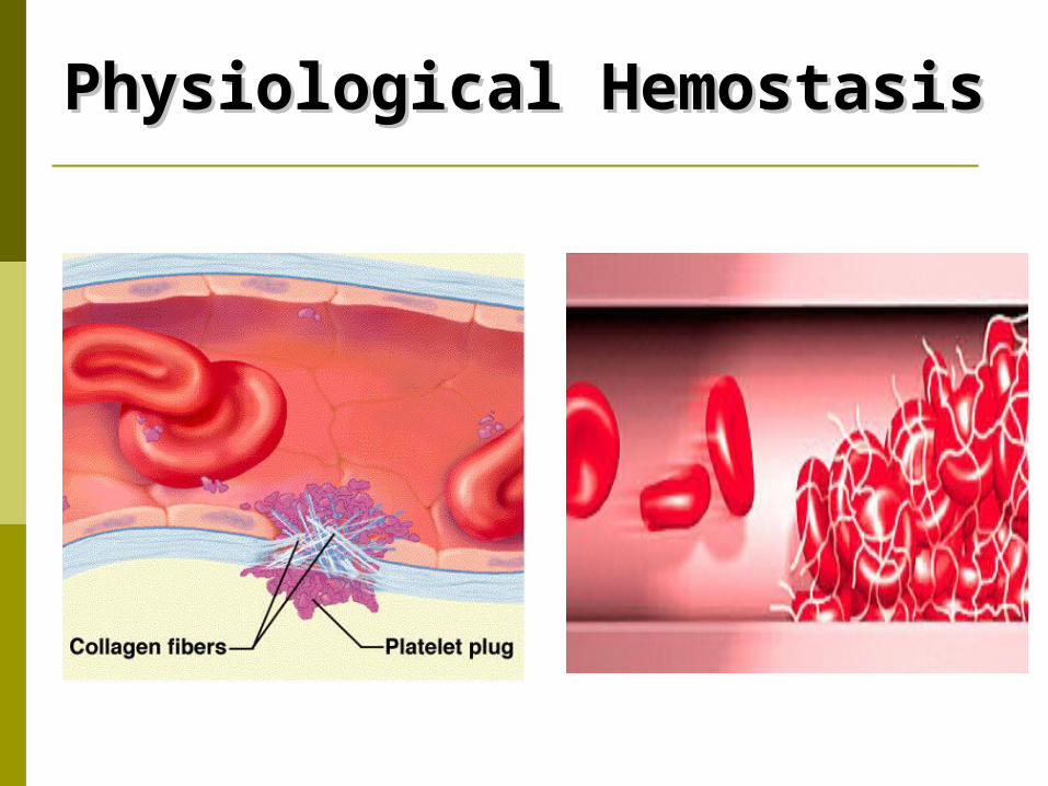
2. Platelet thrombosis forming (made by platelet adhesion, aggregation, release and contraction)
3. fibrin, clot forming and maintenance (made by blood coagulation activation)
Physiological HemostasisPhysiological Hemostasis
1.Endocrine functions of 1.Endocrine functions of vessel endothelial cellsvessel endothelial cells
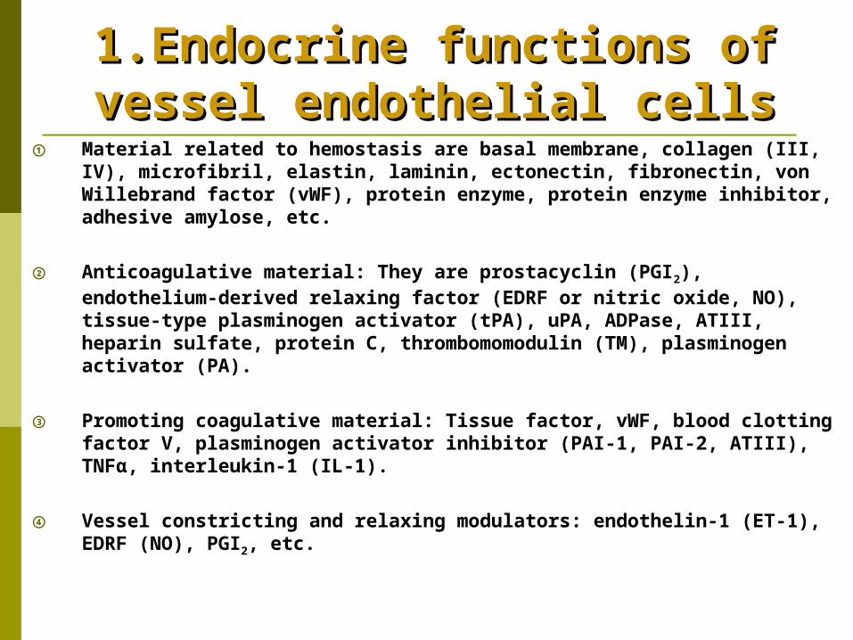
① Material related to hemostasis are basal membrane, collagen (III, IV), microfibril, elastin, laminin, ectonectin, fibronectin, von Willebrand factor (vWF), protein enzyme, protein enzyme inhibitor, adhesive amylose, etc.
② Anticoagulative material: They are prostacyclin (PGI2), endothelium-derived relaxing factor (EDRF or nitric oxide, NO), tissue-type plasminogen activator (tPA), uPA, ADPase, ATIII, heparin sulfate, protein C, thrombomomodulin (TM), plasminogen activator (PA).
③ Promoting coagulative material: Tissue factor, vWF, blood clotting factor V, plasminogen activator inhibitor (PAI-1, PAI-2, ATIII), TNFα, interleukin-1 (IL-1).
④ Vessel constricting and relaxing modulators: endothelin-1 (ET-1), EDRF (NO), PGI2, etc.
Roles of Vessel Endothelial Roles of Vessel Endothelial Cells in Physiological Cells in Physiological
HemostasisHemostasisRoles are close related to its endocrine functionsRoles are close related to its endocrine functions
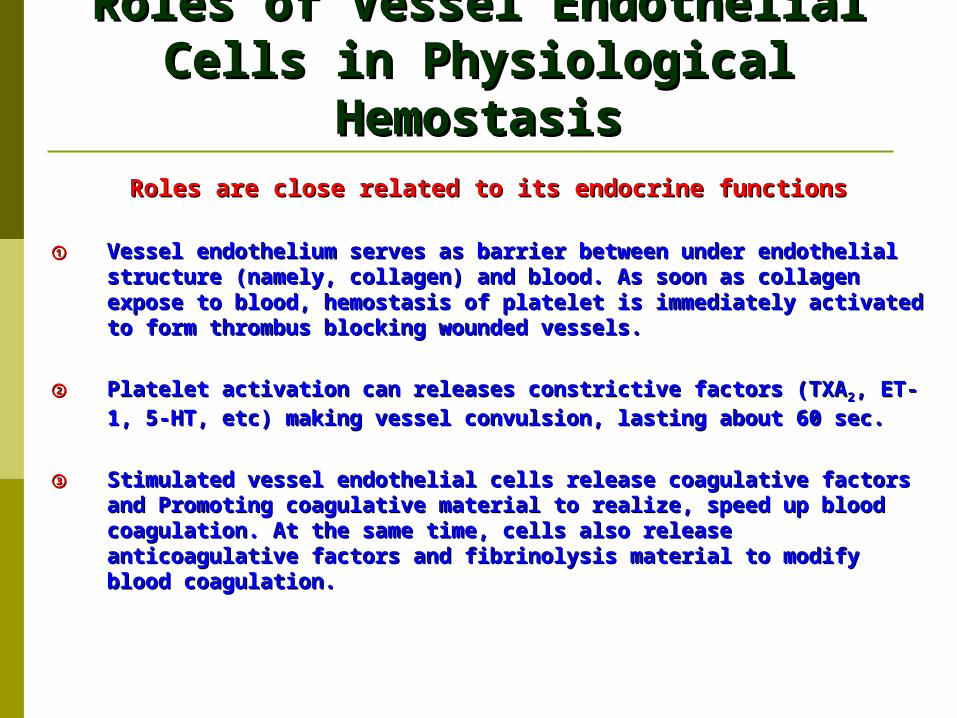
①① Vessel endothelium serves as barrier between under endothelial Vessel endothelium serves as barrier between under endothelial structure (namely, collagen) and blood. As soon as collagen expose structure (namely, collagen) and blood. As soon as collagen expose to blood, hemostasis of platelet is immediately activated to form to blood, hemostasis of platelet is immediately activated to form thrombus blocking wounded vessels.thrombus blocking wounded vessels.
②② Platelet activation can releases constrictive factors (TXAPlatelet activation can releases constrictive factors (TXA22, ET-1, 5-, ET-1, 5-HT, etc) making vessel convulsion, lasting about 60 sec.HT, etc) making vessel convulsion, lasting about 60 sec.
③③ Stimulated vessel endothelial cells release coagulative factors and Stimulated vessel endothelial cells release coagulative factors and Promoting coagulative material to realize, speed up blood Promoting coagulative material to realize, speed up blood coagulation. At the same time, cells also release anticoagulative coagulation. At the same time, cells also release anticoagulative factors and fibrinolysis material to modify blood coagulation.factors and fibrinolysis material to modify blood coagulation.
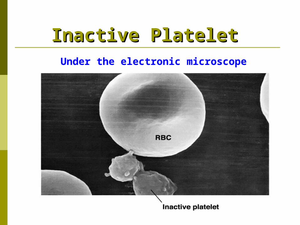
Inactive Platelet Inactive Platelet Under the electronic microscope
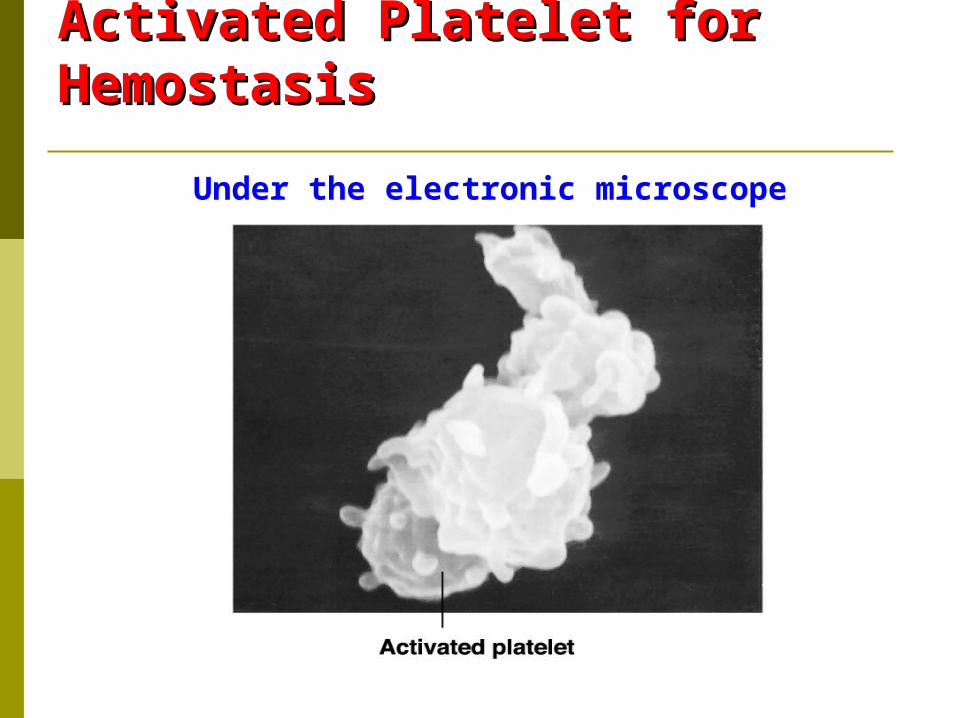
Activated Platelet for Activated Platelet for Hemostasis Hemostasis
Under the electronic microscope
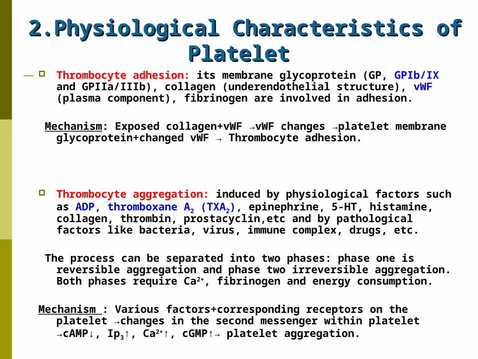
2.Physiological Characteristics of 2.Physiological Characteristics of Platelet Platelet
Thrombocyte adhesion: its membrane glycoprotein (GP, GPIb/IX and GPIIa/IIIb), collagen (underendothelial structure), vWF (plasma component), fibrinogen are involved in adhesion.
Mechanism: Exposed collagen+vWF →vWF changes →platelet
membrane glycoprotein+changed vWF → Thrombocyte adhesion.
Thrombocyte aggregation: induced by physiological factors such as ADP, thromboxane A2 (TXA2), epinephrine, 5-HT, histamine, collagen, thrombin, prostacyclin,etc and by pathological factors like bacteria, virus, immune complex, drugs, etc.
The process can be separated into two phases: phase one is
reversible aggregation and phase two irreversible aggregation. Both phases require Ca2+, fibrinogen and energy consumption.
Mechanism : Various factors+corresponding receptors on the
platelet →changes in the second messenger within platelet →cAMP↓, Ip3↑, Ca2+↑, cGMP↑→ platelet aggregation.
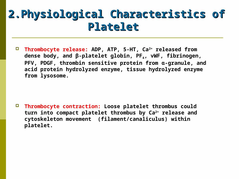
2.Physiological Characteristics of 2.Physiological Characteristics of Platelet Platelet
Thrombocyte release: ADP, ATP, 5-HT, Ca2+ released from dense body, and β-platelet globin, PF4, vWF, fibrinogen, PFV, PDGF, thrombin sensitive protein from α-granule, and acid protein hydrolyzed enzyme, tissue hydrolyzed enzyme from lysosome.
Thrombocyte contraction: Loose platelet thrombus could turn into compact platelet thrombus by Ca2+ release and cytoskeleton movement (filament/canaliculus) within platelet.
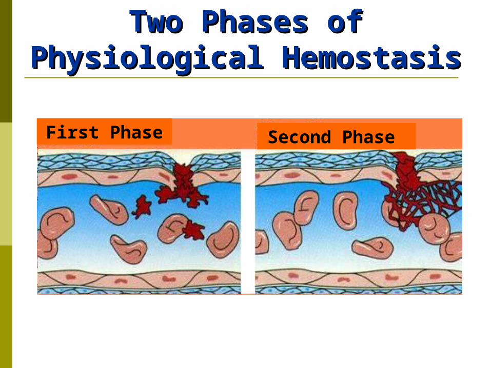
Two Phases of Two Phases of PhysiologicalPhysiological HemostasisHemostasis
First Phase Second Phase
Roles of Platelet in Roles of Platelet in HemostasisHemostasis
Activation of platelet: Stimulus brings about thrombocyte adhesion, aggregation, release and contraction.
Loose platelet thrombus forming: First phase of hemostasis.
Blood coagulation activation by platelet: Fibrin net forming, second phase of hemostasis.

Roles of Platelet in Roles of Platelet in HemostasisHemostasis
*Roles of platelet in hemostasis: 1. Activated platelets supply lecithoid (phospholipid) surface
for blood clotting factor and involve in activating factor X and prothrombin.
2. Surface of platelet membrane combine with many blood
clotting factor, such as fibrinogen, FV, FXI, FXIII to speed up coagulation.
3. Activated platelets release α-granule which contains
fibrinogen to intensify fibrin forming and blood coagulation.
4. Activated platelets contract clot with its contractive
protein to solidify blood coagulation.
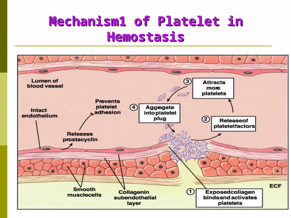
Mechanism1 of Platelet in Mechanism1 of Platelet in HemostasisHemostasis
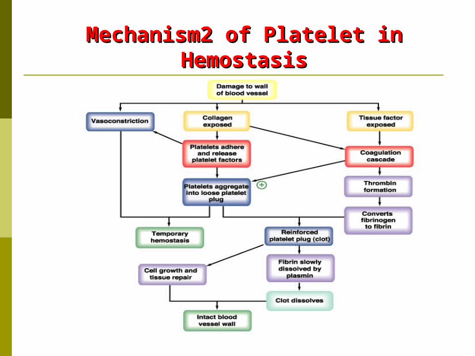
Mechanism2 of Platelet in Mechanism2 of Platelet in HemostasisHemostasis
3.Blood Coagulation3.Blood CoagulationBlood Clotting FactorBlood Clotting Factor
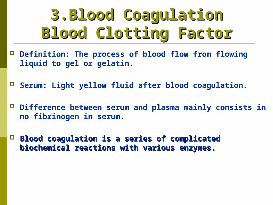
Definition: The process of blood flow from flowing liquid to gel or gelatin.
Serum: Light yellow fluid after blood coagulation.
Difference between serum and plasma mainly consists in no fibrinogen in serum.
Blood coagulation is a series of complicated biochemical Blood coagulation is a series of complicated biochemical reactions with various enzymes.reactions with various enzymes.
3.Blood Coagulation3.Blood CoagulationBlood Clotting FactorBlood Clotting Factor
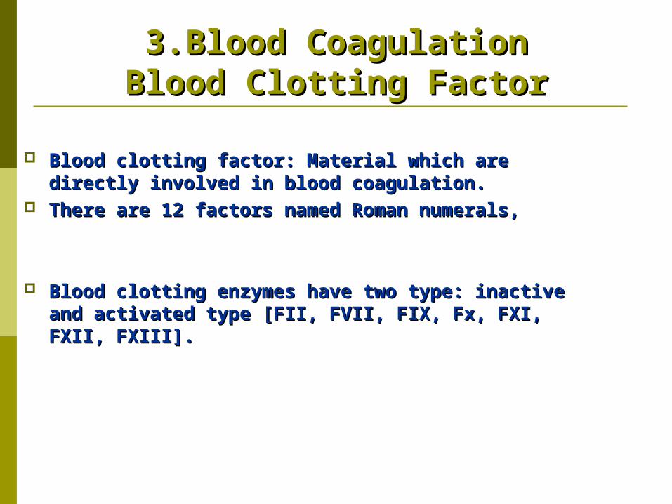
Blood clotting factor: Material which are directly Blood clotting factor: Material which are directly involved in blood coagulation. involved in blood coagulation.
There are 12 factors named Roman numerals, There are 12 factors named Roman numerals,
Blood clotting enzymes have two type: inactive and Blood clotting enzymes have two type: inactive and activated type [FII, FVII, FIX, Fx, FXI, FXII, FXIII].activated type [FII, FVII, FIX, Fx, FXI, FXII, FXIII].
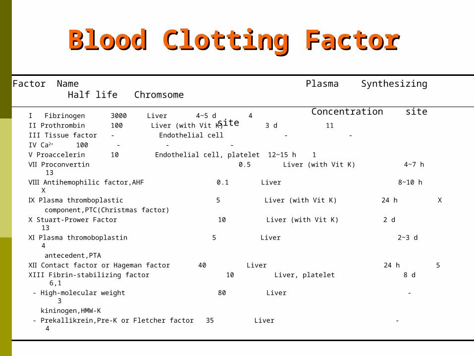
I Fibrinogen 3000 Liver 4~5 d 4II Prothrombin 100 Liver (with Vit K) 3 d 11III Tissue factor - Endothelial cell - -IV Ca2+ 100 - - -V Proaccelerin 10 Endothelial cell, platelet 12~15 h 1Ⅶ Proconvertin 0.5 Liver (with Vit K) 4~7 h 13Ⅷ Antihemophilic factor,AHF 0.1 Liver 8~10 h ⅩⅨ Plasma thromboplastic 5 Liver (with Vit K) 24 h Ⅹ component,PTC(Christmas factor)Ⅹ Stuart-Prower Factor 10 Liver (with Vit K) 2 d 13Ⅺ Plasma thromoboplastin 5 Liver 2~3 d 4 antecedent,PTAⅫ Contact factor or Hageman factor 40 Liver 24 h 5XIII Fibrin-stabilizing factor 10 Liver, platelet 8 d 6,1 - High-molecular weight 80 Liver - 3 kininogen,HMW-K - Prekallikrein,Pre-K or Fletcher factor 35 Liver - 4
Factor Name Plasma Synthesizing Half life Chromsome
Concentration site site
Blood Clotting FactorBlood Clotting Factor
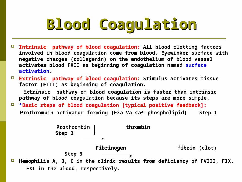
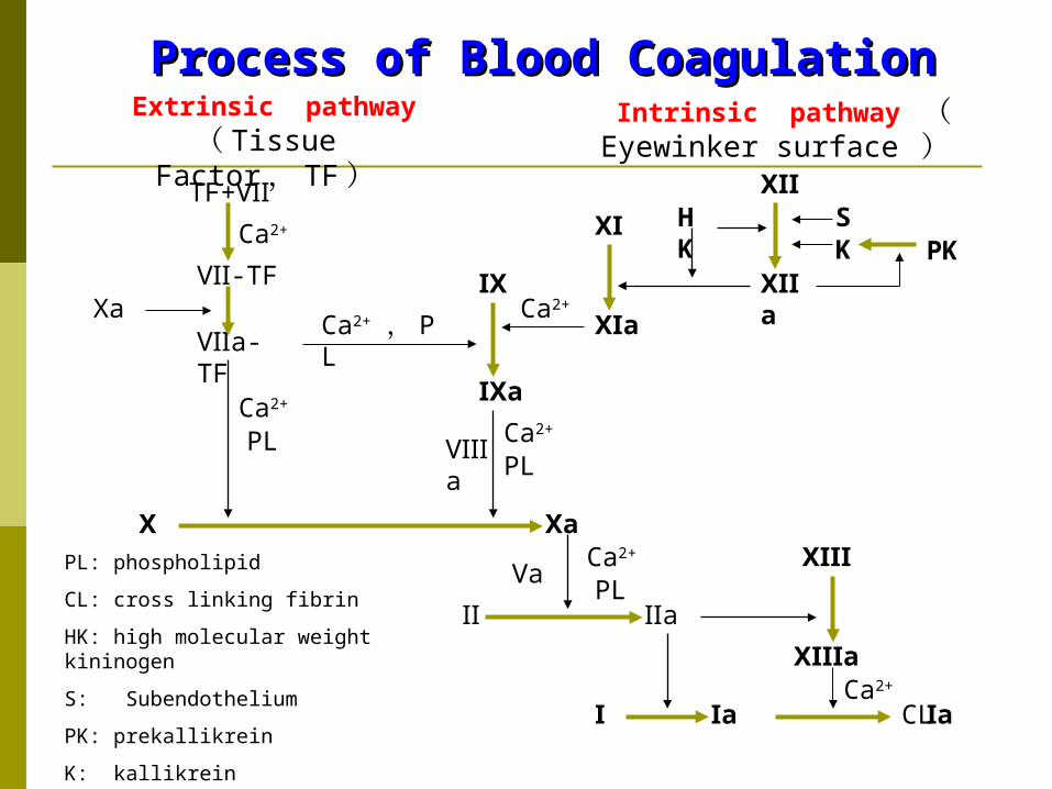
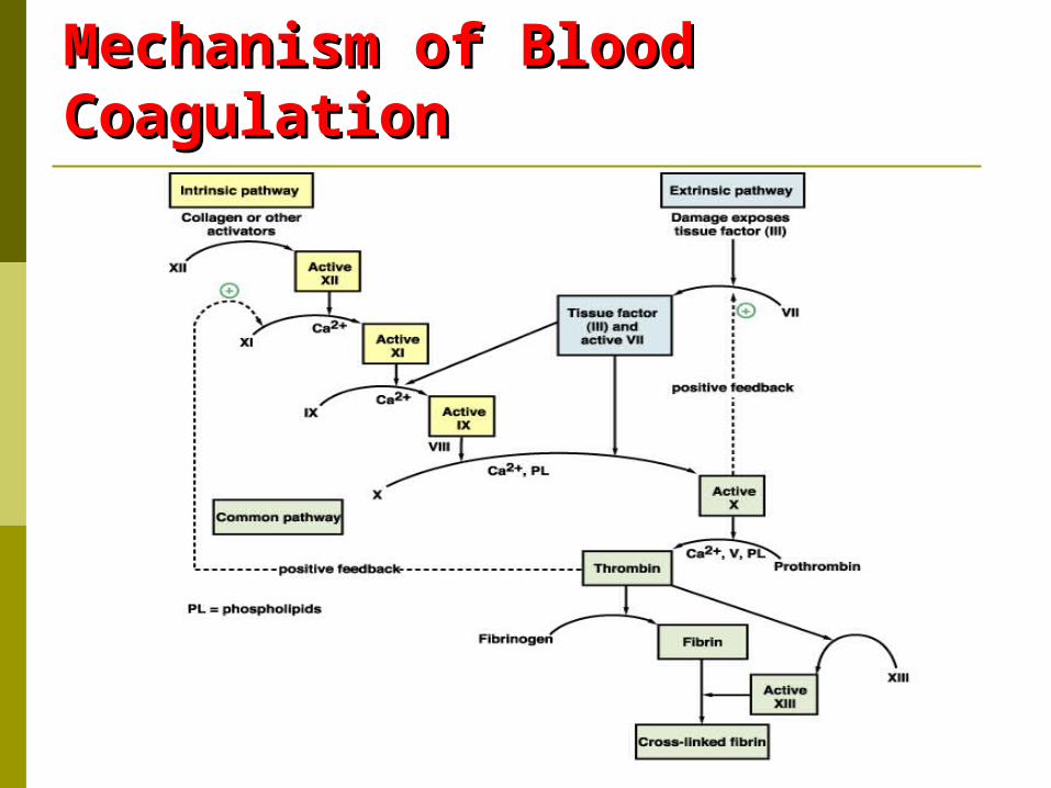
Blood CoagulationBlood Coagulation Intrinsic pathway of blood coagulation: All blood clotting factors
involved in blood coagulation come from blood. Eyewinker surface with negative charges (collagenin) on the endothelium of blood vessel activates blood FXII as beginning of coagulation named surface activation.
Extrinsic pathway of blood coagulation: Stimulus activates tissue factor (FIII) as beginning of coagulation.
Extrinsic pathway of blood coagulation is faster than intrinsic pathway of blood coagulation because its steps are more simple.
*Basic steps of blood coagulation [typical positive feedback]: Prothrombin activator forming [FXa-Va-Ca2+-phospholipid] Step 1 Prothrombin thrombin Step 2 Fibrinogen fibrin (clot) Step 3 Hemophilia A, B, C in the clinic results from deficiency of FVIII, FIX, FXI in the blood, respectively.
Extrinsic pathway ( Tissue Factor , T
F )TF+Ⅶ
Ⅶ-TF
Ⅶa-TF
Ca2+
Ca2+ , PL
Ca2+
Ⅹa
Ca2+
Ⅹ
Ⅺ
Ⅸ
Ⅸa
Ⅹa
Ca2+
Ⅷa
PLPL
Ⅴa
Ⅻ
Ⅱ
Ⅰ
ⅩⅢ
Ⅻa
HK
SK PK
Ⅺa
Ⅰa CLⅠa
Ca2+
PLCa2+
Ⅱa
ⅩⅢa
Intrinsic pathway ( Eyewinker surface )
PL: phospholipid
CL: cross linking fibrin
HK: high molecular weight kininogen
S: Subendothelium
PK: prekallikrein
K: kallikrein
Process of Blood CoagulationProcess of Blood Coagulation
Mechanism of Blood Mechanism of Blood CoagulationCoagulation
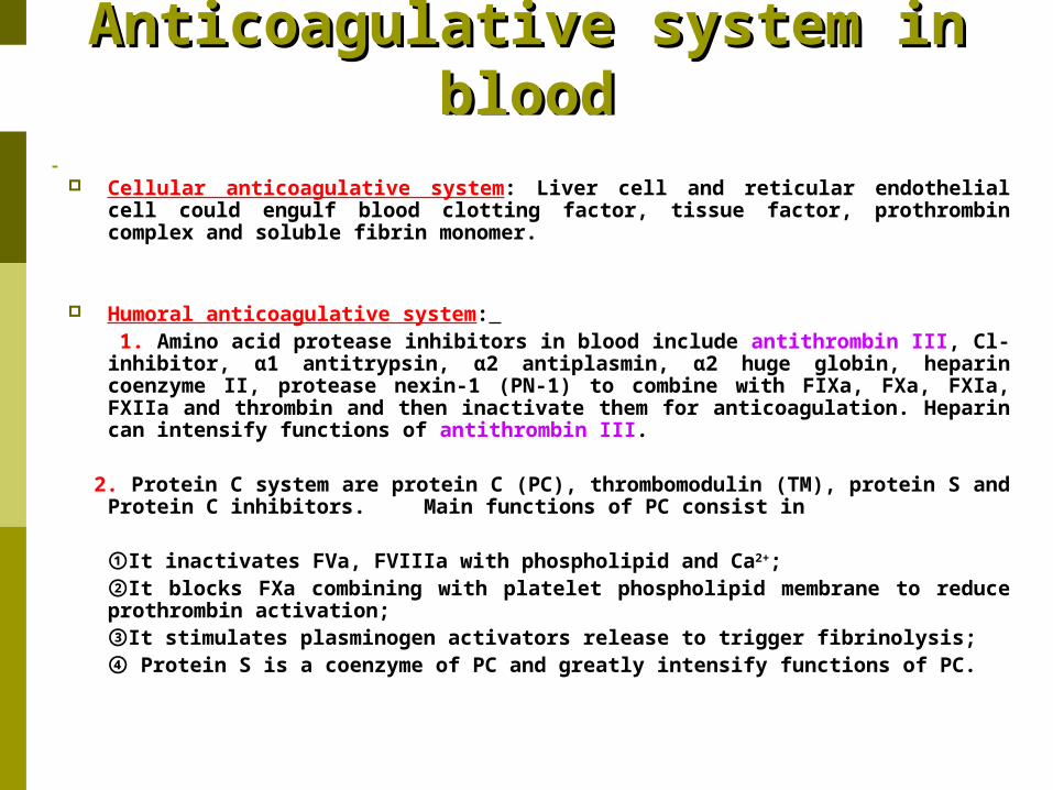
Anticoagulative system in Anticoagulative system in bloodblood
Cellular anticoagulative system: Liver cell and reticular endothelial cell could engulf blood clotting factor, tissue factor, prothrombin complex and soluble fibrin monomer.
Humoral anticoagulative system: 1. Amino acid protease inhibitors in blood include antithrombin III, Cl-
inhibitor, α1 antitrypsin, α2 antiplasmin, α2 huge globin, heparin coenzyme II, protease nexin-1 (PN-1) to combine with FIXa, FXa, FXIa, FXIIa and thrombin and then inactivate them for anticoagulation. Heparin can intensify functions of antithrombin III.
2. Protein C system are protein C (PC), thrombomodulin (TM), protein S and
Protein C inhibitors. Main functions of PC consist in
①It inactivates FVa, FVIIIa with phospholipid and Ca2+; ②It blocks FXa combining with platelet phospholipid membrane to reduce prothrombin activation; ③It stimulates plasminogen activators release to trigger fibrinolysis; ④ Protein S is a coenzyme of PC and greatly intensify functions of PC.
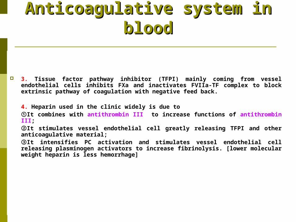
Anticoagulative system in Anticoagulative system in bloodblood
3. Tissue factor pathway inhibitor (TFPI) mainly coming from vessel endothelial cells inhibits FXa and inactivates FVIIa-TF complex to block extrinsic pathway of coagulation with negative feed back.
4. Heparin used in the clinic widely is due to ①It combines with antithrombin III to increase functions of antithrombin III; ②It stimulates vessel endothelial cell greatly releasing TFPI and other anticoagulative material; ③It intensifies PC activation and stimulates vessel endothelial cell releasing plasminogen activators to increase fibrinolysis. [lower molecular weight heparin is less hemorrhage]
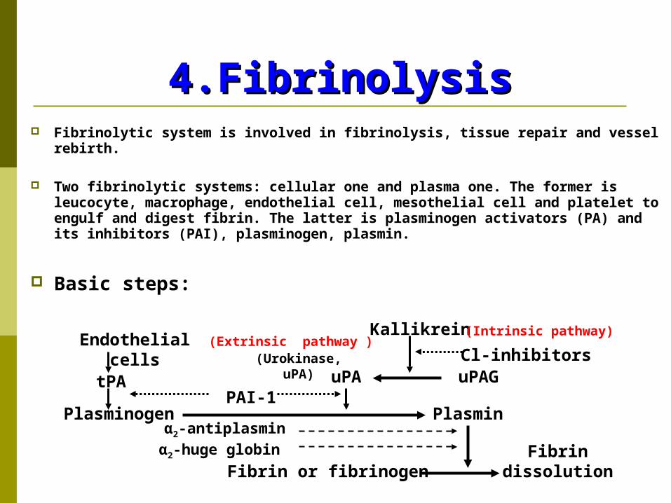
4.Fibrinolysis 4.Fibrinolysis Fibrinolytic system is involved in fibrinolysis, tissue repair and vessel
rebirth.
Two fibrinolytic systems: cellular one and plasma one. The former is leucocyte, macrophage, endothelial cell, mesothelial cell and platelet to engulf and digest fibrin. The latter is plasminogen activators (PA) and its inhibitors (PAI), plasminogen, plasmin.
Basic steps:
Endothelial cells
tPA
Plasminogen
Kallikrein
Cl-inhibitorsuPAGuPA
PlasminPAI-1
Fibrin or fibrinogen Fibrin
dissolution
α2-antiplasminα2-huge globin
(Urokinase, uPA)
(Extrinsic pathway )(Intrinsic pathway)
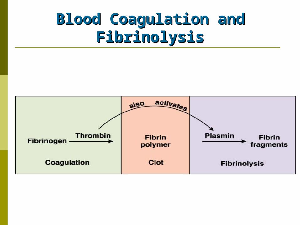
Blood Coagulation and Blood Coagulation and FibrinolysisFibrinolysis
Antifibrinolysis: Antifibrinolysis: Fibrinolytic Inhibitors and Its Fibrinolytic Inhibitors and Its
FunctionsFunctions
Main fibrinolytic inhibitors: They are plasminogen activator inhibitor type-1 (PAI-1, in platelet), α2-antiplasmin (in liver), α2-huge globin, α1-antitrypsin, antithrombin III, alexin C1 inhibitor.
PAI-1 synthesis and release: PAI-1 made by endothelial cell, smooth muscular cell, mesothelial cell, megakaryocyte is stored in platelet with inactive form. Some factors such as thrombin, IL-1, TNFα, etc stimulate its release from platelet.
Antifibrinolysis: Antifibrinolysis: Fibrinolytic Inhibitors and Its Fibrinolytic Inhibitors and Its
FunctionsFunctions
PAI-1 function: It inhibits tPA (tissue-type plasminogen activator) limiting local fibrinolysis of thrombus.
α2-antiplasmin characteristics: (1) Quick effect, (2) Inhibit plasminogen adhering to fibrin; (3) Combine with fibrin αchain and block fibrinolysis
Clinic relation: Innate deficiency of α2-antiplasmin often brings about serious hemorrhage.

Thank you