a bacterial pathogen targets a host rab-family gtpase defense
TRANSCRIPT

Cell Host & Microbe, Volume 19
Supplemental Information
A Bacterial Pathogen Targets a Host Rab-Family
GTPase Defense Pathway with a GAP
Stefania Spanò, Xiang Gao, Sebastian Hannemann, María Lara-Tejero, and Jorge E. Galán

Supplementary Figure S1 related to Figure 2. The S. Typhimurium effector proteins SopD2 and GtgE prevent the recruitment of Rab32 to the S. Typhimurium-containing vacuole. COS-1 cells expressing CFP-Rab32 (green) were infected with the indicated strains of S. Typhi or S. Typhimurium expressing mCherry (red) and 3 h after infection the infected cells were visualized by fluorescence microscopy. Images show maximum-intensity projections of confocal Z-stacks (scale bars, 5 µm). Quantification of this experiment is shown on Fig. 2B.
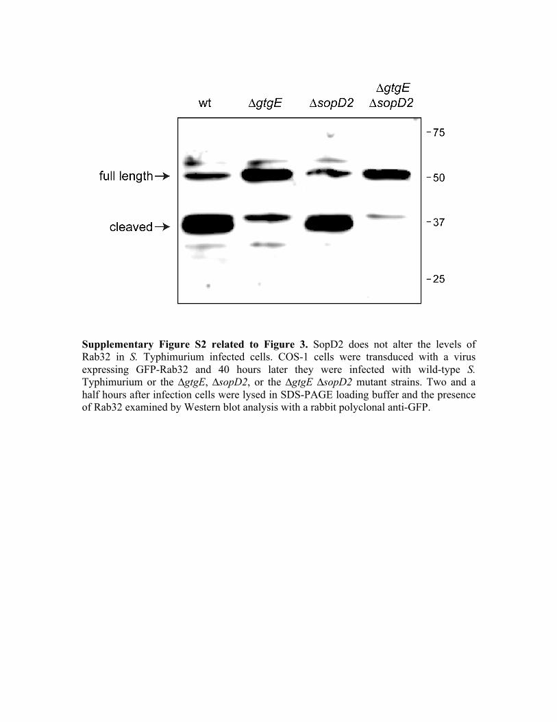
Supplementary Figure S2 related to Figure 3. SopD2 does not alter the levels of Rab32 in S. Typhimurium infected cells. COS-1 cells were transduced with a virus expressing GFP-Rab32 and 40 hours later they were infected with wild-type S. Typhimurium or the ∆gtgE, ∆sopD2, or the ∆gtgE ∆sopD2 mutant strains. Two and a half hours after infection cells were lysed in SDS-PAGE loading buffer and the presence of Rab32 examined by Western blot analysis with a rabbit polyclonal anti-GFP.

Supplementary Figure S3 related to Figure 3. SopD2 interacts with Rab8A, Rab8B, and Rab10. HeLa cells were transiently co-transfected with plasmids expressing FLAG-epitope tagged SopD2 or the unrelated effector PipA along with plasmids expressing GFP-tagged Rab8A, Rab8B, Rab10, or Rab31. Eighteen hours after transfection, cell lysates were analyzed by immunoprecipitation with anti-FLAG and Western immunoblotting with anti-GFP, FLAG and tubulin (as loading control) antibodies.

Supplementary Fig. S4 related to Figure 3. Effect of expression of Rab8 and Rab10 mutants on Rab32 recruitment to the Salmonella-containing vacuole. Henle-407 cells were co-transfected with plasmids expressing CFP-tagged wild type, dominant negative (T22N or T23N), or constitutively active (Q67L or Q68L) forms of Rab8A, Rab8B, Rab10, Rab31, or CFP (control), along with a plasmid encoding YFP-tagged Rab32. Twenty-four hours after transfection, cells were infected with S. Typhimurium ∆gtgE ∆sopD2 expressing mCherry and the infected cells were visualized by fluorescence microscopy. The mean ± SD of the percentage of Salmonella-containing vacuoles that were positive for Rab32 in at least five independent experiments are shown. Values have been normalized according to the percentage of Salmonella-containing vacuoles exhibiting Rab32 in cells transfected with CFP (control), which was considered to be 100% (the actual value for the control was 72 ± 15 %). At least 100 bacteria were counted in each experimental condition. *: p < 0.005 (Student’s t test) for the difference in comparison to control cells (transfected with a plasmid expressing CFP).

Supplementary Figure S5 related to Figure 4. Amino acid sequence of SopD2 depicting the arginine residues targeted for mutagenesis analysis. The change to alanine of the arginine residues depicted in blue did not alter its GAP activity. In contrast, the change to alanine of the arginine residue depicted in red abrogated its GAP activity.

Supplementary Figure S6 related to Figure 4. S. Typhimurium expressing SopD2R315 is unable to prevent the recruitment of Rab32 to its vacuole. Henle-407 cells expressing YFP-tagged Rab32 (green) were infected with S. Typhimurium ∆gtgE ∆sopD2, or the same strain encoding plasmid-borne sopD2, or the catalytic mutant sopD2R315A, as indicated. Two hours after infection, infected cells were fixed, stained with an anti S. Typhimurium LPS antibody, and imaged under a fluorescence microscope. Quantification of this experiment is shown in Fig. 4D.

Supplementary Figure S7 related to Figure 4. Translocation of SopD2 and SopD2R315A into cultured cells during S. Typhimurium infection. Cultured epithelial cells were infected with type III protein secretion competent (T3SS +) or defective (T3SS -) S. Typhimurium strains expressing either SopD2 or SopD2R315A and the presence of these effectors in the insoluble (bacterial associated) or soluble (translocated) fractions was probed by western blot analysis as indicated in Material and Methods.

Table S1: LC-MS/MS analysis of SopD2 interacting proteins
Spectral counts
Accession Number Protein description Biological repeat 1 Biological repeat 2 Q8ZQC8 SopD2 33 50 Q0PD49 RAB8B 3 10 Q4FJL0 RAB10 4 6 B1AX58 Plastin 3 0 6 Q920Q8 Influenza NS1A-binding protein homolog 0 5 Q923T9 Calcium-dependent protein kinase 5 0 Q3UHW5 RAB8A 0 5 Q9JKM7 RAB37 0 4 A2AL34 RAB14 0 4 B2RRN5 RAB4A 0 4 H3BLG3 RAB26 0 4 Q0PD10 RAB43 0 4 Q0PD14 RAB39B 0 4 Q0PD15 RAB39 0 4 Q0PD21 RAB33B 0 4 Q0PD40 RAB15 0 4 Q0PD54 RAB6 0 4 Q0PD66 RAB1B 0 4 Q0PD67 RAB1 0 4 Q9CXS2 RAB3C 0 4 P17751 Triosephosphate isomerase 3 0 Q3V0K9 Plastin-1 0 3 B2KFR5 Serine/threonine kinase 38 3 0 E9Q5D6 Ran-binding protein 9 0 3 Q3TCI7 Putative uncharacterized protein 3 0 Q80UY7 SCY1-like protein 2 0 3 Q8CDE3 Putative uncharacterized protein 3 0

Table S2. List of strains used in this study
Strain Genotype References
Salmonella Typhi: ISP2825 wild type 1
Salmonella Typhimurium: SL1344 wild type 2
SB2519 ∆sipA ∆sptP ∆avrA ∆sopE ∆sopE2 ∆sipF ∆sopB ∆sopD ∆sopD2 ∆slrP ∆gtgE (=∆11) This study
SB2518 ∆gtgE 3
SB2523 gtgEH151A This study
SB2527 ∆gtgE∆ sopD2 This study
SB2731 ∆gtgE∆ slrP∆ sopA This study
SB1343 ∆sopD2 4
SB1227 spiA::kan 3
1 Galán, J. E. & Curtiss III, R. Infect. Immun. 59, 2901-2908 (1991). 2 Hoiseth, S. K. & Stocker, B. A. Nature 291, 238-239 (1981). 3 Spano, S., Liu, X. & Galan, J. E. Proc Natl Acad Sci U S A 108, 18418-18423, doi:10.1073/pnas.1111959108 (2011). 4 Jiang, X. et al. Mol Microbiol 54, 1186-1198, doi:10.1111/j.1365-2958.2004.04344.x (2004).

Table S3. List of plasmids used in this study Plasmid Description References pSB4004 pWSKrpsM-mCherry 1 pVSVG pVSV-G Provided by Dr. W. Mothes pGag/Pol pMLV-Gag-Pol Provided by Dr. W. Mothes pSB4661 pLZRS-CFP-Rab32 This study pSB4830 pWSKlacZ-sopD2 This study pSB4829 pWSKlacZ-sopD2-3xFlag This study pSB5202 pWSKlacZ-pipAH184F-3xFlag Galán laboratory (unpublished) PSB5203 pWSKlacZ-sopAC753A-3xFlag Galan laboratory (unpublished) pSB5298 pWSKlacZ-sopD2R315A This study pCMV-CFP-C2 Clontech pSB4174 pCMV-YFP-Rab32 1 pSB5325 pCMV-CFP-Rab8A This study pSB5327 pCMV-CFP-Rab8A-Q67L This study pSB5326 pCMV-CFP-Rab8A-T22N This study pSB5328 pCMV-CFP-Rab8B This study pSB5322 pCMV-CFP-Rab8B-Q67L This study pSB5323 pCMV-CFP-Rab8B-T22N This study pSB5317 pCMV-CFP-Rab10 This study pSB5318 pCMV-CFP-Rab10-Q68L This study pSB5319 pCMV-CFP-Rab10-T23N This study pSB5329 pCMV-CFP-Rab31 This study pSB3938 pCMV-GFP-Rab8A This study pSB3942 pCMV-GFP-Rab8A-Q67L This study pSB3941 pCMV-GFP-Rab8A-T22N This study pSB5110 pCMV-GFP-Rab8B This study pSB5337 pCMV-GFP-Rab8B-Q67L This study pSB5324 pCMV-GFP-Rab8B-T22N This study pSB5111 pCMV-GFP-Rab10 This study pSB5304 pCMV-GFP-Rab10-Q68L This study pSB5306 pCMV-GFP-Rab10-T23N This study pSB3475 pCMV-GFP-Rab31 This study pSB5253 pRK5-3xFLAG-PipAE181A Galan laboratory (unpublished)

1Spano, S., Liu, X. & Galan, J. E. Proteolytic targeting of Rab29 by an effector protein distinguishes the intracellular compartments of human-adapted and broad-host Salmonella. Proc Natl Acad Sci U S A 108, 18418-18423, doi:10.1073/pnas.1111959108 (2011).