a baseline study of extubation events occurring at the
TRANSCRIPT

oA BASELINE STUDY OF EXTUBATION EVENTS OCCURRING
AT THE INTENSIVE CARE UNIT OF THE KENYATTA
NATIONAL HOSPITAL
A DISSERTATION PRESENTED IN PART FULFILLMENT OF THE REQUIREMENTS
FOR THE AWARD OF THE MASTERS DEGREE IN ANAESTHESIA, UNIVERSITY OF
NAIROBI
J ^ r 'it r i / f .L f&r>
DR. WINNIE WAKIURU MATHANGANI
2010
i

WfflVERsmroF nairoB)
A BASELINE STUDY OF EXTUBATION EVENTS OCCURRING AT THE INTENSIVE
CARE UNIT OF THE KENYATTA NATIONAL HOSPITAL
INVESTIGATOR:
DR. WINNIE WAKIURU MATHANGAM
VIBChB (RPFU)
POST-GRADUATE STUDENT IN ANAESTHESIOLOGY.
DEPARTMENT OF SURGERY
UNIVERSITY OF NAIROBI.
SUPERVISOR
DR. MARK GACII
MBChB, M.MED (ANAESTHESIA)
LECTURER IN ANAESTHESIOLOGY.
DEPARTMENT OF SURGERY
UNIVERSITY OF NAIROBI.
University of NAIROBI Library

DECLARATION.
I declare that this proposal is my original work and has not been submitted for a degree award in
any uni vers i t \ .
RESEARCHER: SIGNATURE DATE
Dr. Winnie W Mathangani
This proposal has been submitted for the degree of Masters of Medicine in Anaesthesiology with
my approval as a university supervisor.
SUPERVISOR: SIGNATURE DATE
Dr. Mark V. Gacii X - t i 'Z o lo
DEPARTMENT CF SURGERY COLLEGE OF HEALTH SCIENCESP. O. B zx 19676 - 00202 KNH NAIROBI
. 2725300. Ext. 43773
iii

DEDICATION:
I dedicate this thesis to my mother. Salome, my biggest supporter and the greatest influence on my life, and to my siblings Angela and Paul for their unwavering and unconditional love and support.
IV

ACKNOWLEDGEMENTS:
I would like to thank my supervisor. Dr. Mark Gacii, as well as my teachers Dr. Thomas Chokwe and Dr. David Misango who provided guidance and support during the development and writing ot
this thesis.
1 owe my deepest gratitude to my sister Angela Mathangani tor her priceless input in streamlining
the rough draft.
Last but not least, I am grateful to all my colleagues in the K..N.H Critical Care Unit who assisted
in the data collection.
May God bless you all.
v

ACKNOWLEDGEMENTS:
I would like to thank my supervisor. Dr. Mark Gacii, as well as my teachers Dr. Thomas Chokwe and Dr. David Misango who provided guidance and support during the development and writing ot
this thesis.
1 owe my deepest aratitude to my sister Angela Vlathangani tor her priceless input in streamlining
the rough draft.
Last but not least, I am grateful to all my colleagues in the K.N.H Critical Care Unit who assisted
in the data collection.
May God bless you all.
v

ABBREVIATIONS
AARC - American Association of Respiratory Clinicians
BIPAP - Bi-phasic intermittent positive airway pressure
CPAP - Continuous positive airway pressure
ETT - Endo tracheal tube
FiO: - Fraction of inspired oxygen
HDU - High Dependency Unit
ICU - Intensive Care Unit
IPPV - Intermittent positive pressure ventilation
K.NH - Kenvatta National Hospital
PEEP - Positive end expiratory pressure
RSBI - Rapid Shallow Breathing Index
SBT - Spontaneous breathing trial
SIMV - Synchronized intermittent positive pressure ventilation
UE - Unplanned Extubation
UoN - University of Nairobi

TABLE OF CONTENTS.
DECLARATION......................................................................................................... (iii)
DEDICATION............................................................................................................. (iv)
ACKNOWLEDGEMENTS....................................................................................... (v)
TABLE OF CONTENTS.......................................................................................... (vi)
LIST OF ABBREVIATIONS.....................................................................................(x)
OPERATIONAL D EFLA TIO N S............................................................................(xi)
LIST OF FIGURES AND TABLES.........................................................................(xiii)
SUMMARY...................................................................................................................(xiv)
1.0 INTRODUCTION...................................................................................................... 1
l . 1 Statement of the problem.......................................................................... 3
2.0 LITERATURE REVIEW............................................................................................4
3.0 STUDY OBJECTIVES............................................................................................... 13
4.0 STUDY JUSTIFICATION......................................................................................... 14
5.0 METHODOLOGY....................................................................................................... 15
6.1 Study site.......................................................................................................... 15
6.2 Study population.............................................................................................. 15
6.3 Study design..................................................................................................... 15
6.4 Sample size....................................................................................................... 15
6.5 Sampling m ethod............................................................................................. 16
6.6 Data collection procedure............................................................................... 17
6.7 Data quality control and management............................................................. 18

UNIVERSITY OF NAIROBIMEDICAL LIBRARY
6.8 Data analysis..................................................................................................... 18
6.9 Inclusion/Exclusion criteria.............................................................................. 18
5.10 Ethical Considerations................................................................................... 19
5.11 Study limitations............................................................................................ 19
6.0 RESULTS......................................................................................................................21
6.1 End point of intubation...................................................................................21
6.2 Extubation m odes............................................................................................ 22
6.3 General outcome of extubation events.......................................................... 23
6.4 Outcomes of extubation events after various modes of extubation.......... 24
6.5 Timing of extubation events............................................................................25
6.6 Adverse events experienced during extubation............... .............................27
6.7 Gender distribution and outcome of extubation events................................ 28
6.8 Age distribution and outcome of extubation events......................................29
6.9 Sub-specialty category and outcome of extubation events........................... 30
6.10 ETT securing methods and extubation m ode................................................ 31
6.11 ETT securing methods in various clinical categories....................................32
6.12 Use of pharmacological and physical restraints.......................................... 33
6.13 SBT performance in predicting extubation outcome.................................. 35
7.0 DISCUSSION.............................................................................................................36
viii
8.0 CONCLUSIONS 41

REFERENCES........................................................................................................................ 43
APPENDIX la: GUARDIAN EXPLANATION FORM.....................................................48
APPENDIX lb: PATIENT EXPLANATION FORM ........................................................ 49
APPENDIX II: CONSENT FORM ..................................................................................... 50
APPENDIX III: SURVEY TOOL.......................................................................................... 51
APPENDIX IV: BUDGET...................................................................................................... 53
APPENDIX V: IMPLEMENTATION PLAN......... .............................................................. 54
9.0 RECOMMENDATIONS................................................................................................42

LIST OF ABBREVIATIONS
A ARC
BIPAP
CPAP
ETT
FiO:
HDU
Hr
ICU
IPPV
KNH
Min
PEEP
RSBI
SBT
SIMV
UE
UoN
- American Association of Respiratory Clinicians
- Bi-phasic intermittent positive airway pressure
- Continuous positive airway pressure
- Endotracheal tube
- Fractional inspired oxygen concentration
- High Dependency Unit
- Hour
- Intensive Care Unit
- Intermittent positive pressure ventilation
- Kenyatta National Hospital
- Minutes
- Positive end expiratory pressure
- Rapid Shallow Breathing Index
- Spontaneous breathing trial
- Synchronized intermittent positive pressure ventilation
- Unplanned Extubation
- University of Nairobi

OPERATIONAL DEFLATIONS:
Intubation: - The placement of an endotracheal tube into the trachea.
Difficult intubation:- Tracheal intubation after more than 2 attempts at intubation.
Extubation: - The complete withdrawal of an endotracheal tube from the trachea.
Planned extubation: •- The elective removal of the endotracheal tube.
Unplanned extubation: - The premature removal of the endotracheal tube.
Self extubation: - The premature removal of the endotracheal tube by action of the patient
Accidental extubation: - The premature removal of the endotracheal tube during care and
Extubation failure:-
manipulation of the patient.
The need for reintubation within 72 hours of an extubation.
Extubation success:- No need for reintubation within 72 hours of an extubation.
Reintubation:- Reinsertion of the endotracheal tube into the trachea after an extubation
incident.
IPPV: - Intermittent positive pressure ventilation: A ventilation mode best used in
heavily sedated, paralyzed or unconscious patients with depressed
respiratory drive. The patient is ventilated at a rate set by the operator.
SIMV: - Synchronized intermittent positive pressure ventilation: A ventilation mode
where the ventilator synchronizes machine breaths with the patient's
attempts at spontaneous breathing. Each machine breath occurs during the
patient’s expiratory pause or self initiated inspiration.
BIPAP:- Bi-phasic intermittent positive airway pressure ventilation: A ventilation
mode that provides two phases of positive pressure during the patient’s
XI

spontaneously generated respiratory cycle. A high pressure level supports
inspiration and a lower level of pressure keeps the alveoli open during
expiration.
CPAP: - Continuous positive airway pressure: A spontaneous breathing mode of
ventilation. It provides positive airway pressure during all phases of the
patient's respiratory cycle increasing the patient’s Functional Residual
Capacity and opening up previously collapsed alveoli;
T- piece: - A T- shaped breathing circuit used to supply a spontaneously breathing
patient with oxygen enriched air. The inhaled oxygen passes down one limb
of the T- piece device and the exhaled gases exit through the second limb
during expiration.
Pressure support: - A spontaneous mode of ventilation whereby the patient initiates every
breath and the ventilator supports each patient generated breath with a preset
pressure value.
NIPPV:- Non invasive positive pressure ventilation. This refers to the administration
of ventilatory support by nasal, face or helmet masks without the use of
invasive endotracheal or tracheostomy tube.
Desaturation:- Fall in arterial oxygen saturation (SaOz) from > 90% to < 90%
Hvpotension:- A Fall in systolic blood pressure to less than 90mmHg for longer than 10
min.
Bradycardia:- A fall in heart rate to below 60 beats/min
Hypoxia:- Partial pressure of oxygen in arterial blood (Pa02) < 8kPa or 60mmHg
Hvpercapnia:- Partial pressure of carbon dioxide in arterial blood (PaC02 ) > 6.0 kPa or
45mmHg
c* •«xii

LIST OF FIGURES AND TABLES
Figures Page
Fig 1. End point of intubation................................................................................................21
Fig 2. Modes of extubation................................................................................................... 22
Fig. 3 Extubation outcome.................................................................................................... 23
Fig 4. Mode of extubation and outcome................................................................................ 24
Fig 5. Schematic of the extubation events in K.NH ICU...................................................... 25
Fig 6. Complications within 30 min of extubation......................
Fig 7. Gender outcome of orally intubated patients....................
Fig 8. Age distribution and outcome of orally intubated patients
Fig 9. Sub-specialty category and outcome.................................
Fig 10. ETT securing method and outcome...................................
Fig 11. ETT securing method and patient sub-specialty category
26
.27
.28
30
31
32
Fig 12. Comparison of extubation outcome in restrained and unrestrained patients........... 34
Table 1 Use of restraints in patients prior to extubation.............................................33
Table 1: SBT performance in predicting extubation outcome.......................................35
xiii

SUMMARY:
Background: Extubation, or the removal of an endotracheal tube from the airway, is ideally
planned and executed once an intubated patient is able to maintain adequate respiratory effort and
a patent airway. However unplanned extubations do occur in intensive care units either
accidentally, during manipulation of the patient, or as self extubation by the intubated patient and
put the patient at increase risk of respiratory failure and reintubation. Planned extubations may also
result in extubation failure and reintubation.
Objective: To study extubation events occurring in a population of intubated patients in KNH
ICU. The primary end points of the study were:
1. The establishment of the incidence of planned and unplanned extubations occurring in
orally intubated ICU patients in KNH and the resultant success and failure rates
experienced with these modes of extubation.
2. The establishment of the complications experienced after extubation.
Methods: A prospective cross-sectional study was proposed. It was carried out in the KNH ICU.
The study population consisted of orally intubated patients who consented to be included in the
study. These patients were prospectively observed for an extubation event and followed up for a
period of up to 72 hrs after the extubation event occurred.
Data collection was done using a survey tool and performed by recruited research assistants. Data
collected will included patient demographic data, details describing the extubation event in terms
of time, place and mode of occurrence, as well as associated adverse events. The occurrence of
extubation failure within the 72 hour follow- up period was also documented in the same tool.
Data analysis: Resulting demographic and clinical data was collated, sorted, entered into the
computer, analysed and interpreted with a view to fulfilling the aforementioned objectives.
INTRODUCTION
God .... breathed into man s nostrils the breath o f life, and man became a living soul. ” Genesis
2:7
It has been long acknowledged that breathing is essential to life. Breathing is the means through
which atmospheric oxygen used in cellular respiration to create energy enters the body and expired
carbon dioxide, a by-product of energy production, leaves the body. Failure to maintain an
adequate level of this basic process of gas exchange is known as respiratory failure and results in
permanent cellular damage and eventually death.
Mechanical ventilation, defined as the use of a mechanical device to repeatedly inflate and deflate
the lungs, is initiated as a life saving intervention in critically ill patients whose ability to maintain
a patent airway, adequate ventilation and alveolar gas exchange is already diminished or lost or in
those at risk of impending respiratory failure. Patients on ventilatory support constitute a majority
of the patient population in KNH ICU.
Mechanical ventilation is commonly used in conjunction with endotracheal cannulation. There are
two main through which the trachea may be cannulated. Namely,
i. Translaryngeally with an endotracheal tube (ETT) inserted via either a nasal or an oral
route.
ii. Via a tracheotomy on the anterior surface of the neck.
This research is concerned with the former approach.
Extubation refers to the process of removing a tube from a hollow organ or passageway, often
from the airway. Ideally, it is a well planned and carefully executed event that takes into account
the clinical condition of each intubated patient and their ability to maintain a patent airway.
However, unplanned extubations are a common occurrence in intensive care units and they are
considered an indicator of quality of care for critically ill patients in ICUs(1}. They may occur
accidentally (e.g. during nursing care procedures or manipulation and transport of the patient), or
as spontaneous self-extubations executed by the intubated patient.(45)
1

In some instances, an unplanned extubation could end up being a successful extubation, whereby
the patient does not require reintubation.(3>) However, unplanned extubations are generally
considered undesirable and should be avoided as they can result in a number o f serious
complications such as difficulty in re-establishing an airway, upper airway trauma, respiratory
insufficiency and respiratory failure. (7'y) These complications, in turn, may lead to cardiac arrest
and even death.
Similarly, planned extubations may sometimes end up in failure, whereby the electively extubatC(j
patient experiences a similar set of complications faced by patients who undergo unplanned
extubation. The following diagram illustrates the above facts:
Fig 1. Schematic of the proposed investigation into the extubation events occurring among
KNH ICU patients.
------------------- "
EXTUBATED' NONEXTUBATED
L~—1-------------- 1 s................
PLANNEDEXTUBATION
2

Extubation failure, variably defined by different studies tJ/'40) as the requirement for reintubation
within 24 - 72 hrs of extubation, is an outcome to be avoided. Reasons for its occurrence, whether
resulting from inappropriate weaning technique during planned extubation or from improper
patient care practices that lead to unplanned extubation. should be identified in order to institute
methods that curb it.
STATEMENT OF THE PROBLEM:
ICU patient records obtained from the K.NH Medical Records Department show that
approximately 1000 patients are admitted per year into the ICU. The exact number of intubated
patients is unknown as current records do not capture this data. However, an estimate by the Senior
Nursing Officer in-charge of K.NH ICU puts the figure at a significant 98% of total admissions.
Similarly, there is no data documenting when or how the endotracheal tubes are removed from
these patients. This implies that the KNH ICU staff manages extubations on a regular basis.
However, the nature and frequency of various challenges presented by the process of extubation is
unknown.
Essential data such as incidence of planned extubation, accidental extubation, self extubation and
extubation failure is not adequately captured although these events are known to occur and impact
significantly on patient outcome and overall cost of care.
3

LITERATURE REVIEW
INTUBATION:
In medicine, intubation refers to the placement of a tube into an external or internal orifice of
body. Although the term can refer to endoscopic procedures, it is most often used to denote
endotracheal intubation. Endotracheal intubation is the placement of a flexible plastic or rubber
tube, into the trachea to protect the patient's airway and provide a means of mechanical ventilatj09.
EXTUB ATION:
Extubation refers to the complete withdrawal of an endotracheal tube from the trachea. It is
accepted practice that after a patient is intubated and an appropriate level of ventilator support
initiated, it should be maintained until the underlying cause of acute respiratory failure andother
complicating issues have shown some sign of reversal.1-1 The clinician may then, in a stepwise
manner, reduce ventilator support from the initial maintenance level to lower support modes. The
progressive transfer of the work of breathing from the ventilator to the patient is referred to as
weaning. Once weaning is complete and mechanical ventilation is no longer required, the
attending clinician must address the separate question of whether or not a patient can tolerate
extubation.
The process of extubation is sometimes erroneously viewed as a routine automatic step that
follows discontinuation from mechanical ventilation. This view is incorrect as the need for
ventilatory support is distinct from the need for an artificial airway.1 J) (e.g clinical syndromes
such as acute epiglottitis and other airway diseases indicate the need for an artificial airway but
ventilator support may not be required.)
Studies reveal that clinicians experience problems during extubation three times more com m only
than they do during intubation: (4.6% versus 12.6%).<4) A closed claims analysis of the American
Society of Anaesthesiologists’ database revealed that death or brain damage with induction of
anaesthesia decreased from 62% of perioperative claims in 1985 - 1992 to 35% in 1993 -1999
This decrease has been attributed to the widespread adoption of difficult airway guidelines which
predominantly address induction of anaesthesia. In contrast, the claims for death or brain damage

CLASSIFICATION OF EXTUBATION:
The Manual of Emergency Airway Management '51 classifies extubation as:
1. Routine.
2. Intermediate risk
3. High risk.
The difference between them is described as follows: "The extubation o f patients who were
easily intubated and in whom no intervening event has occurred to jeopardize their airways is
routine. Those who were easily intubated but at greater risk o f requiring reintubation due to
hypoxemia, hypercapnea. inadequate clearance o f secretions, inability to protect their airway
or airway obstruction are intermediate risk extubations. Those in whom airway management is
likely to be challenging or complex if reintubation is to be required represent high risk
extubations."
The latter group is further considered to include:
1. "Difficult intubations.
2. Those with interval complications (airway oedema, extrinsic compression, glottic
injuries).
3. Those with conditions associated with difficult ventilation or intubation (eg. Morbid
obesity, obstructive sleep apnoea, airway surgery, maxillofacial surgery deep neck
infections and prolonged intubation.)
According to this classification, the majority of intubated ICU patients fall into the intermediate
and high risk extubation categories.
Extubations may also be classified as planned or unplanned. ' 11
associated with extubation remained almost the same. This implies that methods geared at making
the process of extubation safer have not been as effectively employed.
5

Ideally, an extubation is a planned and carefully executed event performed by healthcare workers
after assessments of the patient’s clinical condition and ability to maintain a patent airway are
established as being adequate for extubation.
Unplanned extubations (UE) are unwelcome extubations. UE are classified as accidental
extubations (e.g. occurring during nursing care procedures or manipulation and transport of the
patient), or as self-extubations (when executed by the intubated patient.) (7‘9,10)
Postoperativelv, most patients are extubated soon after the return of consciousness and resumption
of spontaneous respiration. The resolution of neuromuscular block (paralysis) is easily
demonstrated clinically by sustained head lift, good grip strength and ability to protrude the tongue
out of the oral cavity.(1 ” The return of consciousness is demonstrated by the ability to follow
simple commands.
The timing of the withdrawal of an artificial airway from critically ill patients recovering from
respiratory failure is harder to establish and remains one of the more important and challenging
aspects of critical care management. Some critically ill patients may be excellent candidates for
extubation despite an inability to follow the same simple commands <12) and therefore different
criteria should be employed to assess their readiness for extubation.
The decision to extubate involves weighing the risks of prolonged mechanical ventilation and
intubation against the possibility of extubation failure. On the one hand, an overly cautious
approach to extubation will minimize premature discontinuations, but could also unnecessarily
prolong ventilatory support in some patients. On the other hand, over-aggressiveness in removing
ventilator support and the artificial airway predisposes patients to the risk of extubation failure,
with subsequent need for re-intubation and re-institution of ventilatory support.
Prolonged intubation increases a patient's risk of getting sinusitis113,14), laryngeal stenosisll5),
laryngeal and tracheal injuries115,16), pulmonary infections116) among others. This lays bare the fact
that a patient should not remain with an endotracheal tube longer than is absolutely necessary.
6

Recognition of the time-dependent nature of these complications has led clinicians and
investigators to concentrate their efforts on liberating patients from respiratory support and the
ETT as expeditiously as is safely possible.
PLANNED EXTUB ATI ON: ASSESSMENT OF EXTUB ATION READINESS.
Readiness for extubation implies that weaning from mechanical ventilation is completed and that
the patient:
1. Is sufficiently awake with intact airway reflexes
2. Is haemodynamically stable
3. Has manageable secretions.
Clinical judgement:
An early study in extubation observed the process to often be an arbitrary clinical decision based
on a clinician’s judgment and experience.(17) Another study showed that 50% of self extubated
patients did not require reintubation.<l8) This evidence suggests that when using clinical judgment
alone, physicians do not extubate their intubated patients expeditiously.
Protocol-driven extubation:
Critical care teams worldwide have conducted numerous studies in the search for ways to assist
physicians to correctly identify patients on mechanical ventilation who are capable of breathing
spontaneously and ready for extubation.
A randomized controlled trial that compared protocol-directed weaning and physician-directed
weaning found that protocol-directed weaning performed by nurses and respiratory therapists led
to extubation more rapidly than physician-directed weaning.ll9)
7
A separate study found that when physicians were notified that their patients had successfully
completed a spontaneous breathing trial, they were more likely to extubate their patients early.(20)
These two studies show that physicians and their patients benefit when guidelines that aid in the
selection of patients suitable for extubation are implemented.
Quantitative assessments of respiratory function:
Further research performed in the last decade has yielded vital information that is being used to
make the process of assessment of extubation readiness more of a science and less of an art. These
methods of identification of patients suitable for extubation involve the quantitative determination
of clinical weaning parameters that suggest adequate reversal of respiratory failure and that could
reliably support the decision to extubate a patient.
Weaning parameters have been and continue to be investigated as possible predictors of successful
extubation outcome. They range from simple readily available parameters such as respiratory rate,
spontaneous minute ventilation, heart rate and blood pressure level to more sophisticated
measurements such as the determination of oesophageal pressure using an oesophageal balloon.
However, no single parameter has been proven to accurately predict extubation success.*21)
Consequently, investigators developed a variety of integrated indexes from combinations of
individual parameters. Examples of these include the Rapid Shallow Breathing Index (RSBI), a
ratio of respiratory rate and tidal volume (f / Vt) and the CROP index that integrates chest wall
compliance, respiratory rate, oxygenation, and pressure. As yet, none of the currently developed
indexes are sufficiently sensitive and specific to be useful in predicting the success of extubation in
an individual patient.*21' Because of these limitations, the routine use of these parameters and
indexes are not recommended.
Spontaneous Breathing Trials
A multidisciplinary task force set up to produce evidence-based clinical practice guidelines for
managing the ventilator-dependent patient during the discontinuation process concluded that a
spontaneous breathing trial (SBT) provides the most useful information to guide clinical decision
making regarding weaning and extubation.*22' An SBT tests the ability of a patient to sustain
adequate ventilation on minimal respiratory support. It may be performed in one of three ways:
8

1) with low levels of CPAP (l-5cmH20)
2) with low level of pressure support (5-7cmH20)
3) or simply as T- piece breathing.
Multiple studies have found that patients tolerant of SBTs that are 30 to 120 min in length were
found to have successful ventilator discontinuations at least 77% of the time.
The KNH ICU/HDU protocols booklet1'" 1 outlines two weaning protocols that are similar to the
guidelines put forward by the evidence based task force. According to this booklet, extubation in
spontaneously breathing patients may be considered after one of two protocols is followed and
successfully completed. The ‘‘sprint” or "CPAP” protocol uses continuous positive airway pressure
(CPAP) mode of weaning. If a patient tolerates a 2-hour SBT on a CPAP level of 0 cmH20,
extubation is considered. The second protocol known as “The Gentle Work Protocol” proposes the
gradual reduction of pressure support by 2-5 cmH20 per day till a pressure support level of 3-4
cmH20 is achieved. If this low level of pressure support can sustain satisfactory ventilation for 14
hours, the patient is considered to have adequate capacity for spontaneous ventilation and may be
considered for extubation.
PLANNED EXTUBATION: THE PROCEDURE
According to the American Association of Respiratory Clinicians (AARC) 2007 guidelines, the
endotracheal tube should be removed in an environment in which the patient can be
physiologically monitored and in which emergency equipment and appropriately trained health
care providers with airway management skills are immediately available.1-41
Many centres have developed or adopted extubation procedure protocols to standardize the
performance of the extubation procedure. For example an extubation protocol followed by the St
George Hospital, New South Wales, Australia, details the sequence of steps to be followed by
nurses while performing a planned extubation procedure and lists all the necessary equipment that
should be close at hand during the process.1' 51
Notably absent in the KNH ICU/HDU protocol booklet is a description of how elective extubation
procedures in the ICU should be carried out.
9

UNPLANNED EXTUB ATI ON
Generally, unplanned extubations (UE) are considered undesirable and have been reported as a
problem for many institutions worldwide.1 0|.
A significant interest in UE has developed since Coppolo and May first focused attention on the
topic in 1 9 9 0 . That study found that 69% percent of UE were self extubations and that the
majority of these occurred despite use of sedation and restraints. They concluded that self-
extubation is a common occurrence which, despite obvious hazards, is often tolerated well by
adults. Multivariate analyses carried out after 1990, however, have since attributed self extubation
with increased morbidity and mortality.
Unplanned removal of an endotracheal airway represents a potentially life-threatening incident as a
displaced ETT may not be quickly or easily detected by attending staff members of an ICU.
Equipment for emergent re-intubation or assisted ventilation, should it be required, may also not
always be close at hand. A number of serious complications ranging from upper airway trauma,
respiratory insufficiency and respiratory failure may result and in turn lead to cardiac arrest and
even death. Unplanned extubations are thus increasingly being considered an indicator of health
care quality in ICU.(1)
A review of available literature shows that UE occurs with a very varied incidence rate ranging
from 0.87%(24) to 25%.(27'30) Research of the available literature did not reveal an internationally
accepted standard for UE. Rather, each institution carried out an in-house study to determine the
baseline rate of and reasons for UE and then formulated preventive measures and guidelines to
reduce their respective incidence rates to an ‘‘acceptable level”. Several methods geared at
decreasing the rate of UE have been used. These methods range from improving the techniques
employed in securing the endotracheal tube (ETT) to physical and/or chemical restraining of the
intubated patient.
A quality control committee at a tertiary care hospital in Wisconsin, U.S.A, evaluated the
techniques used for securing ETTs at their facility and attributed their high rate ot UE (2.14% -
2.32% in their medical and surgical ICUs respectively) to the variable techniques employed in
10

securing the ETTs. A policy implemented to standardize the ETT securing technique resulted in
the reduction of incidence of unplanned extubatior. in their medical and surgical ICUs to 0.87%
and 1% respectively.(jh
Intubated patients may be chemically and/or physically restrained to prevent them from
performing self-extubating manoeuvers. Stauffer and co-workers who noted high incidence of self-
extubation among their patients, observed that some patients extubated themselves repeatedly
despite arm restraints and careful nursing s t a f f . Ho we v e r , another study by Medina and co
workers showed that it was possible to reduce the incidence of UE by employing not only arm
restraints but also chest restraints."4’
In some instances, a UE could end up being a successful extubation. whereby the patient does not
require reintubation j5). This ultimately suggests that some patients remain intubated for longer
periods than necessary and that planned extubation should have been considered earlier.1"61
EXTUBATION FAILURE AND REINTUBATION
Extubation failure has been variably defined as the need for re-intubation occurring within 24 - 72
hours of an extubation.(37' j8- j9,40,) Re-intubation refers to the re-insertion of an endotracheal tube
into the trachea.
Extubation failure may occur after a planned or unplanned extubation. A review of available
literature reveals that the incidence of extubation failure varies from 2% - 25% depending on the
population studied and the time frame of study with higher incidences reported among paediatric
populations."2' 40’ Extubation failure can occur for various reasons other than inappropriate
discontinuation from ventilatory support. These include upper airway obstruction, inability to
protect the airway and clear secretions.1411
Extubation failure is associated with adverse outcomes, including increased hospital mortality,
prolonged hospital stay, higher costs, and greater need for tracheotomy and transfer to postacute
care.Ij9-40-42' Delayed reintubation and reinstitution of ventilatory support may allow for
deterioration and new organ failure, ultimately contributing to increased mortality and increased
11
However, an urgent reintubation is likely to be more challenging than the original procedure.
Fifty-five studies, totaling approximately 33,000 patients, demonstrate that on average 12.5%
(range: 2% -25%) ofextubated adult patients require reintubation between 24-72 h of endotracheal
tube removal.l4j) They showed that significant clinical deterioration may take place between the
moment of extubation and the re-establishment of ventilatory support, especially when
reintubation is delayed.
Data from a subset of patients who underwent tracheal reintubation was collected for analysis. Of
the reintubations, 93% took place within 2 h of extubation. Of these patients, 72% had
hemodynamic alterations and/or airway-related complications, including hypotension (35%),
tachycardia (30%), hypertension (14%), multiple larvngoscopic attempts (22%), difficult
laryngoscopy (16%), difficult intubations (14%), hypoxemia (14%), and esophageal intubation
(14%).l44' In addition, one surgical airway and one case of "cannot ventilate, cannot intubate"
leading to cardiac arrest and death were recorded. These findings suggest that patients requiring
reintubation will likely do so soon after extubation and that reintubation can be fraught with
significant hemodynamic and airway complications
Noninvasive positive pressure ventilation (NIPPV) has been considered a promising therapy to
avoid reintubation after extubation failure,t46) and is currently gaining increased popularity among
critical care practitioners in developed countries. Several randomized control trials 147' 49) have
shown that NIPPV can be successfully used as a “rescue therapy” to avert extubation failure in
select groups of patients such as obese patients, hypercapnic patients with chronic respiratory
disorders and patients with cardiogenic pulmonary oedema.
However, NIPPV involves the utilization of specialized equipment such as tightly fitting interfaces
(nasal masks, face masks or helmet masks) as well as volume or pressure specialty ventilators
devoted to noninvasive ventilation. This equipment is currently unavailable for use in our setting
thus rendering clinicians in KNH ICU unable to utilize NIPPV to avoid extubation failure.
I ' l l ) . •costs. ' Rapid reintubation or reinstitution of an artificial airway and ventilator support may help
minimize the morbidity and mortality associated with failed extubation.
12

OBJECTIVE OF THE STUDY
Broad objective:
To study both planned and unplanned extubation events occurring in a population of orally
intubated patients admitted into the KNH ICU.
Specific objectives:.
1. To determine the incidence of unplanned extubation in KNH ICU.
2. To determine the success rate of unplanned extubation in KNH ICU.
3. To determine the incidence of extubation failure after planned extubation in KNH ICU.
4. To document any adverse events experienced during extubation of patients in KNH ICU

JUSTIFICATION OF THE STUDY.
Kenyatta National Hospital is a 2000-bed tertiary care and teaching hospital with an ICU bed
capacity of 21. (1%) Majority of the patients admitted to the intensive care unit require a period of
respiratory support involving endotracheal intubation with or without mechanical ventilation.
Extubation in a patient who has been intubated for a prolonged period of time carries significant
risk and a strategy to manage this risk is mandatory.
The bulk of airway management literature is skewed toward developments in the field of tracheal
intubation and the institution of an artificial airway while the period of extubation and reinstitution
of a patient's own naturally functioning airway gets less attention. A casual review' of chapters on
airway management in anaesthesia textbooks revealed that emphasis is made on the anticipation
and preparation for difficult intubations while much less instruction on how to prepare and perform
an extubation is available in the same texts. It appears that extubation is very commonly assumed
to be a 'routine' ‘benign’ reversal of the intubation process.
The period of extubation, however, is fraught with complications and may indeed be far more
treacherous than that of intubation. Studies of the circumstances surrounding extubation events as
well as the contribution of such extubation events to KNH ICU morbidity and mortality rates are
warranted.
14

JUSTIFICATION OF THE STUDY.
Kenyatta National Hospital is a 2000-bed tertiary care and teaching hospital with an ICU bed
capacity of 21. (1%) Majority of the patients admitted to the intensive care unit require a period of
respiratory support involving endotracheal intubation with or without mechanical ventilation.
Extubation in a patient who has been intubated for a prolonged period of time carries significant
risk and a strategy to manage this risk is mandatory.
The bulk of airway management literature is skewed toward developments in the field of tracheal
intubation and the institution of an artificial airway while the period of extubation and reinstitution
of a patient's own naturally functioning airway gets less attention. A casual review of chapters on
airway management in anaesthesia textbooks revealed that emphasis is made on the anticipation
and preparation for difficult intubations while much less instruction on how to prepare and perform
an extubation is available in the same texts. It appears that extubation is very commonly assumed
to be a 'routine' ‘benign’ reversal of the intubation process.
The period of extubation, however, is fraught with complications and may indeed be far more
treacherous than that of intubation. Studies of the circumstances surrounding extubation events as
well as the contribution of such extubation events to K.NH ICU morbidity and mortality rates are
warranted.

METHODOLOGY
5.1 Study Site: The KNH ICU; A 21-bed ICU in a tertiary care hospital in Nairobi, Kenya that
treats a mixed population consisting of general medical, surgical, paediatric and cardiac
patients. The KNH also serves as a major teaching affiliate of the University of Nairobi
Medical School
5.2 Study population: All orally intubated patients who obtained admission into KNH ICU
during the study period were eligible for enrollment.
5.3 Study design: A descriptive cross-sectional study of extubation events occurring in KNH-
ICU. Cross-sectional study design was appropriate because this study focused on describing
the modes of extubation that patients admitted in the KNH ICU underwent and the types of
adverse events they experienced after extubation.
5.4 Sample size:
In this study the sample size was calculated using the formula-20’
n
d2
where:
n is sample size (if the target population is more than 10,000)
z is the standard normal deviation at the required confidence level, in this case its 1.96
p is the proportion in the target population estimated to have characteristics being measured. Since
there is no estimate available of the proportion in the target population assumed to have the
characteristics of interest, 50% (0.5) should be used as recommended by Fisher et alf°
9 is 1-p = 0.5
“ is the level of statistical significance set = 0.05.
15

Therefore; n = (L96)2* (0.5) * (0.5)
(0.05)2
384
Since the study population in this study was less than 10,000. the sample size will be calculated as
follows:
« /=
1+n/N
Where:
n f = the desired sample size (when the population is less than 10,000);
n = the desired sample size (when the population is more than 10,000) which is 384 (from above
calculation);
iV= the estimate of the population size (number of intubated patients in KNH ICU over a 3 month
period i.e. 1000 + 4 = 250)
Therefore:
n f = 384 = 151
1+(384/250)
Therefore the desired sample size for this study was 151.
5.4 Sampling method:
A convenient sampling method was employed in the study. Newly admitted patients were
identified from the admission register and among these, the orally intubated patients were
identified at the bedside by observation. Once ascertained by the investigator or attending clinician
to be orally intubated, they were eligible for enrollment into the study.
16

5.5 Data collection procedure:
Data collection commenced after approval from KNH / UoN' Ethics and Research Committee was
obtained. The attending clinicians of the KNH ICU were recruited and trained as research
assistants in this study to aid in the data collection procedure. Informed consent was obtained from
conscious orally intubated patients or from the respective guardians of orally intubated children
and patients with altered sensorium. A single copy of the data collection tool was assigned to every
orally intubated patient recruited into the study and inserted into the patients file. A yellow sticker
was used to tag the file of a recruited patient on follow-up. This strategy worked to minimize
double participant recruitment. Data relevant to the study such as the patient demographic data as
well as the place, date and time of intubation was obtained from the patient charts and files by the
investigator or the attending clinician on admission and entered into section A of the data
collection tool.
Patients thus recruited were then prospectively followed up and observed for an episode of
extubation. The first extubation event that occured in these patients was documented in section B
of the data collection tool by the investigator or the attending clinician. This section detailed the
manner in which the extubation occurred (i.e. planned, accidental or self- extubation), the time it
occurred as well as the mode of respiratory support the patient received prior to extubation. The
patient was then followed up for a period of 72 hrs. If reintubation in the same patient was
required within seventy two (72) hours of extubation, section C of the survey tool, concerning
information surrounding the re-intubation, was to be filled out. A green sticker was used to identify
files of patients who had experienced an extubation event or had completed the 72 hour follow-up
period. This strategy strived to minimize double participant recruitment.
If an extubated patient required reintubation within seventy two (72) hours of an extubation event,
the incident was classified as failed extubation. Conversely, if an extubated patient did not require
reintubation within seventy two (72) hours of an extubation event, the incident was classified as a
successful extubation. An extubation performed for the purpose of changing blocked or kinked
endotracheal tubes was not considered as an extubation event. This was because it did not fall into
17

either of the planned or unplanned extubation categories described above nor could reintubation
occurring electivelv after such an extubation be quantified as extubation failure.
5.6 Data quality control and management:
The principal investigator made at least two daily follow-up visits to the ICU at 8 am and 5pm,
during the change of shift period, to ensure that the in-coming attending clinicians understood and
were using the data collection tool correctly, to verify and collect completed survey tools. Patient
charts and files were reviewed by the researcher at any time in order to verify information and
correct mistakes or include omissions. Once the data had been verified, it was entered and stored
into the computer as a Microsoft Excel 2007 database.
5.7 Data analysis and presentation: The resulting data was analysed using SPSS version 17.
Demographic characteristics such as age and sex were summarized using mean and proportions
respectively. The modes of extubation, and complications experienced 30 minutes after extubation
were summarized using proportions. Complications experienced during re-intubation were
presented using proportions. The processed data was presented in the form of charts, tables and
graphs.
5.8 Inclusion/Exclusion Criteria
• Inclusion criteria:
i. All orally intubated patients admitted to K.NH ICU and whose consent had been
obtained.
• Exclusion criteria:
i. Non intubated patients.
ii. Nasally intubated patients.
iii. Patients admitted with tracheostomies.
iv. Intubated patients whose next of kin decline to grant consent to the study.s
18

5.9 Ethical considerations
1. The nature of the study was explained to the personnel of the KNH ICU
2. Informed consent was sought from the patient (in the event that he or she was fully
awake) or the guardians or next of kin (of children and patients with altered sensorium)
before a patient was included in this study. In the absence of a guardian, the consultant
in-charge was informed of the recruitment of the patient into the study.
3. No names of the participants were written in forms/documents involved in the study.
Study subjects were coded with serial numbers to ensure confidentiality.
4. The study had no harmful effects on the participants. It did not entail any invasive
procedures, drug administration or omission nor present any hazard whatsoever to the
participants.
5. Permission to conduct the study was sought from Kenyatta National Hospital Ethics
and Research Committee prior to commencement.
6. Study findings were availed to the Ethics and Research Committee of the Kenyatta
National Hospital as well as the personnel of the KNH ICU and were used to make
recommendations geared at improving the management of intubated patients in KNH
ICU.
5.10 Study limitations:
1. Attending health care workers might have been concerned that occurrence of accidental and
self-extubations may be seen as ineptitude or carelessness on their part and this might have
lead to decreased documentation of these occurrences. In order to allay this fear, I reassured
them that confidentiality will be maintained and that this was not a fault finding study.
2. This study relied heavily on the cooperation of on-duty clinicians in the ICU to accurately
document any extubation incidents occurring during the period of time they are on duty.
Participation on their part is completely voluntary. In order to gain their cooperation I
endeavored to explain the significance of my study to each one individually as well as make
the questionnaire as brief and as comprehensive as possible. I also made twice daily follow-up
19

visits to the ICU at 8.00am and 5pm before each shift begins to personally address any
questions that may have arisen concerning use of the data collection tool during the data
collection period.
3. Loss to follow-up limitation was a possibility in the event that after a successful extubation,
the patient was discharged from the ICU before the 72 hour observation period elapsed.
However, this limitation was minimised as the study did not involve a long- lag period. In
addition, the principle investigator continued the follow up of these patients in the general or
private wards to which they were routinely transferred for continued management.
2 0

RESULTS:
A total of 219 patients were admitted into the KNH ICU during the study period. 151 patients wer
orally intubated, 5 were nasally intubated and 63 were not intubated.
6.1 End point of intubation.
Removal o f the ETT tube occurred in either o f 3 instances:
• During planned or unplanned extubation events
• After placement o f a tracheostomy tube
• After the death o f an intubated patient.
An extubation event occurred in 66 patients (43.71%), 67 patients (44.37%) died while intub^
and 18 patients (12%) progressed from endotracheal intubation to tracheostomy.
Fig 1. End point of intubation
21
tWItO
lC.A
l I
IRR
Any

RESULTS:
A total of 219 patients were admitted into the KNH ICU during the study period. 151 patients were
orally intubated, 5 were nasally intubated and 63 were not intubated.
6.1 End point of intubation.
Removal o f the ETT tube occurred in either o f 3 instances:
• During planned or unplanned extubation events
• After placement o f a tracheostomy tube
• After the death of an intubated patient.
An extubation event occurred in 66 patients (43.71%), 67 patients (44.37%) died while intubated
and 18 patients (12%) progressed from endotracheal intubation to tracheostomy.
44%
Death while intubated
■ Tracheostomy
■ Extubation
Fig 1. End point of intubation
2 1

6.2 Extubation modes.
universitym e d ic a l
0F nairrriLIBRARY
O f the 66 patients who experienced an extubation event, 38 (58%) had a planned extubation while
28 had an unplanned extubation. O f the unplanned extubations, 14 (21%) were self-extubations
and 14 (21 %) were accidental extubations.
■ Planned
■ Self
■ Accidental
Fig 2. Modes of extubation
2 2

6.3 General outcome of extubation events.
Successful extubations occurred in 40 patients (61%) while 26 patients (39%) required
reintubation.
66 orally intubated patients experienced an extubation event. Extubation failure was defined in this
study as reintubation within 72 hours of extubation.
Fig. 3 Extubation outcome
24 extubated patients (92% of reintubations) were reintubated within 24 hrs of extubation. 2
patients required reintubation between 24 and 48 hours of extubation. None of the patients who
had successfully completed 48 hours of extubation required reintubation.

6.4 Outcomes of extubation events after various modes of extubation.
O f the 38 planned extubations, 30 patients (78.95%) were extubated successfully while 8 patients
(21.05%) experienced extubation failure and subsequent reintubation.
O f the 14 accidental extubations, 13 (92.86%) o f the 13 resulted in extubation failure and
reintubation while only 1 (7.14%) of the accidental extubations resulted in extubation success.
O f the 14 self-extubations, 9 (64.29%) resulted in extubation success while 5 (35.7%) resulted in
extubation failure and reintubation.
Planned Accidental Self extubationExtubation mode
Fig 4. Outcomes experienced after various modes of extubation.
24

6.5 Timing of extubation events.
Most planned extubations occurred between 9.01 am - 6.00 pm while most self extubations
occurred between 9.01 pm - 3.00 am. All accidental extubations occurred between 9.01 am̂
12.00 am.
12.01- 3.01- 6.01- 9.01- 12.01- 3.01- 6.01- 9.01-3.00am 6.00am 9.00am 12.00pm 3.00pm 6.00pm 9.00pm 12.00am
Time
■ self extubation ■ accidental extubation ■planned
Fig 5. Timing of extubation events

Fig 6. Schematic of extubation events in KNH ICU
The incidence o f unplanned extubation in KNH ICU was 28/151 = 18.5%
The success rate of unplanned extubation in KNH ICU was 10/28 = 35.7%
The incidence o f extubation failure after planned extubation in KNH ICU was 8/38 = 21%
26

6.6 Adverse events experienced during extubation.
The following adverse effects were experienced by patients within 30 minutes of extubation:
Desaturation, laboured breathing, stridor, hypotension, aspiration, bradycardia, hypoxia, cardiac
arrest. No patient experienced hypercapnia and no mortality occurred.
45 out o f 66 extubated patients did not experienced any complication within 30 minutes of
extubation. However 8 o f them were reintubated within 72 hours of extubation.
21 out o f 66 extubated patients experienced one o f more complications within 30 minutes of
extubation. 18 were reintubated within 72 hours.
Type of complication
Fig 7. Complications within 30 min of extubation
27

6.7 Gender distribution and outcome of extubation events.
94 o f the orally intubated patients admitted into the KNH ICU were male and comprised 62.3% of
study participants. 57 of the patients were female and comprised 37.7% of study participants.
36 o f the extubated patients were male while 30 o f the extubated patients were female. As many
females as males underwent self extubation and as many females as males underwent accidental
extubation.
18 males (50%) were extubated successfully while 22 females (73.3 %) were extubated
successfully.
Fig 8. Gender outcome of orally intubated patients

6.8 Age distribution and outcome of extubation events.
35 patients (23.18%) were aged between 0-10 yrs. 5 o f these had a successful extubation while 11
experienced an unsuccessful extubation. 9 patients (5.9%) were aged between 10- 20 yrs. 4 of
them experienced successful extubation while none o f them had an unplanned extubation. 23
patients (15.23%) were aged between 2 1 - 3 0 yrs. 8 o f them experienced a successful extubation
while 3 experienced an un successful extubation. 27 patients (17.88%) were aged between 3 1 - 4 0
yrs. 11 of them had a successful extubation while 5 experienced an unsuccessful extubation. 16
patients (10.59%) were aged between 4 1 - 5 0 yrs. 4 o f these were successful extubations while 6
had unsuccessful extubations. 7 patients (4.63%) were aged between 5 1 - 6 0 yrs. None o f them
experienced an extubation event. 8 patients (5.3%) were aged between 6 1 - 7 0 yrs. 4 of these
experienced a successful extubation while one of them had an unsuccessful extubation. 9 patients
(5.9%) were aged between 7 1 - 8 0 yrs. 3 o f these had a successful extubation while none had an
unsuccessful extubation. 2 patients (1.32%) were aged above 81 yrs. One of these experienced a
successful extubation. The ages o f 15 unextubated adult patients could not be determined.
40
35 ------
30
S 25 va 20 oo 15
10
5
0
Fig 9. Age distribution and outcome of orally intubated patients
II ilr ■ ■ ■ ■ ■
l i - 21- 31- 41-
Age
successful
I unsuccessful
tracheostomy
I death
■
ll
■
51- 61-70 71-60yrs yrs 80yrs
>80yrs
29

6.9 Sub-specialty category and outcome of extubation events.
Fig 10. Sub-specialty category and outcome
None of the intubated bum injury, cardiac medical, or orthopaedic surgery patients experienced an
extubation event.
All extubated cardiac surgical, ENT, maxillofacial, Obstetrics and gynecology and trauma patients
experienced successful extubations.
The rest of the sub-specialty categories experienced varying outcomes o f extubation as follows:
General surgical had 1 failed and 6 successful extubations; General medical had 6 failed and 10
successful extubations; Neurosurgical had 1 failed and 4 successful extubations; Neurotrauma had
10 failed and 5 successful extubations; Paediatric medical had 7 failed and no successful
extubations; Paediatric surgical had 1 failed extubation.
30

6.10 ETT securing methods and extubation mode.
Cloth ties, adhesive tape as well as a combination of these two methods were used to secure ETTs
in the KNH ICU. Cloth ties were used in 92 cases, adhesive tape was used in 51 cases while a
combination of adhesive tape and cloth ties were used in 8 cases. O f the patients whose ETTs were
secured by cloth ties, 16 experienced planned extubation, 9 patients experienced accidental
extubation and 10 patients experienced self extubation. O f the patients whose ETTs were secured
by adhesive tape, 21 experienced planned extubation, 4 patients experienced accidental extubation
and 4 patients experienced self extubation. O f the patients whose ETTs were secured by both
adhesive tape and cloth ties, 1 experienced planned extubation, 1 experienced accidental
extubation and none experienced self extubation. Progress
orally cxtubatcd unplanned self-ex accidental planned intubated
Fig 11. ETT securing method and outcome

6.11 ETT securing methods in various clinical categories.
Cloth ties were used predominantly in neurotrauma (15) and medical (14) patients while adhesi^
tape was used predominantly in surgical (22) and paediatric (5) patients. A combination o f thes^
two methods was used in one general surgical patient and one paediatric patient.
Fig 12. ETT securing method and patient clinical category

6.12 Use of pharmacological and physical restraints.
33 out o f the 66 extubated patients were pharmacologically and or physically restrained at least an
hour prior to extubation. The most widely used form of restraint were wrist restraints which were
used in 27 patients (78.8%). Sedation alone was used in 4 patients (12%) while wrist and leg
restraints, sedation and paralysis as well as sedation, wrist and leg restraints were used in 1 patient
each (3%).
Table I Use of restraints in patients prior to extubation
EXTUBATION
MODE
PLANNED ACCIDENTAL SELF
TOTAL
OUTCOME successful unsuccessful successful unsuccessful successful unsuccessful
Physical restraints:
Wrist restraints
only
14 4 0 3 5 0 26
Wrist & leg
restraints
0 0 0 1 0 0 1
Pharmacological restraints:
Sedation only 3 0 0 0 1 0 4
Sedation &
paralysis
1 0 0 0 0 0 1
Physical and pharmacological restraints:
Sedation with
wrist & leg
restraints
0 0 0 0 1 0 1
No restraints
12 4 1 9 2 5 33
Total 30 8 1 13 9 5 66
33
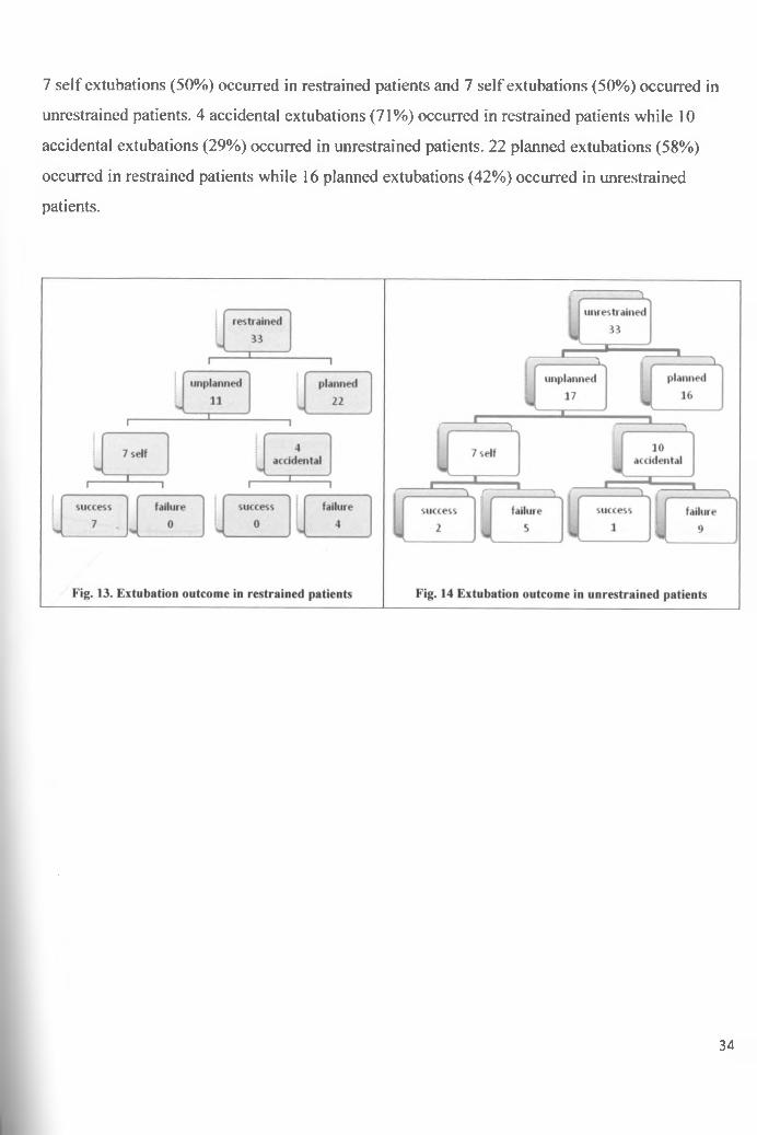
7 self extubations (50%) occurred in restrained patients and 7 self extubations (50%) occurred in
unrestrained patients. 4 accidental extubations (71%) occurred in restrained patients while 10
accidental extubations (29%) occurred in unrestrained patients. 22 planned extubations (58%)
occurred in restrained patients while 16 planned extubations (42%) occurred in unrestrained
patients.
34

6.13 SBT performance in predicting extubation outcome:
26 of the 30 successful planned extubations had successfully completed an SBT according to the
AARC 2007 clinical practice guidelines before extubation while 4 had not.
5 out of 8 unsuccessful planned extubations had had successfully completed an SBT before
extubation while 3 had not. 4 out of 9 successful self extubations had successfully completed an
SBT before extubation while 5 had not. None o f the unsuccessful self extubations had successfully
completed an SBT before extubation. The only successful accidental extubation had successfully
completed an SBT before extubation. 3 o f the 13 unsuccessful accidental extubations had
successfully completed an SBT before extubation while 10 had not.
Table 2: SBT performance in predicting extubation outcome.
Extubation
mode &
outcome
Planned
successful
Planned
unsuccessful
Successful
self
extubation
Unsuccessful
self
extubation
Successful
accidental
Unsuccessful
accidental
Successful Yes No Yes No Yes No Yes No Yes No Yes Nocompletion
of SBT 26 4 5 3 4 5 0 5 1 0 3 1

DISCUSSION :
The main aim of this research was to study the occurrence of both planned and unplanned
extubation events occurring at the KNH ICU and to determine important aspects associated with
each mode in the hope that a better understanding of extubation events will lead to changes geared
at improving the management of intubated patients.
Although the study participant’s ages ranged between 2 months and 93 years, the patient
population was largely a “youthful” population with a mean age of 31.6 vrs. (SD 23.6 years). 69%
of patients were aged 40vrs and below. These results are in stark contrast to findings in western
countries whereby older citizens have been found to consume a disproportionate share of critical
care resources.
This could mean that in our setting, patients younger than 40 years were preferentially admitted to
the ICU or that people aged 40 and below in the greater community were more exposed to factors
that result in greater severity of illness and the need for ICU admission.
THE INCIDENCE OF UNPLANNED EXTUBATION IN KNH ICU
The 18.5% incidence of unplanned extubation is within but on the higher end of the 0.87% - 25%
range reported in other studies.
The use of restraints and the ETT securing methods were examined in this study to determine their
influence on the high rate of unplanned extubation events experienced by patients in our ICU.
Use of restraints.
The study findings revealed that the KNH ICU staff did not routinely physically or
pharmacologically restrain intubated patients. Only 50% of extubated patients had been sedated or
physically restrained while in other studies majority of the patients had been restrained.
The results mirrored the findings first noted by Coppolo and May, in that, self extubations
occurred despite patients being restrained. 33% of restrained patients underwent unplanned
extubation while 51% of unrestrained patients underwent unplanned extubation.
All seven self extubations that occurred in restrained patients were successful while most instances
°fself extubation among unrestrained patients were much less successful.36

The frequency of accidental extubations was noted in this study to be almost 3 times higher in
unrestrained patients than in restrained patients. The reason for this is unclear.
ETT securing methods:
This study revealed that variable techniques (cloth ties, adhesive tape as well as a combination of
cloth ties and adhesive tape) are being employed to secure ETTs in the KNH ICU. Other studies
have shown that the used of variable techniques are a cause of high UE rate. The high UE rate in
the KNH ICU may therefore be attributed to this factor.
Fewer patients who had their ETTs secured with adhesive tape underwent unplanned extubation^
(53%) while the majority of patients who had their ETTs secured with cloth ties underwent
unplanned extubation (68%)
However, paediatric and surgical patients usually had their ETTs secured with adhesive tape while
medical and neurotrauma patients often had their ETTs secured by cloth ties. Earlier studies have
shown that surgical patients have significantly fewer UE than medical patients, (M)'and therefore
selection bias is a possible reason why why fewer unplanned extubations were noted in patients
with who’s ETTs were secured with adhesive tape.
THE SUCCESS RATE OF UNPLANNED EXTUBATION IN KNH ICU
As was the case in earlier studiest35’45,:>2), not all UE experienced by patients in this study were
unsuccessful extubations. 10 out of 28 UEs resulted in success indicating that extubation in these
10 patients should have been considered earlier by the ICU clinician.
Other studies have reported unplanned extubation success rates of between 44- 56%. The KNH
ICU experienced a lower rate of successful unplanned extubation.
9 out of 10 successful unplanned extubations were self-extubations while 1 was accidental. The
extremely low success rate (92%) of accidental extubations indicates that they should be avoided
at all costs.
37

THE INCIDENCE OF EXTUBATION FAILURE AFTER PLANNED EXTUBATION IN KNH
ICU
The overall incidence of extubation failure in the KNH ICU was 39%.
Specific patient groups were found to be especially high risk for extubation failure. These included
male gender (50% failure rate), paediatric medical (100% failure rate), neurotrauma (67% failure
rate), as well as patients who experienced an accidental extubation (93% failure rate).
The incidence of self and accidental extubations did not differ according to gender and thus did not
count as a reason for the apparent successful extubations in women. The singlemost important
factor that predisposed the male gender to higher risk of extubation failure was that male patients
comprised 93% of the extubated neurotrauma patients; a sub-specialty category who were noted to
be especially high risk for extubation failure in this study.
The incidence of failure after planned extubation in this study was 21%
This value in other studies varies from 2 - 25% depending on the population studied and time
period used to determine extubation failure.01’
This researcher did not come across a published ICU study into extubation events that managed as
wide a variety of patients as the KNH ICU did. It appears that in keeping with the wide spectrum
of patients requiring critical care, ICUs have become specialized centers which cater for specific
patient groups. Most ICUs dealt exclusively in adult or paediatric populations. Many were
specialized even further to cater to surgical or medical patients only. A few catered to an even
more exclusive group of patients eg. coronary, neurovascular or bum injury ICUs.
Because of this inter-ICU variability, comparisons of extubation failure are difficult to apply
considering the population studied heavily influences extubation outcomes; e.g. the exclusion of
paediatric patients in our study in order to reflect the results of an adult population would decrease
the planned extubation failure rate from 21% to 19% and the overall extubation failure rate from
' 39% to 30%. Considering adult surgical patients only would give a planned extubation failure rate
of 7% and an overall failure rate of 5% etc.
38

THE INCIDENCE OF EXTUBATION FAILURE AFTER PLANNED EXTUBATION IN KNH
ICU
The overall incidence of extubation failure in the KNH ICU was 39%.
Specific patient groups were found to be especially high risk for extubation failure. These included
male gender (50% failure rate), paediatric medical (100% failure rate), neurotrauma (67% failure
rate), as well as patients who experienced an accidental extubation (93% failure rate).
The incidence of self and accidental extubations did not differ according to gender and thus did not
count as a reason for the apparent successful extubations in women. The singlemost important
factor that predisposed the male gender to higher risk of extubation failure was that male patients
comprised 93% of the extubated neurotrauma patients; a sub-specialty category who were noted to
be especially high risk for extubation failure in this study.
The incidence of failure after planned extubation in this study was 21%
This value in other studies varies from 2 - 25% depending on the population studied and time
period used to determine extubation failure.151 ’
This researcher did not come across a published ICU study into extubation events that managed as
wide a variety of patients as the KNH ICU did. It appears that in keeping with the wide spectrum
of patients requiring critical care, ICUs have become specialized centers which cater for specific
patient groups. Most ICUs dealt exclusively in adult or paediatric populations. Many were
specialized even further to cater to surgical or medical patients only. A few catered to an even
more exclusive group of patients eg. coronary, neurovascular or bum injury ICUs.
Because of this inter-ICU variability, comparisons of extubation failure are difficult to apply
considering the population studied heavily influences extubation outcomes; e.g. the exclusion of
paediatric patients in our study in order to reflect the results of an adult population would decrease
the planned extubation failure rate from 21% to 19% and the overall extubation failure rate from
39% to 30%. Considering adult surgical patients only would give a planned extubation failure rate
of 7% and an overall failure rate of 5% etc.
38

In addition, not only were other ICUs specialized, they were also manned by critical care
physicians from a wide variety of specialty backgrounds including surgery, paediatrics and internal
medicine. This possibly renders them better equipped to handle target patient populations.
All critical care physicians currently managing the KNH ICU during the study period were
casually observed to have a background in anaesthesia. The degree of involvement of other
specialist physicians in the management of their respective ICU patients was not investigated in
this study but is possibly reflected in the extubation outcomes experienced by patients in the
various sub-specialty categories. The low extubation success rates experienced by paediatric,
general medical and neurotrauma patient populations leads to the conclusion that a change in their
management is essential and should be geared towards development and delivery of a more
specialized health care system. A greater degree of involvement of non-anaesthetist specialist
physicians in the KNH ICU should also be sought.
The relatively longer 72 hour period used in this study to define extubation failure could have been
a reason for a higher extubation failure rate. However, most extubation failures occurred within the
first 24 hours of extubation. Only 2 patients who had successfully completed 24 hours of
extubation were reintubated and none of the patients who had successfully completed 48hours of
extubation required reintubation. The study is therefore comparable to studies that used the 24 and
48 hour time frames.
ADVERSE EVENTS EXPERIENCED DURING EXTUBATION IN KNH ICU
The following adverse effects were experienced by patients within 30 minutes of extubation:
Desaturation, laboured breathing, stridor, hypotension, aspiration, bradycardia, hypoxia, cardiac
arrest. No patient was reported to have experienced hypercapnia and no mortality occurred.
21 extubated patients (32%) experienced one or more complications within 30 min of extubation.
18 of them (86%) were reintubated within 72hours of extubation.
45 extubated patients (32%) had no complications within 30 min of extubation. However 8 of them
(18%) were reintubated within 72hours of extubation.

THE ROLE OF SBT IN PREDICTING EXTUBATION SUCCESS IN KNH ICU
From section B item 12 of the surv ey tool it was possible to discern whether patients had
completed a spontaneous breathing trial (SBT) prior to extubation.
SBT completion correctly predicted successful extubation in 31 out of 40 incidences of successful
extubation. In all 9 instances when the absence of an SBT wrongly predicted extubation failure,
that failure can be attributed to iatrogenic error whereby the patient was unnecessarily receiving
substantial ventilatory support at the time of extubation.
The lack of successfully completed SBT correctly predicted extubation failure in 18 out of 26
cases of failed extubation. However successful SBT completion erroneously predicted extubation
success in 8 out of 26 cases. A further review of these 8 cases revealed that 5 of the patients had
been diagnosed as having suffered a severe head injury, while the remaining 3 patients had
respectively been diagnosed as having hypertensive stroke, upper airway obstruction and herbal
intoxication. ,
It thus appears that despite successful completion of an SBT, caution should be exercised while
assessing extubation readiness in patients with depressed neurological function as well as patients
with suspected upper airway obstruction as inability to maintain airway patency and not ventilatory
failure can also be a reason for failed extubation.
40

CONCLUSIONS:
The incidence of unplanned extubation in KNH ICU was 18.5%. This is within but on the higher
end of the range reported in other studies.
The success rate of unplanned extubations is 35.7%. Self extubations are more successful than
accidental extubations, but the general success rate of UE is lower than that of planned
extubations.
Specific patient groups are especially high risk for extubation failure.
High surveillance of extubated patients should be implemented during the first 30 minutes after an
extubation event.
An SBT is a tool used frequently but not routinely by KNH ICU clinicians.
41

tjj
RECOMMENDATIONS:
1. All intubated patients in the KNH ICU should be routinely pharmacologically or physically
restrained.
2. The ETT securing methods used in the KNH ICU should be standardised.
. All intubated patients should have a routine SBT assessment prior to extubation in KNH
ICU.
4. All patients who have completed a successful SBT should be extubated if airway patency
and the ability to protect the airway are not in doubt.
5. A greater degree of involvement of non-anaesthetist specialist physicians in the KNH ICU
should be pursued in order to provide specialized health care to patient groups at increased
risk of extubation failure.
42

REFERENCES
i. Maguire. GP. DeLorenzo. LJ. Moggio. RA. Unplanned extubation in the intensive cQre
unit: a quality-of-care concern. Crit Care Nurs Q 1994; 17.40-47
Richmond AL, Jarog DL, Hanson VM. Unplanned extubation in adult critical care: quality
improvement and education pay off. Crit Care Nurse. 2004;24( 1):32—37.
Coplin WM. Pierson DJ, Cooley K.D. Newell DW, Rubenfeld GD. Implications o f
extubation delay in brain-injured patients meeting standard weaning criteria. Am J Respjr
Crit Care Med. 2000; 161(5): 1530-1536.
4. Asai T. Koga K, Vaughn RS. Respiratory complications associated with tracheal
intubation and extubation. Br J Anaesthesia 1998:80:767-75
5. Peterson GN, Domino KB. Caplan RA, Posner KI, Lee LA, Cheney FW. Management of
the difficult airway, A closed claims analysis. Anaesthesiology 2005;103:33-9
6. Ron M. Walls, Michael F. Murphy. Manual o f Emergency Airway Management. Lippincott
Williams & Wilkins 2008;461:5
7. Grap MJ, Glass C, Lindamood MO. Factors related to unplanned extubation of
endotracheal tubes. Crit Care Nurse. April 1995; 15:57-65.
8. Atkins PM, Mion LC, Mendelson W, Palmer RM, Slomka J, Franko T. Characteristics and
outcomes o f patients who self-extubate from ventilatory support. Chest. 1997; 112; 1317-
1323.
9. Mion LC. Establishing alternatives to physical restraints in the acute care setting: a
conceptual framework to assist nurses' decision making. AACN Clin Issues. 199^;7:592—
602.
10. Krinsley JS, Barone JE. The drive to survive: unplanned extubation in the ICO. Chest.
2005; 128(2):560—566.
11. Aitkenhead AR, Smith G, Rowbotham DJ. Textbook o f anaesthesia. Churchill Livjngstone
Elsevier 2007;6: 93
12. Coplin WM. Pierson DJ, Cooley KD, Newell DW, Rubenfeld GD. Implications o f
extubation delay in brain-injured patients meeting standard weaning criteria. Am J Respir
Crit Care Med. 2000; 161 (5): 1530-1536.
• o 43

13. Holzapfel L. Chevret S. Madinier G, et al. Influence o f long-term oro- or nasotracheal
intubation on nosocomial maxillary sinusitis and pneumonia: results o f a prospective,
randomized, clinical trial. Crit Care Med 1993; 21:1132-8.
14. Holzapfel L, Chastang C, Demingeon G, et al.: A Randomized Study Assessing the
Systematic Search fo r Maxillary Sinusitis in I\as otr ache ally Mechanically Ventilated
Patients. Am. J. Respir. Crit. Care Med., March 1, 1999; 159(3): 695 - 701.
15. Colice GL. Stukel TA. Dain B. Laryngeal complications o f prolonged intubation. Chest
1989;96:877-884.
16. Stauffer, JL, Olson, DE, Petty. TL. Complications and consequences o f endotracheal
intubation and tracheotomy: a prospective study o f 150 critically ill adult patients. Am J
Med 1981 ;70.65-76
17. Sahn SA, Lakshminarayan S. Bedside criteria for discontinuation o f mechanical
ventilation. Chest 1973; 63:1002-1005
18. Tindol GA, DiBennetto RJ, Kosciuk L. Unplanned extubations. Chest 1994; 105:1804-
1807
19. Kollef V1H, Shapiro SD, Silver P, et al.: A randomized, controlled trial o f protocol-directed
versus physician-directed weaning from mechanical ventilation. Crit Care Med
1997;25:567-574
20. Ely EW, Baker AM, Dunagan DP et al. Effect on the duration o f mechanical ventilation o f
identifying patients capable o f breathing spontaneously. N Engl J Med 1996;335:1864-
1869
21. Meade M, Guyatt G, Cook D, et al.: Predicting success in weaning from mechanical
ventilation. Chest 2001, 120(suppl):400S-424S
22. MacIntyre NR, Cook DJ, Ely EW, Epstein SK, Fink JB, Heffner JE et al.: Evidence-based
guidelines for weaning and discontinuing ventilatory support. Chest 2001; 120(6
suppl);375S-395S
23. Kenvatta National Hospital - ICU/HDU Users Committee. KNH- ICU/HDU Protocols
Booklet. 2004;
24. American Association for Respiratory Care. AARC clinical practice guideline: 2007
revision & update: Removal o f the endotracheal tube. Respir Care 2007;52:81-93
44

25. St George Hospital, New South Wales, Australia, ICU nursing procedure manual:
Revised 2003
26. Coppolo. DP. May, JJ. Self extubations: A twelve month experience. Chest 1990; 98:165-
69
27. Kapadia FN. Bajan KB, Raje KV. Airway accidents in intubated intensive care unit
patients: an epidemiological study. Crit Care Med. 2000;28:659-664.
28. Christie JM. Dethlefson M, Cane RD. Unplanned endotracheal extubation in the intensive
care unit. J Clin Anesth. 1996;8: 289-293.
29. Chevron V. Menard JF, Richard JC, Girault C, Leroy J, Bonmarchand G. Unplanned
extubation: risk factors o f development and predictive criteria fo r reintubation. Crit Care
Med. 1998;26:1049-1053.
30. Maguire GP. DeLorenzo LJ, Moggio RA. Unplanned extubation in the intensive care unit:
a quality o f care concern. Crit Care Nurs Q. November 1994;17:40-47.
31. Richmond AL, Jarog DL, Hanson VM. Unplanned extubation in adult critical care: quality
improvement and education pay o ff Crit Care Nurse. 2004;24(l):32-37.
32. Farias. JA, Alia, I, Esteban, A, et al. Weaning from mechanical ventilation in pediatric
intensive care patients. Intensive Care Med 1998;24,1070-1075
33. Stauffer, J, Olson, D, Petty, T, Complications and consequences o f endotracheal
intubation: A prospective study o f 150 critically ill adult patients. Am J Med 1981 ;70:65-
76.
34. Medina M, Beydoun HK, Hsu WW, et al. Reducing unplanned extubations (UEX): the
benefit o f combined chest and arm restrains with sedation [J]. Chest, 1993,103(supply 3) :
273-275.
35. Epstein SK. Nevins ML, Chung J. Effect o f unplanned extubation on outcome o f
mechanical ventilation. Am J Respir Crit Care Med. 2000; 161 :1912 -1916
36. Betbese AJ, Perez M, Bak E, et al. A prospective study o f unplanned extubation intensive
care unit patients. Crit Care Med. 1998;26 :1180 —1 186
37. Epstein SK. Ciubotaru RL. Independent effects o f etiology o f failure and time to
reintubation on outcome for patients failing extubation. Am J Respir Crit Care Med 1998;
158:489-493
38. Epstein SK. Endotracheal extubation. Respir Care Clin N Am 2000; 6:321-360
45
39. Esteban A, Alia I, Tobin MJ. et al. Effect o f spontaneous breathing trial duration on
outcome o f attempts to discontinue mechanical ventilation: Spanish Lung Failure
Collaborative Group. Am J Respir Crit Care Med 1999; 159:512-518
40. Esteban A, Alia I, Gordo F, et al Extubation outcome after spontaneous breathing trials
with T-tube or pressure support ventilation: the Spanish Lung Failure Collaborative
Group. Am J Respir Crit Care Med 1997; 156:459-465
41. Epstein SK. Etiology o f extubation failure and the predictive value o f the rapid shallow
breathing index. Am J Respir Crit Care Vied. 1995:152:545-549.
42. Epstein SK, Ciubotaru RL, Wong JB: Effect o f failed extubation on the outcome o f
mechanical ventilation. Chest 1997, 112:186-192.
43. Rothaar RC, Epstein SK. Extubation failure: magnitude o f the problem. impact on
outcomes, and prevention. Curr Opin Crit Care. 2003;9:59-66.
44. Mort TC. Unplanned tracheal extubation outside the operating room: Anesth Analg.
1998:86:1171-1176
45. Krinsley JS, Barone JE. The drive to survive: unplanned extubation in the ICU. Chest.
2005; 128(2):560-566.
46. American Thoracic Society, European Respiratory Society, European Society of Intensive
Care Medicine, Societe de Reanimation de Langue Franc9aise. International Consensus
Conferences in Intensive Care Medicine: noninvasive positive pressure ventilation in acute
respiratory failure. Am J Respir Crit Care Med 2001;163:283-291
47. El Solh, A. A, Aquilina, A, Pineda, L, Dhanvantri, V, Grant, B, Bouquin, B. Noninvasive
ventilation for prevention o f post-extubation respiratory failure in obese patients. Eur
Respir J 2006; 28:588-595
46

48. Ferrer. M, Sellares, J, Vallencia, M, et al. Noninvasive Ventilation after extubation in
hypercapnic patients with chronic respiratory disorders: A Randomized Trial. Lance*
374: 1082.
49. Masip J, Betbese AJ, Paez J, Vecilla F, Caiiizares R. Padro J, Paz MA, de Otero J, B a^us
Non-invasive pressure support ventilation versus conventional oxygen therapy in acutd
cardiogenic pulmonary oedema: a randomised trial. Lancet 2000;356:2126-2132.
50. Moons P. Sels K, De Becker W. De Geest S. Ferdinande P. Development o f a risk
assessment tool for deliberate self-extubation in intensive care patients. Intensive Cartf
Med. 2004 Jul;30(7): 1348-55. Epub 2004 Mar 25.
51. Edmunds S. Weiss I, Harrison R: Extubation failure in a large pediatric ICC
population. Chest 2001; 119:897-900
52. Curry K, Cobb S, Kutash M, Diggs C. Characteristics Associated With Unplanned
Extubations in a Surgical Intensive Care Unit American Journal ot Critical Care. 200$^’^ '
45-51
47

APPENDIX la: GUARDIAN EXPLANATION FORM
Dear Sir/Madam,
I am a Senior House Officer undertaking a Masters of Medicine (M.Med.) degree in
Anaesthesiology at the University of Nairobi. I am currently conducting a study titled, “A
BASELINE STUDY OF EXTUBATION EVENTS OCCURRING AT THE INTENSIVE
CARE UNIT OF THE KENYATTA NATIONAL HOSPITAL.”
I hereby undertake to explain the nature of my study to you, the guardian(s) of an intubated patient
admitted in the Intensive Care Unit of the Kenvatta National Hospital, in the hope that you will
grant consent for your patient to be included in the study. I understand that this is a difficult time
and I take this opportunity to wish your patient a rapid recovery.
This study intends to observe and document:
1. When and how the endotracheal tube currently inserted into your patient’s airway will be
removed.
2. Whether or not he/she will need to have the endotracheal tube re-inserted after its removal.
3. Whether or not the patient or attending doctor or the patient experiences any problems
during removal or re-insertion of the endotracheal tube.
The study findings will be used to make recommendations geared at improving the management of
intubated patients in KNH ICU. This study does not involve any risks to your patient neither does
it entail the imposition, withholding or withdrawal of any treatment from him/her. Decisions
concerning the management of your patient will be left to the KNH ICU team, in consultation with
you. Your patient will not be denied treatment if, on his behalf, you decline to participate.
Participation in this study is purely voluntary and no financial benefit will be gained. Participation
will be terminated at any time you so wish. Strict confidentiality will be maintained. Feel free to
seek any clarification on matters pertaining to this survey. Thank you for your co-operation.
Dr. Winnie Wakiuru Mathangani (Tel: 0721 624 304)
48V

APPENDIX I b: PATIENT EXPLANATION FORM
Dear Sir/Madam,
I am a Senior House Officer undertaking a Masters of Medicine (M.Med.) degree in
Anaesthesiology at the University of Nairobi. I am currently conducting a study titled, “A
BASELINE STUDY OF EXTUBATION EVENTS OCCURRING AT THE INTENSIVE
CARE UNIT OF THE KENYATTA NATIONAL HOSPITAL.”
I hereby undertake to explain the nature of my study to you, a conscious intubated patient admitted
in the Intensive Care Unit of the Kenyatta National Hospital, in the hope that you will grant
consent to be included in the study. I underhand that this is a difficult time and I take this
opportunity to wish you a rapid recovery.
This studv intends to observe and document:
1. When and how the endotracheal tube currently inserted into your airway will be removed.
2. Whether or not you will need to have the endotracheal tube re-inserted after its removal.
3. Whether or not you or the attending doctor experiences any problems during removal or
re-insertion of the endotracheal tube.
The study findings will be used to make recommendations geared at improving the management of
intubated patients in KNH ICU. This study does not involve any risks to you neither does it entail
the imposition, withholding or withdrawal of any treatment from you. Decisions concerning your
management will be left to the KNH ICU team, in consultation with you. You will not be denied
treatment if you decline to participate. Participation in this study is purely voluntary and no
financial benefit will be gained. Participation will be terminated at any time you so wish. Strict
confidentiality will be maintained. Feel free to seek any clarification on matters pertaining to this
survey. Thank you for your co-operation.
Dr. Winnie Wakiuru Mathangani (Tel: 0721 624 304)
49

APPENDIX II: CONSENT FORM.
I ___________ [initials], hereby consent to be included/ for my patient to be included in the survey
on extubation events at the Kenyatta National Hospital’s Intensive Care Unit.
I confirm that I have been fully informed about the procedure, the confidentiality, and the
voluntary nature of participation in the study. I fully understand the right of withdrawal from the
study at anytime.
I hereby give my informed consent under no duress or coercion whatsoever.
Signature _________________________ Date________________
Principal investigator’s telephone number: 0721 624 304.
FOMU YA KIBALI CHA KUHUSISHWA
Mimi______________ [herufi za majina] nakubali/nakubalia mgonjwa wangu kuhusishwa katika
utafiti huu katika hospitali kuu ya Kenyatta.
Nadhibitisha ya kwamba nimeelezwa kuhusu utafiti huo na ninaelewa haki yangu ya kujiondoa
wakati wowote kutoko kwa utafiti. Ninatia sahihi hii bila ya kusulutishwa.
Sahihi ___________________________ Tarehe _________
Nambari ya simu ya mtafiti mkuu: 0721 624 304

A BASELINE STUDY OF EXTUBATION EVENTS OCCURRING AT KNH ICU.
APPENDIX III: SURVEY TOOL
TOOL NO:
SECTION A: To be filled in for all orally intubated patients.
l.NAME_________________ (imtiais/mmiber) 2. SEX: 1 ] Male I 1 Female
4. Place, Date and time of intubation:
3. AGE:
5. ETT securing method: d Adhesive tape d Cloth ties D n one EH Other
6. ICU Admission date & time 7. Admission diagnosis:
8. End point of intubation: 1 1 Extubation 1 1 Tracheostomy 1 1 Death while intubated.
SECTION B : To be filled in only if the above patient experiences an extubation event.
9. Place, Date and time of extubation:
10. Type of extubation: □ Planned I 1 Accidental I 1 Self - extubation
11. Tick any complications experienced within 30 minutes of extubation:
QNone I I Desaturation I [Laboured breathing EH Stridor I I Hypotension I I Aspiration
QBradycardia I I Hypoxia I iHvpercapnea I ICardiac arrest I I Death I I Other ..................
12. Ventilatory mode prior to extubation:
I- - 1 ippv I- - 1 giiuv EEH niPAP 1 1 ppap 1. —1SIMV
Level of: A) Pressure support
BIPAP
B) PEEP
CPAP
C) Fi02
T-PIECE
% or 0 2 flow lit/min
13. Was the above ventilatory mode maintained for > 30 min prior to extubation? ̂ ̂ Yes ̂ ̂ No
14. Use of physical restraints: EEEH Wrist/Hand I---- 1 Leg EEH other EEH None
15. Administration of sedative drugs 1 hr prior to extubation: H Yes I----- ^No
16. Administration of muscle paralysis 1 hr prior to extubation: dH Yes Ed No
v 51
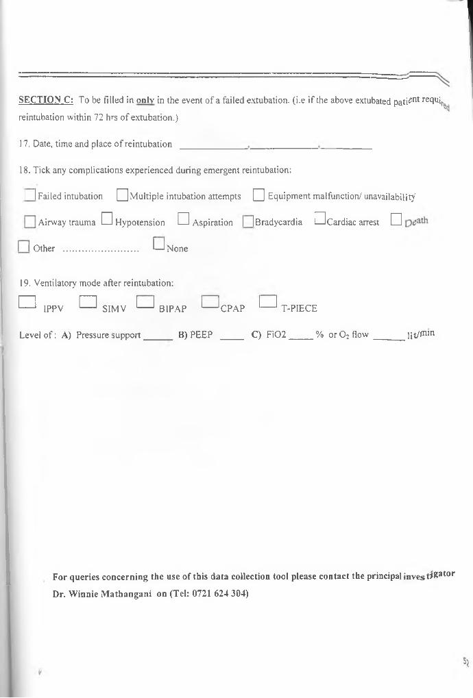
SECTION C: To be filled in only in the event of a failed extubation. (i.e if the above extubated pati^nt recNi,.
reintubation within 72 hrs of extubation.)
17. Date, time and place of reintubation
18. Tick any complications experienced during emergent reintubation:
I | Failed intubation Q Multiple intubation attempts Q Equipment malfunction/ unavailability
Airway trauma D Hypotension dH Aspiration | [Bradycardia HHCardiac arrest Ed
I I Other .............................. t—l None
19. Ventilatory mode after reintubation:
n IPPV n SIMV 1— 1 BIPAP 1— 1 CPAP 1— 1 T-PIECE
Level o f: A) Pressure support_____ B) PEEP ____ C) Fi02____ % or 0 2 flow ______ lit/*™11
For queries concerning the use of this data collection tool please contact the principal invest*Sator
Dr. Winnie Mathangani on (Tel: 0721 624 304)

APPENDIX IV: BUDGET
ITEM UNIT COST
KSh
NUMBER OF
UNITS
TOTAL COST
KSh
Computer 40000 1 40000
Printer/copier 5000 1 5000
Paper 400 4 1600
Internet hours 60 10 600
Statistician 5000 1 5000
Document binding 100 8 800
Sub total 53000
Contingency @ 5% of sub 2650
total
Grand Total 55650
53

APPENDIX V: W O RK PLAN :
ACTIVITY
2009
July
2009
Sept
2009
Oct
2009
Nov
2009
Dec
2010
Jan
2010
Feb
2010
Mar
2010
Apr
2010
May
Proposal
Writing
V v V
Presentation
to Ethical
Review
Committee
V v
Data
Collection
V
Data
Processing
V
Report
Writing
V
Study
Presentationv
54

KENYATTA NATIONAL HOSPITALHospital Rd. along, Ngong Rd.
P.O. Box 20723, Nairobi. Tel: 726300-9
Fax: 725272 Telegrams: MEDSUP", Nairobi.
Email: [email protected]
Ref: KNH-ERC/ A/428 11* March 2010
Dr. Winnie Wakiuru Mathangani Dept, of Surgery/Anaesthesia School of Medicine University of Nairobi
Dear Dr. Mathangani
RESEARCH PROPOSAL: “A BASELINE STUDY OF EXTUBATION EVENTS OCCURING AT THE INTENSIVE CARE UNIT OF KENYATTA NATIONAL HOSPITAL1*________________ (P352/12/2009)
This is to inform you that the KNH/UON-Ethics & Research Committee has reviewed and approved your above revised research proposal for the period 11* March 2010 10* March 2011.
You will be required to request for a renewal of the approval if you intend to continue with the study beyond the deadline given. Clearance for export of biological specimens must also be obtained from KNH/UON-Ethics & Research Committee for each batch.
On behalf of the Committee, I wish you a fruitful research and look forward to receiving a summary of the research findings upon completion of the study.
This information will form part of the data base that will be consulted in future when processing related research study so as to minimize chances of study duplication.
Yours sincerely
DR. L. W. MUCHIRIAG. SECRETARY, KNH/UON-ERC
c.c. Prof. K. M. Bhatt, Chairperson, KNH/UON-ERC The Deputy Director CS, KNH The Dean, School of Medicine, UON The Chairman, Dept.of Surgery, UON The HOD, Records, KNHSupervisor Dr. Mark Gacii, Dept.of Surgery/Anaesthesia, UON