a biomechanical evaluation of standing in high-...
TRANSCRIPT

25
25
A Biomechanical Evaluation of Standing in High- Heeled Shoes
Paula D. Henderson, McNair Scholar, Penn State
Dr. Stephen J. PiazzaDepartments of Kinesiology, Mechanical Engineering, Bioengineering, and
Orthopedics and Rehabilitation Penn State
ABSTRACT
The purpose of this study was to determine the action of the ground reaction force uponthe heels of women standing in high-heeled shoes. The study involved a non-invasivedetermination of the location of the subtalar joint axis, the joint about which the footbends in and out. It was determined where the ground reaction force acts relative to thesubtalar joint axis and whether muscle activity while standing depends on this result. Fivehealthy female volunteers will stand while wearing 2.5” heels, 1” heels, and barefootwhile ground reaction forces and electrical muscle activity were recorded.
INTRODUCTION
Historical Perspective
Stilettos. Boots. Pumps. “Open toed.” Around the age of twelve, girls all overembarked on their first pair of high-heels. Historically, the first form of high heels as weknow started during the 14th century. Gentry and noble men started placing woodenslips, called platens, to the bottom of their footwear to protect them from getting dirty.(Linder and Saltzman, 1998) However, in later days they have become a characteristic offemininity and an accepted custom for women in our society. Through the evolution oftime, several features have made high heels distinguishable in comparison to other shoesworn by women. On the average, shoes have a heel elevation of approximately 1 to 2cm; however, high-heeled shoes can have a heel height greater than 10 cm high. High-heeled shoes also pose a narrow toe box, a rigid heel cap that often protrudes anteriorly(Stephens, 1992) and excessive plantar curvature in the forefoot (Schwartz and Heath,1959). Whether it is to gain a height advantage, look professional, or stay with the trendof fashion, it is not entirely uncommon for a woman to own a hundred pairs of theseshoes at one time. Wearing such footwear can often have deleterious and irreversiblebiomechanical effects. (Linder and Saltzman, 1998) “For 250 years medical scientists

26
26
have propagandized about the health hazard of high heeled shoes, …”. (Linder andSaltzman, 1998)
Despite the uncomfortable feelings that some women experience while wearinghigh heeled shoes, heels are getting higher, inclines are steeper, and toe boxes are morepointed. (Lee et al., 2001) To achieve “toe cleavage” (toes that are perfectly aligned inpointed toed shoes), as it is known to the fashion-conscious world, more than half themembers of the America Orthopedic Foot & Ankle Society are responding to womenrisking permanent disability of cosmetic foot surgery, such as shorten toes, at a cost of$2,300 per toe and collagen injection into the balls of the feet. The collagen serves torestore lost padding caused by frequent high heel usage and costs approximately $500 perinjection. (Harris, 2003) While ample research is done on the biomechanics of high-heeled shoes, research has yet to look at the anatomical differences and biomechanicsprinciples of the subtalar joint axis in relation to its’ effect of standing in high heel shoes.The study will seek to answer the following questions:
1. How does the ground reaction force act upon the heels of women whowear heel-heeled shoes?
2. How does this action vary across subjects in accordance to the location oftheir subtalar joint axis?
3. How does muscle activity patterns correspond to the action of the groundreaction force?
Significance of the study
The data collected in this project is of relevance because the subjects- women area large segment of the world’s population. A census brief entitled “Women in the UnitedStates: A Profile”, projected women to outnumber men by 10 million by the year 2005.(Spraggins, 2005) A 1986 Gallup Organization survey determined that 59% of thewomen surveyed associated wearing high heel shoes for at least one hour to more thaneight hours a day. (The Gallup Organization Inc., 1986) This research can also help assistto educate women as to the effects of standing in high-heeled shoes and may positivelyinfluence the design of high-heeled shoes, which could lead towards a more comfortableand anatomically correct shoe. In addition, the study may clear up assumptions that allwomen should avoid wearing high-heeled shoes and formulate a link to the anatomicalvariations of women and effects of high-heeled shoes.
RELATED LITERATURE
Literature was reviewed in the following topic areas: subtalar joint axis, groundreaction forces, and muscular activity.

27
27
Subtalar Joint Axis
The subtalar joint (figure 1) is a composite joint formed by three separate planearticulations located superiorly to the talus and inferiorly to the calcaneus. Together, thethree surfaces provide a triplanar movement around the single joint axis and one of thefunctional joints of the foot and ankle.
Figure 1. Subtalar joint axis
Often reported as a single axis, a helical screw axis and a bundle of axes, thesubtalar joint is responsible for several movements about the ankle; inversion andeversion in the transverse plane; plantar flexion and dorsiflexion in the sagittal plane; andadduction and abduction in the frontal plane. “Subtalar inversion helps to bring aboutstability of the foot during single-limb stance.”(Backus and Sherry, 1999) Subtalareversion occurs as a mechanism of shock absorption when the foot makes contact with asurface. Five muscles help control inversion of the subtalar joint axis and cross themedial side of the joint: tibialis posterior, tibialis anterior, flexor digitorum longus, flexorhallucis, and soleus. Four muscles are responsible for eversion of the subtalar joint axis:extensor digitorum longus, peroneus tertius, peroneus longus, and peroneus brevis (figure2).
Figure 2. Muscles that cross the subtalar joint axis.
It is the motion at this joint that permits the foot to adapt to a variety of surfaces(Wright et al., 1964). “In addition there is substantial variability in the orientation of thisaxis in normal individuals; thus the relative motions will also vary among normalindividuals.” (Backus and Sherry, 1999) The subtalar joint axis has a wide acceptedrange of deviation and inclination. The range of deviation is 4 degrees to 47 degrees with

28
28
a standard deviation of 11 degrees and a mean deviation of 23 degrees. Subtalarinclination ranges from 20.5 degrees to 68.5 degrees with a standard deviation of 9degrees and mean range being 42 degrees. (Isman and Inman, 1969)
Ground Reaction Forces
Ground reaction force (GRF) is any external reaction force, specifically oneapplied by the ground. Ground reaction force is equal in magnitude and opposite indirection to the force that the body exerts on the supporting surface through the foot.GRFs can be represented by Newton 3rd law of motion, action reaction pair.
Several studies have viewed and reported increased pressure under the forefootwith an increase in heel heights in women wearing high-heeled shoes. However fewstudies have measured GRFs. In a study done by Opila-Correla, K.A. (1990), nosignificant differences in GRFs were found between younger and older wearers orbetween experienced and inexperienced high heeled shoe wearers. Another study,measured vertical, anteroposterior and mediolateral direction of GRFs of a women’s gaitin three different heel heights. The study showed an increase in vertical, anteroposteriorand mediolateral ground reaction forces with increased heel height. The highest heelheight of 7.62 cm showed a pronounced inflection point compared to those of the lowerheels and medium heels tested for vetrical GRFs. Anteroposterior and mediolateral GRFappeared later in the stance and support phases for the higher heel height, but did show asignificant increase.
Muscular Activity
In high heel gait and standing, many muscles located in the lower extremities andthe back are overly worked due to the plantar flexion of the foot. Muscles are at theirpeak for force generation when they are at resting length. When muscle length increasesor decreases beyond its resting length, muscle force production decreases in a bell shapedform. This relationship is seen in high heel wearers. When the heel is raised, as inwearing high-heeled shoes, muscles fibers that innervate the muscles along the leg areshorten. The shortened muscles are now inconsistent with its resting length-tensionrelation resulting in less force production. Esenyel et al. (2001), found “… theexaggerated plantar flexed position of the ankle joint places the gastro-soleus muscle at ashortened and thus less favorable position on its muscle length-tension curve. Undersuch conditions, the plantar flexion musculature is in a less advantageous position forpower and work generation and consequently less propulsive abilities. (Esenyel et al.,2001)

29
29
METHODOLOGY
Five female subjects between the ages of 18-24 years of age were recruited fromthe local Pennsylvania State University community for this study. Their age, height,weight and shoe size were recorded. The subjects reported not having anymusculoskeletal or neuromuscular abnormalities that restrict the range of lower extremitymotion, which might make the wearing of high-heeled shoes painful. All subjects wereexperienced wearers of high-heeled shoes as evidenced by self-reported wearing usage ofat least twice per week. These criteria were established to control variation amongsubjects in their motor control that could result in differences in habitual versus sporadichigh-heeled shoe wearers. All methods were in accordance with the guidelines set forthby the Human Subjects Review Board of The Pennsylvania State University, UniversityPark, Pennsylvania.
Two different shoes (figure 3) were used in this investigation. The first shoe, aflat open toed shoe, had an average heel height of 1 cm and a stacked block heel. Thesecond shoe was a high-heeled open toed shoe with an average heel height of 2.5 cm witha stiletto-spiked heel. Both shoes were commercially available and purchased through aPayless Shoe manufacturer. The shoes were primarily chosen due to their similarity ofconstruction at the forefoot and their access to the calcaneus so that the main differencebetween shoe models was the height and type of heel. Shoe sizes ranged from 7, 7.5, 8,8.5,to 9.
Figure 3 Shoes used in the study. Shoe to the left,the flat, measures with a 1” heel. Shoe on theright, the high heel, measures a heel height of2.5”.
Kinetic data of ground reaction force as well as the point of application of thatforce was collected with a Kistler Instrument. Corporation force plate mounted flush tothe floor. Kinematic data was collected simultaneously with the kinetic data using aVideo-based motion analysis system. An Eagle system (Motion Analysis Corporation.)that consisted of six video cameras tracked the locations of spherical reflective markers inthree dimensions. Electromyographical activity was collected using a telemetered EMGsystem (Noraxon Corporation.). Recording electrodes were connected to a batterypowered transmitting unit worn on the subject’s belt.
Determination of the subtalar joint axis was the first procedure that the subjectundertook. Participants were asked to sit at the edge of a table to perform a non-invasivetechnique for location of the subtalar joint. The applications of eight, 9-mm-diameterreflective markers, using double sided tape were applied to the skin overlying theanterolateral aspect of the tibia and the lateral aspect of the calcaneus of the right leg oneach subject. Four markers each were applied to the anterolateral tibia and the lateralaspest of the calacaneus in an asymmetrically box fashion. This was done to help increase

30
30
the tracking of the cameras. Quiet standing of three seconds was done to record thelocation of the reflective markers to serve as a template for later tracking the location ofthe subtalar joint axis. Palpitation of six anatomical landmarks: the right lateral malleolus,right medial malleolus, right lateral tibial condyle, right medial tibial condyle, right footheel, and the right second metatarsal head were done.
An additional 9-mm-diameter reflective markers were placed on these locations.Inaddition, markers were placed to help computerize the structure of the shank and foot.Three reflective markers were placed on the ground to help digitize the plane coordinatesof the foot. Subjects were asked to stand for three seconds of quiet standing while thecameras recorded the location of the 17 markers (14 markers on the body and three on theground). Participants then had the additional markers from the right lateral malleolus,right medial malleolus, right lateral tibial condyle, right medial tibial condyle, right footheel, right second metatarsal head removed and the three placed on the ground to leavethe remaining eight markers placed on the shank and calcaneus.
Participants were affixed to a Marionette System (figure 4) on the table so thatfeet were dangling. The participant’s thigh was stabilized with a velcro strap and the footwas put into dorsiflexion by pulling up on the Marionette System as the forefoot ishanging from a platform by a velcro strap. Alternately pulling on the strings tension toproduce a slow full side-to-side rocking motion of the foot was done to provide full usageof the subtalar joint axis. The motions of the eight markers were tracked using the Eaglesystem (Motion Analysis Corporation.) video-based motion analysis system. Ten 30-second trails were recorded for each to ensure accuracy.
Figure 4. Non-invasive subtalar joint axis determination.The foot is moved about the subtalar axis and three-dimensional motion of the heel and shank is recorded.
Electromyographical activity of the tibialis anterior, peroneals, lateralgastrocnemius and medial gastrocnemius muscles were collected using surfaceelectrodes. Skin preparation consisted of treating appropriate areas with alcohol swabs toensure dead skin removal and increase EMG activity readings. Self-adhesive circularAg/AgCl electrodes of 1-cm diameter were placed approximately 2 cm apart in themiddle of the muscle bellies in an attempt to minimize cross talk and remain in the sameplacement for data collection of all shoes.

31
31
For each subject the order of the shoe condition was randomly assigned and threestanding trails for five seconds each were collected for each condition. Subjects stoodstill in a normal relaxed position on a wooden platform two inches above the floor.(figure 5) A cutout in the platform contained a wooden block that rested atop the forceplace set in the floor in order to collect heel ground reaction force. The platform itselfwas supported by the floor and not the force plate. Subjects placed just the heel of theright shoe atop the wooden block.
Figure 5. Subject standing in high heels on platform over force plate
Lastly participants performed trails to elicit EMG percent maximal. Two trails foreach muscle will be performed for five seconds each. For tibialis anterior subjects wereasked to first walk on the outer foot borders for five seconds and then invert againstmanual resistance applied by the investigator. For the peroneal muscles, participantswere asked to walk on the inner border of the foot for one trail and then evert againstmanual resistance applied by the investigator for the last trail. The participant was thenasked to push against a wall using maximum force and then perform a resistant toe risefor trails to elicit percent maximum of EMG activity in the gastrocnemius muscle.
A two-way repeated measure, ANOVA, using Sigma Stat software was used tocompare the biomechanical variables (kinematic, kinetic and EMG) between shoeconditions. When a significant difference was found, a Tukey post hoc analysis wasperformed. The level of significance was chosen as p < 0.05.
RESULTS
A one-way analysis of variance test (ANOVA) with repeated measures wasperformed to determine the effect of shoe condition on subtalar joint moment and activityin the tibialis anterior, peroneals, lateral gastrocnemius and medial gastrocnemiusmuscles for each subject.
Mean and standard deviation values of activity of each muscle considered for theshoe conditions of bare, flats and high heels are shown in table 1. There were nosignificant differences (p < 0.050) shown for muscle EMG for the tibialis anterior (p =0.883), peroneals (p = 0.077), lateral gastrocnemius (p = 0.093) or medial gastrocnemius(p = 0.330).

32
32
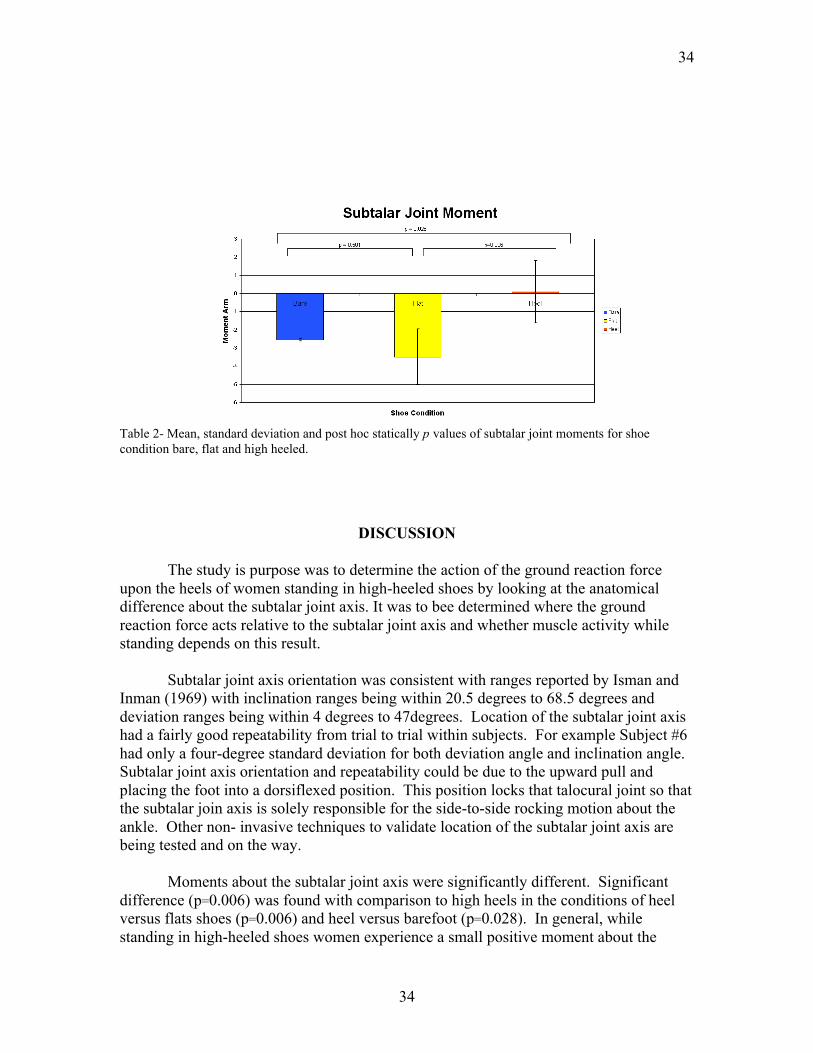
ANOVA testing did reveal significant differences in subtalar joint momentbetween treatments (p = 0.006). Post-hoc mean comparisons (Tukey) were calculated forthe factor of each shoe condition heel versus flat, heel versus bare and bare versus flat(table.2). Comparison showed the high-heeled condition to be different from both thebare and flat conditions with a significant difference of heel versus flat (p = .0.006) andheel versus bare (p = 0.028). No significant difference in mean value for subtalar jointmoment was found for bare versus flat condition (0.501).

33
33
Table.1 - Mean and standard deviation of EMG activity of the tibialis anterior, peroneals, medialgastrocnemius and lateral gastrocnemius for each shoe condition.
34
34
Table 2- Mean, standard deviation and post hoc statically p values of subtalar joint moments for shoecondition bare, flat and high heeled.
DISCUSSION
The study is purpose was to determine the action of the ground reaction forceupon the heels of women standing in high-heeled shoes by looking at the anatomicaldifference about the subtalar joint axis. It was to bee determined where the groundreaction force acts relative to the subtalar joint axis and whether muscle activity whilestanding depends on this result.
Subtalar joint axis orientation was consistent with ranges reported by Isman andInman (1969) with inclination ranges being within 20.5 degrees to 68.5 degrees anddeviation ranges being within 4 degrees to 47degrees. Location of the subtalar joint axishad a fairly good repeatability from trial to trial within subjects. For example Subject #6had only a four-degree standard deviation for both deviation angle and inclination angle.Subtalar joint axis orientation and repeatability could be due to the upward pull andplacing the foot into a dorsiflexed position. This position locks that talocural joint so thatthe subtalar join axis is solely responsible for the side-to-side rocking motion about theankle. Other non- invasive techniques to validate location of the subtalar joint axis arebeing tested and on the way.
Moments about the subtalar joint axis were significantly different. Significantdifference (p=0.006) was found with comparison to high heels in the conditions of heelversus flats shoes (p=0.006) and heel versus barefoot (p=0.028). In general, whilestanding in high-heeled shoes women experience a small positive moment about the

35
35
subtalar joint axis at the ankle. This positive moment causes an inverting torqueexperienced by women standing in high-heeled shoes. During barefoot and flat shoeconditions, a negative moment was seen resulting in a larger everting torque about theankle.
All differences in EMG activity for the tibialis anterior, peroneals, lateralgastrocnemius and medial gastrocnemius were insignificant. However, two trends wereseen in muscle activity by women while standing in high-heeled shoes. Similar to otherstudies, the first trend was that there was more muscle EMG activity of the tibialisanterior peroneals lateral gastrocnemius and medial gastrocnemius with increasing heelheight. More specifically, a second trend was seen toward more activity in the high-heeled shoe condition for the peroneals and lateral gastrocnemius muscles. Both musclesshowed close to significant results with the peroneals having p= 0.007 and lateralgastrocnemius having a p= 0.093.
Both the lateral gastrocnemius and peroneals are muscles that help eversion of theheel. The increase of activity of these muscles could have been a compensating responsefor the more positive- inverting moments of the ground reaction force while wearinghigh-heeled shoes. An increase in peroneal activity is similar to the data presented byStefanyshyn et al., (2000). The increased activity of the peroneal muscle could be due tothe controlling of the increased plantarflexion of the foot when standing in high heels.Another response to the increased activity of the peroneal is its role of protection to thefoot from sudden inversion about the ankle and a stabilizer. The increased activity couldbe a required response, to stabilize the ankle joint when wearing high heels.
The response of the lateral gastrocnemius activity was similar to one other studyconducted by Gefen et al. (2002), which showed the two heads of the gastrocnemius(lateral head and medial head) to respond differently when wearing high-heeled shoes.Other studies agree with no significant differences in EMG activity of the gastrocnemius(Stefanyshyn et al., 2000; Ono, 1969). While other studies showed a progressivenegative linear relationship with increasing heel heights for EMG activity of thegastrocnemius (Lee et al., 1990). The explanation as to why a trend of activity of lateralgastrocnemius increased more in high heels versus that of the medial gastrocnemius isthat the lateral head may act more intensively to produce the forces required tocompensate the positive moment of the ground reaction forces. As seen in previous studyresultant forces generated by the lateral gastrocnemius are transferred down to theAchilles tendon to the calcaneus. Since the foot is plantar flexed, the Achilles tendon isslackened and the lateral gastrocnemius helps to take up this slack, which could result inan inverting moment that acted to incline the foot’s skeleton laterally. (Gefen et al., 2002)
EMG activity of the tibialis anterior was contradictory with results of some otherstudies. Studies conducted by Joseph (1968) and Lee et al. (1990) showed a decrease inEMG activity while standing in high heeled shoes. The tibialis anterior helps withdorsiflexion of the foot and acts as a stabilizer of the ankle. This study concluded whilenot significant a trend of increase muscle activity was seen in high heels for all muscles.Since the subjects were experienced high heel wearers they may no longer experience

36
36
feelings of instability when wearing high heels. The increased EMG activity by theperoneals and lateral gastrocnemius seen to compensate for the inverting moment ofstanding in high heels could possibly not be seen with the tibialis anterior due to islocation on the anterior aspect of the shank.
Limitations
Several limitations could have lead to the results of this study. All five subjectswere college-aged women from the State College, Pennsylvania area. Subjects were alsoexcluded if they did not wear a shoe size from 7, 7.5, 8, 8.5, to 9 comfortably since thesewere the shoes sizes provided by the researcher. Since the shoes were provided for thesubject, new and unfamiliar shoes could have caused a limitation to the study. WhileEMG activities for all four muscles were insignificant a greater sample size could resultin more significant results. Occasional burst of EMG amplitude potential from muscle ofsome subject while standing in high heels due to instability of posture was seen.Basmajian and Bentzon (1954) also found similar burst of activity in musclesaccompanied by women wearing high heels. Placement of Eagle camera could also causea limitation to this study. Eagle cameras were having difficulty tracking the motion ofthe marker located on the anteromedial aspect of the tibia and the lateral aspect of thecalcaneus. Closer and more precise location of the camera could result in better accuracyof the subtalar joint axis location.
Future Research
Future research will include refining this study to test a greater sample sizes andto redefine the non-invasive method of the subtalar joint axis location to gather moreprécis data for analayzation. Other future plans are to test shoes with greater heel heightthan 2.5 cm to look at their effects and to look at the results of ground reaction forcesabout the subtalar joint axis in an individualized way from subject to subject. Futuregoals are to test a design of a biomechanical correct high heel shoe that accounts foranatomical differences of the joint axis.
Conclusions
Subtalar joint axis orientations were consistent with ranges reported by Isman andInman, (1969) and subtalar joint axis location had fairly good repeatability. Momentsabout the subtalar joint axis were significantly different between the shoe conditions heelversus flat shoes, and between heels versus barefoot. High-heel shoes generally had asmall positive inverting moment, but there was a larger everting moment in the barefootand flat conditions seen. All differences in EMG activity for the tibialis anterior,peroneals, lateral gastrocnemius and medial gastrocnemius were insignificant, but with atrend toward more activity in the high-heeled condition for muscles that evert the heel(peroneals and lateral gastrocnemius). These muscles may have been compensating forthe more inverting moments of the ground reaction force while wearing high-heeledshoes.

37
37
REFERENCES
Backus, J., Sherry, 1999. Disorders of the Heel, Rearfoot, and Ankle. 11-13. New York: Harcourt Brace & Company.
Basmajian, J.V., Bentzon, J.W., 1954. An electromyographic study of certain muscles ofthe leg and foot in the standing position. Surg. Gynec. Obster. 98, 662-666.
Esenyel, M., Gitter, A., Walden, G., 2001. Altered Work Distribution in The lower Extremity While Walking in High Heeled shoes. Journal of Biomechanics 29, 405-413.
Gefen, A., Megido-Ravid, M., Itzchak, Y., Arcan, M., 2002. Analysis of muscular fatigue and foot stability during high heeled gait. Gait and Posture 15, 56-63.
Harris, G., 2003, 3 December. “If Shoe Won’t Fit, Fix the Foot? Popular Surgery Raises Concern.”. The New York Times.
Isman,R.E., Inman,V.T., 1969. Anthropometric studies of the human foot and ankle. Bulletin of Prosthetics Research, 97-129
Joseph, J., 1968. Pattern of activity of some muscles in women walking on high heels. Annals of Physical Medicine 9, 295-292.
Lee, CM., Jeong, EH., Freivalds, A., 2001. Biomechanical effects of wearing high-heeled shoes. International Journal of Industrial Ergonomics 2, 321-326.
Lee, K.H., Matteliano, A., Medige, J., Smiehorowski, T., 1990. Electromyographic Changes of Leg Muscles with Heel Lifts in Women: Therapeutic Implications. Archives of Physical Medicine and Rehabilitation 71, 31-33.
Linder, M., Saltzman, C.L., 1998. A history of medical scientists on high heels. International Journal of Health Service 28 (2), 201-225.
Ono, H., 1969. Heel Height and Muscle Activity. Journal of Japanese OrthopedicAssociation 43, 527-547.
Opila-Correia, K.A., 1990. Kinematics of high heeled gait. Archives of Physical Medicine and Rehabilitation 71, 304-309.
Schwartz, R.P., Heath, A.L., 1959. A qualitative analysis of recorder cariable in the walking pattern of normal adults. Journal of Bone and Joint Surgery 41, 1065.
Spraggins, R.E., 2000. A Census Brief entitled Women in the United States: A Profile. Currrent Population Reports.

38
38
Stefanyshyn, D.J., Nigg, B.M., Fisher, V., O’Flynn, B., Liu, W., 2000. The Influence of High Heeled Shoes on Kinematics, Kinetics and Muscle EMG of Normal Female Gait. Journal of Applied Biomechanics 16, 309-319.
Stephens, M.M., 1992. Heel Pain- Shoes, Exertion, and Haglunds Deformity. Physician Sportsmedicine 20 (4).
The Gallup Organization, Incorporation, 1986. “Women’s Attitude on Usage of High Heel Shoes.”.
Wright D.G., Desai S.M., Henderson W.H., 1964. Action of the subtalar and ankle-joint complex during stance phase of walking. Journal of Bone Joint Surgery AM. 46, 361-366.