a case of an infant with bundle branch reentrant ventricular tachycardia
TRANSCRIPT
Journal of Arrhythmia 28 (2012) 117–121
Contents lists available at SciVerse ScienceDirect
Journal of Arrhythmia
1880-42
http://d
n Corr
E-m
journal homepage: www.elsevier.com/locate/joa
Case Report
A case of an infant with bundle branch reentrant ventricular tachycardia
Satoki Fujii, MDa,n, Hiroshi Tasaka, MDa, Yoji Okamoto, MDa, Katsuyuki Takahashi, MTa,Kazushige Kadota, MDa, Kazuaki Mitsudo, MDa, Kenji Waki, MDb, Yoshio Aragaki, MDb
a Department of Cardiology, Kurashiki Central Hospital, 1-1-1 Miwa, Kurashiki 710–8602, Japanb Department of Pediatrics, Kurashiki Central Hospital, Kurashiki, Japan
a r t i c l e i n f o
Article history:
Received 10 April 2010
Received in revised form
17 October 2011
Accepted 15 November 2011Available online 7 April 2012
Keywords:
Bundle branch reentrant ventricular
tachycardia
Catheter ablation
Right bundle branch block
Acute myocarditis
76/$ - see front matter & 2012 Japanese Hea
x.doi.org/10.1016/j.joa.2012.03.010
esponding author. Tel.: þ81 86 422 0210; fax
ail address: [email protected] (S. Fujii).
a b s t r a c t
Catheter ablation of the right bundle branch was performed on an 11-month-old infant for the
treatment of drug-resistant bundle branch reentrant ventricular tachycardia. The occurrence of right
bundle branch block could not be used as an endpoint of treatment because the patient had presented
with incomplete right bundle branch block pattern during sinus rhythm. We performed ablation of the
right bundle branch and utilized changes of duration and morphology of the QRS complex as indicators.
Eight years have passed with no development of any atrioventricular block or tachycardia episode.
& 2012 Japanese Heart Rhythm Society. Published by Elsevier B.V. All rights reserved.
1. Introduction
Bundle branch reentrant ventricular tachycardia occurs inapproximately 5% of ventricular tachycardia [1], and there arerare case reports of patients without underlying heart disease. Inmost cases, it is accompanied by underlying heart disease such asdilated cardiomyopathy or old myocardial infarction [2]. Ventri-cular tachycardia with a left bundle branch block utilizing the leftbundle branch as the retrograde limb and the right bundle branchas the antegrade limb is common, while that with the rightbundle branch block utilizing the right bundle branch as theretrograde limb and the left bundle branch as the antegrade limboccurs only occasionally. In recent years, patients presenting withdrug-resistant arrhythmias can be completely cured using cathe-ter ablation. In our case, we performed catheter ablation of theright bundle branch for an 11-month-old infant who had drug-resistant bundle branch reentrant ventricular tachycardia. Theefficacy of catheter ablation was assessed based on the changes ofduration and morphology of the QRS complex. No recurrence ofventricular tachycardia has been observed during 8 years sincethe procedure was performed. To the extent of our knowledge, noreport has been published regarding infant cases. We herebypresent our report and discussion.
rt Rhythm Society. Published by E
: þ81 86 422 9351.
2. Case report
The patient was an 11-month-old boy. No abnormalities wereobserved throughout the embryonic stage and perinatal period.When he was 11 months and 10 day old, he suddenly developedpallor and poor responsiveness, with his eyes rolled back in hishead, which continued for approximately 1 min. He later devel-oped a fever above 38 1C and was brought to a nearby clinic,continually exhibiting mild clouding of consciousness. When hewas brought to our emergency center on the same day, wesuspected febrile seizure and performed an electroencephalogram(EEG). The results were normal, and he was discharged. Cardiacarrhythmia was detected the following day when he visited theoutpatient unit of the pediatric department of our hospital, withthe electrocardiogram (ECG) showing a wide QRS complex tachy-cardia. The patient was hospitalized for observation and furtherexamination.
Physical findings on admission showed that height was 78 cm,body weight was 9745 g, and blood pressure (during sinusrhythm) was106/60 mmHg, with no other abnormality.
Laboratory findings were as follows: CRP: 0.3 mg/dl; CPK: 52 IU/l;CPK-MB: 14.0 IU/l; TP: 6.7 g/dl; Alb: 4.4 g/dl; GOT: 62 IU/l; GPT: 24IU/l; LDH: 379 IU/l; ALP: 399 IU/l; Cr: 0.24 mg/dl; BUN: 7 mg/dl; Na:138 mEq/l, K: 4.9 mEq/l; Cl: 103 mEq/l; Troponin-T: (þ); BNP:767.0 pg/ml; HANP: 330.0 pg/dl; RBC: 6.02�106; Hct: 42.1; Hgb:12.4; WBC: 14,600 (Segs: 33.0%; Bands: 1.0%; Lymph: 63.0%).
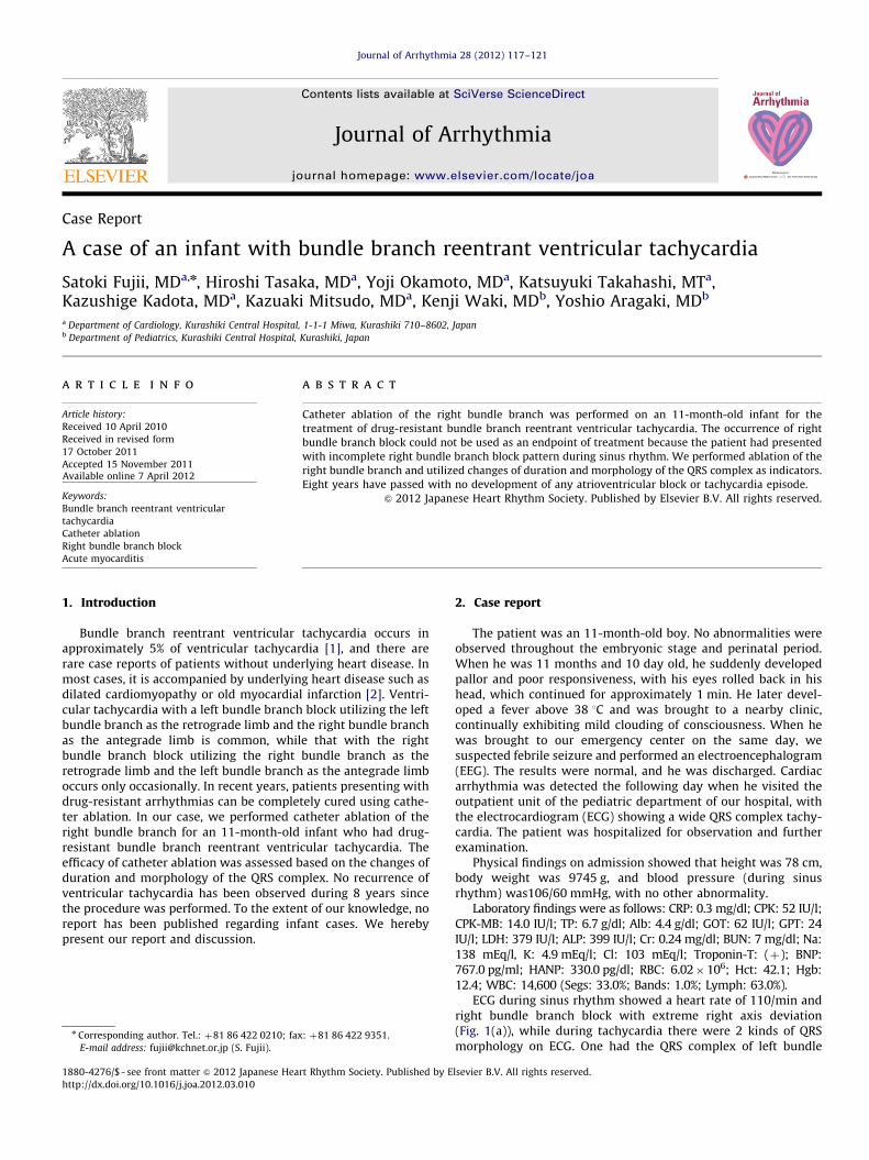
ECG during sinus rhythm showed a heart rate of 110/min andright bundle branch block with extreme right axis deviation(Fig. 1(a)), while during tachycardia there were 2 kinds of QRSmorphology on ECG. One had the QRS complex of left bundle
lsevier B.V. All rights reserved.
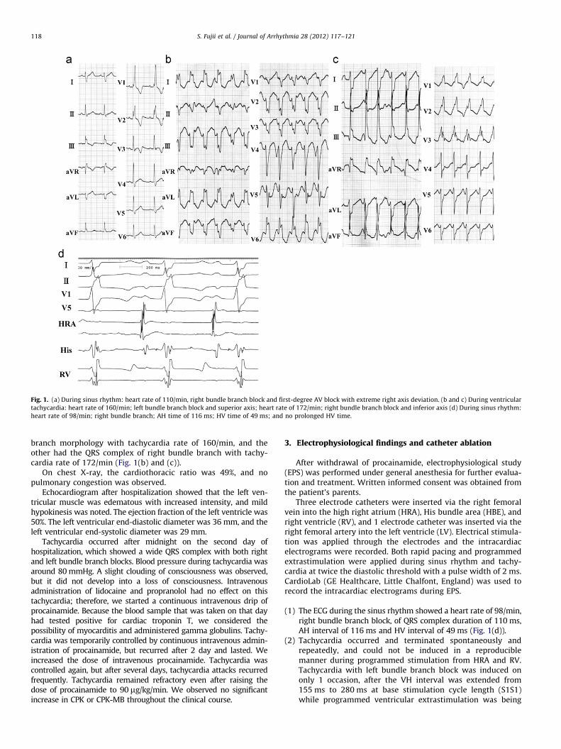
Fig. 1. (a) During sinus rhythm: heart rate of 110/min, right bundle branch block and first-degree AV block with extreme right axis deviation. (b and c) During ventricular
tachycardia: heart rate of 160/min; left bundle branch block and superior axis; heart rate of 172/min; right bundle branch block and inferior axis (d) During sinus rhythm:
heart rate of 98/min; right bundle branch; AH time of 116 ms; HV time of 49 ms; and no prolonged HV time.
S. Fujii et al. / Journal of Arrhythmia 28 (2012) 117–121118
branch morphology with tachycardia rate of 160/min, and theother had the QRS complex of right bundle branch with tachy-cardia rate of 172/min (Fig. 1(b) and (c)).
On chest X-ray, the cardiothoracic ratio was 49%, and nopulmonary congestion was observed.
Echocardiogram after hospitalization showed that the left ven-tricular muscle was edematous with increased intensity, and mildhypokinesis was noted. The ejection fraction of the left ventricle was50%. The left ventricular end-diastolic diameter was 36 mm, and theleft ventricular end-systolic diameter was 29 mm.
Tachycardia occurred after midnight on the second day ofhospitalization, which showed a wide QRS complex with both rightand left bundle branch blocks. Blood pressure during tachycardia wasaround 80 mmHg. A slight clouding of consciousness was observed,but it did not develop into a loss of consciousness. Intravenousadministration of lidocaine and propranolol had no effect on thistachycardia; therefore, we started a continuous intravenous drip ofprocainamide. Because the blood sample that was taken on that dayhad tested positive for cardiac troponin T, we considered thepossibility of myocarditis and administered gamma globulins. Tachy-cardia was temporarily controlled by continuous intravenous admin-istration of procainamide, but recurred after 2 day and lasted. Weincreased the dose of intravenous procainamide. Tachycardia wascontrolled again, but after several days, tachycardia attacks recurredfrequently. Tachycardia remained refractory even after raising thedose of procainamide to 90 mg/kg/min. We observed no significantincrease in CPK or CPK-MB throughout the clinical course.
3. Electrophysiological findings and catheter ablation
After withdrawal of procainamide, electrophysiological study(EPS) was performed under general anesthesia for further evalua-tion and treatment. Written informed consent was obtained fromthe patient’s parents.
Three electrode catheters were inserted via the right femoralvein into the high right atrium (HRA), His bundle area (HBE), andright ventricle (RV), and 1 electrode catheter was inserted via theright femoral artery into the left ventricle (LV). Electrical stimula-tion was applied through the electrodes and the intracardiacelectrograms were recorded. Both rapid pacing and programmedextrastimulation were applied during sinus rhythm and tachy-cardia at twice the diastolic threshold with a pulse width of 2 ms.CardioLab (GE Healthcare, Little Chalfont, England) was used torecord the intracardiac electrograms during EPS.
(1)
The ECG during the sinus rhythm showed a heart rate of 98/min,right bundle branch block, of QRS complex duration of 110 ms,AH interval of 116 ms and HV interval of 49 ms (Fig. 1(d)).(2)
Tachycardia occurred and terminated spontaneously andrepeatedly, and could not be induced in a reproduciblemanner during programmed stimulation from HRA and RV.Tachycardia with left bundle branch block was induced ononly 1 occasion, after the VH interval was extended from155 ms to 280 ms at base stimulation cycle length (S1S1)while programmed ventricular extrastimulation was being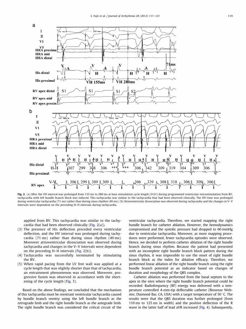
Fig. 2. (a) After the VH interval was prolonged from 155 ms to 280 ms at base stimulation cycle length (S1S1) during programmed ventricular extrastimulation from RV,
tachycardia with left bundle branch block was induced. This tachycardia was similar to the tachycardia that had been observed clinically. The HV-time was prolonged
during ventricular tachycardia (71 ms) rather than during sinus rhythm (49 ms). (b) Atrioventricular dissociation was observed during tachycardia and the changes in V–V
intervals were dependent on the preceding H–H intervals during tachycardia.
S. Fujii et al. / Journal of Arrhythmia 28 (2012) 117–121 119
applied from RV. This tachycardia was similar to the tachy-cardia that had been observed clinically (Fig. 2(a)).
(3)
The presence of His deflection preceded every ventriculardeflection, and the HV interval was prolonged during tachy-cardia (71 ms) rather than during sinus rhythm (49 ms).Moreover atrioventricular dissociation was observed duringtachycardia and changes in the V–V intervals were dependenton the preceding H–H intervals (Fig. 2(b)).(4)
Tachycardia was successfully terminated by stimulatingthe RV.(5)
When rapid pacing from the LV free wall was applied at acycle length that was slightly shorter than that of tachycardia,an entrainment phenomenon was observed. Moreover, pro-gressive fusion was observed in accordance with the short-ening of the cycle length (Fig. 3).Based on the above findings, we concluded that the mechanismof this tachycardia must be reentrant ventricular tachycardia causedby bundle branch reentry using the left bundle branch as theretrograde limb and the right bundle branch as the antegrade limb.The right bundle branch was considered the critical circuit of the
ventricular tachycardia. Therefore, we started mapping the rightbundle branch for catheter ablation. However, the hemodynamicscompromised and the systolic pressure had dropped to 60 mmHgdue to ventricular tachycardia. Moreover, as more mapping proce-dures were performed, fewer tachycardia episodes were observed.Hence, we decided to perform catheter ablation of the right bundlebranch during sinus rhythm. Because the patient had presentedwith an incomplete right bundle branch block pattern during thesinus rhythm, it was impossible to use the onset of right bundlebranch block as the index for ablation efficacy. Therefore, weperformed linear ablation of the right bundle branch using the rightbundle branch potential as an indicator based on changes ofduration and morphology of the QRS complex.
Catheter ablation was performed from the basal septum to theapex in the sites where the right bundle branch potential could berecorded. Radiofrequency (RF) energy was delivered with a tem-perature controlled 4-mm-tip deflectable catheter (Biosense Web-ster, Diamond Bar, CA, USA) with a target temperature of 50 1C. Theresults were that the QRS duration was further prolonged (from110 ms to 125 ms in width) and the positive deflection of the Rwave in the latter half of lead aVR increased (Fig. 4). Subsequently,
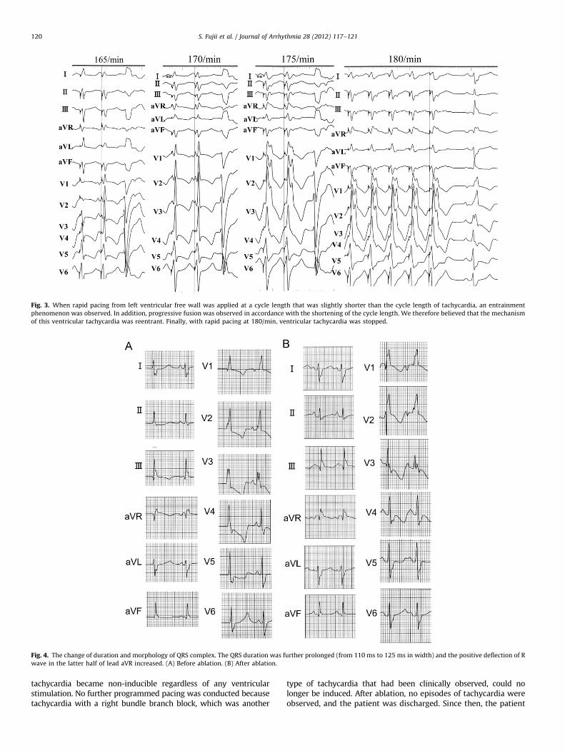
Fig. 3. When rapid pacing from left ventricular free wall was applied at a cycle length that was slightly shorter than the cycle length of tachycardia, an entrainment
phenomenon was observed. In addition, progressive fusion was observed in accordance with the shortening of the cycle length. We therefore believed that the mechanism
of this ventricular tachycardia was reentrant. Finally, with rapid pacing at 180/min, ventricular tachycardia was stopped.
Fig. 4. The change of duration and morphology of QRS complex. The QRS duration was further prolonged (from 110 ms to 125 ms in width) and the positive deflection of R
wave in the latter half of lead aVR increased. (A) Before ablation. (B) After ablation.
S. Fujii et al. / Journal of Arrhythmia 28 (2012) 117–121120
tachycardia became non-inducible regardless of any ventricularstimulation. No further programmed pacing was conducted becausetachycardia with a right bundle branch block, which was another
type of tachycardia that had been clinically observed, could nolonger be induced. After ablation, no episodes of tachycardia wereobserved, and the patient was discharged. Since then, the patient
S. Fujii et al. / Journal of Arrhythmia 28 (2012) 117–121 121
has been visiting the Outpatient Department for regular follow-upfor approximately 8 years, and there has been no recurrence oftachycardia even without administration of antiarrhythmic drugs.Echocardiography shows normal contraction of the LV.
4. Discussion
In this case, acute myocarditis was initially suspected as theunderlying cardiac disease because the onset of symptoms wasrelatively sudden, the results of the Troponin T test were positive,edema was observed throughout the entire cardiac muscle, andcardiac contractility indicated diffuse hypokinesis. However, ahistological diagnosis was not made because the patient was aninfant, and we were unable to obtain the informed consent toconduct a biopsy of the heart muscle from his parents. There havebeen cases reported in which gamma globulins were used for thetreatment of acute myocarditis and successfully improved thesymptoms. Although we acknowledged the necessity of thistreatment, gamma globulin did not have sufficient efficacy forventricular tachycardia control in this case.
Catheter ablation was selected based on the results of EPSbecause ventricular tachycardia, a persistent, fatal arrhythmiaresulting in collapsed hemodynamics, was observed, and thepatient exhibited drug resistance. Therefore, spontaneous recov-ery was not expected.
In EPS, tachycardia was induced only once when programmedpacing was applied. Because both an entrainment phenomenonand progressive fusion were observed during application of rapidpacing from the LV at shorter intervals than the tachycardia cyclelength, we concluded that the mechanism of this tachycardia wasreentry. We also observed a dissociation of the atria and ven-tricles during tachycardia. When ventricular tachycardia relatedto the ordinary ventricular working muscle occurs, the excitationin the right bundle branch and bundle of His is generally delayedcompared to that in the ventricle. Thus, when a slight change isshown in the cycle of ventricular tachycardia, the RB–RB time andH–H time are dependent on the preceding V–V interval. In thepresent case, however, the ventricular electrograms were pre-ceded by the His potential at the bundle of His recording site andthe V–V intervals were defined by the H–H intervals. For thesereasons, the patient was ultimately diagnosed with bundle branchreentrant ventricular tachycardia. In this case, since a high level ofprolongation in the conduction velocity was required so thatanatomical reentry would be possible within the infant’s smallheart, it was presumed that there existed a conduction delay notonly of the His-Purkinje system but of the ventricular muscleas well.
In previous reports, bundle branch reentrant ventriculartachycardia with left bundle branch block has been cured at arate of almost 100% by performing right bundle branch ablation[3,4]. We planned to perform catheter ablation at the site wherethe potential of the right bundle branch could be recorded duringventricular tachycardia. However, because hemodynamics hadbeen compromised, it was impossible to conduct further exam-ination during tachycardia. Therefore, catheter ablation wasperformed along the right bundle branch during sinus rhythm.Because incomplete right bundle branch block pattern wasalready present, the occurrence of a right bundle branch blockin the surface ECG did not provide sufficient evidence for successof complete ablation of the right bundle branch. Hence, weassessed the efficacy of the right bundle branch ablation basedon changes of duration and morphology of the QRS complex.
As a right bundle branch block was originally present in thiscase, it is questionable whether bundle branch reentrant tachy-cardia could actually occur. However, we think that if a right
bundle branch block is not a complete block, but a functionalblock, right bundle branch reentrant ventricular tachycardiacan occur.
In addition, clinically observed tachycardia with a right bundlebranch block and inferior axis was not observed during EPS.We suspected ventricular tachycardia because AV dissociationwas observed, but the details remain unclear. We speculated thatwhile the stimulation was conducted in the antegrade direction inthe anterior left bundle branch, the pathway for the retrogradestimulation was either the posterior left bundle branch or theright bundle branch. We also believed that it was highly likelythat the stimulation was conducted retrogradely in the rightbundle branch, because ventricular tachycardia with both theright and left bundle branch blocks disappeared in clinicalobservation after ablation of the right bundle branch.
The problem pertaining to this treatment is that if theconduction disorder in the left bundle branch develops furtherafter the right bundle branch is ablated, it could result in anadvanced atrioventricular block, which requires implantation of apermanent pacemaker in some cases [5,6]. In our case, althoughthe details of the conduction characteristics in the left bundlebranch could not be tested, we believed that there existed aconduction disorder at least in the posterior left bundle branch,given the presence of a two-bundle block during sinus rhythm.The HV interval after ablation was 67 ms, which was less thanthat before ablation. Cardiac pacemakers are sometimesimplanted preventively when the HV interval exceeds 100 ms.This case was an infant patient whose HV interval after ablationwas mildly prolonged [7]. We decided to follow the patient byoutpatient observation. Eight years have passed since the proce-dure; no AV block has developed and no recurrence of ventriculartachycardia has been recorded. It is said that the prognosis ofbundle branch block syndrome largely depends on the degree ofseverity of any underlying cardiac disease. Tchou et al. reportedthat approximately 30% of patients died due to heart failure [3]. Inour case, the cardiac function has been restored to normal, andthe patient leads a life that is no different from that of otherchildren. We believe that the bundle branch reentrant ventriculartachycardia was a result of transient myocardial damage causedby myocarditis and acute conduction disorder at the bundlebranch level. We hereby present this case because no previousreport on an infant patient has been published and this is theyoungest patient to the extent of our knowledge.
Conflict of interest
All authors have no conflicts of interest that should be disclosed.
References
[1] Caceres J, Yazayeri M, et al. Sustained bundle branch reentry as a mechanismof clinical tachycardia. Circulation 1989;79:256–70.
[2] Fynn SP, Kalman JM, McKinnie J. Bundle branch reentrant tachycardia in apatient with normal ventricular function. J Interv Card Electrophysiol2004;10:255–9.
[3] Tchou P, Blank Z, et al. Mechanism of inducible ventricular tachycardia inpatients with idiopathic dilated cardiomyopathy. J Am Coll Cardiol 1989;13:174A.
[4] Blanck Z, Dhala A, et al. Bundle branch reentrant ventricular tachycardia:Cumulative experience in 48 patients. J Cardiovasc Electrophysiol1993;4:253–62.
[5] Volkmann H, Kuhnert H, Dannberg G, et al. Bundle branch reentranttachycardia treated by transvenous catheter ablation of the right bundlebranch. Pacing Clin Electrophysiol 1989;12:258–61.
[6] Cohen TJ, Chien WW, Lurie KG, et al. Radiofrequency catheter ablation fortreatment of bundle branch reentrant ventricular tachycardia: results andlong-term follow-up. J Am Coll Cardiol 1991;18:1767–73.
[7] Petrac D, Radic B, Vukosavic D. Radiofrequency catheter ablation of the bundlebranch reentrant ventricular tachycardia. Acta Med Austriac 2001;28:16–20.