a case of bilateral adrenal haemorrhage following
TRANSCRIPT

Leong et al. Journal of Intensive Care (2015) 3:4 DOI 10.1186/s40560-015-0073-8
CASE REPORT Open Access
A case of bilateral adrenal haemorrhage followingtraumatic brain injuryMervyn Leong1*, Madhav Pendyala1, Joga Chaganti2 and Suhel Al-Soufi1
Abstract
We report the case of a 57-year-old man who sustained an isolated severe traumatic brain injury (TBI). During hisadmission to the intensive care unit (ICU), he developed marked arterial hypotension of unclear cause. Eventually,the presence of renal angle tenderness on clinical examination and a low random-cortisol level lead to thesuspicion of primary adrenal insufficiency. A computed tomography scan of his abdomen demonstrated newbilateral adrenal haemorrhages. This diagnosis is not unlikely to be missed, as symptoms and laboratory tests areoften nonspecific.
Keywords: Traumatic brain injury, Adrenal haemorrhage, Adrenal insufficiency
BackgroundBilateral adrenal haemorrhages in the context of traumaticbrain injury (TBI) are difficult to diagnose clinically des-pite appropriate symptoms and laboratory parameters asthese are often nonspecific. Serum cortisol levels or theshort synacthen test may not be specific for the diagnosisof primary adrenal insufficiency in critical ill patients, andthere is a lack of data regarding normal cortisol levels incritical illness [1-4] Diagnosis can be made with computedtomography scan of the abdomen. We report a case of bi-lateral adrenal haemorrhages in an isolated TBI.
Case presentationA 57-year-old man sustained an isolated severe trau-matic brain injury as a result of being struck as a pedes-trian by a cyclist.On admission to the emergency department, he had a
Glasgow Coma Scale of eight, equal pupil size and reac-tion to light. He was hypertensive with a systemic bloodpressure of 250/110 mmHg and in a sinus rhythm with80 beats per minute. The patient was intubated for air-way protection as he was vomiting profusely.A computed tomography scan of the brain showed an
acute right frontal subdural hematoma resulting in 1.1 cmof midline shift. In addition, there were bilateral fronto-temporal haemorrhagic contusions, diffuse subarachnoid
* Correspondence: [email protected] Care Unit, St Vincent’s Hospital, Sydney, AustraliaFull list of author information is available at the end of the article
© 2015 Leong et al.; licensee BioMed Central.Commons Attribution License (http://creativecreproduction in any medium, provided the orDedication waiver (http://creativecommons.orunless otherwise stated.
blood along with right-sided parafalcine and uncal hernia-tion. Whole-body computed tomography did not show anyevidence of spinal, thoracic, abdominal or pelvic injury.The patient was taken to theatres for right frontal de-
compressive craniectomy and evacuation of his subduralhematoma. An intra-parenchymal Codman catheter wasinserted to monitor intra-cranial pressure.Following his admission to our intensive care unit, the
patient developed intracranial hypertension, which wascontrolled with deep sedation, continuous paralysis, tar-geted temperature management and infusion of hyper-tonic saline. He required in the first 4 days of his intensivecare unit (ICU) admission infusion of noradrenaline in adose of up to 0.25 mcg/kg/min and concomitant infusionof vasopressin at a dose of up to 0.01 U/min to maintainan adequate cerebral perfusion pressure. The random-cortisol level at this stage was judged to be adequate with452 and 700 nmol/L at two subsequent days.On day 6 of his ICU admission, following the cessation
of vasopressors, he developed autonomic dysfunctionwith severe hypertension, which required temporarytreatment with multiple enteral and intravenous anti-hypertensive medications.In the second week of his admission to the ICU, the pa-
tient became again progressively hypotensive despite cessa-tion of his anti-hypertensive medication. In the presence oflow-grade fever, sepsis was suspected and antibiotics wereempirically started. The patient had ongoing vasopressorrequirements without response to intravascular volume
This is an Open Access article distributed under the terms of the Creativeommons.org/licenses/by/4.0), which permits unrestricted use, distribution, andiginal work is properly credited. The Creative Commons Public Domaing/publicdomain/zero/1.0/) applies to the data made available in this article,
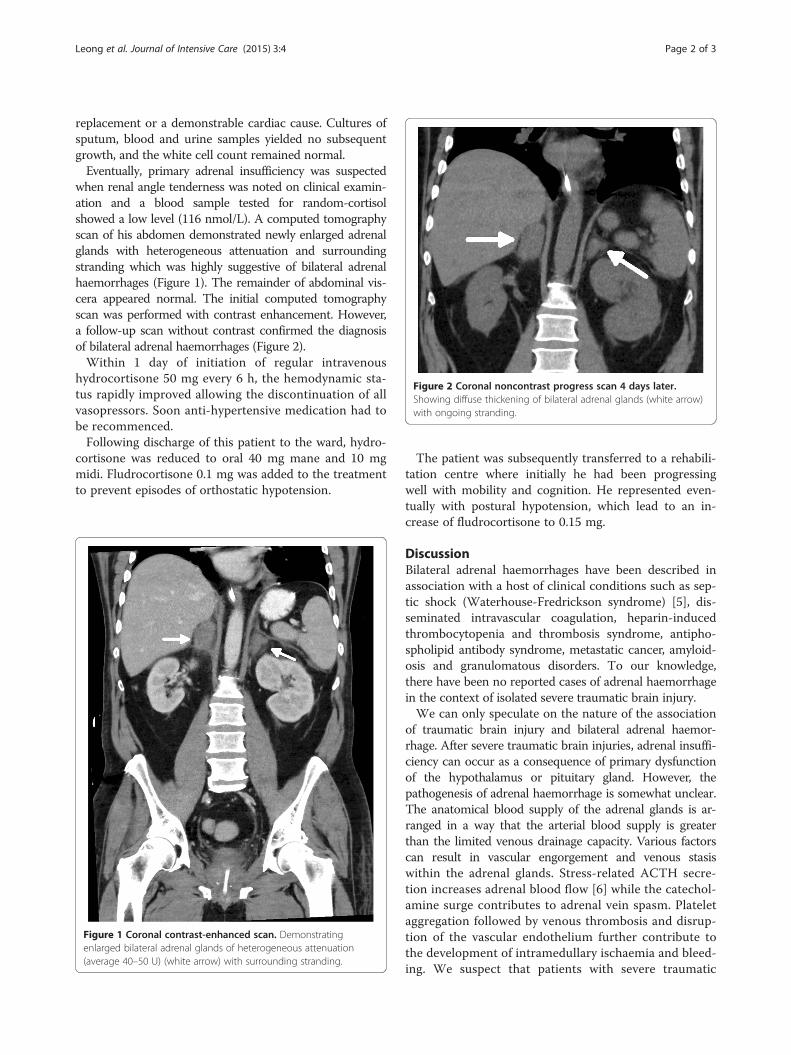
Figure 2 Coronal noncontrast progress scan 4 days later.Showing diffuse thickening of bilateral adrenal glands (white arrow)with ongoing stranding.
Leong et al. Journal of Intensive Care (2015) 3:4 Page 2 of 3
replacement or a demonstrable cardiac cause. Cultures ofsputum, blood and urine samples yielded no subsequentgrowth, and the white cell count remained normal.Eventually, primary adrenal insufficiency was suspected
when renal angle tenderness was noted on clinical examin-ation and a blood sample tested for random-cortisolshowed a low level (116 nmol/L). A computed tomographyscan of his abdomen demonstrated newly enlarged adrenalglands with heterogeneous attenuation and surroundingstranding which was highly suggestive of bilateral adrenalhaemorrhages (Figure 1). The remainder of abdominal vis-cera appeared normal. The initial computed tomographyscan was performed with contrast enhancement. However,a follow-up scan without contrast confirmed the diagnosisof bilateral adrenal haemorrhages (Figure 2).Within 1 day of initiation of regular intravenous
hydrocortisone 50 mg every 6 h, the hemodynamic sta-tus rapidly improved allowing the discontinuation of allvasopressors. Soon anti-hypertensive medication had tobe recommenced.Following discharge of this patient to the ward, hydro-
cortisone was reduced to oral 40 mg mane and 10 mgmidi. Fludrocortisone 0.1 mg was added to the treatmentto prevent episodes of orthostatic hypotension.
Figure 1 Coronal contrast-enhanced scan. Demonstratingenlarged bilateral adrenal glands of heterogeneous attenuation(average 40–50 U) (white arrow) with surrounding stranding.
The patient was subsequently transferred to a rehabili-tation centre where initially he had been progressingwell with mobility and cognition. He represented even-tually with postural hypotension, which lead to an in-crease of fludrocortisone to 0.15 mg.
DiscussionBilateral adrenal haemorrhages have been described inassociation with a host of clinical conditions such as sep-tic shock (Waterhouse-Fredrickson syndrome) [5], dis-seminated intravascular coagulation, heparin-inducedthrombocytopenia and thrombosis syndrome, antipho-spholipid antibody syndrome, metastatic cancer, amyloid-osis and granulomatous disorders. To our knowledge,there have been no reported cases of adrenal haemorrhagein the context of isolated severe traumatic brain injury.We can only speculate on the nature of the association
of traumatic brain injury and bilateral adrenal haemor-rhage. After severe traumatic brain injuries, adrenal insuffi-ciency can occur as a consequence of primary dysfunctionof the hypothalamus or pituitary gland. However, thepathogenesis of adrenal haemorrhage is somewhat unclear.The anatomical blood supply of the adrenal glands is ar-ranged in a way that the arterial blood supply is greaterthan the limited venous drainage capacity. Various factorscan result in vascular engorgement and venous stasiswithin the adrenal glands. Stress-related ACTH secre-tion increases adrenal blood flow [6] while the catechol-amine surge contributes to adrenal vein spasm. Plateletaggregation followed by venous thrombosis and disrup-tion of the vascular endothelium further contribute tothe development of intramedullary ischaemia and bleed-ing. We suspect that patients with severe traumatic

Leong et al. Journal of Intensive Care (2015) 3:4 Page 3 of 3
brain injury who have high catecholamine levels and re-lease tissue thromboplastin from damaged brain are atrisk for bilateral adrenal haemorrhage to occur.In the reported case, there was no evidence of any
blunt trauma apart from isolated traumatic brain injury.The patient did not have a past medical history of malig-nancy or haematological disorder. Repeated coagulationprofiles did not demonstrate any haematological abnor-malities such as disseminated intravascular coagulation.The patient was not prescribed heparin or any otheranticoagulant in the first weeks of his admission as thiswas considered to be contraindicated. We believe there-fore that the bilateral adrenal haemorrhages were a con-sequence of the isolated traumatic brain injury.Bilateral adrenal haemorrhages in the context of trau-
matic brain injury are difficult to recognise clinically, andthe diagnosis might be missed despite dramatic symptoms.Abdominal or flank tenderness on examination of a patientwith isolated severe TBI and shock should raise the suspi-cion of adrenal failure. Further suggestive though unspecificfeatures are fever, anorexia, nausea and confusion, and inparticular, hypotension or shock as are laboratory cluessuch as leukocytosis, hyperkalemia and hyponatremia inthe presence of an unaccountable fall in haemoglobin [7].Symptoms of adrenal insufficiency can be similar to
that of the systemic inflammatory response syndromeand sepsis which are frequently seen in severe traumaticbrain injury [8]. Adrenal insufficiency can be confusedwith other endocrine disorders related to TBI such assyndrome of inappropriate antidiuretic hormone secre-tion and cerebral salt wasting [2-4]. We believe that thelower than expected cortisol level in this patient is con-sistent with adrenal insufficiency caused by bilateral ad-renal haemorrhages. However, serum cortisol levels orthe short synacthen test may not be specific for the diag-nosis of primary adrenal insufficiency in critical ill pa-tients. Fluctuations in serum cortisol levels are frequentand not necessarily indicative of actual damage to theadrenal gland [1-4]. There is also a lack of consensus re-garding normal cortisol levels in critical illness.The diagnosis of adrenal haemorrhage can be made by a
computed tomography scan of the abdomen demonstrat-ing adrenal gland enlargement with high attenuation with-out contrast enhancement. Progress CT scans performedweeks after the acute phase may show progressive adrenalgland atrophy with variable degree of calcification. Ultra-sonographic imaging of the abdomen may aid the diagnosesin the acute situation given that it is a bedside investigationthat is easily accessible. Ultrasonographic examination usu-ally reveals hyperechoic masses with central echogenicareas. Although magnetic resonance imaging may be help-ful to exclude the presence of tumours and provide an esti-mate for the age of haemorrhage, there is very littleevidence found in the literature.
In some cases, there may be spontaneous recoveryeven years after the original injury but patients with bi-lateral adrenal infarctions often require lifelong adrenalreplacement therapy [9].
ConclusionsThis case demonstrates that bilateral adrenal haemorrhagecan be a rare consequence of severe TBI. The diagnosis isdifficult, as symptoms and laboratory test are often unspe-cific, but abdominal or flank tenderness on examination ofa patient with isolated severe TBI and shock should raisethe suspicion of adrenal failure and prompt appropriateimaging.
ConsentWritten and verbal consent was obtained from the pa-tient and his next of kin for publication of this case re-port and use of accompanying images. A copy of thewritten consent is available for review by the Editor inChief of this journal.
Competing interestsThe authors declare that they have no competing interests.
Authors’ contributionsMP and ML performed the literature review and compiled the case report anddiscussion. SA revised the manuscript critically for important intellectual content.JC reviewed and selected the relevant images and interpreted the radiologicalfindings for this case. All authors read and approved the final manuscript.
AcknowledgementsWe would like to acknowledge Dr. Barry Johnston for his contributiontowards proofreading of the manuscript.
Author details1Intensive Care Unit, St Vincent’s Hospital, Sydney, Australia. 2Department ofRadiology, St Vincent’s Hospital, Sydney, Australia.
Received: 30 October 2014 Accepted: 15 January 2015
References1. Olivecrona Z, Dahlqvist P, Koskinen LO. Acute neuro-endocrine profile and
prediction of outcome after severe brain injury. Scand J Trauma ResuscEmerg Med. 2013;21:33.
2. Savaridas T, Andrews PJ, Harris B. Cortisol dynamics following acute severebrain injury. Intensive Care Med. 2004;30:1479–83.
3. Webster JB, Bell KR. Primary adrenal insufficiency following traumatic braininjury: a case report and review of the literature. Arch Phys Med Rehabil.1997;78:314–8.
4. Hwang JJ, Hwang DY. Treatment of endocrine disorders in theneuroscience intensive care unit. Curr Treat Options Neurol. 2014;16:271.
5. Migeon CJ, Kenny FM, Hung W, Voorhess ML. Study of adrenal function inchildren with meningitis. Pediatrics. 1967;40:163–83.
6. Streeten D. Adrenal hemorrhage. Endocrinologist. 1996;6:277.7. Rao RH, Vagnucci AH, Amico JA. Bilateral massive adrenal hemorrhage: early
recognition and treatment. Ann Intern Med. 1989;110:227–35.8. Powner DJ, Boccalandro C. Adrenal insufficiency following traumatic brain
injury in adults. Curr Opin Crit Care. 2008;14:163–6.9. Vella A, Nippoldt TB, Morris 3rd JC. Adrenal hemorrhage: a 25-year experience
at the Mayo Clinic. Mayo Clin Proc. 2001;76:161–8.