a chromosomal profile of polycythemia vera
TRANSCRIPT

A Chromosomal Profile of Polycythemia Vera
Giovanna Rege-Cambrin, Cristina Mecucci, Guido Tricot, Jean-Louis Michaux, Andries Louwagie, Werner Van Hove, Hugo Francart, and Herman Van den Berghe -
ABSTRACT: One hundred four patients with a diagnosis of polycythemia vera and a variable period of follow-up had one or more cytogenetic investigations. Chromosome abnormalities were found in 13% of untreated patients, in 56% of cases treated with radioactive phosphorus (32p) or cytotoxic drugs, and in 85% of patients in which transformation of the disease had oc- curred. Nonrandom chromosome abnormalities found before treatment included +8, +9, 13q- , 2Oq- ; their prognostic value is little, as they are often associated with longstanding, stable disease. In contrast, 5 q - anomaly and the appearance of subclones in patients with an abnormal karyotype were found to be poor prognostic signs, as they are usually coinciden- tal with evolution of the disease to myelofibrosis or leukemia. Chromosomally two patterns of acute leukemia were observed in polycythemia vera patients. The first type resembles de nova acute leukemia, in that the clinical and cytologic characteristics of the disorder are easily defined by FAB criteria and the chromosome changes compatible with the types usu- ally found in those conditions. In the second type, assignment to a FAB morphalogic subgroup was more difficult, myelodysplastic changes were often present, and the karyotype showed complex abnormalities frequently involving chromosomes #5 and #7. All these fea- tures suggest the occurrence of secondary leukemia.
INTRODUCTION
Polycythemia vera (PV) is a monoclonal disorder of the hemopoiet ic stem cell [1] characterized by an abnormal proliferation of the erythroid series associated with a less prominent expansion of the myeloid and megakaryocytic compartment. The disease usual ly has a long clinical history and is well controlled by various thera- peutic regimens including phlebotomies, 32p, or alkylating drugs. Despite the rela- t ively mild course of the disease, myelofibrosis and/or acute leukemia ul t imately develop in some patients; a causative role of treatment in inducing the transforma- tion has been postulated on the basis of a higher incidence of acute leukemia in patients treated with 32p or cytotoxic drugs [2-4]. Chromosome studies in PV have been reported on several occasions [5-13]. Our information about the cytogenetics
From the Division of Human Genetics, Department of Human Biology (G. R-C., C. M., H. F., H. V. d. B.) and Department of Hematology (G. T.), University of Leuven, Service d'H6matologie, Universit~ de Louvain (J-L. M.), Brussels, Department of Hematology (A. L.), A. Z. St. Jan, Brugge, Department of Hematology (W. V. H.), University Hospital, Gent, Belgium
Address requests for reprints to Dr. Herman Van den Berghe, Center for Human Genetics, Herestraat 49, B-3000 Leuven, Belgium.
Received April 4, 1986; June 13, 1986.
233
© 1987 Elsevier Science Publishing Co., Inc. Cancer Genet Cytogenet 25:233-245(1987) 52 Vanderbilt Ave., New York, NY 10017 0165-4608/87/$03.50

234 C. Rege-Cambrin et al.
of this disorder, however, is still incomplete. The present report deals with our cytogenetic experience in 104 PV patients, many of which were repeatedly studied during a long follow-up period. Particular attention will be paid to the prevalence and type of chromosome changes in untreated and treated patients and the dynam- ics of the chromosome changes throughout evolution of the disease, particularly during leukemic transformation.
MATERIALS AND METHODS
One hundred four patients with PV who had a successful chromosomal investiga- tion in the years 1973-1985 were included in this study. The patients were referred by the following Belgian hospitals: Division of Hematology, University of Leuven; Service d'H6matologie, University of Louvain in Brussels; Dienst Hematologie, St. Jan Ziekenhuis, Brugge and Division of Hematology, University of Gent.
The clinical and hematologic data were critically reviewed in all cases. In 104 patients diagnosis of PV was adequately documented; 83 patients completely met the criteria established by the Polycythemia Vera Study Group (PVSG) [14]. Twenty-one patients with a long history of the disease fulfilled all the clinical cri- teria for PV; however, complete laboratory data were not available (these patients, nevertheless, have been included in the study). Twenty-eight other patients exam- ined during the same period could not be included because of insufficient data.
Myelofibrosis (MF) was always documented by bone biopsy. Cytologic prepara- tions of the patients who showed a myelodysplastic or leukemic evolution were reviewed and classified according to the criteria established by the FAB cooperative group [15, 16]. Our group of patients included 56 males and 48 females; age at diagnosis of PV ranged from 18 to 82 years, with a mean age of 60 years. The period of clinical follow-up ranged from less than 1 year to a maximum of 23 years, with a mean of 6 years. Twelve patients were treated only with phlebotomies; these cases will be referred to as "untreated." All other patients received cytotoxic therapy including 32p alone (66 cases), 32p in combination with alkylating drugs (19 cases), or cytotoxic agents such as busulphan, chlorambucil, hydroxyurea, 6-thioguanine and 6-mercaptopurine either alone or in combination (seven cases).
Cytotogenetic investigations were done on bone marrow aspirates and/or periph- eral blood, cultured for approximately 48 hours in the absence of mitogens. The banding method was R-banding using acridine orange. In 63 patients this investi- gation was done at diagnosis, and in five other patients prior to any treatment. The other patients were studied for the first time after they had received cytotoxic treat- ment. Serial chromosome investigations were performed during follow-up; the number of investigations varied between one and ten per case. In seven patients the karyotyping was initially performed when they were in leukemic transformation. Only clonal abnormalities are dealt with. The number of metaphases examined was between 15 and 25. A clone was identified when two cells showed the same super- numerary chromosome or structural rearrangement, or when three cells had the same chromosome missing [17]. Cases 10401, 10691, 7031 [18], 14540, 7111, 8906 [19], and 13788 [20] have been reported previously.
RESULTS
Prevalence and Type of Chromosomal Abnormalities
Cytogenetic and clinical data of the patients with abnormal karyotypes are listed in Tables 1-4. Table 5 includes all the patients who showed leukemic evolution.
Only nine of 68 untreated patients had an abnormal karyotype (13.2%); three

Polycythemia Vera 2 3 5
patients had s tructural anomalies , namely 2 0 q - (Table 1) and 1 3 9 - (Table 2), whereas, in six cases an aneup lo id clone was observed (tr isomy 8 and/or 9; Table 3). In contrast, 24 of 43 treated pat ients s tud ied before leukemic evolut ion had a clonal abnormal karyotype (56%).
The most common chromosomal abnormal i t ies encountered were a dele t ion of the long arm of chromosome #20 ( 2 0 q - ) , an interst i t ial de le t ion of 13q ( 1 3 q - ) , t r isomy 8 and/or t r i somy 9, and the interst i t ial de le t ion of 5q ( 5 q - ) . 5 q - and 1 3 q - chromosomes were found together in two cases, of which one also showed t r isomy 9.
Patients wi th a 2 0 q - anomaly are l is ted in Table 1 . 2 0 q - was the most common sole abnormali ty, being present in six cases. It is to be noted that one of them was untreated. In the other patients, dosage of 32p var ied from one to six doses, and the interval between first 32p therapy and first demonst ra t ion of a 2 0 q - anomaly ranged from 1.5 to 16 years. Patients had a his tory of disease ranging from 4 to 16 years before the finding of 2 0 q - . Evolut ion to myelofibrosis (MF) was observed in two of them.
An interst i t ial de le t ion of the long arm of chromosome #13 was found in nine patients (Tables 2 and 5). Two different delet ions could be defined (Fig. 1): del(13)(q13q31) and del(13)(q13q21); loss of band 13q14 was observed in both types of deletion. 1 3 q - was present as the sole abnormal i ty in three patients, and in two of them it was a l ready found at diagnosis; these pat ients during long fol low-up showed no signs of t ransformation. In contrast, in four of six cases in which 1 3 q - was associated wi th other anomal ies the chromosomal changes were observed at t ransformation into MF, acute non lymphocy t i c leukemia (ANLL), or refractory ane- mia wi th excess of blasts (RAEB). Tr isomy 8 and/or 9 (Table 3) were the most fre- quent abnormal i t ies observed at diagnosis.
5 q - was observed in six treated pat ients (Tables 2, 4 and 5). Three of them had MF, and two evolved into acute leukemia; in two more pat ients leukemic transfor- mat ion was observed. Al l five pat ients d ied soon of their hematologic disease. Pa- t ient 33317 (Table 2) evolved into a RAEB and is alive after 6 months of follow-up.
In Table 4 the pat ients wi th other abnormal i t ies are listed. A dupl ica t ion of the long arm of chromosome #1 was observed in two treated patients; moreover, in
Table 1 Clinical and cytogenetic findings in PV pat ients wi th 2 0 q -
Patient
Disease s t a tus Agea/ and t ime Prev ious
Sex from diagnosis b treatment c Sample Clonal karyotype Cells (%) Follow-up d
14655 59/F PV at d iagnos i s None BM 46,XX 100 PV 4 yr 32p BM 46,XX,del(20)(q11) 100 A, 4 yr
25233 59/M PV 16 hr, MF 32p BM 46,XY,del(20)(q11) 75 D, 2 mo 16386 53/M PV 12 yr 32p, chlorambucil BM 46,XY,del(20)(q11) 60 Lost 37316 66/F PV 4 yr None BM 46,XX,del(20)(q11) 25 A, 4 yr 39851 65/M PV 5 yr 32p BM 46,XY,del(20)(q11) 60
PV 9 yr, MF Hydroxyurea, 6- PB 46,XY,20q- 30 A, 1 mo thyoguanine
26835 54/F PV at diagnosis None BM 46,XX 100 PV 6 yr 32p, hydroxyurea BM 46,XX,del(20)(q11) 60 A, 1 mo
°Age at diagnosis of PV. bPV, polycythemia vera; MF, myelofibrosis; RAEB, refractory anemia with excess of blasts; AL, acute leukemia; AUL, acute undifferentiated leukemia; ANLL, acute nonlymphocytic leukemia. CRadioactive phosphorus (32p) or cytotoxic drugs. dA, alive; D, dead.

Tab
le 2
Pat
ien
t
Cli
nic
al
an
d
cy
tog
en
eti
c f
ind
ing
s in
PV
pa
tie
nts
w
ith
1
3q
-
Dis
ease
sta
tus
Ag
e°/
and
tim
e P
rev
iou
s S
ex
fro
m d
iag
no
sis b
tr
eatm
ent c
1313
2 51
/M
1O4O
1 e
42/M
1069
1 e
67/M
7031
~
37/M
PV
at
dia
gn
osi
s N
on
e P
V 8
yr
32p
PV
12
yr
PV
at
dia
gn
osi
s N
on
e P
V 3
yr
PV
11
yr
32p
PV
15
yr
Sam
ple
5691
9 66
/M
3331
7 68
/F
5700
1 50
/F
BM
B
M
BM
B
M
BM
B
M,
BM
,
Clo
nal
kar
yo
typ
e C
ells
(%
)
PV
10
yr,
MF
32
p B
M,
PV
1 y
r 32
p B
M
4 yr
, R
AE
B
Bu
sulp
ha
n
BM
4 y
r 2
too,
C
yto
sin
e-
BM
R
AE
B
arab
ino
sid
e 5
yr
5 m
o,
BM
R
AE
B
5 y
r 11
mo
, C
yto
sin
e-
BM
R
AE
B
arab
ino
sid
e P
V 1
2 y
r 32
p B
M
12 y
r 5
mo
P
B
PB
P
B
PB
46
,XY
,13
q -
4
6,X
Y,1
3q
-
46
,XY
,13
q -
4
6,X
Y,1
3q
-
46
,XY
,13
q -
4
6,X
Y
46
,XY
,del
(13
)(q
13
q3
1)/
4
6,X
Y,1
3q
-
,del
(11
)(q
22
)
46
,XY
,13
q -
/46
,XY
,13
q -
, in
v(1
2)
46
,XX
4
6,X
X,d
el(1
3)(
q1
3q
31
)/
46
,XX
,in
v(1
2)(
p1
2q
23
) 4
6,X
X,1
3q
-
46
,XX
,del
(5)(
ql
3q
32
),
inv
(12
) 4
6,X
X,1
3q
46
,XX
,del
(12
)(q
14
q2
1)
60
20
95
10
15
100
76/1
8
5O/2
5
25/7
5
See
Tab
le 1
for
foo
tnot
es a
thr
ough
d.
~Pre
viou
sly
publ
ishe
d ca
ses.
46
,XX
,12
q -
/46
,XX
,12
q -
, d
el(1
3)
(q1
3q
21
)
70
50
15
80
60
/30
Fo
llo
w-u
p d
A,
10 y
r
A,
2y
r
D,
afte
r 5
yr
Sta
ble
PV
, lo
st
to f
urt
her
fo
llo
w-u
p
A,
10 m
o
A,
2 m
o
A,
6 m
o
t~

Ta
ble
3
Cli
nic
al
an
d
cy
tog
en
eti
c
fin
din
gs
in P
V
pa
tie
nts
w
ith
tr
iso
my
8
an
d/o
r 9
Dis
ease
sta
tus
Age
~/
and
tim
e P
rev
iou
s P
atie
nt
Sex
fr
om
dia
gn
osi
s b
trea
tmen
t c
Sam
ple
C
lon
al k
ary
oty
pe
Cei
ls (
%)
Fo
llo
w-u
p d
8399
60
/F
PV
5 y
r 32
p B
M,
PB
PV
8 y
r B
usu
lph
an
B
M
PV
14
yr,
MF
B
M
8369
64
/M
PV
at
dia
gn
osi
s N
on
e B
M
PV
5 y
r, M
F
BM
6
11
48
58
/M
PV
at
dia
gn
osi
s N
on
e B
M
4736
3 4
7/F
P
V a
t d
iag
no
sis
No
ne
BM
15
859
52/F
P
V a
t d
iag
no
sis
No
ne
BM
P
V 1
yr
32p
BM
PV
5 y
r B
M,
PB
95
14
56/M
P
V 3
yr
32p
BM
PV
5 y
r B
M
PV
11
yr,
MF
P
B
5083
5 46
/M
PV
2 y
r N
on
e B
M
4005
3 72
/M
PV
at
dia
gn
osi
s N
on
e B
M
47,X
X,
+ C
47
,XX
, +
8
47,X
X,
+ 8
,11
p -
48
,XY
, +
C, +
C
48
,XY
,+ 8
,+ 9
47
,XY
, +
9
47,X
X,
+ 8
48
,XX
, +
9,
+ 2
1 4
6,X
X
46
,XX
45
,XY
, -
18
46
,XY
48
,XY
, +
8,
+ 9
47
,XY
, +
9
48
,XY
,t(7
;19
)(q
21
;q1
2),
+
8,+
9
40
100
95
80
100
100 70
50
10
0 10
0 20
10
0 25
15
50
D,
3y
r
D,
4 y
r A
, 4
mo
A
, 2
yr
6 m
o
A,
3 yr
, M
F
A,
10 m
o
A,
2y
r A
, 1
yr
6 m
o
See
Tab
le 1
for
foo
tnot
es a
thr
ough
d.

t~
Ta
ble
4
Cli
nic
al
an
d
cy
tog
en
eti
c f
ind
ing
s in
PV
pa
tie
nts
w
ith
d
iffe
ren
t ty
pe
s o
f n
um
eri
ca
l a
nd
st
ruc
tura
l c
hro
mo
som
al
ab
no
rma
liti
es
Dis
ease
sta
tus
Age
°/
and
tim
e P
rev
iou
s P
atie
nt
Sex
fr
om
dia
gn
osi
s b
trea
tmen
t c
Sam
ple
C
lon
al k
ary
oty
pe
Cel
ls (
%)
Fo
llo
w-u
p J
8706
66
fM
PV
at
dia
gn
osi
s N
on
e B
M
46
,XY
10
0 P
V 4
yr
32p
BM
4
6,X
Y,d
up
(1)
65
A,
5 yr
, M
F
(q2
1q
ter)
39
129
33/F
P
V 1
5 y
r C
hlo
ram
bu
cil
BM
4
6,X
X,i
ns(
4;1
1)
35/1
5 (q
26
q3
5;p
14
)/
46
,XX
,du
p(1
) (q
21
-~q
ter)
P
V 1
7 y
r 32
p B
M
46
,XX
,in
s(4
;11
] 80
A
, 2
yr
2929
2 58
/F
PV
9 y
r 32
p B
M
46
,XX
10
0
PV
12
yr
BM
4
6,X
,-X
,+M
/ 50
/40
A,
6 m
o
45,X
, -
X
1454
0 °
43/F
P
V 9
yr
32p
BM
4
6,X
X
100
PV
11
yr,
MF
B
usu
lph
an
B
M
44
,X,-
X,d
el(5
) 10
0 (q
14
q3
2),
-
7 P
V 1
1 y
r 3
mo
, P
B
44
,XX
,5q
-,-
7,1
2p
+
100
D,
1 m
o
MF
t(
16
;18
)(q
11
;q1
2)
2492
9 45
/F
PV
2 y
r B
usu
lph
an
B
M
46
,XX
,18
p ÷
10
0 A
, 3
yr,
MF
30
263
62/F
P
V 1
yr
6 m
o 32
p B
M
46
,XX
,del
(7)
100
A,
4 y
r (q
31
qte
r),
18
p ÷
65
46
59/M
P
V a
t d
iag
no
sis
No
ne
BM
46
,XY
, 10
0 P
V 6
yr
32p
BM
, P
B
46
,XY
10
0 P
V 8
yr,
MF
B
usu
lph
an
B
M
46
,XY
,del
(11
)(q
22
) 40
D
, 1
mo
See
Tab
le 1
for
foo
tnot
es a
thr
ough
d.
Polycythemia Vera 239
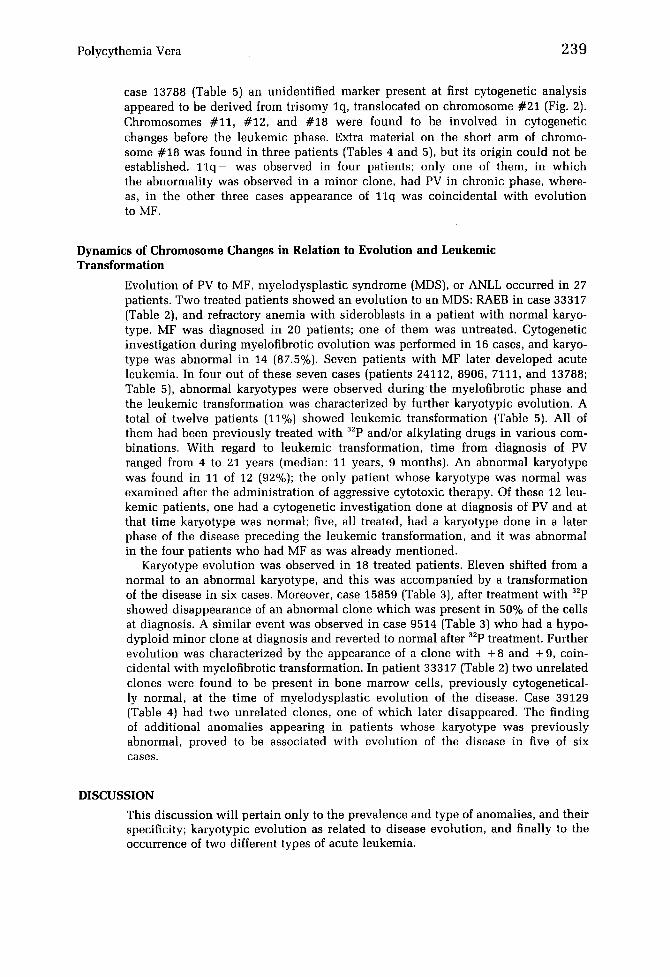
case 13788 (Table 5) an unidentified marker present at first cytogenetic analysis appeared to be derived from trisomy lq, translocated on chromosome #21 (Fig. 2). Chromosomes #11, #12, and #18 were found to be involved in cytogenetic changes before the leukemic phase. Extra material on the short arm of chromo- some #18 was found in three patients (Tables 4 and 5), but its origin could not be established. 11q- was observed in four patients; only one of them, in which the abnormality was observed in a minor clone, had PV in chronic phase, where- as, in the other three cases appearance of 11q was coincidental with evolution to MF.
Dynamics of Chromosome Changes in Relation to Evolution and Leukemic Transformation
Evolution of PV to MF, myelodysplastic syndrome (MDS), or ANLL occurred in 27 patients. Two treated patients showed an evolution to an MDS: RAEB in case 33317 (Table 2), and refractory anemia with sideroblasts in a patient with normal karyo- type. MF was diagnosed in 20 patients; one of them was untreated. Cytogenetic investigation during myelofibrotic evolution was performed in 16 cases, and karyo- type was abnormal in 14 (87.5%). Seven patients with MF later developed acute leukemia. In four out of these seven cases (patients 24112, 8906, 7111, and 13788; Table 5), abnormal karyotypes were observed during the myelofibrotic phase and the leukemic transformation was characterized by further karyotypic evolution. A total of twelve patients (11%) showed leukemic transformation (Table 5). All of them had been previously treated with 32p and/or alkylating drugs in various com- binations. With regard to leukemic transformation, time from diagnosis of PV ranged from 4 to 21 years (median: 11 years, 9 months). An abnormal karyotype was found in 11 of 12 (92%); the only patient whose karyotype was normal was examined after the administration of aggressive cytotoxic therapy. Of these 12 leu- kemic patients, one had a cytogenetic investigation done at diagnosis of PV and at that time karyotype was normal; five, all treated, had a karyotype done in a later phase of the disease preceding the leukemic transformation, and it was abnormal in the four patients who had MF as was already mentioned.
Karyotype evolution was observed in 18 treated patients. Eleven shifted from a normal to an abnormal karyotype, and this was accompanied by a transformation of the disease in six cases. Moreover, case 15859 (Table 3), after treatment with 32p showed disappearance of an abnormal clone which was present in 50% of the cells at diagnosis. A similar event was observed in case 9514 (Table 3) who had a hypo- dyploid minor clone at diagnosis and reverted to normal after 32p treatment. Further evolution was characterized by the appearance of a clone with + 8 and + 9, coin- cidental with myelofibrotic transformation. In patient 33317 (Table 2) two unrelated clones were found to be present in bone marrow cells, previously cytogenetical- ly normal, at the time of myelodysplastic evolution of the disease. Case 39129 (Table 4) had two unrelated clones, one of which later disappeared. The finding of additional anomalies appearing in patients whose karyotype was previously abnormal, proved to be associated with evolution of the disease in five of six cases.
DISCUSSION
This discussion will pertain only to the prevalence and type of anomalies, and their specificity; karyotypic evolution as related to disease evolution, and finally to the occurrence of two different types of acute leukemia.

Tab
le
5 C
lin
ical
an
d
cyto
gen
etic
fi
nd
ing
s in
PV
pat
ien
ts
wh
o
sho
wed
a
leu
kem
ic
evo
luti
on
Dis
ease
sta
tus
Ty
pe
of
Age
a/
befo
re l
euk
emic
le
uk
emia
P
rev
iou
s P
atie
nt
Sex
tr
ansf
orm
atio
n b
(FA
B) b
tr
eatm
ent'
: S
amp
le
Clo
nal
kar
yo
typ
e C
ells
(%
) F
ollo
w-
up
d
5566
0 53
/M
PV
21
yr,
MF
1
AN
LL
(M
2)
Bu
sulp
hau
B
M,
PB
4
7,X
X,+
8
yr,
AL
92
00
41/F
P
V 4
yr,
MF
, A
L
AN
LL
32
p,
BM
, P
B
47
,XX
,+9
b
usu
lph
an
5696
0 73
/F
PV
6 y
r, A
L
AN
LL
(M
5b)
32p,
B
M,
PB
46
,XX
,del
(7)(
p14)
h
yd
rox
yu
rea,
b
usu
lph
an
4798
5 42
/F
PV
10
yr,
MF
, A
L
AN
LL
3z
p P
B
48,X
X,
+ 8,
+ M
24
112
57/F
P
V 8
yr,
MF
32
p B
M
46
,XX
,del
(11
)(q
14
),1
2q
-,
PV
11
yr,
MF
, A
L
AN
LL
1589
3 58
/M
PV
14
yr
32p
PV
15
yr,
AL
A
NL
L (
M4)
B
usu
lph
an
4655
1 42
/F
PV
20
yr,
AL
f A
NL
L
32p
18
p+
P
B
46
,XX
,11
q-
,12
q-
,18p
+ /
47
,XX
,11q
-
,12
q-
,18p
+,
t(1;
9)(q
11;q
11),
+ M
B
M
46,X
Y
BM
43
,XY
, -
5, -
7,
-
11
,- 1
7,+
M1
B
M
44,X
X,d
el(5
)(q1
2q23
),
- 1
2,-
13
,-
17
,+M
100
100
100
100
100
75/2
5
100
100
100
A,
1 yr
, in
re
mis
sio
n
D,
13
mo
D,
3 m
o
D,
6 m
o
D,
1 m
o
D,
2m
o
D,
2m
o

4406
0 63
/F
8906
e 45
/M
7111
~
56/M
1378
8 e
46/F
PV
13
yr,
AL
¢
PV
8 y
r P
V 1
3 yr
, M
F
PV
14
yr,
AL
P
V a
t d
iag
no
sis
PV
6 y
r, M
F
AN
LL
AU
L
PV
7 y
r, M
F,
AL
A
NL
L
PV
9 y
r, M
F
PV
11
yr,
MF
, A
L
AN
LL
Bu
sulp
han
C
hlo
ram
bu
cil,
b
usu
lph
an
32p
No
ne
32p,
hy
dro
- x
yu
rea,
6-
m
erca
pto
- p
uri
ne
32p,
chlo
ram
bu
cil,
b
usu
lph
an
5960
0 68
/M
PV
10
yr,
AL
A
NL
L
Bu
sulp
han
H
yd
rox
yu
rea
BM
BM
P
B
BM
, P
B
BM
B
M
BM
BM
BM
BM
50,X
X,
+ 1,
del(
5)(q
12q3
2),
+8
,+1
0,+
11
47
,XY
, +
9 47
,XY
, +
9/47
,XY
,del
(5)
(q14
;q32
),de
l(8)
(q?)
, +
9,
del(
11)(
q21)
, de
l(13
)(q1
3;q3
1)/
47
,XY
,5q
-, +
9,1
1q
- ,1
3q
- 4
7,X
Y,5
q-,
+ 9
,11
q-,
13
q-
46,X
Y
46,X
Y,d
el(5
) (q
14q3
2),
del(
7)(q
21q3
1)
46
,XY
,5q
-/4
6,X
Y,5
q-,
de
l(13
)(q1
3q31
}
46,X
X,t
(6;2
2)(q
27;q
11},
-
21,
÷ M
1 4
6,X
X,t
(6;2
2),
- 21
, +
M1
/ 4
6,X
X,t
(3q
- ,3
q +
),t(
6;22
), -
7,
-
21
,+M
l,+
M2
46
,XY
100
100
100
7/13
/80
100
100
47
50/5
0
100
50/5
0
100
D,
2 m
o
D,
lmo
D,
1 m
o
D,
1 m
o
D,
5 m
o
D,
2 m
o
See
Tab
le 1
for
foo
tnot
es a
thr
ough
d.
fin
thes
e tw
o pa
tien
ts d
yser
ythr
opoi
esis
and
dys
meg
akar
yopo
iesi
s w
ere
also
obs
erve
d.

242 G. Rege-Cambrin et al.
Figure 1 Two different types of interstitial deletion of chromosome #13.
Prevalence and Types of Anomalies
The prevalence of chromosomal abnormalities in our group of 68 untreated pa- tients, mostly examined at diagnosis, was 13.2%, which is in agreement with pre- viously reported data [5-10] as reviewed by Testa [21]. In the literature, karyotypi- cally abnormal clones were found in 38% of treated cases without ANLL [21]; in our patients they were observed in 56% of the patients studied before leukemic evolution. This higher figure may possibly be related to the length of follow-up, which in our cases was very long. In addition, in contrast with previously pub- lished reports [6, 7, 9-11], all our patients were studied with banding techniques.
Apart from +8, +9, 13q - , and 20q- no other abnormalities were found in untreated patients. These anomalies have been described previously in patients with PV [5-13, 18] and other myeloid disorders [22-25]. Although they are not specific for PV, + 9, 13q- , and 20q- seem to appear with a particularly high fre- quency in this disorder and may very well constitute so-called primary chromo- somal changes in PV, at least in some cases. The prognostic value of such anomalies is little, as they are often observed in patients with long survival and stable disease (up to 10 years).
13q- has been reported to occur in MF [26, 27], preleukemia and myeloid leu- kemia (23, 24], and also in PV [5, 6, 11, 18]; however, its relative frequency in our data (9 cases: 8.5%) is much higher than previously reported. We observed a 13q- in four patients with PV who showed no signs of disease evolution, even during long follow-up; in two of them, 13q- was present at diagnosis. On the other hand,

Polycythemia Vera 243
Figure 2 Partial duplication of the long arm of chromosome #1. The duplicated segment is translocated onto chromosome #21.
when 13q - was accompanied by additional chromosome changes progression of the disease was usually observed. Therefore, our data are in keeping with the hy- pothesis that 13q - might be an initiating step in the neoplastic process, which requires further chromosomal rearrangements for the full development of the malig- nant phenotype in those cases [23]. A recessive antioncogene, characterized during the studies on retinoblastoma [28], is located on band 13q14, and this band is lost in 13q - in our PV patients.
Karyotypic Evolution as Related to Disease Evolution
Chromosome changes were found to accompany transformation of PV from chronic phase into MF and acute leukemia. Deletion of the long arm of chromosome #11, structural rearrangements of chromosome #12, 1 3 q - , and 5 q - were frequent find- ings under those circumstances. An 11q - chromosome has been previously de- scribed in two patients with MF [29, 30]. In our data, the appearance of 11q - seems to be associated, although not invariably, with evolution of PV to MF. A strict as- sociation of 13q - with a given type of transformation in PV could not be estab- lished in this material; however, in keeping with previous observations [26, 27], 13q- was found in two patients who evolved into MF.
5 q - was always found in treated PV patients; its appearance was accompanying marked progression of the disease including evolution to acute leukemia. Thus, in agreement with previous observations [11, 31, 32] 5 q - must be considered to be a sign of poor prognosis. Similarly, further evolution of an already abnormal karyo- type, as a rule, heralded transformation of the disease. In contrast, the appearance of an abnormal karyotype in a previously cytogenetically normal patient was not invariably associated with a progression of the disease. Finally, in four of our pa- tients either the presence of cytogenetically unrelated clones or the disappearance of karyotypically abnormal clones were observed. Such findings have already been described in PV [10, 13] and MDS [33], and are possibly due to the genetic insta- bility of the neoplastic stem cell rather than to the presence of a multiclonal disor- der.
244 G. Rege-Cambrin et al.
Two Types of ANLL Complicating PV?
Data on the cytogenetics of ANLL following PV are scarce [3, 1!, 25, 31]. In our patients, we observed two types of acute transformation, resembling either de novo or secondary leukemia. One type seems to be characterized by morphology easily classifiable according to FAB criteria and has chromosome changes usually found in primitive ANLL. Our cases 55660 and 56960 may constitute examples of this first type of acute leukemia. In the second type of leukemia, assignment to a FAB morphologic subgroup was more difficult, bone marrow myelodysplastic changes were often present and karyotype showed complex abnormalit ies frequently involv- ing chromosome #5 and #7. Such features, which are typical for secondary ANLL [34, 35], were observed in our patients 15893, 46551, and 44060. These two differ- ent categories were also found by Berger et al. [31]. Therefore, cytogenetic data give support to the hypothesis that part of the acute leukemias in PV patients may be induced by the treatment, whereas, other leukemias may be part of the natural dis- ease process.
The authors thank Dr. J. De Vydt, Dr. C. Doyen, Dr. A. Bosly, Dr. J. Bauloye, Dr. P. Brisbois, and Dr. L. Geutjens for providing us with clinical information on their patients.
Dr. Rege-Cambrin is a fellow of Comitato Gigi Ghirotti.
REFERENCES
1. Adamson JW, Fialkow PJ, Scott M, Jaroslaw F, Prchal MD, Steinmann L (1976): Polycy- themia vera: Stem cell and probable clonal origin of the disease. N Engl J Med 295:913- 916.
2. Landaw SA (1976}: Acute leukemia in polycythemia vera. Semin Hematol 13:33-48. 3. Weinfeld A, Westin J, Ridell B, Swolin B (1977}: Polycythemia vera terminating in acute
leukemia. Scand J Hematol 19:225-272.
4. Berk PD, Goldberg JD, Silverstein MN, Weinfeld A, Donovan PB, Ellis JT, Landaw SA, Lazlo J, Najean Y, Pisciotta AV, Wasserman LR {1981}: Increased incidence of acute leu- kemia in polycythemia vera associated with chlorambucil therapy. N Engl J Med 304:441- 447.
5. Shiraishi Y, Hayata I, Sakurai M, Sandberg A (1975}: Chromosome and causation of hu- man cancer and leukemia. XII. Banding analysis of abnormal chromosomes in polycythe- mia vera. Cancer 36:199-202.
6. Wurster-Hill D, Wang-Peng J, Mclntyre OR, Hsu LYF, Hirschhorn K, Modan B, Pisciotta AV, Pierre R, Balcerzack SP, Weinfeld A, Murphy S (1976}: Cytogenetic studies in poly- cythemia vera. Semin Hematol 13:13-32.
7. Westin J, Wahllstom J, Swolin B {1976}: Chromosome studies in untreated polycythemia vera. Scand J Hematol 17:183-196.
8. Zech L, Garthon C, Killander D, Franzen S, Haglund U (1976}: Specific chromosomal ab- errations in polycythemia vera. Blood 48:687-696.
9. Nowell P, Finan J (1978): Chromosome studies in preleukemic states. IV. Myeloprolifera- tive versus cytopenic disorders. Cancer 42:2254-2261.
10. Lawler SD (1980): Cytogenetic studies in Philadelphia chromosome-negative myeloprolif- erative disorders, particularly polycythemia rubra vera. Clin Hematol 9:159-174.
11. Testa JR, Kanofsky JR, Rowley JD, Baron JM, Vardiman JW (1981): Karyotypic patterns and their clinical significance in polycythemia vera. Am J Hematol 11:29-45.
12. Wurster-Hill DH, Mclntyre OR (1978): Chromosome studies in polycythemia vera. Vir- chows Arch B Cell Path 29:39-44.
13. Berger R, Bernheim A, Le Coniat M, Vecchione D, Flandrin G, Dresch C, Najean Y (1984): Chromosome studies in polycythemia vera. Cancer Genet Cytogenet 12:217-223.
P o l y c y t h e m i a Vera 245
14. Wasserman LR (1976): The treatment of polycythemia vera. Semin Hematol 13:57-78.
15. Bennett JM, Catovsky D, Daniel MT, Flandrin G, Galton DAG, Gralnick HR, Sultan .C (1976): Proposal for the classification of the acute leukemias. Br J Hematol 33:451-458.
16. Bennett JM, Catovsky D, Daniel MT, Flandrin G, Galton DAG, Gralnick HR, Sultan C (1982): Proposal for the classification of the myelodysplastic syndromes. Br J Hematol 51:189-199.
17. ISNC (1978): An international system for human cytogenetic nomenclature. Birth Defects: Original articles series, XVI, 8. The National Foundation, NY.
18. Kohno S, Van den Berghe H, Sandberg A (1979): Chromosome and causation of human cancer and leukemia. D q - deletions and their significance in proliferative disorders. Can- cer 43:1350-1357.
19. Van den Berghe H, Broeckaert-Van Orshoven A, Louwagie A, Verwilghen R, Michaux JL, Sokal G (1979): Transformation of polycythemia vera to myelofibrosis and late appearance of a 5 q - chromosome anomaly. Cancer Genet Cytogenet 1:157-167.
20. Mecucci C, Vermaelen K, Tricot G, Louwagie A, Michaux JL, Bosly A, Thomas J, Barbieri D, Van den Berghe H (1983): 3 q - , 3q+ anomaly in malignant proliferations in humans. Cancer Genet Cytogenet 9:367-382.
21. Testa JR (1980): Cytogenetic pattern in polycythemia vera. Cancer Genet Cytogenet 1:207- 215.
22. Davis MP, Dewald GW, Pierre R, Hoagland HC (1984): Hematological manifestation asso- ciated with deletions of the long arm of chromosome 20. Cancer Genet Cytogenet 12:63- 71.
23. Morgan R, Hecht F (1985): Deletion of chromosome band 13q14: A primary event in pre- leukemia and leukemia. Cancer Genet Cytogenet 18:243-249.
24. Johnson D, Dewald GW, Pierre RV, Letendre C, Silverstein MN (1985): Deletions of chro- mosome 13 in malignant hematologic disorders. Cancer Genet Cytogenet 18:235-241.
25. Sandberg AA (1980): The Chromosomes in Human Cancer and Leukemia. Elsevier North- Holland, NY.
26. Borgstrom GH, Knuutila S, Ruutu T, Pakkala A, Lahtinen R, de la Chapelle A (1984): Abnormalities of chromosome 13 in myelofibrosis. Scand J Hematol 33:15-21.
27. Carbone P, Barbata G, Mirto S, Marcena R, Leone S, Granata G (1984): Cytogenetic studies in five patients with myelofibrosis and myeloid metaplasia. Cancer Genet Cytogenet 6:213-221.
28. Murphee AL, Benedict WF (1984): Retinoblastoma: Clues to human oncogenesis. Science 223:1028-1033.
29. Kovacs G, Freund M, Georgii A (1985): Trisomy lq and deletion of 11q in acute myelofi- brosis. Acute Haemat 73:179-180.
30. Sessarego M, Ajmar F, Ravazzolo R, Bianchi Scarr~ L, Garrb L, Boccaccio P (1983): Coin- cidence between fragile site expression and interstitial deletion of chromosome 11 in a case of myelofibrosis. Human Genet 63:299-301.
31. Berger R, Bernheim A, Flandrin G, Dresh C, Najean Y (1984): Cytogenetic studies on acute nonlymphocytic leukemia following polycythemia vera. Cancer Genet Cytogenet 11:441- 451.
32. Swolin B, Weinfeld A, Ridell B, Waldestrom J, Westin J (1981): On the 5 q - deletion: clinical and cytogenetic observations in ten patients and review of the literature. Blood 58:986-993.
33. Mecncci C, Rege-Cambrin G, Michaux JL, Tricot G, Van den Berghe H (1986): Multiple chromosomally distinct cell populations in myelodysplastic syndromes and their possible significance in the evolution of the disease. Br J Haematol (in press).
34. Berger R, Bernheim A, Daniel MT, Valensi F, Flandrin G (1982): Leuc6mies "induites." Aspects cytog~n~tique et cytologique. Comparaison avec des leuc~mies primitives. Nouv Rev Fr H~matol 23:275-284.
35. Fourth International Workshop on Chromosomes in Leukemia, 1982 (1984): Morphology of therapy-related acute nonlymphocytic leukemia. Cancer Genet Cytogenet 11:280-281.