a comparative study for interpenetrating polymeric network
TRANSCRIPT
African Journal of Pharmacy and Pharmacology, Vol. 4(2) pp. 035-054, February 2010 Available online http://www.academicjournals.org/ajpp ISSN 1996-0816 © 2010 Academic Journals Full Length Research Paper
A comparative study for interpenetrating polymeric network (IPN) of chitosan-amino acid beads for
controlled drug release
Manjusha Rani1, Anuja Agarwal1, Tungabidya Maharana2 and Yuvraj Singh Negi2*
1Department of Chemistry, J. V. Jain College, Saharanpur (U. P.) India. 2Polymer Science and Technology Program, Department of Paper Technology,
Saharanpur Campus, Indian Institute of Technology, Roorkee, Saharanpur (U. P.) India.
Accepted 13 January, 2010
The paper addresses development of novel pH sensitive interpenetrating polymeric network (IPN) beads composed of chitosan-glycine-glutamic acid cross linked with glutaraldehyde and their use for controlled drug release. A comparative study has been carried out on these IPN beads with the beads that of chitosan, chitosan-glycine and chitosan-glutamic acid cross linked with glutaraldehyde. The beads were characterized by FTIR to confirm the cross linking reaction and drug interaction with cross linked polymer in beads, scanning electron microscopy (SEM) to understand the surface morphology and internal structure and DSC to find out the thermal stability of beads. The swelling behavior of the beads at different time intervals was monitored in solutions of pH 2.0 and pH 7.4. The release experiments were performed in solutions of pH 2.0 and pH 7.4 at 37°C using chlorpheniramine maleate (CPM) as a model drug. The swelling behavior and release of drug were observed to be dependent on pH, degree of cross linking and their composition. The results indicate that the newly constructed cross linked IPN beads of chitosan-glycine-glutamic acid might be useful as a vehicle for controlled release of drug. The kinetics of drug release from beads was best fitted by Higuchi’s model in which release rate is largely governed by rate of diffusion through the matrix. Key words: Cross-linked beads, chitosan, chlorpheniramine maleate, glycine, glutamic acid, controlled drug release.
INTRODUCTION Polymers from natural resources have been studied in the recent past as the important material for biotech-nological and biomedical application owing to their unique characteristics such as biological compatibility with natural environment, non-toxicity and biodegradability. Deacetylated product of chitin provides a polysaccharide (1� 4) 2 amino-2 deoxy � - D glucan which is known as the chitosan and is one of the well known biodegradable polymers metabolized by human enzymes. Chitosan can be prepared as hydrogel beads, having a positive charge at metabolic and physiological pH, bioadhesivity and water holding capacity enhanced in tissues of human body for extended period of time. Three dimensional hydro- *Corresponding author. E-mail: [email protected]. Tel: +91-132-2714328.
philic polymer network of hydrogel beads are capable of retaining large amount of water or bio fluids. Hydrogels are thermodynamically compatible with water and exhibit swelling in aqueous media. Hydrogels has resemblance with natural living tissues due to their high water retention capacity. Cross linked hydrogel network can be obtained by cross linking chitosan by using a cross linker like glutaraldehyde. Their properties depend mainly on the cross linked density (the ratio of moles of cross linking agent to the moles of polymer repeating units). Formation of hydrogel network requires a critical number of cross links per chain and it forms porous struc-ture whose pore size depends upon swelling of beads which in turn depends on external environment.
Currently, chitosan is the preferred material for con-trolled drug delivery devices (Machida et al., 1989; Chein and Yie, 1983; Yao et al., 1994; Yuji et al., 1996; Chandy and Sharma, 1992; Chandy and Sharma, 1993; Hou et al.,
036 Afr. J. Pharm. Pharmacol.
Table 1. Composition of IPN beads and alignment of the column (Glutaraldehyde %)
Bead type Chitosan (g) Glycine (g) Glutamic acid (g) 2% acetic acid (ml) Glutaraldehyde (%) A1 A2 A3 A4 A5 A6 A7
1.0 1.0 1.0 1.0 1.0 1.0 1.2
- -
1.0 0.5 0.5 0.6 0.5
- 1.0 -
0.5 0.5 0.4 0.5
40 40 40 40 40 40 40
12.5 12.5 12.5 12.5 25.0 12.5 12.5
1985; Miyazaki et al., 1981; Lee et al., 1997). The use in the development of oral sustained release preparation is based on the intra gastric floating tablets of chitosan (Sheth and Tossounian, 1984; Inouye et al., 1988). Moreover, the antacid and anti ulcer charac-teristics of chitosan prevents or weaken drug irritation in the stomach (Hou et al., 1985). Therefore, chitosan has great potential for its use as a suitable carrier in controlled drug delivery systems. However, there have been some reports on chitosan based beads cross linked with glutaraldehyde as oral drug delivery system com-posed of chitosan and one of the amino acids like glycine (Gupta and Ravi Kumar, 2000), glycine, glutamic acid (Kumari and Kundu, 2008) and alanine (Kumari and Kundu, 2007) to obtain beads for oral drug delivery. Our present study is an attempt to develop cross linked beads composed of chitosan and two amino acids as spacer groups cross linked with glutaraldehyde for sustained release of chlorpheniramine maleate as a model drug and to compare it with cross linked beads of chitosan and chitosan-amino acid. We have prepared four types of beads cross linked with glutaraldehyde (a) chitosan (b) chitosan-glutamic acid (c) chitosan-glycine (d) chitosan-glycine-glutamic acid having different composition to investigate the comparative swelling behavior and modeling drug release properties. MATERIALS AND METHODS Chitosan was purchased by India Sea Food, Kerala and was used as received. Its percentage of deacetylation after drying was 89%. Chlorpheniramine maleate (CPM), C16H19ClN2C4H4O4 was obtained as a gift sample from Sarthak Biotech Pvt. Ltd., HSIDC, Haryana, India. Glutaraldehyde, glycine and monosodium glutamate were procured from SD Fine Chemicals Ltd., Mumbai, India, Sisco Research Laboratories Pvt. Ltd., India and Reidal Chemicals, India respectively. All other chemicals used were of analytical grade. Double distilled water was used in throughout the studies. Preparation of semi-interpenetrating polymer network (IPN) beads Different IPN beads (A1 - A7) varying in composition were prepared separately. Their composition is described in Table 1. Weighed quantity of chitosan and amino acid were dissolved in 40 ml of 2%
acetic acid by weight and stirred for three hours using magnetic stirrer at room temperature. The homogeneous mixture was extru-ded in the form of droplets using a syringe into NaOH-methanol solution (1:20 w/w) under stirring condition at 400 rpm. The beads were washed with hot and cold water respectively. The resultant beads were allowed to react with glutaraldehyde solution as given in Table 1 at 50°C for about 10 min. Finally, the cross linked IPN beads were successively washed with hot and cold water followed by air drying.
Drug loaded beads of same composition were also prepared separately by adding a known amount of CPM (150 mg, 200 mg) respectively to the chitosan, amino acid mixture before extruding into the NaOH- methanol solution. Swelling studies Swelling behavior of chitosan beads (A1 - A7) were studied in different pH (2.0 and 7.4) solutions. The percentage of swelling for each sample at time t was calculated using the following formula. Percentage of swelling = {(Wt -Wo)/Wo} x 100 Where; Wt = weight of the beads at time t after emersion in the solution. Wo = weight of the dried beads. Drug loading assay Accurately weighed (0.1 g) drug loaded sample was kept in 100 ml of 2% acetic acid for 48 h. After centrifugation the CPM in the supernatant was assayed by Spectrophotometer at 193.5 nm. Drug release studies The drug release experiments were performed at 37°C under unstirred condition in acidic (pH 2.0) and basic (pH 7.4) solution. Beads (0.1 g) containing known amount of the drug were added to the release medium (30 ml). At pre decided intervals, samples of 2 ml aliquots were withdrawn, filtered and assessed by recording the absorbance at 193.5 nm. The cumulative CPM release was measured as a function of time. Kinetic analysis of drug release A fair amount of work has been included in literature on kinetics of drug release (Agnihotri et al., 2004; Laszlo et al., 2006). A large number of modified release dosage forms contain some sort of matrix system and the drug dissolves from this matrix. The diffusion
Rani et al. 037
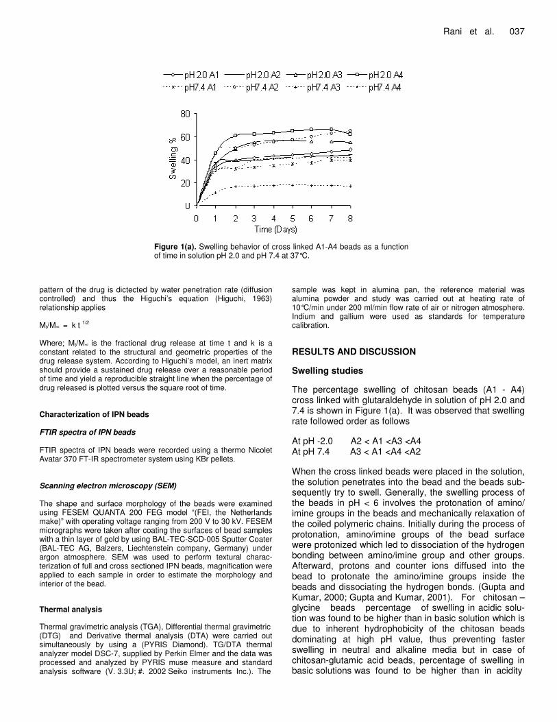
Figure 1(a). Swelling behavior of cross linked A1-A4 beads as a function of time in solution pH 2.0 and pH 7.4 at 37°C.
pattern of the drug is dictected by water penetration rate (diffusion controlled) and thus the Higuchi’s equation (Higuchi, 1963) relationship applies Mt/M� = k t 1/2 Where; Mt/M� is the fractional drug release at time t and k is a constant related to the structural and geometric properties of the drug release system. According to Higuchi’s model, an inert matrix should provide a sustained drug release over a reasonable period of time and yield a reproducible straight line when the percentage of drug released is plotted versus the square root of time. Characterization of IPN beads FTIR spectra of IPN beads FTIR spectra of IPN beads were recorded using a thermo Nicolet Avatar 370 FT-IR spectrometer system using KBr pellets. Scanning electron microscopy (SEM) The shape and surface morphology of the beads were examined using FESEM QUANTA 200 FEG model “(FEI, the Netherlands make)” with operating voltage ranging from 200 V to 30 kV. FESEM micrographs were taken after coating the surfaces of bead samples with a thin layer of gold by using BAL-TEC-SCD-005 Sputter Coater (BAL-TEC AG, Balzers, Liechtenstein company, Germany) under argon atmosphere. SEM was used to perform textural charac-terization of full and cross sectioned IPN beads, magnification were applied to each sample in order to estimate the morphology and interior of the bead. Thermal analysis Thermal gravimetric analysis (TGA), Differential thermal gravimetric (DTG) and Derivative thermal analysis (DTA) were carried out simultaneously by using a (PYRIS Diamond). TG/DTA thermal analyzer model DSC-7, supplied by Perkin Elmer and the data was processed and analyzed by PYRIS muse measure and standard analysis software (V. 3.3U; #. 2002 Seiko instruments Inc.). The
sample was kept in alumina pan, the reference material was alumina powder and study was carried out at heating rate of 10°C/min under 200 ml/min flow rate of air or nitrogen atmosphere. Indium and gallium were used as standards for temperature calibration. RESULTS AND DISCUSSION Swelling studies The percentage swelling of chitosan beads (A1 - A4) cross linked with glutaraldehyde in solution of pH 2.0 and 7.4 is shown in Figure 1(a). It was observed that swelling rate followed order as follows At pH -2.0 A2 < A1 <A3 <A4 At pH 7.4 A3 < A1 <A4 <A2 When the cross linked beads were placed in the solution, the solution penetrates into the bead and the beads sub-sequently try to swell. Generally, the swelling process of the beads in pH < 6 involves the protonation of amino/ imine groups in the beads and mechanically relaxation of the coiled polymeric chains. Initially during the process of protonation, amino/imine groups of the bead surface were protonized which led to dissociation of the hydrogen bonding between amino/imine group and other groups. Afterward, protons and counter ions diffused into the bead to protonate the amino/imine groups inside the beads and dissociating the hydrogen bonds. (Gupta and Kumar, 2000; Gupta and Kumar, 2001). For chitosan – glycine beads percentage of swelling in acidic solu-tion was found to be higher than in basic solution which is due to inherent hydrophobicity of the chitosan beads dominating at high pH value, thus preventing faster swelling in neutral and alkaline media but in case of chitosan-glutamic acid beads, percentage of swelling in basic solutions was found to be higher than in acidity
038 Afr. J. Pharm. Pharmacol.
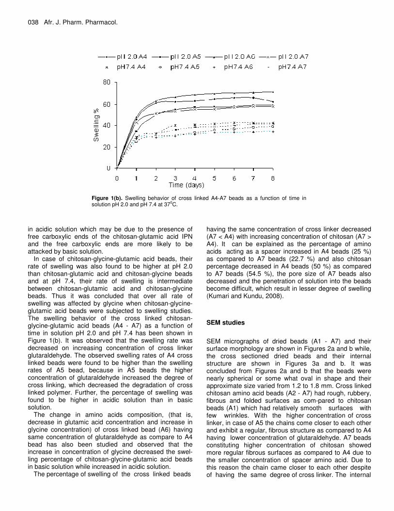
Figure 1(b). Swelling behavior of cross linked A4-A7 beads as a function of time in solution pH 2.0 and pH 7.4 at 370C.
in acidic solution which may be due to the presence of free carboxylic ends of the chitosan-glutamic acid IPN and the free carboxylic ends are more likely to be attacked by basic solution.
In case of chitosan-glycine-glutamic acid beads, their rate of swelling was also found to be higher at pH 2.0 than chitosan-glutamic acid and chitosan-glycine beads and at pH 7.4, their rate of swelling is intermediate between chitosan-glutamic acid and chitosan-glycine beads. Thus it was concluded that over all rate of swelling was affected by glycine when chitosan-glycine-glutamic acid beads were subjected to swelling studies. The swelling behavior of the cross linked chitosan-glycine-glutamic acid beads (A4 - A7) as a function of time in solution pH 2.0 and pH 7.4 has been shown in Figure 1(b). It was observed that the swelling rate was decreased on increasing concentration of cross linker glutaraldehyde. The observed swelling rates of A4 cross linked beads were found to be higher than the swelling rates of A5 bead, because in A5 beads the higher concentration of glutaraldehyde increased the degree of cross linking, which decreased the degradation of cross linked polymer. Further, the percentage of swelling was found to be higher in acidic solution than in basic solution.
The change in amino acids composition, (that is, decrease in glutamic acid concentration and increase in glycine concentration) of cross linked bead (A6) having same concentration of glutaraldehyde as compare to A4 bead has also been studied and observed that the increase in concentration of glycine decreased the swel-ling percentage of chitosan-glycine-glutamic acid beads in basic solution while increased in acidic solution.
The percentage of swelling of the cross linked beads
having the same concentration of cross linker decreased (A7 < A4) with increasing concentration of chitosan (A7 > A4). It can be explained as the percentage of amino acids acting as a spacer increased in A4 beads (25 %) as compared to A7 beads (22.7 %) and also chitosan percentage decreased in A4 beads (50 %) as compared to A7 beads (54.5 %), the pore size of A7 beads also decreased and the penetration of solution into the beads become difficult, which result in lesser degree of swelling (Kumari and Kundu, 2008). SEM studies
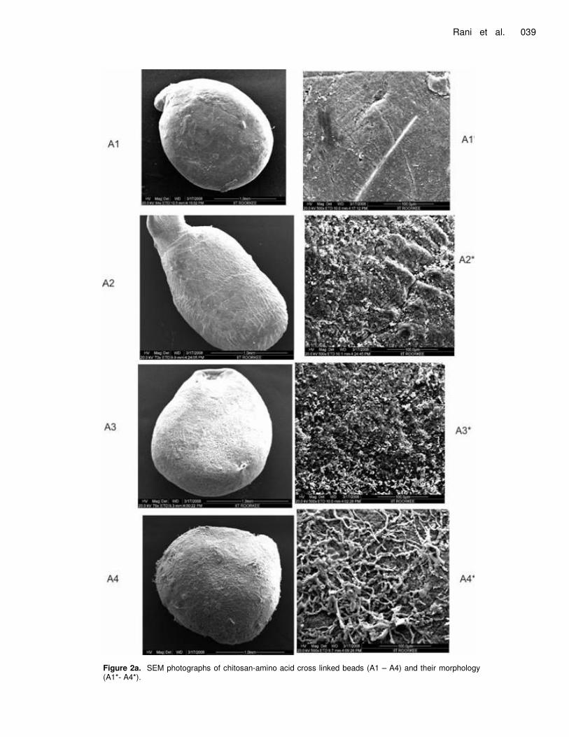
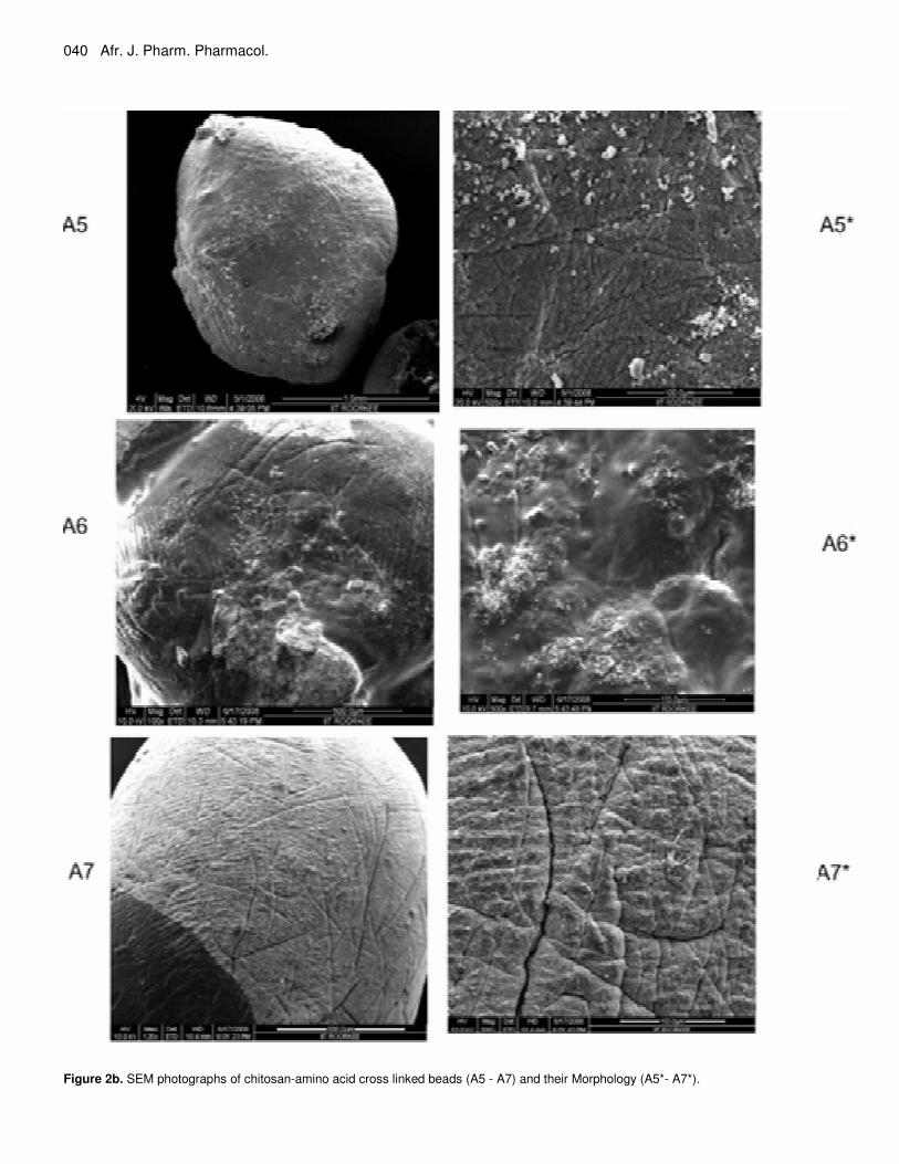
SEM micrographs of dried beads (A1 - A7) and their surface morphology are shown in Figures 2a and b while, the cross sectioned dried beads and their internal structure are shown in Figures 3a and b. It was concluded from Figures 2a and b that the beads were nearly spherical or some what oval in shape and their approximate size varied from 1.2 to 1.8 mm. Cross linked chitosan amino acid beads (A2 - A7) had rough, rubbery, fibrous and folded surfaces as com-pared to chitosan beads (A1) which had relatively smooth surfaces with few wrinkles. With the higher concentration of cross linker, in case of A5 the chains come closer to each other and exhibit a regular, fibrous structure as compared to A4 having lower concentration of glutaraldehyde. A7 beads constituting higher concentration of chitosan showed more regular fibrous surfaces as compared to A4 due to the smaller concentration of spacer amino acid. Due to this reason the chain came closer to each other despite of having the same degree of cross linker. The internal
Rani et al. 039
Figure 2a. SEM photographs of chitosan-amino acid cross linked beads (A1 – A4) and their morphology (A1*- A4*).
040 Afr. J. Pharm. Pharmacol.
Figure 2b. SEM photographs of chitosan-amino acid cross linked beads (A5 - A7) and their Morphology (A5*- A7*).
Rani et al. 041
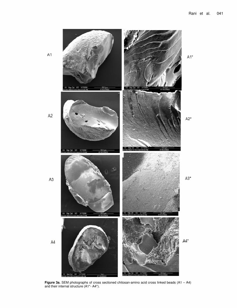
Figure 3a. SEM photographs of cross sectioned chitosan-amino acid cross linked beads (A1 – A4) and their internal structure (A1*- A4*).
042 Afr. J. Pharm. Pharmacol.
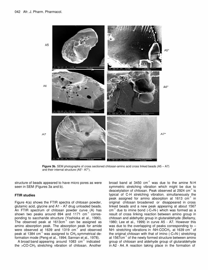
Figure 3b. SEM photographs of cross sectioned chitosan-amino acid cross linked beads (A5 – A7) and their internal structure (A5*- A7*).
structure of beads appeared to have micro pores as were seen in SEM (Figures 3a and b).
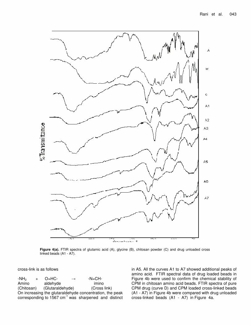
FTIR studies Figure 4(a) shows the FTIR spectra of chitosan powder, glutamic acid, glycine and A1 - A7 drug unloaded beads. An FTIR spectrum of chitosan powder curve (A) has shown two peaks around 894 and 1171 cm-1 corres-ponding to saccharide structure (Yoshioka et al., 1990). The observed peak at 1613cm-1 can be assigned as amino absorption peak. The absorption peak for amide were observed at 1639 and 1319 cm-1 and observed peak at 1384 cm-1 was assigned to CH3 symmetrical de-formation mode (Peng et al., 1994; Sannan et al., 1978).
A broad band appearing around 1083 cm-1 indicated the >CO-CH3 stretching vibration of chitosan. Another
broad band at 3450 cm-1 was due to the amine N-H symmetric stretching vibration which might be due to deacetylation of chitosan. Peak observed at 2924 cm-1 is typical of C-H stretching vibration. simultaneously the peak assigned for amino absorption at 1613 cm-1 in original chitosan broadened or disappeared in cross linked beads and a new peak appearing at about 1567 cm-1 due to imine bond (-C=N-) which was formed as a result of cross linking reaction between amino group in chitosan and aldehydic group in glutaraldehyde (Bellamy, 1980; Lee et al., 1999) in curve A5 - A7. However this was due to the overlapping of peaks corresponding to –NH- stretching vibrations in -NH-COCH3 at 1639 cm-1 of the original chitosan with that of imino (-C=N-) stretching at 1567cm-1 of the newly formed structure between amino group of chitosan and aldehyde group of glutaraldehyde in A2 - A4. A reaction taking place in the formation of
Rani et al. 043
Figure 4(a). FTIR spectra of glutamic acid (A), glycine (B), chitosan powder (C) and drug unloaded cross linked beads (A1 - A7).
cross-link is as follows -NH2 + O=HC- � -N=CH- Amino aldehyde imino (Chitosan) (Glutaraldehyde) (Cross link) On increasing the glutaraldehyde concentration, the peak corresponding to 1567 cm-1 was sharpened and distinct
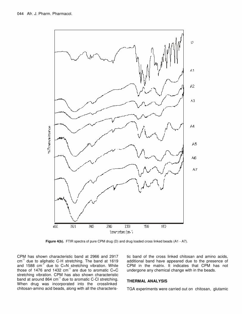
in A5. All the curves A1 to A7 showed additional peaks of amino acid. FTIR spectral data of drug loaded beads in Figure 4b were used to confirm the chemical stability of CPM in chitosan amino acid beads. FTIR spectra of pure CPM drug (curve D) and CPM loaded cross-linked beads (A1 - A7) in Figure 4b were compared with drug unloaded cross-linked beads (A1 - A7) in Figure 4a.
044 Afr. J. Pharm. Pharmacol.
Figure 4(b). FTIR spectra of pure CPM drug (D) and drug loaded cross linked beads (A1 - A7).
CPM has shown characteristic band at 2966 and 2917 cm-1 due to aliphatic C-H stretching. The band at 1619 and 1588 cm-1 due to C=N stretching vibration. While those of 1476 and 1432 cm-1 are due to aromatic C=C stretching vibration. CPM has also shown characteristic band at around 864 cm-1 due to aromatic C-Cl stretching. When drug was incorporated into the crosslinked chitosan-amino acid beads, along with all the characteris-
tic band of the cross linked chitosan and amino acids, additional band have appeared due to the presence of CPM in the matrix. It indicates that CPM has not undergone any chemical change with in the beads. THERMAL ANALYSIS
TGA experiments were carried out on chitosan, glutamic
Rani et al. 045
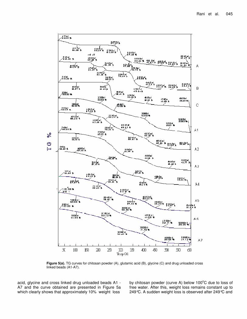
Figure 5(a). TG curves for chitosan powder (A), glutamic acid (B), glycine (C) and drug unloaded cross linked beads (A1-A7).
acid, glycine and cross linked drug unloaded beads A1 - A7 and the curve obtained are presented in Figure 5a which clearly shows that approximately 10% weight loss
by chitosan powder (curve A) below 100oC due to loss of free water. After this, weight loss remains constant up to 249°C. A sudden weight loss is observed after 249°C and
046 Afr. J. Pharm. Pharmacol.
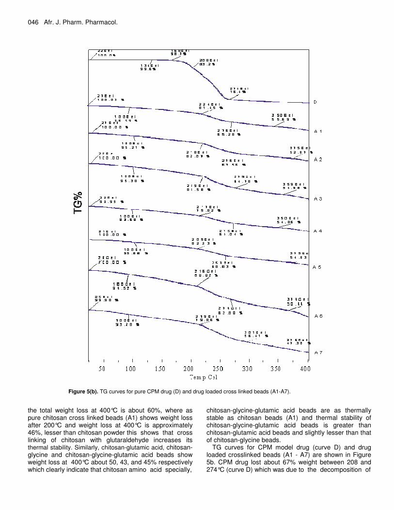
Figure 5(b). TG curves for pure CPM drug (D) and drug loaded cross linked beads (A1-A7).
the total weight loss at 400°C is about 60%, where as pure chitosan cross linked beads (A1) shows weight loss after 200°C and weight loss at 400°C is approximately 46%, lesser than chitosan powder this shows that cross linking of chitosan with glutaraldehyde increases its thermal stability. Similarly, chitosan-glutamic acid, chitosan-glycine and chitosan-glycine-glutamic acid beads show weight loss at 400°C about 50, 43, and 45% respectively which clearly indicate that chitosan amino acid specially,
chitosan-glycine-glutamic acid beads are as thermally stable as chitosan beads (A1) and thermal stability of chitosan-glycine-glutamic acid beads is greater than chitosan-glutamic acid beads and slightly lesser than that of chitosan-glycine beads.
TG curves for CPM model drug (curve D) and drug loaded crosslinked beads (A1 - A7) are shown in Figure 5b. CPM drug lost about 67% weight between 208 and 274°C (curve D) which was due to the decomposition of
Rani et al. 047
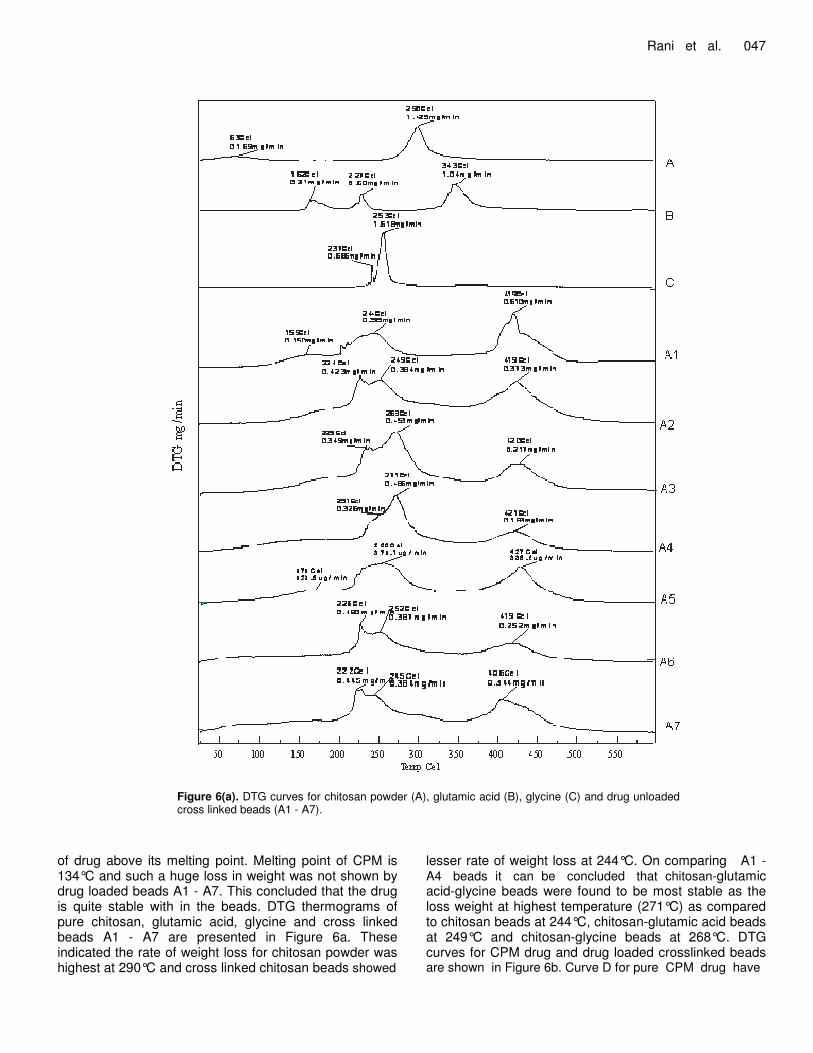
Figure 6(a). DTG curves for chitosan powder (A), glutamic acid (B), glycine (C) and drug unloaded cross linked beads (A1 - A7).
of drug above its melting point. Melting point of CPM is 134°C and such a huge loss in weight was not shown by drug loaded beads A1 - A7. This concluded that the drug is quite stable with in the beads. DTG thermograms of pure chitosan, glutamic acid, glycine and cross linked beads A1 - A7 are presented in Figure 6a. These indicated the rate of weight loss for chitosan powder was highest at 290°C and cross linked chitosan beads showed
lesser rate of weight loss at 244°C. On comparing A1 - A4 beads it can be concluded that chitosan-glutamic acid-glycine beads were found to be most stable as the loss weight at highest temperature (271°C) as compared to chitosan beads at 244°C, chitosan-glutamic acid beads at 249°C and chitosan-glycine beads at 268°C. DTG curves for CPM drug and drug loaded crosslinked beads are shown in Figure 6b. Curve D for pure CPM drug have
048 Afr. J. Pharm. Pharmacol.
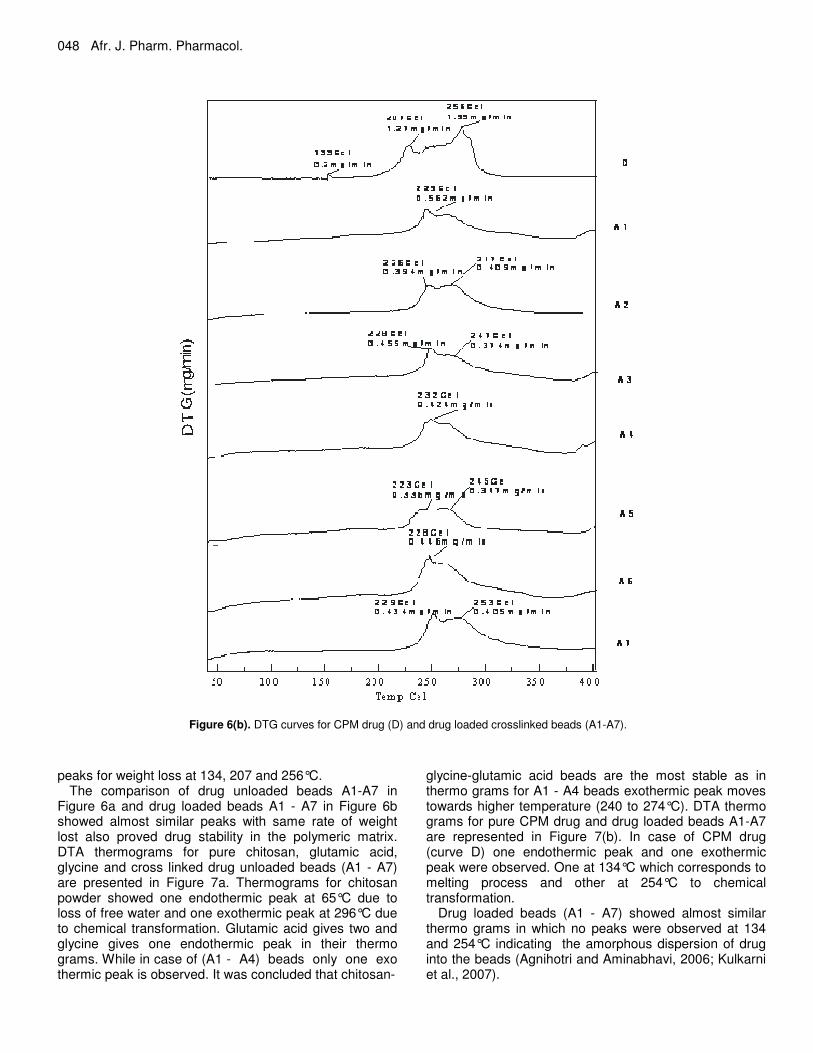
Figure 6(b). DTG curves for CPM drug (D) and drug loaded crosslinked beads (A1-A7).
peaks for weight loss at 134, 207 and 256°C.
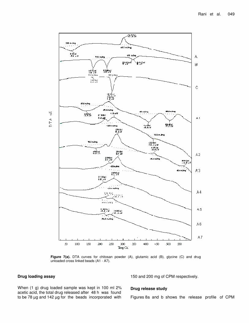
The comparison of drug unloaded beads A1-A7 in Figure 6a and drug loaded beads A1 - A7 in Figure 6b showed almost similar peaks with same rate of weight lost also proved drug stability in the polymeric matrix. DTA thermograms for pure chitosan, glutamic acid, glycine and cross linked drug unloaded beads (A1 - A7) are presented in Figure 7a. Thermograms for chitosan powder showed one endothermic peak at 65°C due to loss of free water and one exothermic peak at 296°C due to chemical transformation. Glutamic acid gives two and glycine gives one endothermic peak in their thermo grams. While in case of (A1 - A4) beads only one exo thermic peak is observed. It was concluded that chitosan-
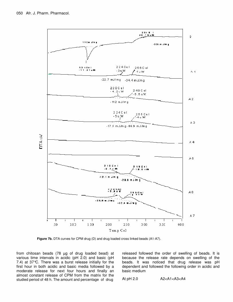
glycine-glutamic acid beads are the most stable as in thermo grams for A1 - A4 beads exothermic peak moves towards higher temperature (240 to 274°C). DTA thermo grams for pure CPM drug and drug loaded beads A1-A7 are represented in Figure 7(b). In case of CPM drug (curve D) one endothermic peak and one exothermic peak were observed. One at 134°C which corresponds to melting process and other at 254°C to chemical transformation.
Drug loaded beads (A1 - A7) showed almost similar thermo grams in which no peaks were observed at 134 and 254°C indicating the amorphous dispersion of drug into the beads (Agnihotri and Aminabhavi, 2006; Kulkarni et al., 2007).
Rani et al. 049
Figure 7(a). DTA curves for chitosan powder (A), glutamic acid (B), glycine (C) and drug unloaded cross linked beads (A1 - A7).
Drug loading assay When (1 g) drug loaded sample was kept in 100 ml 2% acetic acid, the total drug released after 48 h was found to be 78 µg and 142 µg for the beads incorporated with
150 and 200 mg of CPM respectively. Drug release study Figures 8a and b shows the release profile of CPM
050 Afr. J. Pharm. Pharmacol.
Figure 7b. DTA curves for CPM drug (D) and drug loaded cross linked beads (A1-A7).
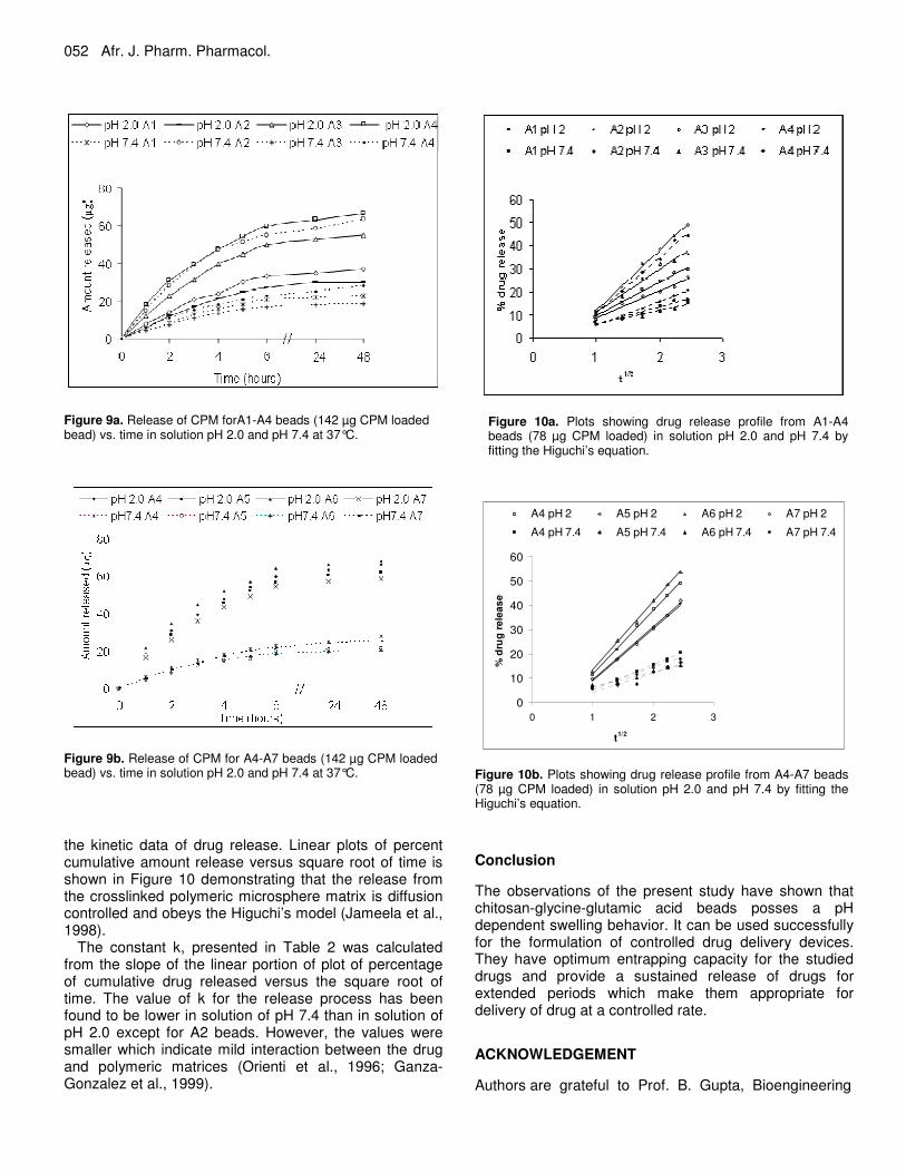
from chitosan beads (78 µg of drug loaded bead) at various time intervals in acidic (pH 2.0) and basic (pH 7.4) at 37°C. There was a burst release initially for the first hour in both acidic and basic media followed by a moderate release for next four hours and finally an almost constant release of CPM from the matrix for the studied period of 48 h. The amount and percentage of drug
released followed the order of swelling of beads. It is because the release rate depends on swelling of the beads. It was noticed that drug release was pH dependent and followed the following order in acidic and basic medium At pH 2.0 A2<A1<A3<A4
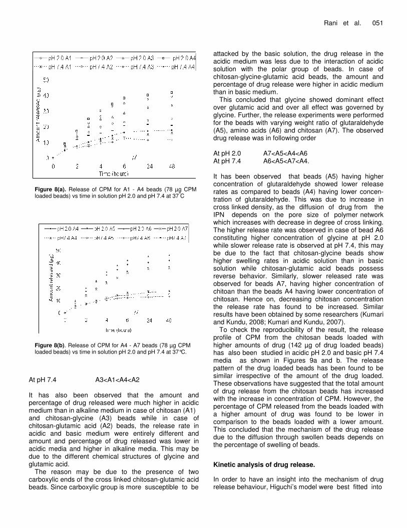
Figure 8(a). Release of CPM for A1 - A4 beads (78 µg CPM loaded beads) vs time in solution pH 2.0 and pH 7.4 at 37°C
Figure 8(b). Release of CPM for A4 - A7 beads (78 µg CPM loaded beads) vs time in solution pH 2.0 and pH 7.4 at 37°C.
At pH 7.4 A3<A1<A4<A2 It has also been observed that the amount and percentage of drug released were much higher in acidic medium than in alkaline medium in case of chitosan (A1) and chitosan-glycine (A3) beads while in case of chitosan-glutamic acid (A2) beads, the release rate in acidic and basic medium were entirely different and amount and percentage of drug released was lower in acidic media and higher in alkaline media. This may be due to the different chemical structures of glycine and glutamic acid.
The reason may be due to the presence of two carboxylic ends of the cross linked chitosan-glutamic acid beads. Since carboxylic group is more susceptible to be
Rani et al. 051 attacked by the basic solution, the drug release in the acidic medium was less due to the interaction of acidic solution with the polar group of beads. In case of chitosan-glycine-glutamic acid beads, the amount and percentage of drug release were higher in acidic medium than in basic medium.
This concluded that glycine showed dominant effect over glutamic acid and over all effect was governed by glycine. Further, the release experiments were performed for the beads with varying weight ratio of glutaraldehyde (A5), amino acids (A6) and chitosan (A7). The observed drug release was in following order At pH 2.0 A7<A5<A4<A6 At pH 7.4 A6<A5<A7<A4. It has been observed that beads (A5) having higher concentration of glutaraldehyde showed lower release rates as compared to beads (A4) having lower concen-tration of glutaraldehyde. This was due to increase in cross linked density, as the diffusion of drug from the IPN depends on the pore size of polymer network which increases with decrease in degree of cross linking. The higher release rate was observed in case of bead A6 constituting higher concentration of glycine at pH 2.0 while slower release rate is observed at pH 7.4, this may be due to the fact that chitosan-glycine beads show higher swelling rates in acidic solution than in basic solution while chitosan-glutamic acid beads possess reverse behavior. Similarly, slower released rate was observed for beads A7, having higher concentration of chitoan than the beads A4 having lower concentration of chitosan. Hence on, decreasing chitosan concentration the release rate has found to be increased. Similar results have been obtained by some researchers (Kumari and Kundu, 2008; Kumari and Kundu, 2007).
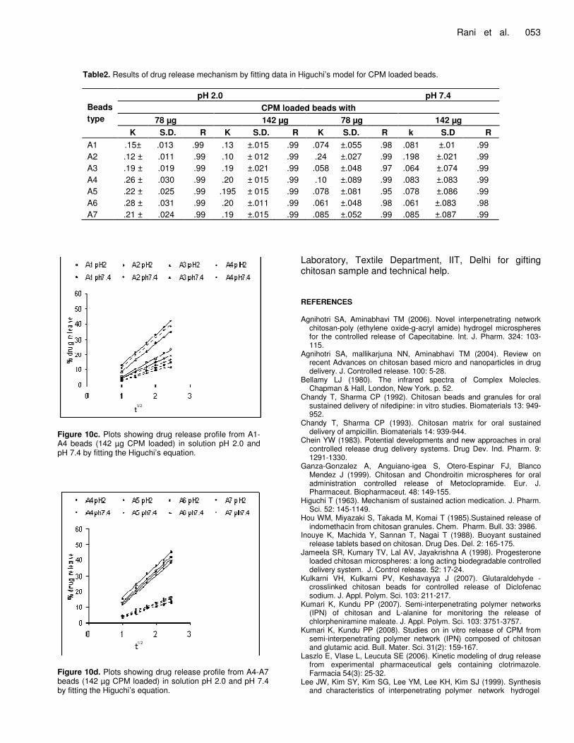
To check the reproducibility of the result, the release profile of CPM from the chitosan beads loaded with higher amounts of drug (142 µg of drug loaded beads) has also been studied in acidic pH 2.0 and basic pH 7.4 media as shown in Figures 9a and b. The release pattern of the drug loaded beads has been found to be similar irrespective of the amount of the drug loaded. These observations have suggested that the total amount of drug release from the chitosan beads has increased with the increase in concentration of CPM. However, the percentage of CPM released from the beads loaded with a higher amount of drug was found to be lower in comparison to the beads loaded with a lower amount. This concluded that the mechanism of the drug release due to the diffusion through swollen beads depends on the percentage of swelling of beads. Kinetic analysis of drug release. In order to have an insight into the mechanism of drug release behaviour, Higuchi’s model were best fitted into
052 Afr. J. Pharm. Pharmacol.
Figure 9a. Release of CPM forA1-A4 beads (142 µg CPM loaded bead) vs. time in solution pH 2.0 and pH 7.4 at 37°C.
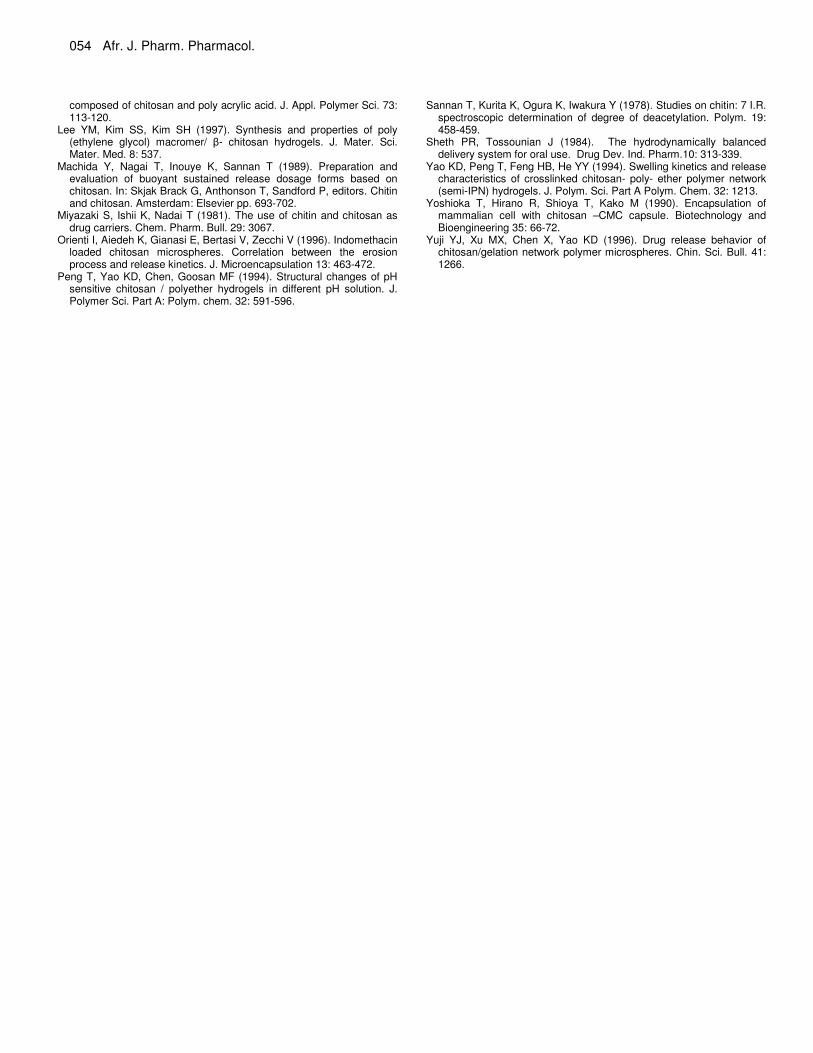
Figure 9b. Release of CPM for A4-A7 beads (142 µg CPM loaded bead) vs. time in solution pH 2.0 and pH 7.4 at 37°C. the kinetic data of drug release. Linear plots of percent cumulative amount release versus square root of time is shown in Figure 10 demonstrating that the release from the crosslinked polymeric microsphere matrix is diffusion controlled and obeys the Higuchi’s model (Jameela et al., 1998).
The constant k, presented in Table 2 was calculated from the slope of the linear portion of plot of percentage of cumulative drug released versus the square root of time. The value of k for the release process has been found to be lower in solution of pH 7.4 than in solution of pH 2.0 except for A2 beads. However, the values were smaller which indicate mild interaction between the drug and polymeric matrices (Orienti et al., 1996; Ganza- Gonzalez et al., 1999).
Figure 10a. Plots showing drug release profile from A1-A4 beads (78 µg CPM loaded) in solution pH 2.0 and pH 7.4 by fitting the Higuchi’s equation.
0
10
20
30
40
50
60
0 1 2 3
t1/2
% d
rug
rel
ease
A4 pH 2 A5 pH 2 A6 pH 2 A7 pH 2
A4 pH 7.4 A5 pH 7.4 A6 pH 7.4 A7 pH 7.4
Figure 10b. Plots showing drug release profile from A4-A7 beads (78 µg CPM loaded) in solution pH 2.0 and pH 7.4 by fitting the Higuchi’s equation. Conclusion The observations of the present study have shown that chitosan-glycine-glutamic acid beads posses a pH dependent swelling behavior. It can be used successfully for the formulation of controlled drug delivery devices. They have optimum entrapping capacity for the studied drugs and provide a sustained release of drugs for extended periods which make them appropriate for delivery of drug at a controlled rate. ACKNOWLEDGEMENT Authors are grateful to Prof. B. Gupta, Bioengineering
Rani et al. 053
Table2. Results of drug release mechanism by fitting data in Higuchi’s model for CPM loaded beads. Beads type
pH 2.0 pH 7.4 CPM loaded beads with
78 µg 142 µg 78 µg 142 µg K S.D. R K S.D. R K S.D. R k S.D R
A1 A2 A3 A4 A5 A6 A7
.15± .12 ± .19 ± .26 ± .22 ± .28 ± .21 ±
.013 .011 .019 .030 .025 .031 .024
.99 .99 .99 .99 .99 .99 .99
.13
.10
.19
.20 .195 .20 .19
±.015 ± 012 ±.021 ± 015 ± 015 ±.011 ±.015
.99
.99
.99
.99
.99
.99
.99
.074 .24
.058 .10
.078
.061
.085
±.055 ±.027 ±.048 ±.089 ±.081 ±.048 ±.052
.98
.99
.97
.99
.95
.98
.99
.081
.198
.064
.083
.078
.061
.085
±.01 ±.021 ±.074 ±.083 ±.086 ±.083 ±.087
.99
.99
.99
.99
.99
.98
.99
Figure 10c. Plots showing drug release profile from A1-A4 beads (142 µg CPM loaded) in solution pH 2.0 and pH 7.4 by fitting the Higuchi’s equation.
Figure 10d. Plots showing drug release profile from A4-A7 beads (142 µg CPM loaded) in solution pH 2.0 and pH 7.4 by fitting the Higuchi’s equation.
Laboratory, Textile Department, IIT, Delhi for gifting chitosan sample and technical help. REFERENCES Agnihotri SA, Aminabhavi TM (2006). Novel interpenetrating network
chitosan-poly (ethylene oxide-g-acryl amide) hydrogel microspheres for the controlled release of Capecitabine. Int. J. Pharm. 324: 103-115.
Agnihotri SA, mallikarjuna NN, Aminabhavi TM (2004). Review on recent Advances on chitosan based micro and nanoparticles in drug delivery. J. Controlled release. 100: 5-28.
Bellamy LJ (1980). The infrared spectra of Complex Molecles. Chapman & Hall, London, New York. p. 52.
Chandy T, Sharma CP (1992). Chitosan beads and granules for oral sustained delivery of nifedipine: in vitro studies. Biomaterials 13: 949-952.
Chandy T, Sharma CP (1993). Chitosan matrix for oral sustained delivery of ampicillin. Biomaterials 14: 939-944.
Chein YW (1983). Potential developments and new approaches in oral controlled release drug delivery systems. Drug Dev. Ind. Pharm. 9: 1291-1330.
Ganza-Gonzalez A, Anguiano-igea S, Otero-Espinar FJ, Blanco Mendez J (1999). Chitosan and Chondroitin microspheres for oral administration controlled release of Metoclopramide. Eur. J. Pharmaceut. Biopharmaceut. 48: 149-155.
Higuchi T (1963). Mechanism of sustained action medication. J. Pharm. Sci. 52: 145-1149.
Hou WM, Miyazaki S, Takada M, Komai T (1985).Sustained release of indomethacin from chitosan granules. Chem. Pharm. Bull. 33: 3986.
Inouye K, Machida Y, Sannan T, Nagai T (1988). Buoyant sustained release tablets based on chitosan. Drug Des. Del. 2: 165-175.
Jameela SR, Kumary TV, Lal AV, Jayakrishna A (1998). Progesterone loaded chitosan microspheres: a long acting biodegradable controlled delivery system. J. Control release. 52: 17-24.
Kulkarni VH, Kulkarni PV, Keshavayya J (2007). Glutaraldehyde - crosslinked chitosan beads for controlled release of Diclofenac sodium. J. Appl. Polym. Sci. 103: 211-217.
Kumari K, Kundu PP (2007). Semi-interpenetrating polymer networks (IPN) of chitosan and L-alanine for monitoring the release of chlorpheniramine maleate. J. Appl. Polym. Sci. 103: 3751-3757.
Kumari K, Kundu PP (2008). Studies on in vitro release of CPM from semi-interpenetrating polymer network (IPN) composed of chitosan and glutamic acid. Bull. Mater. Sci. 31(2): 159-167.
Laszlo E, Vlase L, Leucuta SE (2006). Kinetic modeling of drug release from experimental pharmaceutical gels containing clotrimazole. Farmacia 54(3): 25-32.
Lee JW, Kim SY, Kim SG, Lee YM, Lee KH, Kim SJ (1999). Synthesis and characteristics of interpenetrating polymer network hydrogel
054 Afr. J. Pharm. Pharmacol. composed of chitosan and poly acrylic acid. J. Appl. Polymer Sci. 73: 113-120.
Lee YM, Kim SS, Kim SH (1997). Synthesis and properties of poly (ethylene glycol) macromer/ �- chitosan hydrogels. J. Mater. Sci. Mater. Med. 8: 537.
Machida Y, Nagai T, Inouye K, Sannan T (1989). Preparation and evaluation of buoyant sustained release dosage forms based on chitosan. In: Skjak Brack G, Anthonson T, Sandford P, editors. Chitin and chitosan. Amsterdam: Elsevier pp. 693-702.
Miyazaki S, Ishii K, Nadai T (1981). The use of chitin and chitosan as drug carriers. Chem. Pharm. Bull. 29: 3067.
Orienti I, Aiedeh K, Gianasi E, Bertasi V, Zecchi V (1996). Indomethacin loaded chitosan microspheres. Correlation between the erosion process and release kinetics. J. Microencapsulation 13: 463-472.
Peng T, Yao KD, Chen, Goosan MF (1994). Structural changes of pH sensitive chitosan / polyether hydrogels in different pH solution. J. Polymer Sci. Part A: Polym. chem. 32: 591-596.
Sannan T, Kurita K, Ogura K, Iwakura Y (1978). Studies on chitin: 7 I.R.
spectroscopic determination of degree of deacetylation. Polym. 19: 458-459.
Sheth PR, Tossounian J (1984). The hydrodynamically balanced delivery system for oral use. Drug Dev. Ind. Pharm.10: 313-339.
Yao KD, Peng T, Feng HB, He YY (1994). Swelling kinetics and release characteristics of crosslinked chitosan- poly- ether polymer network (semi-IPN) hydrogels. J. Polym. Sci. Part A Polym. Chem. 32: 1213.
Yoshioka T, Hirano R, Shioya T, Kako M (1990). Encapsulation of mammalian cell with chitosan –CMC capsule. Biotechnology and Bioengineering 35: 66-72.
Yuji YJ, Xu MX, Chen X, Yao KD (1996). Drug release behavior of chitosan/gelation network polymer microspheres. Chin. Sci. Bull. 41: 1266.