a comparative translational study ... - dott. pietro...
TRANSCRIPT

SM
A Comparative Translational Study: The Combined
Use of Enhanced Stromal Vascular Fraction and
Platelet-Rich Plasma Improves Fat Grafting
Maintenance in Breast Reconstruction
PIETRO GENTILE,aAUGUSTO ORLANDI,
bMARIA GIOVANNA SCIOLI,
bCAMILLA DI PASQUALI,
a
ILARIA BOCCHINI,aCRISTIANO BENIAMINO CURCIO,
aMICOL FLORIS,
aVALERIA FIASCHETTI,
c
ROBERTO FLORIS,cVALERIO CERVELLI
a
Key Words. Adipose • Adult stem cells • Autologous stem cell transplantation •Clinical translation • Stem cell transplantation • Tissue-specific stem cells
aPlastic and ReconstructiveSurgery Department,bInstitute of AnatomicPathology, and cDepartmentof Diagnostic Imaging,University of Rome TorVergata, Rome, Italy
Correspondence: Pietro Gentile,M.D., San Salvatore in LauroPlace, no. 15, 00186 Rome, Italy.Telephone: 393388515479; e-mail:[email protected]
Received January 3, 2012;accepted for publication March5, 2012; first published online inSCTM EXPRESS April 13, 2012.
©AlphaMed Press1066-5099/2012/$20.00/0
http://dx.doi.org/10.5966/sctm.2011-0065
ABSTRACT
The use of autologous fat grafting is ideal in breast reconstruction. However, published data on
long-termoutcomes and instrumental results of fat grafting to the breast are lacking. The purpose of
this studywas to review the authorsQ experience of fat grafting, evaluating the effects related to the
use of enhanced stromal vascular fraction (e-SVF) and fat grafting with platelet-rich plasma (PRP) in
themaintenance of fat volume in breast reconstruction, comparing the results with a control group.
Twenty-three patients aged 19W60 years affected by breast soft tissue defects were analyzed at the
Plastic and Reconstructive Department of the University of Rome Tor Vergata. Ten patients were
treated with SVF-enhanced autologous fat grafts, and 13 patients were treated with fat grafting !platelet-rich plasma. The patients in the control group (n " 10) were treated with centrifuged fat
grafting injection according to ColemanQs procedure. The patients treatedwith SVF-enhanced autol-
ogous fat grafts showed a 63% maintenance of the contour restoring and of three-dimensional
volume after 1 year compared with the patients of the control group treated with centrifuged fat
graft, who showed a 39% maintenance. In those patients who were treated with fat grafting and
PRP, we observed a 69%maintenance of contour restoring and of three-dimensional volume after 1
year. As reported, the use of either e-SVF or PRP mixed with fat grafting produced an improvement
in maintenance of breast volume in patients affected by breast soft tissue defect. STEM CELLSTRANSLATIONAL MEDICINE 2012;1:341–351
INTRODUCTION
The popularization of fat grafting is the result ofan increased demand for breast augmentationwith stromal vascular fraction (SVF) enhancedautologous fat graft. The immediate gratificationand the absence of breast implants have becomepowerful motivators that have enticed patientsto request this regenerative surgery.
During the last 5 years, an increasing numberof publications have reported on this topic. Theareas in which stromal vascular fraction cellshave been used include radiotherapy-based tis-sue damage after mastectomy [1], breast aug-mentation [2], postmastectomy breast recon-struction [3], breast implant complications [4],calvarial defects [5], Crohn’s fistulas and complexperianal fistula [6–9], damaged skeletal muscle[10], Parry-Romberg disease and facial lipoatro-phy [11], scarring, gluteal soft tissue defect, pec-tus excavatus, dermatofibrosis [12], and vocalfold augmentation [13, 14].
Visceral and subcutaneous adipose tissuehas been demonstrated to contain progenitorcells able to differentiate inmultiple cell lineages[10, 15]. After centrifugation of collagenase-di-gested adult adipose tissue, a heterogeneous cellpopulation named stromal-vascular fraction isobtained [15, 16]. This population contains adultstem cells named adipose-derived stromal cells(ASCs) [17]. ASCsmight improve tissue outcomesby increasing vascularity and through the secre-tion of growth factors that improve tissue sur-vival. Recently, the authors published works onthe use of fat grafting in the lipostructure tech-nique as described by Coleman (purified fat ob-tained after centrifugation at 3,000 rpm for 3minutes) [18, 19]mixedwith platelet-rich plasma(PRP) in plastic surgery [20], in lower chronic ex-tremity ulcers [21], and in hemifacial atrophy[22]. Now, we present out experience using re-generative surgerywith the SVF-enhanced autol-ogous fat grafting in breast reconstruction. In
TISSUE ENGINEERING AND REGENERATIVE MEDICINE
STEM CELLS TRANSLATIONAL MEDICINE 2012;1:341–351 www.StemCellsTM.com
by guest on July 3, 2012stem
cellstm.alpham
edpress.orgD
ownloaded from

this report, we present our studies using either SVF or PRPmixedwith fat grafting and patient self-assessment of the outcomes asan additional parameter supporting the results of clinical assess-ment.
MATERIALS AND METHODS
Patients
A total of 23 patients aged 19–60 years were treated from Jan-uary 2008 to February 2012 at the Department of Plastic andReconstructive Surgery of the University of Rome Tor Vergata.
Thirteen patients affected by breast soft tissue defects (3patients affected by unilateral breast hypoplasia, 8 patients af-fected by outcomes of breast cancer reconstruction, and 2 pa-tients after prosthesis removal) were treated with fat graft !PRP (supplemental online Table 1) for breast reconstruction (Fig.1A, 1C). The purified fat was obtained after centrifugation at3,000 rpmandplaced in 1-ml syringes, and itwas then asepticallyreinserted using specific microcannulas for implanting. The 1 mlof centrifuged fat tissue was also mixed with 0.4 ml of PRP. Theselection of location destined to receive the implant was deter-mined taking into account the diversity in the lesions. To implantthe fat tissue, small tunnels were previously created forcing thecannulas of 1.5 mm diameter with accurate and controlledmovements. Once the fat tissue had been implanted at differentlevels, the access incisions were closed using 5-0 nylon stitches,and no compressive bandage was applied.
Ten patients (2 patients affected by unilateral breast hyp-oplasia, 7 patients affected by outcomes of breast cancer recon-struction, and 1 patient after prosthesis removal) were treatedwith SVF-enhanced autologous fat grafts (supplemental online
Table 2), obtained using the Celution System (Fig. 2A). The pa-tients were subjected to additional wash and centrifugation cy-cles, after which 5 ml of the enhanced SVF (e-SVF) suspensionwas extracted from the system. The e-SVF (5 ml) was added tothe tissue collection container with the liposuction. Subsequentto the carrying out of a washing step, the e-SVF suspension wasadded andmixedwith thewashed fat graft. Using specific micro-cannulas for implantation, the SVF-enhanced fat graft was trans-ferred into 10-ml syringes and aseptically reinjected into the softtissue defect. The preoperative evaluation included a completeclinical examination, a photographic assessment, nuclear mag-netic resonance imaging (MRI) (Fig. 3A, 3B) of the soft tissue, andultrasound. In addition, in the more complex cases, such as thecase with absence of pectoralis muscle and Poland syndrome, ahigh-resolution computed tomography scan with three-dimen-sional imaging was performed. Postoperative follow-up tookplace at 2, 7, 15, 21, and 36 weeks and then annually.
To establish the effects of their treatment, we compared ourresults with a control groupmade up of 10 patients (supplemen-tal online Table 3). The control group comprised 10 females aged21–65 years, all affected by breast soft tissue defects (3 patientsaffected by unilateral breast hypoplasia, 5 patients affected byoutcomes of breast cancer reconstruction, and 2 patients afterprosthesis removal). This sample group was treated with centri-fuged fat grafting injection according to the Coleman procedure.
Exclusion criteria were divided into two types: local and sys-temic. The systemic criteria include platelet disorders, thrombo-cytopenia, antiaggregating therapy, bone marrow aplasia, un-compensated diabetes, sepsis, and cancer. The local criteriainclude cancer loss of substance. We did not use tobacco use orgenetic disorders as exclusion criteria. This study is part of a
Figure 1. Breast reconstruction with fat graft ! platelet-rich plasma. (A): Preoperative in frontal projection. (B): Postoperative in frontalprojection after 1 year. (C): Preoperative in 3⁄4 right projection. (D): Postoperative in 3⁄4 right projection after 1 year.
342 e-SVF or PRP in Breast Reconstruction
STEM CELLS TRANSLATIONAL MEDICINE
by guest on July 3, 2012stem
cellstm.alpham
edpress.orgD
ownloaded from

research project approved by Tor Vergata, and all procedureswere performed under written patient informed consent andaccording to the guidelines of the local committee on humanresearch.
Harvest Region and Preparation of the SVF-Enhanced
Autologous Fat Graft
The cell and tissue preparation procedure mainly exhibited twophases. Phase 1 started with a syringe liposuction (715.4 ml av-erage in all patients; range, 250–1,080 ml) in the abdominal re-gion using 3-mm cannulas (supplemental online Fig. 1A ). Whileaseptic technique was maintained, the plunger of the 60-ml sy-ringe was removed, and the tip was closedwith a cap. Half of thelipoaspirate (234.46 ml average) was placed into the tissue col-lection container of the Celution 800/CRS System (Cytori Thera-peutics Inc., SanDiego, http://www.cytoritx.com) (supplementalonline Fig. 1B). Blood and free lipid was removed from the tissue(supplemental online Fig. 1D) through a wash cycle (supplemen-tal online Fig. 1C), and the Celase 835/CRS Reagent (Cytori Ther-apeutics) was added to enzymatically digest the tissue, whichreleased SVF (supplemental online Fig. 1E). After additionalwash(supplemental online Fig. 1F) and centrifugation cycles (supple-mental online Fig. 1G), 4–5 ml of the SVF suspension was ex-tracted from the system (supplemental online Fig. 1H). In thesecond phase, the remaining part of the lipoaspirate was addedto the tissue collection container (supplemental online Fig. 2A)and a washing step was automatically carried out (supplementalonline Fig. 2B). Once completed, the 4–5 ml of SVF suspensionwas added (supplemental online Fig. 2C) and mixed with thewashed fat graft (supplemental online Fig. 2D), resulting in ap-
proximately 429.61 ml (range, 60–620 ml) of SVF-enhanced fattissue for grafting (supplemental online Fig. 2E). This newly pro-cessed cell-enhanced fat graft typically consists of 25%–30%wa-ter, which will be reabsorbed by the body in the postoperativeperiod. This overall process was controlled through automatedsensors and processing algorithms that ensured standard han-dling of the tissue and cells, and the process was completedwithin 160 minutes. The SVF-enhanced fat graft was transferredinto 10-ml syringes (supplemental Fig. 2F) and aseptically rein-jected into the patient using specific microcannulas for implan-tation (supplemental Fig. 2G, 2H).
The donor site region was infiltrated with a cold saline solu-tion containing 1 ml of adrenaline per 500 ml of saline solutionwithout lidocaine or carbocaine. Adipose tissue was removedafter 5 minutes using a 3-mm-diameter cannula and a 60-mlToomey syringe.We reinjected the SVF-enhanced adipose tissueusing specific microcannulas (1–2mm in diameter) for implanta-tion.
Surgical Technique and Location of Implantation
The area destined to receive the graft was determined on thebasis of the necessary corrections. Based on the necessarycorrections, the harvested material was implanted for breastaugmentation prevalently into three areas: inferior breastrim, superior and inferior regions of the areola, and the supe-rior lateral quadrant.
After pretunnelling, fat tissue was implanted (395.4 mlaverage [range, 80–600 ml]; 197.7 ml average for eachbreast) at different levels using a delivery cannula (1–2 mm in
Figure 2. Patients treated with stromal vascular fraction-enhanced autologous fat grafts. (A): Preoperative in frontal projection. (B): Post-operative in frontal projection after 1 year. (C): Preoperative in 3⁄4 right projection. (D): Postoperative in 3⁄4 right projection after 1 year.
343Gentile, Orlandi, Scioli et al.
www.StemCellsTM.com
by guest on July 3, 2012stem
cellstm.alpham
edpress.orgD
ownloaded from
diameter) with precise, controlled movements. Small quanti-ties of the SVF-enhanced fat graft were injected while thecannula was withdrawn, to create a large grid to increasesurvival of the transplanted tissue. Several layers were laiddown to increase the contact surface between the receivingtissue and the implant; this technique is of fundamental im-portance to allow each layer deposited to survive by diffusionduring the few days necessary for growth of blood vessels thatwill nourish the implant permanently [1, 2, 4, 6]. The incisionswere closed with 5-0 nylon sutures, and no compressive ban-dage was applied.
Stromal Vascular Fraction Nucleated Cell Isolation and
Counting
For manual SVF extraction, liposuction aspirates were washedthree times with phosphate-buffered saline (PBS) and sus-pended in an equal volume of PBS and 0.1% collagenase type I(C130; Sigma-Aldrich, Milan, Italy, http://www.sigmaaldrich.com) prewarmed to 37°C. Adipose tissue was placed in a shakingwater bath at 37°C with continuous agitation for 60 minutes andcentrifuged for 10 minutes at 600g at room temperature. Thesupernatant, containing mature adipocytes, was aspirated. TheSVF pellet was resuspended in erythrocyte lysis buffer (155 mMNH4Cl, 10 mM KHCO3, and 0.1 mM EDTA) and incubated for 5minutes at room temperature. After centrifugation at 1,100 rpmfor 5 minutes, the pellet was resuspended in few microliters ofgrowth medium (Dulbecco’s modified Eagle’s medium [DMEM]supplemented with 10% fetal bovine serum [FBS], 2 mM L-glu-
tamine, 100 U/ml penicillin, 100 !g/ml streptomycin, and 0.25!g/ml amphotericin B [Fungizone; Invitrogen, Milan, Italy,http://www.invitrogen.com]) and passed through a 100-!mFal-con strainer (Becton, Dickinson and Company, Sunnyvale, CA,http://www.bd.com). Then, to evaluate SVF extraction, cellularsuspension was counted by using a hemocytometer with trypanblue staining exclusion. Cell viability by trypan blue exclusionwasconsistently more than 98%.
PRP Preparation
The types of PRP preparation may be divided in two categories:manualmethods andmethods that rely onuseof semiautomatic orautomatic devices. Briefly, the process of preparing PRP consists offour phases: blood collection, centrifugation for platelet concentra-tion, induction of gelation (if the PRP is used in the gel form), andactivation.
The manual preparation of PRP consisted of a slow centrifu-gation, which allows the platelets to remain suspended in theplasma while the leukocytes and erythrocytes are displaced tothe bottom of the tube. A rapid centrifugation can cause me-chanical forces and elevate the temperature, thus inducingchanges in the ultrastructure of platelets that, in turn, can initi-ate a partial activation, with a subsequent loss of their granularcontent [20]. The current systems for preparation of plateletconcentrations routinely report the use of various centrifugationrates (we used a 1,100g centrifuge). After centrifugation, thebuffy coat layer, consisting of platelets andwhite blood cells, wassequestered in a volume of 9 ml of plasma.
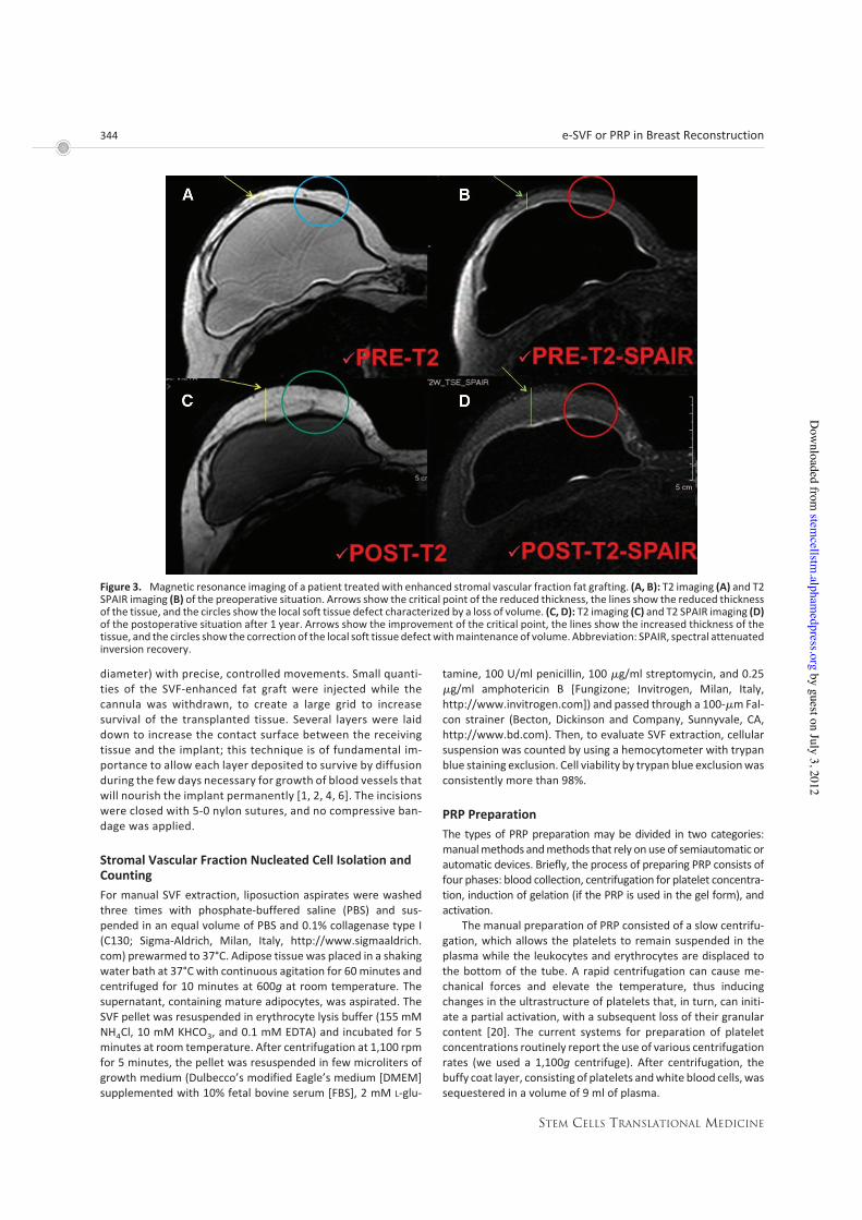
Figure 3. Magnetic resonance imaging of a patient treatedwith enhanced stromal vascular fraction fat grafting. (A, B): T2 imaging (A) and T2SPAIR imaging (B) of the preoperative situation. Arrows show the critical point of the reduced thickness, the lines show the reduced thicknessof the tissue, and the circles show the local soft tissue defect characterized by a loss of volume. (C, D): T2 imaging (C) and T2 SPAIR imaging (D)of the postoperative situation after 1 year. Arrows show the improvement of the critical point, the lines show the increased thickness of thetissue, and the circles show the correction of the local soft tissue defectwithmaintenance of volume. Abbreviation: SPAIR, spectral attenuatedinversion recovery.
344 e-SVF or PRP in Breast Reconstruction
STEM CELLS TRANSLATIONAL MEDICINE
by guest on July 3, 2012stem
cellstm.alpham
edpress.orgD
ownloaded from

Standard cell separators and salvage devices can be used toproduce platelet-rich plasma. These devices operate on a unit ofblood and typically use continuous-flow centrifuge bowl or con-tinuous-flow disk separation technology and both a hard (fast)and a soft (slow) spin, yielding platelet concentrations from twoto four times baseline [23, 24]. Such devices include the CATS(Fresenius, Wilmington, DE), Sequestra (Medtronic, Minneapo-lis, MN, http://www.medtronic.com), Cell Saver 5 (HaemoneticsCorp., Braintree, MA, http://www.haemonetics.com), and oth-ers [23–25]. Many surgical procedures require use of relativelysmall volumes of platelet-rich plasma [26]. Consequently, small,compact office systems have been developed that produce ap-proximately 6 ml of platelet-rich plasma from 45–60ml of blood[26, 27]. There are many such systems, including the GPS (Bi-omet, Warsaw, IN, http://www.biomet.com/), the PCCS (Im-plant Innovations, Inc., PalmBeachGardens, FL, http://biomet3i.com), the Symphony II (DePuy, Warsaw, IN, http://www.depuy.com), the SmartPReP (Harvest Technologies Corp., Norwell, MA,http://www.harvesttech.com), and the Magellan (Medtronic)[23, 26, 27]. Althoughall operateona small volumeof drawnblood(45–60 ml) and on the principle of centrifugation, these systemsdifferwidely in their ability tocollect andconcentrateplatelets,withapproximately 30%–85% of the available platelets collected andfrom a less than twofold to an approximately eightfold increase inthe platelet concentration over baseline [23, 24].
There are several devices for the PRP preparation, such asFibrinet (Cascade Medical Enterprises, Plymouth, U.K.), Regen(Regen Lab, Le Mont-sur-Lausanne, Switzerland, http://www.regenlab.com), Plateltex (Plateltex S.R.O., Bratislava, Slovakia,http://www.plateltex.com/), and Vivostat (Vivostat A/S, Al-leroed, Denmark, http://www.vivostat.com). Generally, we pre-pared the PRP according to the Cascade method and in all casesunder a protocol approved by our institution’s transfusion ser-vice.
In general, most systems, whether large or small volume, donot concentrate the plasma proteins of the coagulation cascade[23, 25]. The concentration of plasma protein levels above base-line can be achieved through secondary ultrafiltration, as is donewith the Ultra Concentrator (Interpore Cross, Irvine, CA), and theAccess System (Interpore Cross), in which the buffy coat col-lected from a centrifugation stage is passed through hollow fi-bers with an effective pore size of 30 kDa. This system removesby filtration up to two-thirds of the aqueous phase; thus, theconcentrations of the plasma proteins retained and the ele-ments formed are increased substantially [28].
In our procedure, PRP was prepared in the presence of atransfusional service doctor froma small volumeof blood (18ml)according to the method of the Cascade-Esforax system [20](supplemental online Fig. 3E), a commercially approved formu-lation. Briefly, to prepare PRP, bloodwas taken froma peripheralvein using sodium citrate as an anticoagulant. This system forpreparing platelet concentrations uses centrifugation of 1,100gfor 10 minutes (supplemental online Fig. 3E). The PRP protocolusesCa2! to induceplatelet activationandexocytosis of the"gran-ules. We added Ca2! when the fat was centrifuged. The final aimwas to obtain a platelet pellet (supplemental online Fig. 3F, 3G),although the preparation was not selective and included leuko-cytes. The secretion of growth factor begins with platelet ac-tivation.
Fat Graft Centrifugation According to the Coleman
Procedure and Mix with PRP
Before proceeding to activationof PRP, under general anesthesiawe harvested fat tissue from the abdominal region using somespecific cannulas. Maintaining asepsis, we took the plungers offthe syringes; after closing them with a cap, we positioned themflat in the sterile centrifuge (supplemental online Fig. 3A). Thesyringeswere processed for 3minutes at 3,000 rpm (supplemen-tal online Fig. 3B). This procedure obtained purified fat tissue(supplemental online Fig. 3C), preserving the integrity of the adi-pocytes but separating the fluid fat portion from the serous-bloody part (supplemental online Fig. 3D). We mixed 0.5 ml ofPRP with 1 ml of centrifuged fat tissue (supplemental online Fig.3I, 3L). The purified body fat mixed with PRP was put in 1-mlsyringes (supplemental online Fig. 3M, 3N) and aseptically rein-serted using the specific microcannulas for implanting.
Clinical Evaluation Method
Twomethods for the clinical evaluation of outcomes were used:(a) team evaluation, and (b) patient self-evaluation. The teamevaluation is an evaluation method based on clinical observa-tion, using a scale of six values (excellent, good, discreet enough,poor, inadequate). The patient-based self-evaluation uses thesame six values mentioned above. The factors/variables thatwere taken into account were pigmentation, vascularization, pli-ability, thickness, itching, and pain.
The percentage of maintenance restored was clinically eval-uated with two different criteria. The first was the subjectiveevaluation, and the second one was the objective evaluation.The subjective evaluation was based on the personal score ofeach patient focused on the following parameters: (a) presenceof asymmetry, deformity, irregularity, dyschromia, dysesthesia,paraesthesia, and pain; (b) results of the supero-external quad-rant, infero-external quadrant, supero-internal quadrant, and in-fero-internal quadrant; (c) resorption of fat in one or more re-gions; (d) time of stabilization of the transplanted fat; and (e)need for retreatment.
For each parameter, patients gave a yes/no or positive/neg-ative evaluation, and percentage of maintenance of restoredwas calculated as the mean of all calculated single percentages.The objective evaluation was made on the analysis of the preop-erative and postoperative photos. The photos were of the samesize, brightness, and even contrast. According to parameters re-ported above, the operator similarly calculated the percentageof restoration. Finally, the mean between patient and operatorevaluations was calculated.
Instrumental Imaging Evaluation Method
The percentage of maintenance restored was imaging evaluatedwithMRI (Figs. 3, 4). The timingwas as follows: preoperative (Fig.3A, 3B), after 6 and 12 months (Fig. 3C, 3D), and then annually.
MRI showed that transplanted fat tissue survived andformed a significant thickness of the fatty layer not only subcu-taneously on and around themammary glands but also betweenthemammary glands and the pectoralis muscles. Although smallcystic formation and macrocalcification were detected in onecase, the macrocalcification was easily distinguished from thatassociated with breast cancer, and the overall cosmetic resultswere generally satisfactory and encouraging. Almost all the pa-tients were satisfied with their enlarged and soft breasts with anatural contour.
345Gentile, Orlandi, Scioli et al.
www.StemCellsTM.com
by guest on July 3, 2012stem
cellstm.alpham
edpress.orgD
ownloaded from
ASC Differentiation Potential and Growth Curves
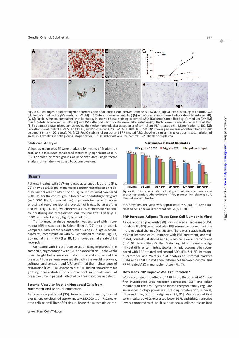
To demonstrate the differentiation capacity of cultured ASCs,adipogenic (Fig. 5A, 5B) and osteogenic (Fig. 5C, 5D) differentia-tion was verified in third-passage confluent cells, according topreviously published methods [20]. Briefly, for adipogenesis,ASCs were cultured in DMEM supplemented with 10% FBS, 100!M L-ascorbic acid, 1 !M dexamethasone, 0.5 mM 1-methyl-3-iso-butylxanthine, 100 !M indomethacin, and 10 !g/ml humanrecombinant insulin (Sigma-Aldrich). Control was cultured inDMEM plus 10% FBS. Medium was changed every 3 days for 3weeks, and adipogenesis was assessed by Oil Red O staining.Osteogenic differentiation was induced in DMEM supplementedwith 10% FBS, 200 !M L-ascorbic acid, 0.1 !M dexamethasone,and 10 mM #-glycerol phosphate (Sigma-Aldrich). Control wascultured in DMEM plus 10% FBS. Medium was changed every 3days for 21 days. To assess mineralization corresponding to os-teogenic differentiation, intracellular calcium deposits werestained with von Kossa stain (Fig. 5C, 5D). Images were obtainedat amagnification of"200 through a digital camera (Dxm1200F;Nikon, New York, http://www.nikon.com) connected to a com-puter using Nikon ACT-1 software. For growth curves (Fig. 5G),third-passage ASCs were seeded at 2,500 cells per cm2, serumstarved overnight, and maintained in DMEM plus 10% FBS (con-trol) or DMEM ! 10% FBS ! PRP (5% vol/vol) for 6 days. Every 2days, cells were trypsinized and counted using a hemocytome-ter. In some experiments, epidermal growth factor receptor(EGFR) and ErbB2 selective inhibitors were used at 5 !M(AG1478 and AG879; Sigma-Aldrich). The inhibition of ASC pro-
liferation was expressed as the percentage of reduction com-pared with PRP-control (mean # SE). Each experiment was per-formed in triplicate.
Immunofluorescence
Control and PRP-treated ASCs (after 6 days of treatment) werefixed in 4% paraformaldehyde for 5 minutes at 4°C and thenincubated with mouse monoclonal antibody anti-CD44 and anti-CD90 (Santa Cruz Biotechnology Inc., Santa Cruz, CA, http://www.scbt.com) for 1 hour at room temperature. Anti-mousesecondary antibody (Nordic Immunological Laboratories, Til-burg, The Netherlands, http://www.nordiclabs.nl) was added,and then cells were incubated with fluorescent streptavidin(R&D Systems Inc., Minneapolis, http://www.rndsystems.com).Hoechst was used for nuclear staining.
Western Blot Analysis
After extraction and quantification of total cell lysates, proteinswere separated by gradient sodium dodecyl sulfate-polyacryl-amide gel electrophoresis, blotted to nitrocellulose transfermembranes, and incubated with anti-CD44 (Santa Cruz Biotech-nology), anti-CD90 (Santa Cruz Biotechnology), anti-EGFR (CellSignaling Technology, Beverly, MA, http://www.cellsignal.com),anti-c-erbB-2 (Upstate, Charlottesville, VA, http://www.upstate.com; Millipore, Billerica, MA, http://www.millipore.com), anti-phosphorylated ErbB2 (pTyr1248) (Sigma-Aldrich), and anti-"-tubulin antibody (Sigma-Aldrich). Each experiment wasperformed in triplicate.
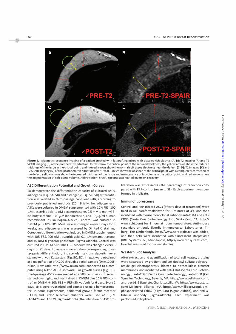
Figure 4. Magnetic resonance imaging of a patient treated with fat grafting mixed with platelet-rich plasma. (A, B): T2 imaging (A) and T2SPAIR imaging (B) of the preoperative situation. Circles show the critical point of the reduced thickness, the yellow arrows show the reducedthickness of the tissue in the critical point, and the red arrows show the normal soft tissue thickness near the defect. (C, D): T2 imaging (C) andT2 SPAIR imaging (D) of the postoperative situation after 1 year. Circles show the absence of the critical point with a completely correction ofthe defect, yellow arrows show the increased thickness of the tissue andmaintenance of fat volume in the critical point, and red arrows showthe augmentation of soft tissue volume. Abbreviation: SPAIR, spectral attenuated inversion recovery.
346 e-SVF or PRP in Breast Reconstruction
STEM CELLS TRANSLATIONAL MEDICINE
by guest on July 3, 2012stem
cellstm.alpham
edpress.orgD
ownloaded from
Statistical Analysis
Values as mean plus SE were analyzed by means of Student’s ttest, and differences considered statistically significant at p $.05. For three or more groups of univariate data, single-factoranalysis of variation was used to obtain p values.
RESULTS
Patients treated with SVF-enhanced autologous fat grafts (Fig.2B) showed a 63% maintenance of contour restoring and three-dimensional volume after 1 year (Fig. 6, red column) comparedwith 39% for the control group treated with centrifuged fat graft(p$ .0001; Fig. 6, green column). In patients treated with recon-structing three-dimensional projection of breast by fat graftingand PRP (Fig. 1B, 1D), we observed a 69% maintenance of con-tour restoring and three-dimensional volume after 1 year (p $.0001 vs. control group; Fig. 6, blue column).
Transplanted fat tissue resorption was analyzed with instru-mental MRI as suggested by Salgarello et al. [29] and ultrasound.Compared with breast reconstruction using autologous centri-fuged fat, reconstruction with SVF-enhanced fat tissue (Fig. 2B,2D) and fat graft! PRP (Fig. 1B, 1D) showed a smaller rate of fatresorption.
Compared with breast reconstruction using implants of thesame size, augmentation with SVF-enhanced fat tissue showed alower height but a more natural contour and softness of thebreasts. All the patients were satisfiedwith the resulting texture,softness, and contour, and MRI confirmed the maintenance ofrestoration (Figs. 3, 4). As reported, e-SVF and PRPmixedwith fatgrafting demonstrated an improvement in maintenance ofbreast volume in patients affected by breast soft tissue defect.
Stromal Vascular Fraction Nucleated Cells from
Automatic and Manual Extraction
As previously published [30], from adipose tissue, by manualextraction, we obtained approximately 250,000# 34,782 nucle-ated cells per milliliter of fat tissue. Using the automatic extrac-
tor, however, cell yield was approximately 50,000 # 6,956 nu-cleated cells per milliliter of fat tissue (p $ .01).
PRP Increases Adipose Tissue Stem Cell Number In Vitro
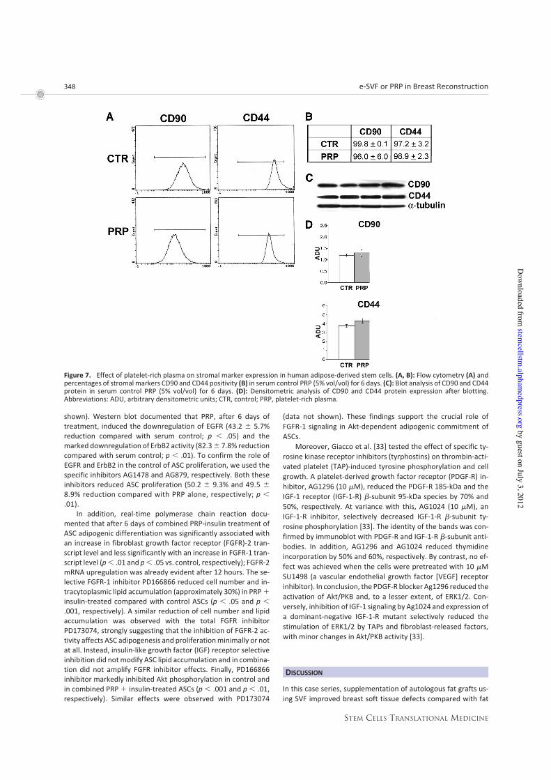
As we reported previously [20], PRP induced an increase of ASCnumber (Fig. 5G) compared with 10% serum control without anymorphological changes (Fig. 5E, 5F). There was a statistically sig-nificant increase of cell number with PRP treatment, approxi-mately fourfold, at days 4 and 6, when cells were preconfluent(p $ .02). In addition, Oil Red O staining did not reveal any sig-nificant difference in intracytoplasmic lipid accumulation com-pared with PRP-treated and control ASCs (Fig. 5H, 5I). Immuno-fluorescence and Western blot analysis for stromal markersCD44 and CD90 did not show differences between control andPRP-treated ASC immunophenotype (Fig. 7).
How Does PRP Improve ASC Proliferation?
We investigated the effects of PRP in proliferation of ASCs: wefirst investigated ErbB receptor expression. EGFR and othermembers of the ErbB tyrosine kinase receptor family regulateseveral cell biology processes, including proliferation, survival,differentiation, and tumorigenesis [31, 32]. We observed thatserum-cultured ASCs expressed lower EGFR and ErbB2 transcriptlevels compared with adult subcutaneous adipose tissue (not
Figure 5. Adipogenic and osteogenic differentiation of adipose-tissue-derived stem cells (ASCs). (A, B): Oil Red O staining of control ASCs(Dulbecco’s modified Eagle’s medium [DMEM]! 10% fetal bovine serum [FBS]) (A) and ASCs after induction of adipocyte differentiation (B).(C, D): Nuclei were counterstained with hematoxylin and von Kossa staining in control ASCs (Dulbecco’s modified Eagle’s medium [DMEM]plus 10% fetal bovine serum [FBS]) (C) and ASCs after induction of osteogenic differentiation (D). Nuclei were counterstained with Fast Red.(E, F): Contrast phasemicrographs showing the similar morphological appearance of control and PRP-treated cells. Magnification,"100. (G):Growth curve of control (DMEM! 10%FBS) and PRP-treatedASCs (DMEM! 10%FBS! 5%PRP) showing an increase of cell numberwith PRPtreatment (!, p $ .02; t test). (H, I): Oil Red O staining of control and PRP-treated ASCs showing a similar intracytoplasmic accumulation ofsmall lipid droplets in both groups. Magnification, "100. Abbreviations: ctr, control; PRP, platelet-rich plasma.
Figure 6. Clinical evaluation of fat graft volume maintenance inbreast restoration. Abbreviations: PRP, platelet-rich plasma; SVF,stromal vascular fraction.
347Gentile, Orlandi, Scioli et al.
www.StemCellsTM.com
by guest on July 3, 2012stem
cellstm.alpham
edpress.orgD
ownloaded from
shown). Western blot documented that PRP, after 6 days oftreatment, induced the downregulation of EGFR (43.2 # 5.7%reduction compared with serum control; p $ .05) and themarked downregulation of ErbB2 activity (82.3# 7.8% reductioncompared with serum control; p $ .01). To confirm the role ofEGFR and ErbB2 in the control of ASC proliferation, we used thespecific inhibitors AG1478 and AG879, respectively. Both theseinhibitors reduced ASC proliferation (50.2 # 9.3% and 49.5 #8.9% reduction compared with PRP alone, respectively; p $.01).
In addition, real-time polymerase chain reaction docu-mented that after 6 days of combined PRP-insulin treatment ofASC adipogenic differentiation was significantly associated withan increase in fibroblast growth factor receptor (FGFR)-2 tran-script level and less significantly with an increase in FGFR-1 tran-script level (p$ .01 and p$ .05 vs. control, respectively); FGFR-2mRNA upregulation was already evident after 12 hours. The se-lective FGFR-1 inhibitor PD166866 reduced cell number and in-tracytoplasmic lipid accumulation (approximately 30%) in PRP!insulin-treated compared with control ASCs (p $ .05 and p $.001, respectively). A similar reduction of cell number and lipidaccumulation was observed with the total FGFR inhibitorPD173074, strongly suggesting that the inhibition of FGFR-2 ac-tivity affects ASC adipogenesis and proliferationminimally or notat all. Instead, insulin-like growth factor (IGF) receptor selectiveinhibition did notmodify ASC lipid accumulation and in combina-tion did not amplify FGFR inhibitor effects. Finally, PD166866inhibitor markedly inhibited Akt phosphorylation in control andin combined PRP ! insulin-treated ASCs (p $ .001 and p $ .01,respectively). Similar effects were observed with PD173074
(data not shown). These findings support the crucial role ofFGFR-1 signaling in Akt-dependent adipogenic commitment ofASCs.
Moreover, Giacco et al. [33] tested the effect of specific ty-rosine kinase receptor inhibitors (tyrphostins) on thrombin-acti-vated platelet (TAP)-induced tyrosine phosphorylation and cellgrowth. A platelet-derived growth factor receptor (PDGF-R) in-hibitor, AG1296 (10 !M), reduced the PDGF-R 185-kDa and theIGF-1 receptor (IGF-1-R) #-subunit 95-kDa species by 70% and50%, respectively. At variance with this, AG1024 (10 !M), anIGF-1-R inhibitor, selectively decreased IGF-1-R #-subunit ty-rosine phosphorylation [33]. The identity of the bands was con-firmed by immunoblot with PDGF-R and IGF-1-R #-subunit anti-bodies. In addition, AG1296 and AG1024 reduced thymidineincorporation by 50% and 60%, respectively. By contrast, no ef-fect was achieved when the cells were pretreated with 10 !MSU1498 (a vascular endothelial growth factor [VEGF] receptorinhibitor). In conclusion, the PDGF-R blocker Ag1296 reduced theactivation of Akt/PKB and, to a lesser extent, of ERK1/2. Con-versely, inhibition of IGF-1 signaling by Ag1024 and expression ofa dominant-negative IGF-1-R mutant selectively reduced thestimulation of ERK1/2 by TAPs and fibroblast-released factors,with minor changes in Akt/PKB activity [33].
DISCUSSION
In this case series, supplementation of autologous fat grafts us-ing SVF improved breast soft tissue defects compared with fat
Figure 7. Effect of platelet-rich plasma on stromal marker expression in human adipose-derived stem cells. (A, B): Flow cytometry (A) andpercentages of stromal markers CD90 and CD44 positivity (B) in serum control PRP (5% vol/vol) for 6 days. (C): Blot analysis of CD90 and CD44protein in serum control PRP (5% vol/vol) for 6 days. (D): Densitometric analysis of CD90 and CD44 protein expression after blotting.Abbreviations: ADU, arbitrary densitometric units; CTR, control; PRP, platelet-rich plasma.
348 e-SVF or PRP in Breast Reconstruction
STEM CELLS TRANSLATIONAL MEDICINE
by guest on July 3, 2012stem
cellstm.alpham
edpress.orgD
ownloaded from

grafting alone. Although small cystic formation and microcalcifi-cationwere detected in one case, themicrocalcificationwas eas-ily distinguished, by checking with ultrasound, from that associ-ated with breast cancer, and the overall cosmetic results weregenerally satisfactory and encouraging. In this case, we did notperform a biopsy because the clinical features and ultrasoundimages of the microcalcification confirmed the diagnosis of fatnecrosis-associated microcalcification.
Lipofilling procedures can modify radiologic images; how-ever, this interference has been studied in the literature [34–36],and radiologic studies suggest that imaging technologies (ultra-sound, mammography, and MRI) can identify the microcalcifica-tions caused by fat injection [37]. Moreover, recent follow-upstudies have demonstrated the safety of the procedure, detect-ing no increase in new disease or tumor recurrence [3, 34, 38].Almost all the patientswere satisfiedwith their enlarged and softbreastswith a natural contour.MRI showed that transplanted fattissue survived and formed a significant thickness of the fattylayer not only subcutaneously on and around the mammaryglands but also between themammary glands and the pectoralismuscles. Maximum breast augmentation using the techniquedescribed varied among the patients and appeared to be 80–180ml. Although these volumesmay be smaller than those achievedwith large artificial implants, a definite advantage is that patientsneed not be concerned about postoperative complications in-duced by artificial implants, such as rupture, infection, capsularcontracture, unnatural contour, hardness, neurologic symp-toms, and immune response. Compared with patients who un-derwent conventional autologous fat graft to the breasts, aug-mentation effects were apparently higher with SVF-enhancedfat.
For each injection, a 0.5–1.2-cm increase in breast soft tissuevolume was common with the conventional procedure, com-pared with the 1.2–2.8-cm increase seen in this trial of SVF-en-hanced fat, although the augmentation effect varied among pa-tients. The measurement system we recently devised may helpto quantify the difference in augmented volume in the future.
The potential benefit of SVF supplementation could be ex-plained by the ability of cells to secrete various growth factorsthat improve survival and increased vascularization [18, 22],leading to increased survival of the graft as shown a rodent study[19]. In light of this concept, we propose the chain of eventsleading to regeneration of the tissue to be as follows: targeting ofdamaged areas, release of angiogenic and antiapoptotic factors,and then formation of new vessels and oxygenation.
SVFs might indeed improve fat graft survival and mainte-nance, which is supported by observations from other surgicalprocedures, such asmaxillofacial surgery for a calvarial defect [2]and breast reconstruction after partial mastectomy with radio-therapy damage [39]. Implanted adipose tissuemust survive by asimple diffusion mechanism until an active blood supply is rees-tablished. Thus survival of the graft, particularly of a larger vol-ume graft, is balanced between this process and hypoxia-in-duced cell death [40]. Prosurvival factorsmay therefore promotelong-term retention and hence durability of the graft. In an ani-mal study, this effect was achieved by using gene therapy todeliver VEGF (a potent proangiogenic factor) to the graft. Thisresulted in increased blood vessel density within the graft and asignificant improvement in graft retention at 15 weeks [41].
The traditional preparation of growth factors contained inPRP consisted of a slow centrifugation,which allows the platelets
to remain suspended in the plasma while the leukocytes anderythrocytes are displaced to the bottom of the tube. The cur-rent systems for preparing platelet concentrations use variouscentrifuges. The final aim was to obtain a platelet pellet, al-though the preparation is not selective and includes leukocytes.The secretion of growth factor begins with platelet activation.The PRP protocol uses Ca2! to induce platelet activation andexocytosis of the " granules. A rapid centrifugation can causemechanical forces and can raise the temperature, thus inducingchanges in the ultrastructure of platelets that, in turn, can initi-ate a partial activation,with a consequent loss of its content [20].
Calcium acts as a necessary cofactor for platelet aggregation[20]. When Ca2! is used to induce platelet activation, the secre-tion of the growth factors contained in the granules is slow [20].To optimize the secretion process, the optimum concentrationof Ca2 was previously determined [20]. When a rapid activationand coagulation is required, endogenous thrombin could beused.
Anitua et al. [42] reported the use of two centrifugationrates. Blood was collected into 3.8% (wt/vol) sodium citrate andcentrifuged at 4,500g for 12 minutes at 4°C to obtain platelet-poor (PP) plasma or at 460g for 8minutes to obtain PRP. Calciumchloridewas added to PP and PRP at a final concentration of 22.8mM. The secretion of growth factor begins with platelet activa-tion.
In an interesting work, Mazzucco et al. described the differ-ent growth factor concentrations that are obtained through dif-ferent devices (Fibrinet, Plateltex, and Regen) and a homemademethod [43]. PDGF-BB, transforming growth factor-# (TGF-#),and IGF-1 were detected in lower concentrations with the use ofFibrinet. In contrast, the Regen displayed high concentrations ofTGF-#, basic fibroblast growth factor, and IGF-1, whereas thePlateltex showed a high level of epidermal growth factor [43].
SVF can favor neoangiogenic vascularization and fibrogenicactivity of fibroblasts that favor adipose tissue survival andthree-dimensional organization. Compared with traditional fatgrafting, the survival of the graft is more probable and fat necro-sis is reduced, potentially because of improved vascular develop-ment in the implanted area. Results of this study offer an in vivotissue-engineering approach that provides an optimized mi-croenvironment, supporting the correct architectural adipocytedistribution, better cell-to-cell interaction, adipose tissue sur-vival, and maybe limited differentiation from SVF; this could of-fer early protection fromsurrounding inflammatory events. Also,the early establishment of new microcapillary networks, whichdeliver the proper nutrients and oxygen to the implant, mightcontribute to the improved outcomes observed [1, 44, 45]. Infact, in the adipose tissue, ASCs reside between adipocytes or inthe extracellular matrix, especially around vessels, and contrib-ute to the turnover of adipose tissue, which is known to be veryslow (2 years or more) [46].
However, adipose grafts probably turn over during the first2–3 months after transplantation because they experience tem-porary ischemia followed by reperfusion injury. This turnover,the replacement process of the adipose tissue, is conducted bytissue-specific progenitor cells, which are ASCs. The relative de-ficiency of ASCs in aspirated fat may affect the replacement pro-cess and lead to postoperative atrophy of grafted fat, which isknown to occur commonly during the first 6 months after lipoin-jection.
349Gentile, Orlandi, Scioli et al.
www.StemCellsTM.com
by guest on July 3, 2012stem
cellstm.alpham
edpress.orgD
ownloaded from

After transplantation, ASCs may interact with other cells,such as vascular endothelial cells, and supplementation with theSVF may be superior to supplementation with ASCs alone in thistreatment. However, additional studies are needed to elucidatethe synergistic effects of ASCs with other cells contained in thegraft.
In this preliminary study, satisfactory clinical results weregenerally achieved without any major complications. Thus, wecan conclude that SVF-enhanced fat graft is sufficiently safe forcontinuation of the study, although controlled investigations andaccumulated long-term results are needed to elucidate the over-all safety and efficacy of the treatment. A variety of innovations,including stem cell technology, may be developed and may con-tribute to the improvement of autologous tissue transplantationand regeneration. Further improvements of the technique maycause autologous tissue transfer to become the first choice forbreast augmentation in the future.
Furthermore, the use of other types of scaffolds for breastreconstruction should be noted. Colwell et al. [47], in a retro-spective review of 331 consecutive breast reconstructions withacellular dermal matrix, demonstrated that this matrix offers acost-effective reconstruction with a low complication rate. Inaddition, a recent review showed that acellular dermal matrix intwo-stage expander/implant reconstruction offers a safety pro-file similar to that of standard submuscular techniques [48]. Sev-eral studies have been published on the use of acellular matrix,showing both the advantages and disadvantage of this technique
[49, 50]. We believe that this matrix may be the procedure ofchoice in select patients.
CONCLUSION
We conclude that engineered fat grafting based on the addictionof SVF or PRP is a reliable alternative to breast implant based onsome initial indications. (a) A preliminary study with follow-up at30 months showed with instrumental imaging the absence ofcalcification or microcalcification. (b) This absence suggests thatengineered fat grafting is effective and safe. (c) Autologous fattissue can be used as a scaffold. (d) PRP and SVF favor adiposetissue survival. Additional study is necessary to evaluate the ef-ficacy of this method further.
AUTHOR CONTRIBUTIONS
P.G.: conception and design, manuscript writing; C.D.P., I.B.,C.B.C., andM.F.: data analysis and interpretation; M.G.S.: collec-tion and assembly of data; A.O.: conception and design, provi-sion of study material; V.F. and R.F.: instrumental imaging anal-ysis; V.C.: administrative support, final approval of themanuscript.
DISCLOSURE OF POTENTIAL CONFLICTS OF INTEREST
The authors indicate no potential conflicts of interest.
REFERENCES
1 Rigotti G, Marchi A, Galie M et al. Clinicaltreatment of radiotherapy tissue damage bylipoaspirate transplant: A healing process me-diated by adipose-derived adult stem cells.Plast Reconstr Surg 2007;119:1409–1422; dis-cussion 1423–1404.2 Yoshimura K, Sato K, Aoi N et al. Cell-as-
sisted lipotransfer for cosmetic breast aug-mentation: Supportive use of adipose-derivedstem/stromal cells. Aesthetic Plast Surg 2008;32:48–55; discussion 56–47.3 Rigotti G, Marchi A, Stringhini P et al. De-
termining the oncological risk of autologous li-poaspirate grafting for post-mastectomybreast reconstruction. Aesthetic Plast Surg2010;34:475–480.4 Yoshimura K, Asano Y, Aoi N et al. Progen-
itor-enriched adipose tissue transplantation asrescue for breast implant complications.Breast J 2010;16:169–175.5 Lendeckel S, Jodicke A, Christophis P et al.
Autologous stem cells (adipose) and fibrin glueused to treat widespread traumatic calvarialdefects: Case report. J Craniomaxillofac Surg2004;32:370–373.6 García-OlmoD,Garcia-ArranzM,Herreros
D et al. A phase I clinical trial of the treatmentof Crohn’s fistula by adipose mesenchymalstem cell transplantation. Dis Colon Rectum2005;48:1416–1423.7 García-Olmo D, Herreros D, De-La-Quin-
tana P et al. Adipose-derived stem cells inCrohn’s rectovaginal fistula. Case Report Med2010;2010:961758.8 Garcia-Olmo D, Herreros D, Pascual I et al.
Expanded adipose-derived stem cells for the
treatment of complex perianal fistula: A phaseII clinical trial. Dis Colon Rectum 2009;52:79–86.9 Garcia-Olmo D, Herreros D, Pascual M et
al. Treatment of enterocutaneous fistula inCrohn’s disease with adipose-derived stemcells: A comparison of protocols with andwith-out cell expansion. Int J Colorectal Dis 2009;24:27–30.10 Peterson B, Zhang J, Iglesias R et al. Heal-
ing of critically sized femoral defects, using ge-netically modified mesenchymal stem cellsfrom human adipose tissue. Tissue Eng 2005;11:120–129.11 Yoshimura K, Sato K, Aoi N et al. Cell-
assisted lipotransfer for facial lipoatrophy: Ef-ficacy of clinical use of adipose-derived stemcells. Dermatol Surg 2008;34:1178–1185.12 Tiryaki T, Findikli N, Tiryaki D. Staged
stem cell-enriched tissue (SET) injections forsoft tissue augmentation in hostile recipientareas: A preliminary report. Aesthetic PlastSurg 2011;35:965–971.13 Lo Cicero V, Montelatici E, Cantarella G
et al. Do mesenchymal stem cells play a role invocal fold graft survival? Cell Prolif 2008;41:460–473.14 Cantarella G, Mazzola RF, Domenichini
E et al. Vocal fold augmentation by autolo-gous fat injection with lipostructure proce-dure. Otolaryngol Head Neck Surg 2005;132:239–243.15 Oedayrajsingh-Varma MJ, van Ham
SM, Knippenberg M et al. Adipose tissue-de-rived mesenchymal stem cell yield andgrowth characteristics are affected by thetissue-harvesting procedure. Cytotherapy2006;8:166–177.
16 Prunet-Marcassus B, Cousin B, Caton Det al. From heterogeneity to plasticity in adi-pose tissues: Site-specific differences. Exp CellRes 2006;312:727–736.17 Gimble JM, Katz AJ, Bunnell BA. Adipose-
derived stem cells for regenerative medicine.Circ Res 2007;100:1249–1260.18 Coleman SR. Facial recontouring with li-
postructure. Clin Plast Surg 1997;24:347–367.19 Coleman SR. Long-term survival of fat
transplants: Controlled demonstrations. Aes-thetic Plast Surg 1995;19:421–425.20 Cervelli V, Gentile P, Scioli MG et al. Ap-
plication of platelet-rich plasma in plastic sur-gery: Clinical and in vitro evaluation. Tissue EngPart C Methods 2009;15:625–634.21 Cervelli V, Gentile P, Grimaldi M. Regen-
erative surgery: Use of fat grafting combinedwith platelet-rich plasma for chronic lower-ex-tremity ulcers. Aesthetic Plast Surg 2009;33:340–345.22 Cervelli V, Gentile P. Use of cell fat mixed
with platelet gel in progressive hemifacial atro-phy. Aesthetic Plast Surg 2009;33:22–27.23 Kevy SV, Jacobson MS. Comparison of
methods for point of care preparation of autol-ogous platelet gel. J Extra Corpor Technol2004;36:28–35.24 SiebrechtMA, De Rooij PP, ArmDMet al.
Platelet concentrate increases bone ingrowthinto porous hydroxyapatite. Orthopedics 2002;25:169–172.25 Waters JH, Roberts KC. Database review
of possible factors influencing point-of-careplatelet gel manufacture. J Extra Corpor Tech-nol 2004;36:250.26 Man D, Plosker H, Winland-Brown JE.
The use of autologous platelet-rich plasma
350 e-SVF or PRP in Breast Reconstruction
STEM CELLS TRANSLATIONAL MEDICINE
by guest on July 3, 2012stem
cellstm.alpham
edpress.orgD
ownloaded from

(platelet gel) and autologous platelet-poorplasma (fibrin glue) in cosmetic surgery. PlastReconstr Surg 2001;107:229.27 Lozada JL, Caplanis N, Proussaefs P et al.
Platelet-rich plasma application in sinus graftsurgery: Part I. Background and processingtechniques. J Oral Implantol 2001;27:38.28 HoodAG, ArmDM. Topical application of
autogenous tissue growth factors for augmen-tation of structural bone graft fusion. Paperpresented at: American Society of Extra-Cor-poreal Technology 11th Annual Symposium onNew Advances in Blood Management; April20–23, 2004; Las Vegas, NV.29 Salgarello M, Visconti G, Rusciani A.
Breast fat grafting with platelet-rich plasma: Acomparative clinical study and current state ofthe art. Plast Reconstr Surg 2011;127:2176–2185.30 Cervelli V, Gentile P, De Angelis B et al.
Application of enhanced stromal vascular frac-tion and fat grafting mixed with PRP in post-traumatic lower extremity ulcers. Stem CellRes 2011;6:103–111.31 Citri A, Yarden Y. EGF-ERBB signalling:
Towards the systems level. Nat Rev Mol CellBiol 2006;7:505–516.32 Schneider MR, Wolf E. The epidermal
growth factor receptor ligands at a glance.J Cell Physiol 2009;218:460–466.33 Giacco F, Perruolo G, D’Agostino E et al.
Thrombin-activated platelets induce prolifera-tion of human skin fibroblasts by stimulatingautocrine production of insulin-like growthfactor-1. FASEB J 2006;20:2402–2404.34 Petit JY, Lohsiriwat V, Clough KB et al.
The oncologic outcome and immediate surgi-cal complications of lipofilling in breast cancerpatients: A multicenter study: Milan-Paris-
Lyon experience of 646 lipofilling procedures.Plast Reconstr Surg 2011;128:341–346.35 Pulagam SR, Poulton T, Mamounas EP
Long-term clinical, radiologic results with au-tologous fat transplantation for breast aug-mentation: Case reports, review of the litera-ture. Breast J 2006;12:63–65.36 Kwak JY, Lee SH, Park HL et al. Sono-
graphic findings in complications of cosmeticbreast augmentation with autologous fat ob-tained by liposuction. J Clin Ultrasound 2004;32:299–301.37 Gutowski KA; ASPS Fat Graft Task Force.
Current applications and safety of autologousfat grafts: A report of the ASPS fat graft taskforce. Plast Reconstr Surg. 2009;124:272–280.38 Fraser JK, Hedrick MH, Cohen SR. Onco-
logic risks of autologous fat grafting to thebreast. Aesthet Surg J 2011;31:68–75.39 Nedelec B, Shankowsky HA, Tredget
EE. Rating the resolving hypertrophic scar:Comparison of the Vancouver Scar Scale andscar volume. J Burn Care Rehabil 2000;21:205–212.40 Lin K, Matsubara Y, Masuda Y et al. Char-
acterization of adipose tissue-derived cells iso-lated with the Celution system. Cytotherapy2008;10:417–426.41 Rehman J, Traktuev D, Li J et al. Secretion
of angiogenic and antiapoptotic factors by hu-man adipose stromal cells. Circulation 2004;109:1292–1298.42 Anitua E, Sanchez M, Nurden AT et al.
Autologous fibrin matrices: A potential sourceof biological mediators that modulate tendoncell activities. J Biomed Mater Res A 2006;77:285–293.43 Mazzucco L, Balbo V, Cattana E et al. Not
every PRP-gel is born equal. Evaluation ofgrowth factor availability for tissues through
four PRP-gel preparations: Fibrinet, RegenPRP-Kit, Plateltex and one manual procedure. VoxSang 2009;97:110–118.44 Cao Y, Sun Z, Liao L et al. Human adipose
tissue-derived stem cells differentiate into en-dothelial cells in vitro and improve postnatalneovascularization in vivo. Biochem BiophysRes Commun 2005;332:370–379.45 Zhu M, Zhou Z, Chen Y et al. Supple-
mentation of fat grafts with adipose-derivedregenerative cells improves long-termgraft retention. Ann Plast Surg 2010;64:222–228.46 Strawford A, Antelo F, Christiansen M et
al. Adipose tissue triglyceride turnover, denovo lipogenesis, and cell proliferation in hu-mansmeasuredwith 2H2O. Am J Physiol Endo-crinol Metab 2004;286:E577–E588.47 Colwell AS, Damjanovic B, Zahedi B et al.
Retrospective review of 331 consecutive im-mediate single-stage implant reconstructionswith acellular dermal matrix: Indications, com-plications, trends, and costs. Plast ReconstrSurg 2011;128:1170–1178.48 Sbitany H, Serletti JM. Acellular dermis-
assisted prosthetic breast reconstruction: Asystematic and critical review of efficacy andassociated morbidity. Plast Reconstr Surg2011;128:1162–1169.49 Kim JY, Davila AA, Persing S et al. A
meta-analysis of human acellular dermis andsubmuscular tissue expander breast recon-struction. Plast Reconstr Surg 2012;129:28–41.50 Hoppe IC, Yueh JH, Wei CH et al. Compli-
cations following expander/implant breast re-construction utilizing acellular dermal matrix:A systematic review andmeta-analysis. Eplasty2011;11:e40.
See www.StemCellsTM.com for supporting information available online.
351Gentile, Orlandi, Scioli et al.
www.StemCellsTM.com
by guest on July 3, 2012stem
cellstm.alpham
edpress.orgD
ownloaded from
DOI: 10.5966/sctm.2011-00652012;1;341-351; originally published online April 13, 2012;Stem Cells Trans Med
Floris and Valerio CervellBocchini, Cristiano Beniamino Curcio, Micol Floris, Valeria Fiaschetti, Roberto
Pietro Gentile, Augusto Orlandi, Maria Giovanna Scioli, Camilla Di Pasquali, Ilariain Breast Reconstruction
MaintenanceVascular Fraction and Platelet-Rich Plasma Improves Fat Grafting A Comparative Translational Study: The Combined Use of Enhanced Stromal
This information is current as of July 3, 2012
& ServicesUpdated Information
http://stemcellstm.alphamedpress.org/content/1/4/341including high-resolution figures, can be found at:
Supplementary Material
sctm.2011-0065.DC1.htmlhttp://stemcellstm.alphamedpress.org/content/suppl/2012/04/12/Supplementary material can be found at:
by guest on July 3, 2012stem
cellstm.alpham
edpress.orgD
ownloaded from