a compendium of scientific literature - … compendium of scientific literature. ... spray diathermy...
TRANSCRIPT
Evidence Supporting the Efficacy and Safety
of the SURGICEL® Family of Absorbable Hemostats
A Compendium of
Scientific Literature


General Surgery• Use of haemostatic agents and glues during laparoscopic partial nephrectomy:
a multi-institutional survey from the United States and Europe of 1347 cases (Breda)...............................5
• A simple technique to control iatrogenic solid organ injury haemorrhage (Hadi).........................................6
• The use of oxidised cellulose as a topical haemostatic dressing on a bleeding stomal wound (Lawrentschuk) ........................................................................................................................................7
• Control of port-site bleeding from smaller incisions after laparoscopic cholecystectomy surgery (Rastogi) ...................................................................................................................................................8
• Use of knitted oxidized cellulose (Nu-knit) for the definitive packing of grade III liver fracture (Theuer)........9
• A simplified approach to techniques of splenic salvage (Trooskin)............................................................10
Cardiothoracic Surgery• Surgicel Nu-Knit hemostat for bleeding control of fragile sternum (Mair)..................................................12
ENT Surgery• Selective Surgicel packing for the treatment of posterior epistaxis (Bhatnagar) ......................................14
• Evaluation of Surgicel Nu-knit, Merocel and Vasolene gauze nasal packs: a randomized trial (Shinkwin) .............................................................................................................................................15
Obstetric/Gynecologic Surgery• Abdominal packing for intractable obstetrical and gynecologic hemorrhage (Awonuga).........................18
• Laparoscopic oxidized cellulose (Surgicel) application for small uterine perforations (Sharma)................19
• Successful management of uterine incision hemorrhage in caesarean section with topical oxidized regenerated cellulose (Surgicel Nu-Knit): a case report (Sharma)...........................................................21
Orthopedic Surgery• The use of local agents: Surgicel and Surgifoam (Sabel)........................................................................23
Urologic Surgery • Use of oxidized cellulose hemostats (SurgicelTM) to support parenchymal closure and achieve
hemostasis following partial nephrectomy (Abou-Elela)..........................................................................26
• Laparoscopic partial nephrectomy with “on-demand” clamping reduces warm ischemia time (Bollens)................................................................................................................................................27
Plastic Surgery• Use of oxidized regenerated cellulose to stop bleeding after a facelift procedure (Bassetto)...................29
In Vivo Studies• Antibacterial activity of oxidized regenerated cellulose (Dineen)..............................................................31
• The effect of oxidized regenerated cellulose on experimental infected splenotomies (Dineen) ................32
• The effect of oxidized regenerated cellulose on experimental intravascular infection (Dineen).................33
• Effects of absorbable hemostats on intraabdominal sepsis (Kuchta)......................................................34
In Vitro Study• In vitro antimicrobial activity of oxidized regenerated cellulose against antibiotic-resistant
microorganisms (Spangler) ....................................................................................................................37
Bibliography .........................................................................................................................................40
Full Prescribing Information .........................................................................................................41
Table of Contents

General Surgery
Ge
ne
ral S
urg
ery

General Surgery 5
Title Use of haemostatic agents and glues during laparoscopic partial nephrectomy: a multi-institutional survey from the United States and Europe of 1347 cases
Author Breda A, Stepanian SV, Lam, JS, et al.
Source European Urology. 2007;52(3):798-803.
Study Objectives
The authors sought to investigate the practice patterns of urologists in the US and Europe, and their acceptance of renal hemostatic agents and glues in patients undergoing laparoscopic partial nephrectomy.
Methods
A e-mail survey was sent to 26 centers in the US and Europe that perform a high volume of laparoscopic renal surgeries; 18 responded. Data requested during a retrospective review of patient charts for individuals who underwent laparoscopic partial nephrectomy (LPN) included:
• Indications for hemostatic agent and/or fibrin glue usage
• The type of hemostatic agent and/or fibrin glue used
• Use of concomitant suturing/bolstering
• Type of laparascopic tools used to perform the tumor resection
• Total number of LPN’s performed
• Tumor size and position
Results
Suitable surveys from 1347 LPN cases reported from 18 major academic centers were returned.
Mean tumor size was 2.8 cm (range 2 to 4 cm).
16 centers reported always using hemostatic agents and/or glues (n=1042, 77%); 2 centers never used hemostatic agents and/or glues. 16 centers also always used parenchymal suturing over absorbable bolsters of SURGICEL® Absorbable Hemostat and/or central hemostatic suture. 1 center reported variable use of a bolster suture and/or central hemostatic suture depending on the depth of the lesion. 1 center only used sealants and never performed suturing/bolstering. 15 centers performed tumor resection with the use of cold scissor, 2 used a harmonic scalpel, and 1 used bipolar forceps.
For the 1042 cases where hemostatic agents and/or glues were used, complications included postoperative bleeding requiring transfusion in 28 patients (2.7%) and urine leakage in 20 (1.9%).
Conclusions
The authors note that use of hemostatic agents and glues is becoming increasingly standardized in most centers performing laparoscopic partial nephrectomy. Based on these self-reported survey results, the use of hemostatic agents and/or fibrin glues is routine in most centers performing laparoscopic partial nephrectomy.

Title A simple technique to control iatrogenic solid organ injury haemorrhage
Author Hadi H, Maw A, Hay D
Source Surgeon: Journal Royal Colleges of Surgeons of Edinburgh and Ireland. 2004;2(6):339-340.
Study Objectives
The objective of this study was to describe the use of SURGICEL® Absorbable Hemostat and a Hanna-Belfast suction diathermy device to control minor solid organ hemorrhage.
Methods
Following iatrogenic solid organ injury (primarily gall bladder bed and spleen) during abdominal surgery, the authors combined the use of a device allowing simultaneous application of suction and diathermy with SURGICEL Hemostat. This technique involved first applying a layer of SURGICEL Hemostat over the area of injury and including a 2 to 3 cm margin on both sides. Suction from the Hanna-Belfast device was applied to keep the area dry and then the spray diathermy was used to “weld” the SURGICEL Absorbable Hemostat onto the surface of the organ. Spray diathermy was used in the non-contact mode in order to prevent the SURGICEL Hemostat from sticking to the instrument.
Results
This paper represents a “how I do it” description of an innovative technique. While no specific outcome measures were noted, the authors report using this technique without complication on more than 40 surgical cases in the gallbladder bed and in minor splenic injury. No adverse events using SURGICEL Hemostat in combination with suction diathermy were reported.
General Surgery 6

Title The use of oxidised cellulose as a topical haemostatic dressing on a bleeding stomal wound
Author Lawrentschuk N, Hewitt PM
Source Journal of Wound Care. 2002;11(9):344-345.
Objectives
This case report describes the use of oxidized regenerated cellulose (SURGICEL® Absorbable Hemostat) in the emergency care of a 20-year-old male patient with known ulcerative colitis who presented to the emergency department with a hemorrhaging ileostomy following revision of an existing stoma for acute intussusceptions.
Methods
Oxidized regenerated cellulose was utilized as an aid to control hemorrhage from an edema-tous, fragile mucosa and skin edge at the peripheral surface of the stoma.
Discussion
The ileostomy had been present for 2 years. 1 month prior to this event, conservative management was utilized to treat an episode of sub-acute small bowel obstruction. The patient returned, however, with signs and symptoms of obstruction consistent with a stomal intussusception. Upon presentation to the emergency department, an attempted non-operative reduction of the stoma was unsuccessful due to the degree of edema and swelling. At surgery, the intussuscepted portion of the stoma was removed and a new stoma refashioned. Bleeding was unable to be controlled by peristomal sutures, exerting direct pressure for extend-ed periods of time, or electrocautery, due to edema at the site. Oxidized cellulose was applied around the stoma at the mucocutaneous junction where the bleeding originated. Hemostasis was achieved “within seconds” of application to the site. The oxidized cellulose dressing was removed 24 hours later, no further surgery was necessary, and no further bleeding occurred. The patient was discharged with a functioning ileostomy 2 days after the event.
The authors cite this as an unusual case that required an immediate response. Oxidized cellulose was a readily available and effective solution.
General Surgery 7

Title Control of port-site bleeding from smaller incisions after laparoscopic cholecystectomy surgery
Author Rastogi V, Dy V
Source Surgical Laparoscopy, Endoscopy & Percutaneous Techniques. 2002;12(4):224–226.
Study Objectives
The authors describe a technique to rapidly control bleeding from difficult to approach small port-site incisions from the inside, utilizing SURGICEL® Absorbable Hemostat (oxidized regenerated cellulose (ORC); endoscopic size, 2.54 cm x 8.89 cm as a plug.
Methods
Of 207 patients undergoing laparoscopic cholecystectomy between July 1998 and June 2000, 20 patients experienced bleeding from 1 or more small (5-mm) port-site at the conclusion of the procedure. These 20 comprised the study group.
Once the laparoscopic procedure was completed, the 5-mm ports were removed. When bleeding was noted along the port-site, the port was reintroduced through the same incision. A grasper was introduced from the 10-mm subxiphoid port along with SURGICEL Hemostat. A second grasper was introduced through the 5-mm port at the port-site that was bleeding. Through this site, the ORC was grasped in the middle and pulled through the port incision, thereby snugly fitting through the hole to provide occlusion. The port and grasper were removed and excess SURGICEL Hemostat cut so that it was flush with the skin incision. The skin incision was approximated with interrupted subcuticular 4.0 Coated VICRYL* (polyglactin 910) Suture and ¼-inch steristrips. Patients were followed 7 days after the procedure by the surgeon during office visits.
Results
The authors report cessation of bleeding in all patients who were treated with this technique. No visible wound hematomas or port-site infections were noted during follow-up. Although not a blinded or comparative trial, and involving a small number of patients, the authors concluded that this technique, utilizing SURGICEL Hemostat at the port-site just prior to closure, was a simple and time-saving technique to control bleeding from small abdominal incisions.
*Trademark
General Surgery 8

Title Use of knitted oxidized cellulose (Nu-knit) for the definitive packing of grade III liver fracture
Author Theuer CP, Imagawa Dk
Source Injury: International Journal of the Care of the Injured. 1999;30:137-140.
Objective
This case report describes the utility of knitted oxidized cellulose (SURGICEL® NU-KNIT® Absorbable Hemostat) for the definitive packing of a grade III liver fracture that rebled following the removal of laparotomy pads as an initial attempt at hemorrhage control.
Case Report
A 23-year-old woman presented with a grade III liver laceration and a right renal fracture as a result of being an unrestrained back seat passenger in a high-speed motor vehicle accident. Following nephrectomy, continuous bleeding was noted from segment VII of the liver, and could not be controlled with cautery, argon beam coagulation, or ligatures. The patient appeared coagulopathic, and a damage control approach was selected. The liver was effectively packed with 5 laparotomy pads.
The second postoperative day, the patient was returned to the operating room to unpack her liver. A large subcapsular hematoma, which was not present on initial exploration and encompassed the entire anterior surface of the liver, made liver mobilization hazardous. Persistent bleeding was again noted from segment VII of the liver and remained refractory to cautery, argon beam coagulation, or ligatures.
5 large sheets of knitted oxidized regenerated cellulose (SURGICEL NU-KNIT Hemostat) were packed into the fracture and, after ensuring that hemostasis was complete, fibrin glue was sprayed over the knitted oxidized regenerated cellulose packing. All laparotomy pads were then removed and the patient was closed after placement of 2 closed suction drains.
Results
Postoperatively, the patient remained hemodynamically stable with a hemoglobin of 10 mg/dL and required no additional blood products. A follow-up CT scan 2 months after injury showed resolution of the retroperitoneal air and partial resolution of the subcapsular hematoma. At 11 months after injury she continued to do well and attained her preoperative weight.
Conclusion
Knitted oxidized cellulose (SURGICEL NU-KNIT Hemostat) was a useful agent for definitive packing of a grade III liver fracture and may prove useful for the management of solid organ injuries.
General Surgery 9

Title A simplified approach to techniques of splenic salvage
Author Trooskin SZ, Flancbaum L, Boyarsky AH et al.
Source Surgery, Gynecology and Obstetrics. 1989;168:546-458.
Objectives
This paper describes a simple method for controlling splenic parenchymal hemorrhage after splenic trauma or during partial splenectomy utilizing oxidized regenerated cellulose (ORC) as a suture bolster.
Methods
Perform a sharp dissection of the splenic attachments to the diaphragm, colon, and left kidney to mobilize spleen from the left upper quadrant, leaving the spleen suspended by the splenic hilar and short gastric vessels. This will allow for easy visual inspection of splenic surface and avoidance of capsular tears caused by traction.
For partial splenectomy, the spleen is mobilized in the standard fashion, taking care to ligate the short gastric vessels and control the splenic hilum. Ischemic tissue is readily identified by its deep blue color and sharply excised. Bleeding is minimized by placing a noncrushing clamp at the junction of the viable and nonviable splenic tissue, and the nonviable tissue is excised using a scalpel. Suturing the splenic parenchyma is simplified by passing the suture through bolsters consisting of pledgets of folded sheets of oxidized cellulose (SURGICEL® Absorbable Hemostat or SURGICEL® NU-KNIT® Absorbable Hemostat). 2-0 absorbable suture is placed as a horizon-tal mattress through the bolsters to compress the raw or oozing splenic surface and prevent the suture from tearing or cutting through the splenic capsule.
For splenic salvage after trauma, nonviable splenic tissue is sharply debrided. Bleeding from lacerations deep into the splenic parenchyma are controlled by compression of splenic tissue between bolsters of folded sheets of oxidized cellulose placed parallel to the laceration and sutured. Sutures are placed through the spleen in the manner of a horizontal mattress and tied snugly.
Results
The approach described was successfully used in 14 patients, 11 of whom underwent extensive splenic repair, and 3 of whom had partial splenectomies. There were no episodes of postoperative bleeding. The technique of placing mattress sutures through absorbable pledgets of oxidized cellulose was felt to facilitate teaching splenic salvage techniques to surgical residents.
General Surgery 10

Ca
rdio
tho
rac
ic S
urg
ery
Cardiothoracic Surgery

Title Surgicel Nu-knit hemostat for bleeding control of fragile sternum
Author Mair H, kaczmarek I, Oberhoffer M, et.al.
Source The Journal of Thoracic and Cardiovasular Surgery. 2005;130(2):605–606.
Objective
Bone wax has been utilized to control bleeding from sternal bone marrow after sternotomy because of efficacy and decreased cost. However, bone wax inhibits osseous fusion and promotes infection. In this case report, the authors describe an alternative technique to control bleeding from sternal bone marrow utilizing SURGICEL® NU-KNIT® Absorbable Hemostat, a form of oxidized regenerated cellulose (ORC).
Methods
This case study describes a 73-year-old woman with a history of unstable angina, insulin- dependent diabetes, chronic obstructive lung disease, and obesity (body mass index, 32.4), who was referred for urgent surgical revascularization and mitral valve repair. The median sternotomy revealed a fragile sternum and severe osteoporosis with excessive bleeding from the bone marrow. Electrocautery was used sparingly to control bleeding. Patches of SURGICEL NU-KNIT Hemostat were placed on each side of the sternum before the sternal retractor was inserted. Full heparinization was then initiated for the extracorporeal circulation and routine coronary arterial bypass grafting (3 grafts) and mitral valve repair were performed. The sternum was closed in a standard fashion.
Results
Hemostasis was achieved within 2 to 3 minutes after application of the SURGICEL NU-KNIT Hemostat. The intraoperative and postoperative course of the patient was uneventful. No enhanced bleeding, infection, or sternal wound-healing complications occurred postoperatively.
Additionally, the authors report they have subsequently used this technique in 53 patients who were at high risk for unstable sternum and infection. Intraoperative blood control in all cases was reported as good or at least satisfactory. In-house mortality was not observed. No patient required reoperation because of bleeding or unstable sternum and no patient had a deep sternal wound infection.
Based on this case study and the aforementioned experience, the authors conclude that temporary administration of ORC patches can be a safe and effective alternative to bone wax to avoid sternal bleeding complications.
Cardiothoracic Surgery 12

ENT Surgery
EN
T S
urg
ery

ENT Surgery 14
Title Selective Surgicel packing for the treatment of posterior epistaxis
Author Bhatnagar Rk, Berry S
Source Ear, Nose & Throat Journal. 2004;83(9):633-634.
Study Objectives
In cases of posterior epistaxis, intranasal manipulation for electrocautery may be painful, difficult, or inadvisable. This pilot study was intended to evaluate the efficacy of selectively packing a bleeding site within the nasal cavity with SURGICEL® Absorbable Hemostat to control intranasal hemorrhage.
Methods
Over a 10-month period, 33 patients were admitted to the Aberdeen Royal Infirmary with a diagnosis of acute epistaxis. Endoscopic examination of the nasal cavity within 12 hours of admission confirmed posterior bleeding in 8 patients. Once the bleeding site was confirmed, SURGICEL Hemostat was tightly packed into the bleeding site:
• If from the inferior lateral wall SURGICEL Hemostat was packed under the inferior turbinate
• If from the roof SURGICEL Hemostat was packed between the roof of the nasal septum and the middle turbinate
• 1 patient had bleeding from the middle meatus which was packed with SURGICEL Hemostat
All patients were discharged home within 12 hours. No attempt was made to remove the SURGICEL Hemostat during follow-up visits to the outpatient department. The procedure was regarded as successful if patients experienced no further bleeding within 3 months of leaving the hospital.
Results
The cause of bleeding was idiopathic in all 8 cases. Following immediate successful treatment with SURGICEL Hemostat, the patient’s average hospital stay was less than 24 hours compared to an average stay following conventional packing of 3.25 days. Patients reported they could breathe despite the packing and experienced no pain. 1 patient with multiple bleeding sites experienced another episode of epistaxis within 3 months. He responded well to embolization.
Conclusion
This single site pilot study demonstrated with a limited number of patients that selectively packing the bleeding site with SURGICEL Hemostat is an effective technique for controlling idiopathic posterior epistaxis.

Title Evaluation of Surgicel Nu-knit, Merocel and Vasolene gauze nasal packs: a randomized trial
Author Shinkwin CA, Beasley N, Simo R, et al.
Source Rhinology. 1996;34:41-43.
Study Objectives
The authors use 3 types of nasal pack to evaluate patient assessment of discomfort both in situ and upon removal of the packing.
Methods
This is a single-center, unblended study of 60 patients with bilateral nasal disease, all seeing the same consultant and having the same surgery performed on each side of the nose. The mean age was 49 years (range 16-70 yrs). 63% (n=38) underwent bilateral intranasal polypectomy and antral washouts; 22% (n=13) underwent bilateral functional endoscopic sinus surgery including ethmoidectomy.
At the end of surgery, patients were randomly assigned to have SURGICEL® NU-KNIT® Absorbable Hemostat inserted in 1 nostril and either Merocel packs or Vasolene gauze inserted into the other nostril (n=30 in each group). The side into which SURGICEL NU-KNIT Hemostat was inserted was randomly allocated. The packs were sutured together to prevent aspiration.
Prior to pack removal and following pack removal, patients recorded their level of nasal discom-fort on a 100 mm visual analog scale. Nasal packs were removed 24 hours post-operatively by a ward staff nurse. Ice packs were placed on the nose following pack removal.
Any hemorrhage was recorded by measuring the length of time to stop bleeding and by esti-mating the amount of blood loss (less than usual, usual, or more than usual). Intranasal compli-cations were recorded at a 6-week follow up visit.
Results
SURGICEL NU-KNIT Hemostat/Merocel group (n=30)
29 patients completed the trial. Neither pack controlled primary hemorrhage following bilateral intranasal polypectomy in 1 patient and they were replaced by Vasolene gauze packs.
ENT Surgery 15
SURGICEL NU-KNIT Merocel P-value (n=29) (n=29)
Mean pain score with pack in situ (100 mm VAS) 18.41 23.07 0.13 (ns)
Mean pain score on pack removal (100 mm VAS) 33.21 50.72 0.011

ENT Surgery 16
SURGICEL NU-KNIT Hemostat/Vasolene gauze group
30 patients completed the trial. 1 patient required general anesthetic to remove the SURGICEL NU-KNIT Hemostat pack that fragmented upon attempted removal, so pain upon pack removal data is available for only 29 patients.
SURGICEL NU-KNIT Hemostat resulted in significantly less bleeding compared to Merocel and Vasolene gauze (p<0.01 for both). Length of bleeding was significantly longer in both Merocel group (p<0.001) and Vasolene gauze group (p=0.02) than the SURGICEL NU-KNIT Hemostat group.
7 of the 60 SURGICEL NU-KNIT Hemostat packs (12%) fragmented on removal. The packs that fragmented had been inserted dry and soaking the pack in sterile water prior to insertion overcame this problem. No nasal complications were noted at the 6-week follow-up.
SURGICEL NU-KNIT Hemostat nasal packs resulted in significantly less bleeding on pack removal than Vasolene gauze or Merocel nasal packs. SURGICEL NU-KNIT Hemostat nasal packs caused patients significantly less discomfort than Vasolene gauze packs while in place and on removal and were significantly less painful upon removal than Merocel nasal packs. Neither SURGICEL NU-KNIT Hemostat nasal packs or Merocel nasal packs are recommend-ed in cases where there is major primary hemorrhage at the time of surgery.
SURGICEL NU-KNIT Merocel P-value (n=30) gauze (n=29)
Mean pain score with pack in situ (100 mm VAS) 14.23 24.77 <0.01
Mean pain score on pack removal (100 mm VAS) 33.03 51.52 <0.01

Ob
stetric
/Gyn
eco
log
ic S
urg
ery
Obstetric/GynecologicSurgery

Title Abdominal packing for intractable obstetrical and gynecologic hemorrhage
Author Awonuga AO, Merhi ZO, khulpateea N
Source International Journal of Gynecology & Obstetrics. 2006;93(2):160-163.
Objectives
This technique paper describes a modification of the traditional packing procedure during gynecological surgery that eliminates the need to return the patient to the operating room for removal of the packing material and decreases the chance of re-bleeding upon removal of gauze packing material.
Methods
Ribbon gauze (2 in x 3 yd) was draped over a layer of SURGICEL® Absorbable Hemostat (2 in x 14 in) oxidized regenerated cellulose (ORC) and then threaded through a 1-inch Penrose drain tightly folded several times to maintain pressure over the bleeding area. The other end of the Penrose drain, with the ribbon gauze visible within it, was inserted through a stab incision on the ipsilateral side of the lower abdomen.
Results
This case series describes 1 patient following cesarean hysterectomy and 3 patients following debulking surgery for advanced gynecologic cancer who had the ORC/Penrose pack placed with good results. All had undergone ligation of the uterine or hypogastric arteries for bleeding to no avail. Estimated blood loss at surgery ranged from 2500 to 6000 mL and the patients received several units of blood and coagulation products. Drain effluents were observed to quantify post-op blood loss.
48 hours after surgery the ORC/Penrose drain was removed at bedside in 3 patients. The fourth had the pack removed under laparoscopic guidance 96 hours following surgery for fear of injury to the left ureter, which had been dissected free from the tumor on its entire pelvic course. All patients were reported to have done well.
Obstetric Gynecologic Surgery 18

Obstetric Gynecologic Surgery 19
Study Objectives
This is a prospective study to evaluate the efficacy and safety of laparoscopic oxidized cellulose application at the site of uterine perforation.
Methods
30 cases of 1786 women requesting concurrent surgical termination of pregnancy and laparoscopic sterilization, all with iatrogenic uterine perforations, were enrolled in this prospective study between January 1999 and July 2002.
Surgical termination was offered in the first trimester only, up to 12 weeks from last menstrual period. Patients were eligible for study inclusion if the perforation was <10 mm with moderate bleeding and no injury to intestines or any other organ as seen by laparoscopy.
Oxidized cellulose (SURGICEL® Absorbable Hemostat), was applied to the perforation site, pressure was applied for approximately 2 minutes, then checked again for hemostasis. In the event that hemostasis was not achieved with local SURGICEL Hemostat application, polyglactin 910 sutures (Vicryl*) were placed. Patients were hospitalized for at least 24 hours to observe hemodynamic stability and abdominal distension. Patients who required laparotomy were kept for 5 days as per hospital protocol.
Results
Laparoscopic application of oxidized cellulose was successful in achieving hemostasis in all cases of perforation in the fundal, anterior and posterior wall, and upper lateral uterine wall. 2 cases of lower lateral uterine wall perforation did not respond to laparoscopic oxidized cellulose application despite being only 3 mm and 4 mm in size. The uterine artery had given way with excessive bleeding in 1 case and the oxidized cellulose fell off. A broad ligament hematoma in the second case extending from the lower lateral uterine wall perforation occurred with no place to apply oxidized cellulose. Laparotomy was performed in these cases and hemostasis was achieved utilizing sutures. All 28 women in which oxidized cellulose application was successful were discharged in good condition on the next day with no postoperative complications. No patient in this group required laparotomy.
Title Laparoscopic oxidized cellulose (Surgicel) application for small uterine perforations
Author Sharma JB, Malhotra M, Pundir P
Source International Journal of Gynecology and Obstetrics. 2003;83:271-275.

Obstetric Gynecologic Surgery 20
Conclusion
Oxidized cellulose was applied to bleeding uterine perforations with excellent results. Only 2 cases of lower lateral uterine wall perforations were not controlled by application of oxidized cellulose.
The present study also confirms that location of perforation is at least as important as size in assessing risk of serious sequelae from uterine perforation. Failed cases had only 3 mm and 4 mm perforations; however, their lateral position in proximity to the vascular injuries and excessive hemorrhage precluded success of topical treatment with oxidized cellulose.
Laparoscopic application of oxidized cellulose appears to be safe and effective for treating small uterine perforations with moderate bleeding and in the absence of injury to intraperitoneal organs.
*Trademark

Title Successful management of uterine incision hemorrhage in caesarean section with topical oxidized regenerated cellulose: (Surgicel Nu-knit): a case report
Author Sharma JB, Malhotra M
Source Archives of Gynecology and Obstetrics. 2006;274:115-116.
Objectives
The objective of this paper was to describe and document the use of SURGICEL® NU-KNIT® Absorbable Hemostat to treat excessive hemorrhage from a uterine incision site at the time of caesarean section, when traditional treatments were unsuccessful.
Methods
In this case report, surgeons attempted to treat constant oozing of blood from the uterine C-section incision in a 28-year-old female at 38 weeks of her second pregnancy. Despite good approximation, an oxytocin drip, intravenous ergometrine, and prostaglandin F2 alpha, bleeding persisted. A 15.2 x 22.9 cm2 piece of SURGICEL NU-KNIT Hemostat was placed on the incision. It was stitched on the bleeding site with 2 interrupted VICRYL* (polyglactin 910) sutures and local pressure was applied. The site was observed for 10 minutes, and then the abdomen was closed in layers.
Results
The observed bleeding stopped with the use of SURGICEL NU-KNIT Hemostat. There was no post-partum hemorrhage. The patient’s postoperative period was uneventful, and she and her baby were discharged on day 5. Both were doing well at a 1-month follow-up visit. No adverse events were reported.
*Trademark
Obstetric Gynecologic Surgery 21

Orth
op
ed
ic S
urg
ery
Orthopedic Surgery
Orthopedic Surgery 23
Objective
This paper focuses on technical aspects of the application of absorbable porcine gelatin and regenerated oxidized cellulose. New application forms such as SURGICEL® FIBRILLAR™ Absorbable Hemostat and SURGIFOAM® Absorbable Gelatin Powder confer different handling options to control bleeding during a standard intraspinal procedure, including bleeding from bone and epidural venous oozing.
Overview
In the majority of intraspinal, extradural procedures, bleeding is caused by venous vessels. Bleeding of only a few millimeters within the spinal canal may cause devastating neurological damage. In addition, continuous bleeding may impede visualization and identification of target structures. The standard methods to control hemostasis, direct pressure and ligature, are not applicable in intraspinal surgery. Complete occlusion of the vessel lumen via bipolar cautery may compromise perfusion of neural tissues and dissipation of heat may induce thermal injury to nerve roots or neural structures.
• Bone wax is commonly used to stop bleeding from oozing bone. A disadvantage of bone wax includes difficulty in molding the wax to the contours of the bleeding areas utilizing a dissector
• Absorbable gelatin sponges tend to stick to surgical instruments. In addition, sponges do not form a tight bond with the source of bleeding and may become easily dislodged
• Proper utilization and handling of gelatin sponges is essential to minimize the potential for engorgement and a mass effect
• Oxidized cellulose granulomata may mimic tumor recurrence or postoperative abscess on both CT and sonography when the patient undergoes imaging early in the postoperative period. Magnetic resonance imaging may be helpful in differentiating SURGICEL FIBRILLAR™ Hemostat from an abscess, and therefore, in preventing unnecessary attempts at re-operation
Title The use of local agents: Surgicel and Surgifoam
Author Sabel M, Stummer W
Source European Spine Journal. 2004;13(Suppl. 1):S97-S101.

Orthopedic Surgery 24
Discussion
Chemical hemostatic agents can control bleeding without occluding the vessel lumen and cause no thermal injuries to adjacent structures. Based on the authors’ experience, topical application of these agents can effectively control diffuse capillary oozing.
• Regenerated oxidized cellulose (SURGICEL Hemostat) offers superior handling characteristics as compared to gelatin sponges
—SURGICEL FIBRILLAR Hemostat knitted strips may be easily trimmed to any size. SURGICEL FIBRILLAR Hemostat conforms well to shapes. It is easily manipulated and does not stick to instruments
—SURGICEL FIBRILLAR Hemostat, a layered three-dimensional wafer resembling cotton, may be peeled off in the desired amount, facilitating placement of the customized pieces
• Bipolar cautery can be directly applied to the SURGICEL FIBRILLAR Hemostat surface to produce more effective hemostasis
• Direct application of suction to SURGICEL FIBRILLAR Hemostat may be performed, eliminating the need for an overlying cotton patty, which can significantly obscure visualization of the operative site
• SURGICEL Hemostat can swell and should be removed from the site. Over-liberal use of the hemostatic agents should be avoided and excess amounts should be removed once hemostasis is achieved to prevent spinal cord compression in all intraspinal and perispinal procedures
Conclusions
The authors stated that the hemostats allow for effective control of bleeding of microsurgical procedures. These agents should be used primarily when bipolar cautery is either ineffective or dangerous. Absorbable porcine gelatin and regenerated oxidized cellulose in intraspinal surgery can be considered safe and beneficial. Their appropriate use requires an understanding of their advantages, limitations, and the nature of complications associated with application.

Uro
log
ic S
urg
ery
Urologic Surgery

Urologic Surgery 26
Study Objective
The authors describe their experience with absorbable oxidized cellulose hemostats in optimizing the outcomes in nephron-sparing surgery.
Methods
25 patients with renal masses underwent tumor resection by open partial nephrectomy. The median age was 56 years (range 24 to 71 years). The median tumor diameter was 3.5 cm (range 1.8 to 9.0 cm) and in 19 of the 25 patients, the tumor was at least 50% exophytic on preoperative CT scan.
During surgery, the renal capsule around the tumor was incised with a safety margin around the tumor and the tumor was bluntly dissected and excised with a margin of normal tissue. In situ hypothermia was performed in cases with central or large tumors where the anticipated time of warm ischemia could exceed 30 minutes. Following tumor resection, visible blood vessels within the renal defect were ligated or closed with interrupted 5-0 polypropylene vascular sutures. The collecting system was closed with running absorbable sutures. Absorbable oxidized cellulose hemostats (SURGICEL® Absorbable Hemostat) were transferred into the defect and tightened with a running suture. The renal capsule was slightly approximated with interrupted sutures. A percutaneous drain was placed to monitor bleeding and urinary leakage.
Results
The mean time of cold perfusion and warm ischemia was 59 min (range: 27 to 125 min) and 18 min (range: 6 to 28 min) respectively. Median operating time was 175 min (range: 90 to 230 min and the median intraoperative blood loss was 350 cc (range: 100 to 750 cc). Generally, it took 5 to 10 minutes to fix the SURGICEL Hemostat. No additional hemostatic agent was used and there were no significant changes in preoperative and postoperative hematocrits.
Postoperative bleeding occurred in one patient from a segmental renal artery following resection of an angiomyolipoma on postoperative day 2 and blood transfusion was required.
Conclusions
Although this case series had no comparator group, the authors conclude based on their experience that oxidized cellulose hemostats (SURGICEL Hemostat) have proven to be a highly effective and easy-to-use instrument for preventing postoperative bleeding and urinary fistula complications.
Title Use of oxidized cellulose hemostats (Surgicel™) to support parenchymal closure and achieve hemostasis following partial nephrectomy
Author Abou-Elela A, Morsy A, Badawy H, et al.
Source Surgical Technology International. 2009;18:75-79.

Urologic Surgery 27
Title Laparoscopic partial nephrectomy with “on-demand” clamping reduces warm ischemia time
Author Bollens R, Rosenblatt A, Espinoza B, et al.
Source European Urology. 2007;52(3):804-809.
Study Objectives
To investigate the impact of “on-demand” clamping on warm ischemia time during laparoscopic partial nephrectomy.
Methods
The authors performed a retrospective review of 39 consecutive patients with single sporadic renal masses who underwent laparoscopic partial nephrectomy using the transperitoneal approach. All operations were performed by a single surgeon at one institution.
During laparoscopy, an 11 mm port was introduced caudally and slightly inferior to the umbilicus to place an extracorporeal 5 cm Satinsky clamp for vascular control of renal pedicle. The pedicle was only clamped when bleeding interfered with safe removal of the tumor.
In addition, when a renal calyx was opened a VICRYL* (polyglactin 910) 2-0 running suture was used to close the defect. Interrupted “U-shaped” sutures of VICRYL* (polyglactin 910) 0 were placed through the Gerota and the renal parenchyma. 2 SURGICEL® Absorbable Hemostat 10x20 cm bolsters were placed under the loose loops of the suture to fill in the defect and help with hemostasis. The knot was tied carefully to avoid tearing the parenchyma and after tying the first suture the clamp was opened. Further bleeding was controlled by placing additional “U-shaped” sutures.
Results
Median operative time was 120 min (range 60-240 min). The renal pedicle was clamped in 31 cases (79.5%). Vessels were clamped en bloc in 23 cases (59%). The median warm ischemia time was 9 min (range 6-40 min) with a median estimated blood loss of 150 mL (range 30 to 1500 mL). 8 patients required blood transfusion (20.5%) and 2 were converted to an open procedure. [Note: this study was not powered or specifically intended to study SURGICEL Hemostat].
* Trademark

Pla
stic S
urg
ery
Plastic Surgery

Title Use of oxidized regenerated cellulose to stop bleeding after a facelift procedure
Author Bassetto F, Vindigni V, Scarpa C, et al.
Source Aesthetic Plastic Surgery. 2008;32:807-809
Objectives
This case report suggests another method/use of oxidized regenerated cellulose (SURGICEL® Absorbable Hemostat ), as an adjunct to hemostasis to prevent or treat bleeding complications associated with facelift (rhytidectomy).
Methods:
This case report details the care of a 52-year-old woman who underwent a facelift using the SMAS technique for soft tissue laxity after a 67 kg weight loss. The deep plane facelift was performed under local anesthesia with mepivacaine plus 1:300,000 epinephrine supplemented by sedation. 3 hours post-procedure taut, painful hematomas in the pre- and retro-auricular areas were noted. At reoperation, diffuse bleeding not attributable to any specific vessels was found. After local hemostasis where possible with electrocautery, oxidized regenerated cellulose (Tabotamp®*) was placed and then left in situ.
Results:
Hemostasis was complete. No rebleeding was noted. Small seromas were evacuated with pressure. No local pain or skin damage was present at a 2-week follow-up visit.
Conclusions:
Hematoma is the most frequent complication after rhytidectomy. The authors report that ORC could be a good ancillary therapy for controlling local bleeding associated with this type of surgery.
*SURGICEL Hemostat
Plastic Surgery 29

In Vivo Studies
In V
ivo S
tud
ies

In Vivo Studies 31
Title Antibacterial activity of oxidized regenerated cellulose
Author Dineen P
Source Surgery, Gynecology & Obstetrics. 1976;142(4):481-486.
Study Objective
The objective of this study was to evaluate how well or how poorly oxidized regenerated cellulose (SURGICEL® Absorbable Hemostat), absorbable gelatin sponge, and topical thrombin would support bacterial growth in both in vitro and in vivo models.
Methods
For in vitro testing, test tubes were prepared with each of the test materials or control (unoxidized regenerated cellulose), 9.8 mL of trypticase soy broth, and 0.2 mL of a 103 dilution of an 18 hour culture of the bacterial organism to be evaluated. 10 different strains of bacterial organisms were used. At various times from 30 minutes to 48 hours, samples were removed from each tube and plated. The plates were then incubated and colonies counted after 48 hours. This test was also repeated using concentrated inoculums (1 mL). Zones of inhibition were evaluated with plates containing 2 mL of culture and either 1 cm2 of SURGICEL Hemostat, 1 cm2 of absorbable gelatin sponge, or 0.5 mL of topical thrombin. The plates were then incubated and at 48 hours were measured.
The in vivo section of this study involved 100 male albino guinea pigs who under general anesthesia received 2 parallel 5 cm linear incisions on the dorsum. The incisions were carried through the fascia. After the incision, 3 mL of a concentrated 18 hour culture of 1 of 3 species of bacteria were placed in the wounds. Absorbable 5 cm strips of either gelatin sponge or SURGICEL Hemostat were placed in the wounds. Control wounds had only culture inserted. The guinea pigs were divided into 2 groups. Group 1 was observed for 10 days for the development of sepsis. Wounds were inspected daily and when sepsis was noted, the time and degree was recorded. In Group 2, the wounds were opened at varying times (24h, 72h, 96h) and tissue or fluid removed and plated.
The number of bacterial colonies was counted as the primary outcome measure both in vitro and in vivo.
Results
The in vitro tests demonstrated significant antibacterial activity for SURGICEL Hemostat but not for the absorbable gelatin sponge or topical thrombin. When concentrated culture from any of the 10 test organisms was exposed to SURGICEL Hemostat for 24 hours, no growth was apparent. Zones of inhibition around wells made in agar plates showed an average of 6 mm with SURGICEL Hemostat and 0 mm with absorbable gelatin sponge and topical thrombin. Also, when pour plates were streaked with test organisms and then a strip of SURGICEL Hemostat or absorbable gelatin sponge was placed on the surface, a zone of inhibition only appeared around the SURGICEL Hemostat.

Title The effect of oxidized regenerated cellulose on experimental infected splenotomies
Author Dineen P
Source Journal of Surgical Research. 1977;23:114-116.
Study Objectives
The purpose of this study was to further characterize the antibacterial activity of oxidized regenerated cellulose (ORC) in an in vivo model (canine splenic injury).
Methods
25 large mongrel dogs of either sex were anesthetized, a midline incision made, and the spleen exposed. 2 splenotomies were made each measuring 2 x 2 cm, and a core of tissue was removed. A plug of either SURGICEL® Absorbable Hemostat ORC or an absorbable gelatin sponge was placed in the splenotomy site. The splenic capsule was reapproximated and hemostasis obtained. In 5 of the animals a third core of splenic tissue was removed to be used as a baseline of untreated tissue.
After suturing and achieving hemostasis, the animals were challenged with penicillin-resistant bacteria-contaminated normal saline (104 culturable units of S. aureus (Giorgio)/mL intravenously. Each animal served as its own control. Evaluations were conducted immediately following closure of the abdomen (time zero) up to 20 days.
After sacrifice and under aseptic conditions, the spleen was removed. The splenotomy site and an edge of approximately 2 mm of surrounding tissue were dissected and placed in a homogenizing tube. Specimens were prepared for each of the splenotomy sites from all animals including the third untreated sample from 5. After incubation for 48 hours, the number of colonies was counted.
Outcomes measured
Count of bacterial colonies in cultured splenic tissue.
Results
The number of organisms isolated from the SURGICEL Hemostat splenotomy sites remained low, while the control sites (the absorbable gelatin sponge) exhibited high (106/mL) colony counts.
In Vivo Studies 32
6
5
4
3
2
10
Adapted from Dineen et al.
2 4 6 8 10Days
AGS siteORC site
Log
num
ber
of
cultu
rab
le u
nits
12 14 16 18 20

In Vivo Studies 33
Title The effect of oxidized regenerated cellulose on experimental intravascular infection
Author Dineen P
Source Surgery. 1977;82(5):576-579.
Study Objective
The purpose of this study was to characterize the antibacterial activity of oxidized regenerated cellulose (SURGICEL® Absorbable Hemostat) in an in vivo model of intravascular infection.
Methods
Large mongrel dogs of either sex were anesthetized, a midline incision made, and a small ellipse of infrarenal aorta approximately 0.25 by 2.0 cm removed. A Teflon patch was then sutured in place. In the treatment group a double layer of SURGICEL Hemostat was wrapped around the aorta in tight contact with the patch area. Control animals were not wrapped.
After closing the wound, 67 animals (37 treatment, 30 control) were challenged with penicillin-resistant bacteria-contaminated normal saline (104 culturable units of S. aureus (Giorgio)/mL) intravenously over 20 minutes. An additional 8 dogs were exposed to 100 mL Klebsiella aerogenes (103/mL) in the same manner. Sacrifice was conducted immediately following closure of the abdomen (time zero) up to 30 days. Specimens were prepared from the area of the patch and 1 mm of surrounding aorta. After incubation for 48 hours, the number of colonies was counted.
Outcomes measured included the number of culturable units of S. aureus and Klebsiella aerogenes isolated from Teflon aortic patches in dogs after bacterial challenge was measured, from immediately following operation up to 30 days.
Results
The animals whose patch was wrapped with SURGICEL Hemostat had significantly lower bacterial colony counts (of either strain) when the patch was removed and cultured than did those with no wrapping following a challenge. These differences were most apparent immedi-ately following the challenge. For the S. aureus exposed animals, no organisms were isolated in dogs sacrificed at time zero in the SURGICEL Hemostat group. This low result continued for at least 30 days. The counts exceeded 15 colonies in only 2 dogs (sacrificed on days 7 and 10). In the untreated group, the bacterial population grew from the time of challenge and stayed high for approximately 1 week. Most of the untreated infected Teflon patches showed significant amounts of inflammatory reaction in surrounding tissues. This was not the case in the SURGICEL Hemostat wrapped aortic patches.

In Vivo Studies 34
Title Effects of absorbable hemostats on intraabdominal sepsis
Authors kuchta N, Dineen P
Source Infections in Surgery. 1983;441-444.
Study Objectives
An in vivo animal simulation model was utilized to compare the bactericidal activity of various hemostatic agents. Since the spleen has no endogenous flora, this makes it possible to define the lethal innoculum size with a specific pathogen.
Methods
140 guinea pigs weighing 250 to 400 g were anesthetized and the abdomen was opened through a left subcostal incision. An injury to the spleen was created by ligation of the splenic artery, causing the spleen to infarct, creating a necrotic organ similar to a large blood clot. The spleen of each animal was injected with 0.5 mL of a culture of Klebsiella pneumoniae in a 10-4 dilution, a common postoperative pathogen.
Animals were divided into 4 groups: Group 1—Control Group: after injection, the wound was closed and the animals returned to their cages; Group 2—Absorbable gelatin sponge group: an absorbable sponge (2 x 2 cm) was tied to the anterior and posterior surfaces of the spleen. The spleen was returned to the abdomen and the wound was closed; Group 3—Microfibrillar collagen hemostatic agent group: 250 mg of powder was placed on the surface of the spleen and tapped into position to form a cast. The spleen was returned to the abdomen and the incision was closed; Group 4—Oxidized regenerated cellulose (ORC) group: ORC (1 x 3 cm) was wrapped around the spleen. The spleen was returned to the abdomen and the wound was closed.
3 animals from each group were sacrificed and autopsy on postoperative days 1 and 3 was performed. Splenic tissues were cultured. The study protocol also included autopsies and cultures on postoperative days 10, 20, and 30.
Results
Survival: All animals in groups 1, 2, and 3 were dead at 5 days; 90% of the animals in the ORC group were alive at 30 days. Deaths occurring in Group 4 were believed to be related to technique. No animal from the ORC group died after 3 days.
Autopsy: All animals in groups 1, 2, and 3 had evidence of frank peritonitis and/or abscess formation. No sign of infection was seen in the splenic bed of the animals in the ORC group. Fibrous tissue was present where the necrotic spleen had been absorbed.
Cultures: Animals in groups 1, 2, and 3 with abscess had cultures positive for Klebsiella pneumoniae.
1 animal in the ORC group grew 0 to 1 Klebsiella pneumoniae organisms.

In Vivo Studies 35
Conclusions
Survival results clearly demonstrate that the use of oxidized regenerated cellulose (ORC, SURGICEL® Absorbable Hemostat) is effective in preventing Klebsiella pneumoniae infections, fatal peritonitis, and/or abdominal abscess in this experimental model.
The paper speculates as to the mechanism. ORC may reduce the size of the challenge inoculum to a manageable number secondary to a low pH, allowing host defense mechanisms to control the infection.
Other absorbable hemostatic agents do not appear to prevent bacterial proliferation. The lack of effectiveness demonstrates that this phenomenon cannot be due to hemostasis alone.

In V
itro S
tud
y
In Vitro Study

In Vitro Study 37
Title In vitro antimicrobial activity of oxidized regenerated cellulose against antibiotic-resistant microorganisms
Authors Spangler D, Rothenburger S, Nguyen k, et al.
Source Surgical Infections (Larchmt). 2003;4(3):255-262.
Study Objectives
The objective of this in vitro study was to examine the bactericidal activity of oxidized regenerated cellulose (ORC) products against antibiotic-resistant organisms.
Methods
This study was designed to evaluate the effect of 3 ORC product samples against a microbial challenge over a period of 24 hours. 3 different ORC products were evaluated: SURGICEL® Absorbable Hemostat, SURGICEL® NU-KNIT® Absorbable Hemostat, and SURGICEL® FIBRILLAR™ Absorbable Hemostat. Microorganisms utilized included 4 clinical isolates from the Robert Wood Johnson Pharmaceutical Research Institute and 5 American Type Culture Collection strains. The samples were prepared and pour plated for the challenge. This procedure was carried out in triplicate at time 0 (immediately following inoculation), 1, 6, and 24 hours after inoculation. Positive controls of all test strains were run alongside active samples and were plated at times 0 and 24 hours. All plates were incubated at 30°C to 35°C for 24 hours. Plates were read and results recorded in colony forming units (CFU)/mL.
Outcomes Measured
Microorganism counts were recorded in colony forming units (CFU)/mL. Reduction in pH level in both the presence and absence of a bacterial inoculum was noted.
Results
Study results indicated antimicrobial activity of all 3 SURGICEL Hemostat products against the 9 challenge organisms. All products were significantly more antibacterial compared to the corresponding untreated controls.

In Vitro Study 38
T-test P-values were calculated using the Student’s t-test assuming unequal variances with one-tail recorded. ATCC, American Type Culture Collection; MRSA, methicillin-resistant S. aureus; MRSE, methicillin-resistant S. epidermis; PRSP, penicillin-resistant S. pneumonie; VRE, vancomycin-resistant enterococci.
All 3 SURGICEL Hemostat products reduced pH levels from approximately 7.3 to a range of 3.7 to 4.5 over the 24-hour test period as compared to controls. Of the 3, SURGICEL FIBRILLAR Hemostat showed the most immediate reduction in pH to a level of 4.8 at time zero, continuing to decrease and ultimately to be maintained at a level below pH 4 throughout the 24-hour testing period.
Since low pH affects a relatively broad spectrum of bacteria and does not act in a mechanism-specific manner, as do antibiotics, antibiotic-resistant strains of bacteria are unlikely to resist the ORC pH effect. Results of this in vitro assessment support the hypothesis that the antimicrobial activity of ORC is effective against antibiotic-resistant microorganisms.
Microorganism Source ID no. Percent kill (P-value) after 24 h exposure
Staphylococcus aureus Clinical isolate OC-4159 99.9999 99.9999 99.9999 (MRSA) (0.00002) (0.00001) (0.00009)
Streptococcus pneumonia Clinical isolate OC-4409 99.9999 99.9999 99.9996 (PRSP) (0.00008) (0.00001) (0.0003)
Enterococcus faecium Clinical isolate OC-4345 99.9565 99.9998 99.9413 (VRE) (0.004) (0.001) (0.00003)
Enterococcus faecalis Clinical isolate OC-4238 99.9405 99.9735 99.9992 (VRE) (0.0003) (0.003) (0.003)
Enterococcus faecium ATCC 700221 35.15 63.0939 99.9723 (VRE) (0.001) (0.001) (0.002)
Staphylococcus epidermidis ATCC 51625 99.9999 99.9999 99.9999 (MRSE) (0.00007) (0.00004) (0.00004)
Staphylococcus aureus ATCC 6538 99.9999 99.9999 99.9999 (0.00004) (0.00004) (0.00001)
Staphylococcus aureus ATCC 33591 99.9147 99.9831 99.9999 (MRSA) (0.0001) (0.00007) (0.00007)
Pseudomonas aeruginosa ATCC 9027 99.9999 99.9997 99.9999 (0.00007) (0.0003) (0.0003)
SURGICEL®
Original Absorbable Hemostat
SURGICEL® FIBRILLAR™ Absorbable Hemostat
SURGICEL® NU-KNIT® Absorbable Hemostat
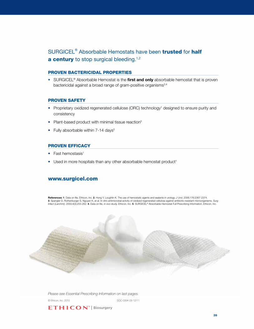
SURGICEL® Absorbable Hemostats have been trusted for half a century to stop surgical bleeding.1,2
PROVEN BACTERICIDAL PROPERTIES
• SURGICEL® Absorbable Hemostat is the first and only absorbable hemostat that is proven bactericidal against a broad range of gram-positive organisms3,4
PROVEN SAFETy
• Proprietary oxidized regenerated cellulose (ORC) technology1 designed to ensure purity and consistency
• Plant-based product with minimal tissue reaction5
• Fully absorbable within 7-14 days5
PROVEN EFFICACy
• Fast hemostasis1
• Used in more hospitals than any other absorbable hemostat product1
www.surgicel.com
References: 1. Data on file, Ethicon, Inc. 2. Hong Y, Loughlin K. The use of hemostatic agents and sealants in urology. J Urol. 2006;176:2367-2374. 3. Spangler D, Rothenburger S, Nguyen K, et al. In vitro antimicrobial activity of oxidized regenerated cellulose against antibiotic-resistant microorganisms. Surg Infect (Larchmt). 2003;4(3):255-262. 4. Data on file, in vivo study. Ethicon, Inc. 5. SURGICEL® Absorbable Hemostat Full Prescribing Information, Ethicon, Inc.
Please see Essential Prescribing Information on last pages.
© Ethicon, Inc. 2010 SGC-0394-09-12/11
39

Abou-Elela A, Morsy A, Badawy H, et al. Use of oxidized cellulose hemostats (Surgicel™) to support parenchymal closure and achieve hemostasis following partial nephrectomy. Surg Technol Int. 2009;18:75-79.
Awonuga AO, Merhi ZO, Khulpateea N. Abdominal packing for intractable obstetrical and gynecologic hemorrhage. Int J Gynecol Obstet. 2006;93(2):160-163.
Bhatnagar RK, Berry S. Selective Surgicel packing for the treatment of posterior epistaxis. Ear Nose Throat J. 2004;83(9):633-634.
Bassetto F, Vindigni V, Scarpa C, et al. Use of oxidized regenerated cellulose to stop bleeding after a facelift procedure. Aesthetic Plastic Surgery. 2008;807-809.
Bollens R, Rosenblatt A, Espinoza B, et al. Laparoscopic partial nephrectomy with “on-demand” clamping reduces warm ischemia time. Eur Urol. 2007;52(3):804-809.
Breda A, Stepanian SV, Lam JS, et al. Use of haemostatic agents and glues during laparoscopic par-tial nephrectomy: a multi-institutional survey from the United States and Europe of 1347 cases. Eur Urol. 2007;52(3):798-803.
Dineen P. Antibacterial activity of oxidized regenerated cellulose. Surg Gynecol Obstet. 1976;142(4):481-486.
Dineen P. The effect of oxidized regenerated cellulose on experimental intravascular infection. Surg. 1977;82(5):576-579.
Dineen P. The effect of oxidized regenerated cellulose on experimental infected splenotomies. J Surg Res. 1977;23:114-116.
Hadi HA, Maw A, Hay DJ. A simple technique to control iatrogenic solid organ injury haemorrhage. Surgeon: J R Coll Surg Edinb Irel. 2004;2(6):339-340.
Kuchta N, Dineen P. Effects of absorbable hemostats on intraabdominal sepsis. Infect in Surg. 1983:441-444.
Lawrentschuk N, Hewitt PM. The use of oxidised cellulose as a topical haemostatic dressing on a bleeding stomal wound. J Wound Care. 2002;11(9):344-345.
Mair H, Kaczmarek I, Oberhoffer M, et al. Surgicel Nu-Knit hemostat for bleeding control of fragile sternum. J Thorac Cardiovasc Surg. 2005;130(2):605-606.
Rastogi V, Dy V. Control of port-site bleeding from smaller incisions after laparoscopic cholecystectomy surgery. Surg Laparosc Endosc Percutan Tech. 2002;12(4):224–226.
Sabel M, Stummer W. The use of local agents: Surgicel and Surgifoam. Eur Spine J. 2004;13(Suppl.1):S97-S101.
Sharma JB, Malhotra M, Pundir P. Laparoscopic oxidized cellulose (Surgicel) application for small uterine perforations. Int J Gynecol Obstet. 2003;83(3):271-275.
Sharma JB, Malhotra M. Successful management of uterine incision hemorrhage in caesarean section with topical oxidized regenerated cellulose (Surgicel Nu-knit): a case report. Arch Gynecol Obstet. 2006; 274:115-116.
Shinkwin CA, Beasley N, Simo R, et al. Evaluation of Surgicel Nu-knit, Merocel and Vasolene gauze nasal packs: a randomized trial. Rhinol. 1996;34(1):41-43.
Spangler D, Rothenburger S, Nguyen K, et al. In vitro antimicrobial activity of oxidized regenerated cellulose against antibiotic-resistant microorganisms. Surg Infect (Larchmt). 2003;4(3):255-262.
Theuer C, Imagawa DK. Use of knitted oxidized cellulose (Nu-knit) for the definitive packing of grade III liver fracture. Inj, Int J Care Injur. 1999;30(2):137-140.
Trooskin SZ, Flancebaum L, Boyarsky A, et al. A simplified approach to techniques of splenic salvage. Surg Gynecol Obstet. 1989;168:546-548.
Bibliography
40

SURGICEL® ORIGINAL, SURGICEL® NU-KNIT® and SURGICEL® FIBRILLAR™Absorbable Hemostats (oxidized regenerated cellulose)
FOR SURGICAL USE(For dental application of this product, reference should
be made to the dental use section of this insert.)
DESCRIPTIONSURGICEL Absorbable Hemostat is a sterile absorbable knitted fabric prepared by the controlled oxidation of regeneratedcellulose. The fabric is white with a pale yellow cast and has a faint, caramel-like aroma. It is strong and can be suturedor cut without fraying. It is stable and should be stored at controlled room temperature. A slight discoloration may occurwith age, but this does not affect performance. The SURGICEL FIBRILLAR form of the product allows the surgeon to grasp with forceps any amount of SURGICEL FIBRILLAR Hemostat needed to achieve hemostasis at a particular bleeding site. The SURGICEL FIBRILLAR form may be more convenient than the knitted form for hard to reach or irregularly shaped bleeding sites. Although it is easy to pull the desired amount of SURGICEL FIBRILLAR Hemostat from the entire supply, the group of selected fibers continue to cohere to one another and application to the bleeding siteis easily controlled. Unwanted dispersal over the operative site does not occur.
ACTIONSThe mechanism of action whereby SURGICEL Absorbable Hemostat accelerates clotting is not completely understood, but it appears to be a physical effect rather than any alteration of the normal physiologic clotting mechanism. After SURGICEL Absorbable Hemostat has been saturated with blood, it swells into a brownish or black gelatinous mass which aids in the formation of a clot, thereby serving as a hemostatic adjunct in the control of local hemorrhage. Whenused properly in minimal amounts, SURGICEL Absorbable Hemostat is absorbed from the sites of implantation with practically no tissue reaction. Absorption depends upon several factors including the amount used, degree of saturationwith blood, and the tissue bed.In addition to its local hemostatic properties, SURGICEL Absorbable Hemostat is bactericidal in vitro against a wide rangeof gram positive and gram negative organisms including aerobes and anaerobes. SURGICEL Absorbable Hemostat isbactericidal in vitro against strains of species including those of:methicillin-resistant Staphylococcus aureus (MRSA)penicillin-resistant Streptococcus pneumoniae (PRSP)vancomycin-resistant Enterococcus (VRE)methicillin-resistant Staphylococcus epidermidis (MRSE)Staphylococcus aureus Bacillus subtilisStaphylococcus epidermidis Proteus vulgarisMicrococcus luteus Corynebacterium xerosisStreptococcus pyogenes Group A Mycobacterium phleiStreptococcus pyogenes Group B Clostridium tetaniStreptococcus salivarius Clostridium perfringensBranhamella catarrhalis Bacteroides fragilisEscherichia coli EnterococcusKlebsiella aerogenes Enterobacter cloacaeLactobacillus sp. Pseudomonas aeruginosaSalmonella enteritidis Pseudomonas stutzeriShigella dysenteriae Proteus mirabilisSerratia marcescensStudies conducted in animals show that SURGICEL Absorbable Hemostat in contrast to other hemostatic agents does notenhance experimental infection. (1-4)
INDICATIONSSURGICEL Absorbable Hemostat (oxidized regenerated cellulose) is used adjunctively in surgical procedures to assist inthe control of capillary, venous, and small arterial hemorrhage when ligation or other conventional methods of control areimpractical or ineffective. SURGICEL ORIGINAL, SURGICEL FIBRILLAR and SURGICEL NU-KNIT Hemostats can be cutto size for use in endoscopic procedures.
CONTRAINDICATIONSAlthough packing or wadding sometimes is medically necessary, SURGICEL Absorbable Hemostat should not be used inthis manner, unless it is to be removed after hemostasis is achieved (See WARNINGS and PRECAUTIONS).SURGICEL Absorbable Hemostat should not be used for implantation in bone defects, such as fractures, since there is apossibility of interference with callus formation and a theoretical chance of cyst formation.When SURGICEL Absorbable Hemostat is used to help achieve hemostasis in, around, or in proximity to foramina inbone, areas of bony confine, the spinal cord, or the optic nerve and chiasm, it must always be removed after hemostasisis achieved since it will swell and could exert unwanted pressure.SURGICEL Absorbable Hemostat should not be used to control hemorrhage from large arteries.SURGICEL Absorbable Hemostat should not be used on non-hemorrhagic serous oozing surfaces, since body fluids otherthan whole blood, such as serum, do not react with SURGICEL Absorbable Hemostat to produce satisfactory hemostaticeffect.SURGICEL Absorbable Hemostat is an absorbable hemostat, and should not be used as an adhesion prevention product.
WARNINGSSURGICEL Absorbable Hemostat is supplied sterile and as the material is not compatible with autoclaving or ethylene oxidesterilization, SURGICEL Absorbable Hemostat should not be resterilized.SURGICEL Absorbable Hemostat is not intended as a substitute for careful surgery and the proper use of sutures and ligatures.Closing SURGICEL Absorbable Hemostat in a contaminated wound without drainage may lead to complications and shouldbe avoided.The hemostatic effect of SURGICEL Absorbable Hemostat is greater when it is applied dry; therefore it should not bemoistened with water or saline.SURGICEL Absorbable Hemostat should not be impregnated with anti-infective agents or with other materials such asbuffering or hemostatic substances. Its hemostatic effect is not enhanced by the addition of thrombin, the activity ofwhich is destroyed by the low pH of the product.Although SURGICEL Absorbable Hemostat may be left in situ when necessary, it is advisable to remove it once hemostasisis achieved. It must always be removed from the site of application when used in, around, or in proximity to foramina in bone, areas of bony confine, the spinal cord, and/or the optic nerve and chiasm regardless of the type of surgical procedure because SURGICEL Hemostat, by swelling, may exert pressure resulting in paralysis and/or nerve damage. Dislodgement of SURGICEL Absorbable Hemostat could possibly occur by means such as repacking, further intraopera-tive manipulation, lavage, exaggerated respiration, etc. There have been reports that in procedures such as lobectomy,laminectomy and repair of a frontal skull fracture and lacerated lobe that SURGICEL Absorbable Hemostat, when left in the patient after closure, migrated from the site of application into foramina in bone around the spinal cord resulting in paralysis and, in another case, the left orbit of the eye, causing blindness. While these reports cannot be confirmed,special care must be taken by physicians, regardless of the type of surgical procedure, to consider the advisability of removing SURGICEL Absorbable Hemostat after hemostasis is achieved.Although SURGICEL Absorbable Hemostat is bactericidal against a wide range of pathogenic microorganisms, it is not intended as a substitute for systemically administered therapeutic or prophylactic antimicrobial agents to control orprevent post-operative infections.
PRECAUTIONSUse only as much SURGICEL Absorbable Hemostat as is necessary for hemostasis, holding it firmly in place until bleeding stops. Remove any excess before surgical closure in order to facilitate absorption and minimize the possibilityof foreign body reaction.In urological procedures, minimal amounts of SURGICEL Absorbable Hemostat should be used and care must be exercised to prevent plugging of the urethra, ureter, or a catheter by dislodged portions of the product.Since absorption of SURGICEL Absorbable Hemostat could be prevented in chemically cauterized areas, its use shouldnot be preceded by application of silver nitrate or any other escharotic chemicals.If SURGICEL Absorbable Hemostat is used temporarily to line the cavity of large open wounds, it should be placed so asnot to overlap the skin edges. It should also be removed from open wounds by forceps or by irrigation with sterile wateror saline solution after bleeding has stopped.Precautions should be taken in otorhinolaryngologic surgery to assure that none of the material is aspirated by the patient. (Examples: controlling hemorrhage after tonsillectomy and controlling epistaxis.)Care should be taken not to apply SURGICEL Absorbable Hemostat too tightly when it is used as a wrap during vascularsurgery (see Adverse Reactions).Endoscopic procedures should be performed only by persons having adequate training and familiarity with endoscopictechniques. Consult medical literature relative to techniques, complications and hazards prior to performance of any endoscopic procedure.A thorough understanding of the principles and techniques involved in laporoscopic laser and electrosurgical proceduresis essential to avoid shock and burn hazards to both patient and medical personnel and damage to the device or other
medical instruments. Refer to appropriate electrosurgical system users manual for use indications and instructions to ensure that all safety precautions are followed. When endoscopic instruments and accessories from different manufacturers are employed together during a procedure, verify their compatability prior to initiation of the procedure and ensure that isolation or grounding is not compromised.
ADVERSE REACTIONS“Encapsulation” of fluid and foreign body reactions have been reported.There have been reports of stenotic effect when SURGICEL Absorbable Hemostat has been applied as a wrap during vascular surgery. Although it has not been established that the stenosis was directly related to the use of SURGICEL Absorbable Hemostat, it is important to be cautious and avoid applying the material tightly as a wrapping.Paralysis and nerve damage have been reported when SURGICEL Absorbable Hemostat was used around, in, or in proximity to foramina in bone, areas of bony confine, the spinal cord, and/or the optic nerve and chiasm. While most ofthese reports have been in connection with laminectomy, reports of paralysis have also been received in connection withother procedures. Blindness has been reported in connection with surgical repair of a lacerated left frontal lobe whenSURGICEL Absorbable Hemostat was placed in the anterior cranial fossa (5) (See WARNINGS and PRECAUTIONS).Possible prolongation of drainage in cholecystectomies and difficulty passing urine per urethra after prostatectomy have been reported. There has been one report of a blocked ureter after kidney resection, in which postoperative catheterization was required.Occasional reports of “burning” and “stinging” sensations and sneezing when SURGICEL Absorbable Hemostat has beenused as packing in epistaxis, are believed due to the low pH of the product.Burning has been reported when SURGICEL Absorbable Hemostat was applied after nasal polyp removal and after hemorrhoidectomy. Headache, burning, stinging, and sneezing in epistaxis and other rhinological procedures, and stinging when SURGICEL Absorbable Hemostat was applied on surface wounds (varicose ulcerations, dermabrasions, anddonor sites) also have been reported.
DOSAGE AND ADMINISTRATIONSterile technique should be observed in removing SURGICEL Absorbable Hemostat from its sterile container. Minimalamounts of SURGICEL Absorbable Hemostat in appropriate size are laid on the bleeding site or held firmly against the tissues until hemostasis is obtained.Opened, unused SURGICEL Absorbable Hemostat should be discarded, because it cannot be resterilized.
HOW SUPPLIEDSterile SURGICEL® FIBRILLAR™ Absorbable Hemostat is supplied as staple fiber in envelopes of the following sizes.Code No. 1961 1 in. x 2 in. (2.5 cm. x 5.1 cm.)Code No. 1962 2 in. x 4 in. (5.1 cm. x 10.2 cm.)Code No. 1963 4 in. x 4 in. (10.2 cm. x 10.2 cm.)Sterile SURGICEL® ORIGINAL Absorbable Hemostat (oxidized regenerated cellulose) is supplied as knitted fabric stripsin envelopes in the following sizes.Code No. 1951 2 in. x 14 in. (5.1 cm. x 35.6 cm.)Code No. 1952 4 in. x 8 in. (10.2 cm. x 20.3 cm.)Code No. 1953 2 in. x 3 in. (5.1 cm. x 7.6 cm.)Code No. 1955 0.5 in. x 2 in. (1.3 cm. x 5.1 cm.)Sterile SURGICEL® NU-KNIT® Absorbable HemostatCode No. 1940 1 in. x 1 in. (2.5 cm. x 2.5 cm.)Code No. 1941 1 in. x 3.5 in. (2.5 cm. x 8.9 cm.)Code No. 1943 3 in. x 4 in. (7.6 cm. x 10.2 cm.)Code No. 1946 6 in. x 9 in. (15.2 cm. x 22.9 cm.)
STORAGEStore at controlled room temperature 15° - 30°C (59° - 86°F).
CAUTIONFederal law restricts this device to sale by or on the order of a physician.
CLINICAL STUDIESSURGICEL Absorbable Hemostat (oxidized regenerated cellulose) has been found useful in helping to control capillary orvenous bleeding in a variety of surgical applications, including abdominal, thoracic, neurosurgical, and orthopedic, aswell as in otorhinolaryngologic procedures. Examples include gallbladder surgery, partial hepatectomy, hemorrhoidectomy,resections or injuries of the pancreas, spleen, kidney, prostate, bowel, breast or thyroid, and in amputations. (6,8)SURGICEL Absorbable Hemostat has been applied as a surface dressing on donor sites and superficial open wounds,
controlling bleeding adequately, and causing no delay in healing or interference with epithelization. (9,11) It also has beenapplied after dermabrasion, punch biopsy, excision biopsy, curettage, finger and toenail removal, and to traumatic wounds.In the foregoing applications, bleeding was controlled and the SURGICEL Absorbable Hemostat was absorbed from the sites where it was applied. (10) In cardiovascular surgery, investigators have found SURGICEL Absorbable Hemostat useful in helping to control bleeding from implanted textile grafts, including those of the abdominal aorta. (7,12) Such grafts may leak or weep considerably, even when pre-clotted, but this seepage can be controlled by covering the graft with a layer or two of SURGICEL Absorbable Hemostat after the graft is in place and before releasing the proximal and distal clamps. When the flow has been reestablished and all the bleeding controlled, the fabric either can be removed or left in situ, since absorption of SURGICEL Absorbable Hemostat has been shown to occur without constriction of the graft or other untoward incident when proper wrapping technique is employed.Otorhinolaryngologic experience with SURGICEL Absorbable Hemostat includes adjunctive use in controlling bleedingresulting from epistaxis, tonsillectomy, adenoidectomy, removal of nasal polyps, repair of deviated septum, tympanoplasty,Stapes surgery, surgery for sinusitis, and removal of tumors. (13,14) SURGICEL Absorbable Hemostat has been reported useful as a hemostatic adjunct in such gynecologic procedures asoophorectomy, hysterectomy, conization of the cervix, and repair of cystorectocele. (6,15)
ANIMAL PHARMACOLOGYThe effects of SURGICEL Absorbable Hemostat, absorbable gelatin sponge, and microfibrillar collagen hemostat werecompared in a standardized infection model consisting of intra-abdominal and intrahepatic abscesses in mice.This infection mimics the common characteristics of human infection with nonspore-forming anaerobic bacteria, including a chronic and progressive course. SURGICEL Absorbable Hemostat did not increase the infectivity of normallysubinfectious inocula of mixed anaerobic species in mice. With the other hemostatic agents, microfibrillar collagen hemostat and absorbable gelatin sponge, an enhancement of infectivity of anaerobic mixtures has been shown. SURGICEL Absorbable Hemostat, in contrast to these hemostatic agents, did not enhance or provide a site for bacterialgrowth.It was also found that aerobic pathogens did not grow in the presence of SURGICEL Absorbable Hemostat. In these studies (1), SURGICEL Absorbable Hemostat was placed in contaminated incisions of guinea pigs and markedly reducedbacterial growth of three different strains of common pathogens. In a dog model (2), it was shown that bacterial contamination of implanted Teflon™ patches in the aorta could be reduced by wrapping the area of the patch with SURGICEL Absorbable Hemostat prior to pathogen challenge. Also, in another study (3), SURGICEL Absorbable Hemostat and a gelatin sponge were placed in two splenotomy sites in large mongrel dogs and the animals were then challenged intravenously and the number of organisms from the splenotomy sites were measured over a period of time. The number of organisms at the site of SURGICEL Absorbable Hemostat were significantly lower than those in thecontrol, or the absorbable gelatin sponge site.
DIRECTIONS FOR USING SURGICEL ORIGINAL, SURGICEL FIBRILLAR AND SURGICEL NU-KNIT HEMOSTATS IN ENDOSCOPIC PROCEDURES:
SURGICEL ORIGINAL, SURGICEL FIBRILLAR and SURGICEL NU-KNITAbsorbable Hemostats should be cut to the appropriate size for endoscopic placement. Standard endoscopic procedures should be usedup to the point of placement of the absorbable hemostat. Grasp the SURGICEL ORIGINAL, SURGICEL FIBRILLA, or SURGICEL NU-KNIT Absorbable Hemostat at one corner. With a steady backward motion, pullthe material into the operating channel until the material is enclosed inthe end of the laparoscope.
41

A. Place the laparoscope back into the patient via the sleeve and reposition the scope over the area of desired application.Slowly push the grasping instrument and material into the cavity.
B. With the use of grasping instruments in a second and/or third auxiliary site, placement can be made and the materialpositioned in place.
REFERENCES:1. Dineen, P.: Antibacterial activity of oxidized regenerated cellulose. Surgery, Gynecology and Obstretrics 142:481-
486, 1976.2. Dineen, P.: The effect of oxidized regenerated cellulose on experimental intravascular infecton. Surgery 82:576-579,
1977.3. Dineen, P.: The effect of oxidized regenerated cellulose on experimental infected splenotomies. Journal of Surgical
Research 23:114-116, 1977.4. Kuchta, N. and Dineen, P.: Effects of absorbable hemostats on intra abdominal sepsis. Infections in Surgery
2:441-444, June 1983.5. Dutton, J., Tse, D., and Anderson, R.: Compressive optic neuropathy following use of intra-cranial oxidized cellulose
hemostat. Ophthalmic Surgery 14(6): 487-490, June 1983.6. Degenshein, G., Hurwitz, A., and Ribacoff, S.: Experience with regenerated oxidized cellulose. New York State
Journal of Medicine 63(18): 2639-2643, 1963.7. Hurwitt, E.: A new absorbable hemostatic packing. Bulletin de la Societe Internationale de Chirurgie XXI
(3):237-242, 1962.8. Venn, R.: Reduction of postsurgical blood-replacement needs with SURGICEL hemostasis. Medical Times
93(10):1113-1116, 1965.9. Miller, J., Ginsberg, M., McElfatrick, G. and Johnson, H.: Clinical experience with oxidized regenerated cellulose.
Experimental Medicine and Surgery 19(2-3):202-206, (June-Sept.) 1961.10. Blau, S., Kanof, N., and Simonson, L.: Absorbable hemostatic gauze SURGICEL in dermabrasions and dermatologic
surgery. Acta Dermato-Venerelogica 40:358-361, 1960.11. Shea, P., Jr.: Management of the donor site: a new dressing technic. Journal of the Medical Association of Georgia
51(9):437-440, 1962.12. Denck, H.: Use of resorbable oxycellulose in surgery. Chirurg 33(11): 486-488, 1962.13. Tibbels, E. Jr.: Evaluation of a new method of epistaxis management. Laryngoscope LXXIII (30):306-314, 1963.14. Huggins, S.: Control of hemorrhage in otorhinolaryngologic surgery with oxidized regenerated cellulose. Eye, Ear,
Nose and Throat Monthly 48(7): (July) 1969.15. Crisp, W.E., Shalauta, H. and Bennett, W.A.: Shallow conization of the cervix. Obstetrics and Gynecology
31(6):755-758, 1968.
U.S. customers: to order product,call 1-800-255-2500;for product quality and technical questions,call 1-877-384-4266.
Manufacturer:ETHICON SARL,Neuchatel, Switzerland
A division of ETHICON, INC.Somerville, NJ 08876, USA
Made in Switzerland© ETHICON INC 2007
SURGICEL ORIGINAL, SURGICEL NU-KNIT, and SURGICEL FIBRILLARAbsorbable Hemostats (oxidized regenerated cellulose)
For Dental Use(For surgical applications of this product, reference should
be made to the surgical use section of this insert.)
DESCRIPTIONSURGICEL Absorbable Hemostat is an absorbable knitted fabric prepared by the controlled oxidation of regenerated cellulose.The fabric is white with a pale yellow cast and has a faint, caramel-like aroma. It is strong and can be suturedor cut without fraying. It is stable and can be stored at controlled room temperature. A slight discoloration may occur withage, but this does not affect performance. The SURGICEL FIBRILLAR form of the product allows the physician to graspwith forceps any amount of SURGICEL FIBRILLAR Hemostat needed to achieve hemostasis at a particular bleeding site.The fibrillar form may be more convenient than the knitted form for hard to reach or irregularly shaped bleeding sites. Although it is easy to pull the desired amount of SURGICEL FIBRILLAR Hemostat from the entire supply, the group of selected fibers continue to cohere to one another and application to the bleeding site is easily controlled. Unwanted dispersal over the operative site does not occur.
ACTIONSThe mechanism of action whereby SURGICEL Absorbable Hemostat accelerates clotting is not completely understood but it appears to be a physical effect rather than any alteration of the normal physiologic clotting mechanism. After SURGICEL Absorbable Hemostat has been saturated with blood, it swells into a brownish or black gelatinous mass which aids in the formation of a clot, thereby serving as a hemostatic adjunct in the control of local hemorrhage. Whenused properly in minimal amounts, SURGICEL Absorbable Hemostat is absorbed from the sites of implantation with practically no tissue reaction. Absorption depends upon several factors, including the amount used, degree of saturationwith blood, and the tissue bed.
INDICATIONSSURGICEL Absorbable Hemostat (oxidized regenerated cellulose) is indicated for adjunctive use to assist in the controlof bleeding in exodontia and oral surgery. It may also be used to help achieve hemostasis after single or multiple toothextractions, alveoloplasty, gingival hemorrhage, impactions, biopsies, and other procedures in the oral cavity.
CONTRAINDICATIONSAlthough packing or wadding sometimes is medically necessary, SURGICEL Absorbable Hemostat should not be used inthis manner unless it is to be removed after hemostasis is achieved.SURGICEL Absorbable Hemostat should not be used for implantation in bone defects, such as fractures, since there is apossibility of interference with callus formation and a theoretical chance of cyst formation.
WARNINGSSURGICEL Absorbable Hemostat is supplied sterile and should not be autoclaved because autoclaving causes physicalbreakdown of the product.SURGICEL Absorbable Hemostat is not intended as a substitute for careful surgery and the proper use of sutures and ligatures.Closing SURGICEL Absorbable Hemostat in a contaminated wound without drainage may lead to complications and shouldbe avoided.The hemostatic effect of SURGICEL Absorbable Hemostat is greater when it is applied dry; therefore it should not bemoistened with water or saline.SURGICEL Absorbable Hemostat should not be impregnated with anti-infective agents or with other materials such asbuffering or hemostatic substances. Its hemostatic effect is not enhanced by the addition of thrombin, the activity ofwhich is destroyed by the low pH of the product.Although SURGICEL Absorbable Hemostat may be left in situ when necessary, it is advisable to remove it once hemostasisis achieved.
PRECAUTIONSUse only as much SURGICEL Absorbable Hemostat as is necessary for hemostasis, holding it in place until bleedingstops. Remove any excess before surgical closure in order to facilitate absorption and minimize the possibility of foreign body reaction. SURGlCEL Absorbable Hemostat should be applied loosely against the bleeding surface. Waddingor packing should be avoided, especially within rigid cavities, where swelling may interfere with normal function or possibly necrosis. Precautions should be taken to assure that none of the material is aspirated by the patient.
ADVERSE REACTIONSEncapsulation of fluid and foreign body reactions have been reported.
DOSAGE AND ADMINISTRATIONSterile technique should be observed in removing SURGICEL Absorbable Hemostat from its envelope. Minimal amountsof SURGICEL Absorbable Hemostat of appropriate size are laid on the bleeding site or held firmly against the tissues untilhemostasis is obtained.Opened, unused SURGICEL Absorbable Hemostat should be discarded, because it cannot be resterilized.
DO NOT USE IF PACK IS DAMAGED/OPENED. DO NOT RESTERILIZE/REUSE. THE USE BY DATE OF THIS PRODUCT ISPRINTED ON THE PACKAGING.
42