a comprehensive view of the epigenetic landscape part i ... · 123 (2005) revealed that a...
TRANSCRIPT

REVIEW
A Comprehensive View of the Epigenetic Landscape Part I: DNAMethylation, Passive and Active DNA Demethylation Pathwaysand Histone Variants
Anna Sadakierska-Chudy • Richard M. Kostrzewa •
Małgorzata Filip
Received: 11 August 2014 / Revised: 7 October 2014 / Accepted: 16 October 2014 / Published online: 2 November 2014
� The Author(s) 2014. This article is published with open access at Springerlink.com
Abstract In multicellular organisms, all the cells are
genetically identical but turn genes on or off at the right
time to promote differentiation into specific cell types. The
regulation of higher-order chromatin structure is essential
for genome-wide reprogramming and for tissue-specific
patterns of gene expression. The complexity of the genome
is regulated by epigenetic mechanisms, which act at the
level of DNA, histones, and nucleosomes. Epigenetic
machinery is involved in many biological processes,
including genomic imprinting, X-chromosome inactiva-
tion, heterochromatin formation, and transcriptional regu-
lation, as well as DNA damage repair. In this review, we
summarize the recent understanding of DNA methylation,
cytosine derivatives, active and passive demethylation
pathways as well as histone variants. DNA methylation is
one of the well-characterized epigenetic signaling tools.
Cytosine methylation of promoter regions usually represses
transcription but methylation in the gene body may have a
positive correlation with gene expression. The attachment
of a methyl group to cytosine residue in the DNA sequence
is catalyzed by enzymes of the DNA methyltransferase
family. Recent studies have shown that the Ten-Eleven
translocation family enzymes are involved in stepwise
oxidation of 5-methylcytosine, creating new cytosine
derivatives including 5-hydroxymethylcytosine, 5-formyl-
cytosine, and 5-carboxylcytosine. Additionally, histone
variants into nucleosomes create another strategy to regu-
late the structure and function of chromatin. The replace-
ment of canonical histones with specialized histone
variants regulates accessibility of DNA, and thus may
affect multiple biological processes, such as replication,
transcription, DNA repair, and play a role in various dis-
orders such as cancer.
Keywords Cytosine variants � DNA methylation � DNA
methyltransferases � Histone variants � Passive and active
demethylation � TET family enzymes
Introduction
In the last decade, epigenetics has become an important
topic of genetic research. The classical definition of epi-
genetics refers to the mitotically and/or meiotically heri-
table changes in gene activity that does not involve
alterations in DNA sequence. This definition emphasizes
the heritability of the cellular phenotype, and therefore, it
only includes changes in the germ line that can be passed
down from generation to generation and changes in
dividing cells that can be transferred to daughter cells.
Currently, we know that epigenetic changes can be induced
by environmental factors at different times in life and are
potentially reversible. In 2007, Brenda Weis proposed the
broader term of epigenetics that refers to ‘‘the study of
regulation of gene activity that is not dependent on gene
sequence and includes heritable and non-heritable
A. Sadakierska-Chudy (&) � M. Filip
Laboratory of Drug Addiction Pharmacology, Institute of
Pharmacology Polish Academy of Sciences, Smetna Street 12,
31-343 Krakow, Poland
e-mail: [email protected]
R. M. Kostrzewa
Department of Biomedical Sciences, Quillen College of
Medicine, East Tennessee State University, Johnson City,
TN 37614, USA
M. Filip
Department of Toxicology, Faculty of Pharmacy, Jagiellonian
University, Medical College, Medyczna 9, 30-688 Krakow,
Poland
123
Neurotox Res (2015) 27:84–97
DOI 10.1007/s12640-014-9497-5

alterations in gene activity and transcriptional potential of a
cell’’ (Brenda Weis at the ‘‘Diet, Epigenetic Events, and
Cancer Prevention Symposium’’ on September 27th, 2007,
in Washington, D.C./http://prevention.cancer.gov/files/
news-events/100908_epigenetics%20meeting%20report%
20Sept%202007.pdf).
Epigenetic control operates on three major levels, i.e.,
on DNA, histones, and nucleosomes. The relationships
among these various epigenetic elements are currently
being extensively investigated. In this review, data from
the literature are analyzed to discuss the significance of
DNA methylation and demethylation, cytosine derivatives
as well as histone variants in the epigenetic regulation of
the genome.
DNA Level
DNA Methylation
DNA methylation is a biochemical process crucial for
normal development in higher organisms, and it is the most
thoroughly studied epigenetic mark. Methylation entails
the covalent attachment of a methyl (CH3) group to the C5
position of a cytosine residue, forming 5-methylcytosine
(5mC).
In some organisms, this modification is so frequent that
it is denoted as the fifth nucleotide. The methyl group is
transferred from S-adenosyl-L-methionine (SAM) to cyto-
sine by the DNA methyltransferase (DNMT) family of
enzymes: DNMT1, DNMT2, DNMT3A, DNMT3B, and
DNMT3L (Jin et al. 2011). DNMT1 preferentially meth-
ylates hemimethylated cytosines in CpG dinucleotide
sequences, maintaining the methylation pattern during
replication (Probst et al. 2009). In contrast to DNMT1,
DNMT3A and 3B prefer unmethylated CpG dinucleotides
and perform de novo methylation in early development (Li
2002). Thus, DNMT1 acts primarily as a maintenance
methyltransferase during DNA synthesis, and DNMT3A
and DNMT3B act as de novo enzymes in development. A
growing body of evidence suggests that DNMT1 may also
be necessary for de novo methylation of genomic DNA
(Egger et al. 2006) and that DNMT3A and DNMT3B are
also responsible for the maintenance of methylation during
cell replication (Riggs and Xiong 2004). It is worth noting
that DNMT2 displays weak DNA methyltransferase
activity but actually functions as an RNA methyltransfer-
ase. The DNMT2 enzyme specifically methylates cytosine-
38 in the anticodon loop of aspartic acid transfer RNA that
protects tRNAs from cleavage under stress conditions (Goll
et al. 2006; Schaefer et al. 2010).
A recent finding has suggested that DNMT2 might be
involved in the mammalian paramutation pathway, by
protecting small RNA molecules against endonucleolytic
cleavage (Adams and Meehan 2013; Kiani et al. 2013), and
thus it might induce heritable epigenetic phenotypes.
DNMT3L, although it shares homology with DNMT3A
and 3B, has no catalytic activity. Instead, DNMT3L
increases the ability of DNMT3A and B to bind to methyl
groups, thus facilitating methylation in vivo (Bird 2002; Jin
et al. 2011). Moreover, DNMT3L recognizes nucleosomes
with an unmethylated histone H3 lysine 4 (H3K4) and
recruits DNMT3A and DNMT3B to their targets (Saitou
et al. 2012). Structural and functional domains of mam-
malian DNMTs are shown in Fig. 1.
The level of 5mC affects gene expression, and deregu-
lation of cytosine methylation may play a role in devel-
opment, cellular differentiation, or disease (Santos-Rebouc
and Pimentel 2007; Aguilera et al. 2010; Hackett and
Surani 2013). The DNA methylation level can affect
transcriptional activities, hypermethylation (a surplus of
methyl groups) of promoter regions, and is generally
associated with transcriptional silencing, but hypomethy-
lation (a deficit of methyl groups) causes an increased level
of gene expression (Crider et al. 2012). Approximately,
2–8 % of the cytosines in the mammalian genome are
methylated, mostly in CpG sequences (Zhu 2009; Varriale
2014). In the human genome, CpG dinucleotides are dis-
tributed asymmetrically among GC-rich and -poor DNA
regions, and not all CpG sites are methylated. The pattern
of DNA methylation varies in different cell types and is
tissue specific. For example, in differentiated mammalian
cells, cytosine residues in GC-rich regions (which typically
contain more than 50 % GC) are usually methylated. DNA
regions that contain a high frequency of CpG sites are so-
called CpG islands (CGIs) and represent an important
feature of the mammalian genome. They are located in
promoters, preferentially near the transcription start sites
(TSSs) of [50 % of human genes. CGI methylation is
lower at promoters and higher in gene bodies and inter-
genic regions. CGI-rich promoters are largely free of DNA
methylation due to the abundance of GC-rich transcription
factor-binding sites (Deaton and Bird 2011). Methylation
of DNA cytosine residues at promoter regions usually
correlates with a higher order of chromatin state and
repression mRNA transcription. However, Niesen et al.
Neurotox Res (2015) 27:84–97 85
123

(2005) revealed that a sequence-specific DNA-binding
protein can facilitate transcriptional activation of methyl-
ated promoter. Interestingly, recent findings suggest that in
undifferentiated stem cells, cytosines outside of CpG sites
can be methylated as well, and this process is particularly
important for the proper regulation of gene expression in
embryonic stem cells (ESCs) (Lister et al. 2009). As pre-
viously mentioned, gene bodies are highly methylated but
the role of methylation remains largely unresolved. Some
studies have begun to decipher molecular implications of
gene body methylation. For example, methylation in the
gene body contributes to the suppression of transcriptional
noise (Huh et al. 2011) and might stimulate transcription
elongation (Jones 2012). A recent study has suggested that
exons are methylated at higher levels than introns and
possibly play a role in the regulation of mRNA splicing
(Laurent et al. 2010). More details about genomic locations
of DNA methylation and its consequence can be found in
excellent recent reviews (Estecio and Issa 2011; Moore
et al. 2013).
DNA methylation has been considered a stable, persis-
tent and heritable mark; therefore, methyl groups are added
but not removed. Recent data have indicated that tran-
scription factors and related proteins not only protect
sequences from methylation but also initiate active DNA
demethylation (Stadler et al. 2011). Both passive demeth-
ylation during replication and active demethylation take
place in eukaryotic cells. For example, DNA methylation
patterns undergo reprogramming during the establishment
of primordial germ cells (PGCs) as well as after fertiliza-
tion (Branco et al. 2011; Saitou et al. 2012). Surprisingly,
the establishment of DNA methylation patterns occurs
during development and differentiation of the central ner-
vous system, where it has been implicated in synaptic
plasticity, learning, and memory. In the human brain, DNA
methylation changes are strongly correlated with age
(Hernandez et al. 2011). In turn, pathological activation of
DNMTs and aberrant 5mC formation may cause neurode-
gradation and apoptotic neuronal death (Chestnut et al.
2011; Hernandez and Singleton 2012).
DNA methylation influences gene expression not only
by impeding the binding of specific transcription factors
but also by recruiting chromatin-modifying proteins. DNA
methylation also determines the histone modification pat-
terns and the DNMTs and methyl-CpG-binding domain
(MBD) proteins that help to recruit repressor complexes
containing histone deacetylases (HDACs) (Fuks et al.
2003). Conversely, interactions between DNMT1, G9a
Fig. 1 Schematic structure of mammalian DNMT family members.
DNMT1, the first described methyltransferase, preferentially methyl-
ates hemimethylated DNA (Robertson 2001). DNMT2 lacks the
N-terminal domain, while C-terminal domain contains the full set of
sequence motifs but has not been shown to have transmethylase
activity (Bestor 2000). DNMT3A and DNMT3B have similar domain
arrangements and an equal preference for hemimethylated and
unmethylated DNA (Robertson 2001). DNMT3L, being closely
related to the catalytic domain of DNMT3A/3B, lacks canonical
DNA cytosine-methyltransferase motifs (Bestor 2000). Its N-termi-
nal regulatory domains exhibit little similarity but the catalytic
domains of DNMTs are conserved. The N-terminal domain possesses:
PBD—proliferating cell nuclear antigen (PCNA) binding domain,
NLS—nuclear localization signal, TRF—targeting replication foci,
CXXC—cysteine rich, zinc finger DNA-binding motif, PBH—
polybromo homology domain, PWWP—tetrapeptide domain contain-
ing proline-tryptophan-tryptophan-proline motif. The N-terminal
and C-terminal domains are linked by dinucleotide repeats: GK—
glycine-lysine repeat. The C-terminal domain consists of six most
conserved amino acid motifs (motif I and X form SAM binding site,
motif IV binds cytosine at the active site). Mapped interaction sites of
DNMTs and HDACs (histone deacetylases) are indicated in the above
diagrams. The borders of the DNMT1 domains are marked according
Song et al. (2011)
86 Neurotox Res (2015) 27:84–97
123

(methyltransferase H3K9), and the replication complex
lead to dimethylation of histone H3 lysine 9 (H3K9me2), a
repressive epigenetic mark. Methylated H3K9 is bound by
heterochromatin protein 1 (HP1), which interacts directly
with DNMT1, resulting in cytosine methylation (Small-
wood et al. 2007; Saitou et al. 2012). The interaction of the
H3K9 methyltransferases (SUV39H1 and ESET) with
DNMT3A and DNMT3B can also cause DNA methylation
at H3K9me2 (Fuks et al. 2003). Notably, chromatin orga-
nization differs between CpG and non-CpG promoters.
GC-rich DNA is preferentially bound by CXXC domain
proteins that can recruit chromatin modifiers, including the
CXXC finger protein 1 (Cfp1) subunit of the H3K3me3
methyltransferase complex and KDM2A, an H3K36me2
demethylase (Vavouri and Lehner 2012). In addition to
participating in the histone modifications, DNA methyla-
tion may influence the incorporation of histone variant
H2A.Z into nucleosomes. A growing body of evidence
suggests that the H2A.Z is excluded from methylated DNA
and the global anticorrelation between DNA methylation
and H2A.Z is observed (Conerly et al. 2010; Weber and
Henikoff 2014).
Taken together, DNA methylation affects the interaction
between the histone and DNA, resulting in either activation
or repression of transcription. It is well known that the
disruption of methylation patterns can cause many diseases
including cancer, autoimmune disease, as well as chro-
mosomal instability, and mental retardation syndromes
(Dobrovic 2010; Javierre et al. 2011). In humans, muta-
tions in genes, including DNMTs and methyl-CpG binding
proteins (MBPs), could have profound impact on specific
DNA methylation patterns leading to epigenetic diseases
(Santos-Rebouc and Pimentel 2007). Up to now, more
studies have signified that life style and environmental
factors, such as nutrient supply, drugs, pollutants, patho-
gens, sex hormones, radiation, heavy metals, and early
stress can modulate DNA methylation (Javierre et al. 2011;
Lim and Song 2012). Interestingly, certain dietary
constituents (e.g., folate and bioactive components) may
alter genomic and gene-specific DNA methylation levels
during embryonic development and adult life (Aguilera
et al. 2010; Choi and Friso 2010; McKay and Mathers
2011). Concerning the reversible nature of DNA methyla-
tion, it seems to be attractive for epigenetic modulation
(Egger et al. 2004; Yang et al. 2010).
Cytosine Variants
It has long been known that cytosine can exist in one of two
functional states, unmethylated or methylated. Moreover,
mechanisms of DNA methylation are among the best
understood epigenetic phenomena. Recently, several cyto-
sine variants, including 5-hydroxymethylcytosine (5hmC),
5-formylcytosine (5fC), 5-carboxylcytosine (5caC), and
3-methylcytosine (3mC), were identified.
5-Hydroxymethylcytosine (5hmC)
5-Hydroxymethylcytosine was discovered 60 years ago in
T2 bacteriophage (Wyatt and Cohen 1952), and 20 years
later Penn et al. found 5hmC base in mammalian cells
(Penn et al. 1972). These early findings could not be rep-
licated in later studies until 2009, when two independent
groups showed that 5hmC exists in mouse Purkinje neurons
(Kriaucionis and Heintz 2009) and in ESCs (Tahiliani et al.
2009). Currently, 5hmC is regarded as the ‘‘sixth’’ base of
the genome of higher organisms (Munzel et al. 2010). The
levels of 5hmC in the genome are relatively low and
account for *0.4 % of all cytosines compared to the
*10 % that are 5mC (Branco et al. 2011). 5hmC consti-
tutes approximately 40 % of the modified cytosines in
mouse brain, and the amount increases during maturation
in both the hippocampus and the cerebellum (Szulwach
et al. 2011). Recently, it has been confirmed that 5hmC is
generated by the Ten-Eleven Translocation (TET) enzymes
that are Fe(II) and a-oxoglutarate-dependent dioxygenases.
Neurotox Res (2015) 27:84–97 87
123

The TET subfamily, including TET1, TET2, and TET3,
catalyzes the conversion of 5mC–5hm in vitro and in vivo
(Ito et al. 2010; Branco et al. 2011) and may be engaged in
the further oxidation of 5hmC–5fC and 5caC (He et al.
2011; Ito 2011) (Fig. 2). The TET proteins contain iron and
oxyglutarate domains as well cysteine-rich regions that are
most likely involved in DNA binding (Iyer et al. 2009).
Moreover, TET1 and TET3 contain CXXC zinc finger
domains, which allow binding to unmethylated, methylated
and hydroxymethylated DNA.
Other CXXC-containing proteins, for example DNMT1,
almost solely bind to unmethylated DNA; therefore, poor
recognition of 5hmC could lead to passive demethylation
(Valinluck and Sowers 2007). The level of 5hmC in adult
tissues is between 0.03 and 0.69 % with the highest levels
(0.4–0.7 %) in the central nervous system (Globisch et al.
2010). The biological role of 5hmC is still unclear. It has
been postulated that 5hmC could be an intermediate in
active DNA demethylation, and it may play an important
role in gene regulation (Tahiliani et al. 2009; Wu and
Zhang 2010). It has been observed that 5hmC is enriched in
the body of the active genes and at the TSSs of transcrip-
tionally inactive genes (Song et al. 2010; Wu et al. 2011a).
In vitro analysis revealed that 5hmC in the gene body
prevents the binding of MBD proteins, which act as tran-
scriptional repressors (Valinluck et al. 2004; Jin et al.
2010). The level of 5hmC in the gene body might modify
the accessibility of chromatin to the transcriptional
machinery. Nestor et al. have demonstrated that 5hmC
patterns are tissue specific. The global content of 5hmC
varies markedly between tissues and does not correlate
with global 5mC levels (Nestor et al. 2012). Chen et al.
(2012) have demonstrated that aging increases both global-
and locus-specific 5hmC content in the mouse
hippocampus.
It is possible that 5hmC initiates the pathway of passive
or active DNA demethylation by excluding DNMT1 and
the MBD proteins from methylating cytosine, and it may
recruit other unknown 5hmC-specific effector proteins
(Stroud et al. 2011). Recent in vitro studies have revealed
that TET proteins could contribute to the removal of
methylated cytosine (He et al. 2011; Ito et al. 2011;
Matarese et al. 2011). This enzyme family has the capacity
to oxidize 5mC not only to 5hmC but also to 5-formylcy-
tosine and 5-carboxylcytosine. Other researchers have
shown that thymine-DNA glycosylase (TDG) belonging to
the uracil-DNA glycosylase (UDG) superfamily can rec-
ognize and excise 5fC and 5caC; thus, the base excision
repair (BER) system could be a trigger (Ooi and Bestor
2008; He et al. 2011; Matarese et al. 2011). The crystal
structure of human TDG revealed a binding pocket that can
accommodate 5caC which facilitates its cleavage (Zhang
et al. 2012; Kohli and Zhang 2013). Furthermore, TDG can
remove T:G or hmU:G mismatches generated by enzy-
matic deamination of 5mC to thymine and 5hmC to
5-hydroxymethyluracil (5hmU) (Shen et al. 2014). In
addition, alternative UDG glycosylases including methyl-
CpG-binding domain protein 4 (MBD4) and single-strand-
selective monofunctional uracil-DNA glycosylase 1
(SMUG1) can be involved in active DNA demethylation
pathway (Shen et al. 2014). Recent studies have reported
that the hydroxylation of 5mC mediated by the Tet1 protein
promotes active DNA demethylation in the adult brain by
deaminating cytosine residue to uracil by the activation-
induced deaminase (AID)/apolipoprotein B mRNA-editing
enzyme complex (APOBEC) family, and then deaminated
cytosine residue is excised by DNA glycosylases and
repaired by the BER pathway (Guo et al. 2011). Potential
mechanisms responsible for passive and active demethyl-
ation are presented in Fig. 2.
Fig. 2 Passive and active DNA demethylation pathways. Passive
DNA demethylation is caused by a reduction in activity or absence of
DNMTs (yellow arrows). Active demethylation via oxidation path-
way (green arrows): TET enzymes can hydroxylate methylcytosine
(5mC) to form 5-hydroxymethylcytosine (5hmC); further oxidation
produces 5-formylcytosine (5fC) and 5-carboxylcytosine (5caC). 5fC
and 5caC can be actively removed by the DNA glycosylases. In
addition, a putative deformylase may convert 5fC to C and
decarboxylase convert 5caC to C. Active demethylation via deam-
ination pathway (red arrows): AID/APOBEC family members can
deaminate 5mC or 5hmC to form thymidine or 5-hydroxymethylura-
cil (5hmU). These intermediates are replaced by cytosine during base
excision repair (BER) mediated by the uracil-DNA glycosylase
(UDG) family, like TDG or SMUG1 as well as MBD4 (specifically
recognize thymine and 5hmU). AID activation-induced deaminase,
APOBEC apolipoprotein B mRNA-editing enzyme complex, BER—
base excision repair, DNMT1/3A/3B—DNA methyltransferase,
MBD4—methyl-binding domain protein 4, SMUG1—single-strand
specific monofunctional uracil-DNA glycosylase, TET1/2/3—ten-
eleven methylcytosine dioxygenase family, TDG—thymine-DNA
glycosylase (Color figure online)
88 Neurotox Res (2015) 27:84–97
123
5-Formylcytosine (5fC)
5-Formylcytosine is one of the DNA base variants pro-
duced by oxidation of 5hmc by the TET family of enzymes
(Ito et al. 2011). Thin layer chromatography and tandem
liquid chromatography-mass spectrometry has revealed
5fC in mouse ESCs and in brain cortex (Raiber et al. 2012).
The levels of 5fC are estimated to be from 0.02 to 0.002 %
of the genomic DNA of ES cells and are 10- to 100-fold
lower than the levels of 5hmC (Ito et al. 2011; Pfaffeneder
et al. 2011). These levels seem reasonable because TET1
and TET2 are highly expressed and most likely play roles
in DNA methylation reprogramming and cell differentia-
tion (Koh et al. 2011). Indeed, during differentiation, levels
of 5fC decrease, suggesting its participation in develop-
ment and germ cell programming (Pfaffeneder et al. 2011).
A recent study has reported that CGI promoters were more
enriched in 5fC levels than in 5hmC or 5mC levels, which
correlated with active gene expression. Moreover, TDG
was shown to be actively involved in the removal of 5fC
marks in CGIs, exons, and promoter regions (Raiber et al.
2012). Therefore, 5fC excision may help to establish cor-
rect methylation patterns during cell-specific develop-
mental programs. Surprisingly, 5fC-enriched promoter
regions overlap with H3K4me3, suggesting cross-talk
between these marks in transcriptionally active genes.
5-Carboxylcytosine (5caC)
5-Carboxylcytosine is one of the intermediates in active
DNA demethylation and is produced by TET-mediated
enzymatic oxidation from 5fC. The TET3 protein is most
likely responsible for this conversion (Gu et al. 2011). To
date, 5caC has been found in ESCs and in mouse pre-
implantation embryos (Inoue et al. 2011; He et al. 2011).
Alioui and co-workers have shown that 5caC is detectable
in the somatic cells of amphibian ovaries and is primarily
localized to gene-rich euchromatic regions similar to 5hmC
(Alioui et al. 2012). This study also demonstrated that TDG
glycosylase can initiate the BER pathway and cleave 5caC
both in vitro and in vivo, but the MBD4 enzyme exhibited
no activity toward 5caC. Interestingly, 5caC levels
increased when TDG was depleted in mouse ES cells; thus,
TDG is most likely not the only enzyme capable of pro-
cessing 5caC (He et al. 2011). It is not known whether
TDG is able to recognize and excise 5caC from duplex
DNA and whether additional enzymes might be engaged in
the conversion of 5caC in mammalian cells.
3-Methylcytosine (3mC)
3-Methylcytosine is a DNA adduct created by spontaneous
exposure to endogenous or environmental alkylating
agents, leading to cytotoxicity and carcinogenesis. This
mutagenic lesion can be directly repaired with the partic-
ipation of the ABH3 or ABH2 DNA dioxygenases through
the BER pathway in humans, or it can be dealkylated by
AlkB in bacteria (Koivisto et al. 2004; Yi et al. 2012).
Biochemical experiments indicate that ALKBH2 prefers
double-stranded DNA (dsDNA) substrates, while ALKBH3
prefers single-stranded DNA (ssDNA) substrates, which
are generated by the activating signal cointegrator complex
(ASCC) (Dango et al. 2011; Yi et al. 2012). Dango et al.
(2011) demonstrated that loss of ALKBH3 or ASCC3
significantly reduced cell proliferation in vitro and in vivo
in xenograft models. Concurrently, the accumulation of
endogenous 3meC in genomic DNA was observed. Addi-
tionally, ALKBH2 has been shown to play an efficient role
in pediatric brain tumors during chemotherapy treatment,
and the combination of an ALKBH2 knockdown and cis-
platin chemotherapy seems to improve the efficacy of
treatment (Cetica et al. 2009; Wu et al. 2011b). Taken
together, these findings indicate an important role for
alkylation repair in removing environmentally induced
DNA lesions as well as in maintaining genome integrity
and stability.
Histone Variants
Histones are small, basic, and highly conserved proteins
that serve as structural scaffolds for DNA packaging
(Cooper 2000). A DNA molecule (*147 bp in length)
wrapped around the octamer of a histone (two dimers of
H2A–H2B and a heterotetramer (H3–H4)2) constitutes a
nucleosome, the fundamental repeating unit of eukaryotic
chromatin (Cooper 2000). Histone H1 binds to linker DNA
(*50 bp) between nucleosomes, forming a macromolec-
ular structure to help in further compaction of genomic
DNA (Sancho et al. 2008). Histone proteins have a tri-
partite structure consisting of a central globular domain
flanked by N- and C-terminal parts (Fig. 3). The unstruc-
tured tail located at the N-terminal portion protrudes away
from the nucleosome and, therefore, is prone to a variety of
post-translational modifications (PTMs) (Kouzarides
2007). The highly conserved globular domain, termed a
helix-turn-helix, contains three a–helices separated by loop
regions and is involved in histone–histone and histone-
DNA interactions (Luger 2001). The C-terminal domains
of all histones except histone H1 and H2A are relatively
short (Vogler et al. 2010).
Histone tails have many positively charged amino acids
(especially lysine and arginine), which facilitate their
binding to the negatively charged DNA molecule and in-
tranucleosomal interaction (Hansen 2002). The N-terminal
histone tails have been studied extensively, but little is
Neurotox Res (2015) 27:84–97 89
123
known about the function of the C-terminal part. Vogler
et al. have shown that the H2A C-terminal tail plays a
pivotal role in regulating chromatin structure and dynamics
(Vogler et al. 2010). These experiments revealed that the
H2A C-terminus is required for efficient nucleosome
translocation by chromatin remodelers and acts as a novel
recognition module for linker histone H1 (Vogler et al.
2010). It appears that the H2A C-terminal tail has a dual
function. On the one hand, it provides stabilization of the
nucleosomal core particle, and on the other hand, it par-
ticipates in interactions with proteins that control chro-
matin dynamics and conformation.
There are highly similar forms of histones termed ‘his-
tone variants’. It has been estimated that approximately
937 different variants of linker and core histones exist in
various species. In humans, 57 histone variants are encoded
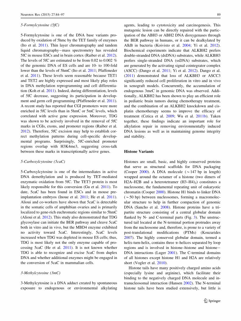
Fig. 3 Schematic structure of the five histone proteins. The N-ter-
minal part is flexible and positively charged and protrudes from the
nucleosome. Two short helices, a-1 and a-2 have a length of 10–14
amino acid residues; central a-2 helix comprises *28 amino acid
residues (Luger 2001). The H2A-docking domain spans amino acids
82–119 and is implicated in both structural and functional properties
of the nucleosome (Shukla et al. 2011). It stabilizes the wrapping of
one helical turn of DNA around the histone octamer (Shukla et al.
2011) and the binding of H2A–H2B dimers to (H3–H4)2 tetramers
(Bonisch and Hake 2012). In addition, the H2A C-terminus has also
been found to be crucial for binding of the linker histone H1 to
nucleosome (Vogler et al. 2010). a helices and b strands of the
histone fold extensions are shown as open boxes and arrows,
respectively
Table 1 Variants of histone H1
in humans
a Gene expressed in a
replication-dependent mannerb Gene expressed in a
replication-independent manner
Variants Name of
genes
Genomic
location
(Ensembl)
Protein length
(aa) (Swiss-
Prot)
Function (reference)
H1.0b H1F0 22q13.1 194 Nucleosome spacing and chromatin
compaction
H1.1a HIST1H1A 6p22.2 215 Linker histones inhibit sliding of histone
octamers, and it is postulated that they can
inhibit chromatin remodeling (Clausell
et al. 2009)
H1.2a HIST1H1C 6p22.2 213
H1.3a HIST1H1D 6p22.2 221 However, a recent study suggested that H1.2–
H1.5 are depleted from active promoters
and gene regulatory elements but enriched
at regions carrying repressive histone marks
(Izzo et al. 2013). H1 binding might be
more sensitive to initiation of transcription
than to transcriptional elongation (Izzo
et al. 2013). Linker histones may operate in
conjunction with a ‘network’ of other
chromatin-binding proteins so as to define
permissive (euchromatin) and repressive
(heterochromatin) DNA domains (Ausio
2006).
H1.4a HIST1H1E 6p22.2 219
H1.5a HIST1H1B 6p22.1 226
H1xb H1FX 3q21.3 213
H1oo H1FOO 3q22.1 346
H1t HIST1H1T 6p22.2 207
Testis-
specific H1
H1FNT 12q13.11 255
Spermatid-
specific H1
HILS1 17q21.33 231
90 Neurotox Res (2015) 27:84–97
123

by 94 genes, the majority of them being present in four
clusters: cluster 1 on chromosome 6 (6p22), cluster 2 on
chromosome 1 (1p21), cluster 3 on chromosome 1 (1q42),
and cluster 4 on chromosome 12 (12p12–13). The incor-
poration of specific histone variants into nucleosomes has
significant impacts on gene expression, heterochromatiza-
tion, and the formation of specialized regions of the
chromatin (Kamakaka and Biggins 2005; Pusarla and
Bhargava 2005). The histone variants have recently
emerged as important factors in regulating chromatin states
and also DNA repair in response to genotoxic treatments
(Malik and Henikoff 2003). Moreover, it is likely that
histone variants, as potential drivers of cancer initiation
and/or progression, thus may be utilized as prognostic
indicators of cancer (Vardabasso et al. 2014).
Histone H1
Histone H1 proteins consist of 194–346 amino acid resi-
dues, depending on the variant. Approximately 126 dif-
ferent members of the H1 family have been reported from
diverse species thus far (http://www.actrec.gov.in/histome/
). Eleven variants of histone H1 have been described in
humans; these are coded by a single gene that exhibits
either replication-dependent or replication-independent
expression (Table 1). Three of the variants are testis-spe-
cific (i.e., HIST1H1T, H1FNT, and HILS1), one of them is
oocyte-specific (H1foo), and the others are somatic vari-
ants. Linker histone H1 is involved in chromatin compac-
tion and plays a role in the formation of higher-order
chromatin structures (Millan-Arino et al. 2014). The
Table 2 Variants of histone H2A in humans
Variants Name of genes Genomic
location
(Ensembl)
Protein length
(aa) (Swiss-Prot)
Function (reference)
H2A type 1 HIST1H2AI 6p22.1 130 Stabilization of the histone core octamer (Ausio 2006)
HIST1H2AK
HIST1H2AL
HIST1H2AM
HIST1H2AG
H2A1 type 1-A HIST1H2AA 6p22.2 131
H2A type1-B/E HIST1H2AE 6p22.2 130
HIST1H2AB
H2A type 1-C HIST1H2AC 6p22.2 130
H2A type 1-D HIST1H2AD 6p22.2 130
H2A type 1-H HIST1H2AH 6p22.1 128
H2A type 1-J HIST1H2A 6p22.1 128
H2A type 2-A HIST2H2AA4 1q21.2 130
HIST2H2AA4
H2A type 2-B HIST2H2AB 1q21.2 130
H2A type 2-C HIST2H2AC 1q21.2 129
H2A type 3 HIST3H2A 1q42.13 130 Unknown function
H2A-Bbd type 1 H2AFB1 Xq28 115 Transcription activation (Tolstorukov et al. 2012)
H2A-Bbd type 2/3 H2AFB2 Xq28 115 Transcription activation (Tolstorukov et al. 2012)
H2AFB3
H2A.J H2AFJ 12p12.3 129 unknown function
H2A.X H2AFX 11q23.3 143 Genome integrity: DNA repair regulation (Pusarla and
Bhargava 2005)
H2A.Z.1 H2AFZ 4q23 128 Maintenance of heterochromatin, transcription
repression and activation (Fan et al. 2004; Guillemette
et al. 2005; Raisner et al. 2005)H2A.Z.2 H2AFV 7p13 128
macroH2A.1 H2AFY 5q31.1 372 Silencing: enriched in inactivated chromosome X
(Pusarla and Bhargava 2005)
macroH2A.2 H2AFY2 10q22.1 372 Silencing: enriched in inactive X-chromosome
chromatin and in senescence-associated
heterochromatin (Pusarla and Bhargava 2005)
Neurotox Res (2015) 27:84–97 91
123

specific role of histone H1 variants is still far from clear,
and genomic distribution of H1 is challenging due to the
lack of variant-specific antibodies (Izzo et al. 2013).
Histone H2A
Histone H2A proteins are composed of *130 amino acid
residues, but atypical variants (macroH2As, H2A.X and
H2A-Bbd) differ in size. Approximately 265 different
members of histone H2A were identified from a variety of
species (http://www.actrec.gov.in/histome/). In humans,
nineteen variants of histone H2A encoded by 26 genes
were reported (Table 2).
Histone H2B
Except for four variants, the variants of histone H2B con-
tain 126 amino acid residues. The histone H2B family
contains 214 different members described from diverse
species (http://www.actrec.gov.in/histome/). Histone H2B
forms a dimer with histone H2A in nucleosome cores.
Histone H2B has 19 variants encoded by 23 genes in
humans, the majority of which are assembled in cluster 1
(i.e., 6p22.1–22.2) (Table 3). There are relatively few
PTMs identified among the amino acid residues of histone
H2B compared to other core histones.
Histone H3
Histone H3 consists of *136 amino acid residues; only the
centromere protein A (CENP-A) is a longer variant. The
histone H3 family contains 216 different members char-
acterized from various species (http://www.actrec.gov.in/
histome/). In humans, 20 genes encode 8 variants of his-
tone H3, most of which are clustered on chromosome 6
(Table 4). Histone H3 is the most extensively post-trans-
lationally modified of the five histones.
Histone H4
Histone H4 contains only 103 amino acid residues and forms a
heterotetramer (H3–H4)2 with histone H3. The histone H4
family consists of 116 members reported from different
organisms (http://www.actrec.gov.in/histome/). Interestingly,
humans have a single histone H4 protein encoded by 14 genes,
eleven of which are clustered on chromosome 6 (Table 5).
Table 3 Variants of histone H2B in humans
Variants Name of genes Genomic
location
(Ensembl)
Protein length
(aa) (Swiss-Prot)
Function (references)
H2B type 1-A HIST1H2BA 6p22.2 127 Specific role of H2B variants is poorly understood. It
is probable that they specialize in chromatin
compaction and transcription repression,
particularly during gametogenesis (Kamakaka and
Biggins 2005)
H2B type 1-B HIST1H2BB 6p22.2 126
H2B type 1-c/E/F/G/I HIST1H2BG 6p22.2 126
HIST1H2BF
HIST1H2BE
HIST1H2BI
HIST1H2BC
H2B type 1-D HIST1H2BD 6p22.2 126
H2B type 1-H HIST1H2BH 6p22.2 126
H2B type 1-J HIST1H2BJ 6p22.1 126
H2B type 1-K HIST1H2BK 6p22.1 126
H2B type 1-L HIST1H2BK 6p22.1 126
H2B type 1-M HIST1H2BM 6p22.1 126
H2B type 1-N HIST1H2BN 6p22.1 126
H2B type 1-O HIST1H2BN 6p22.1 126
H2B type 2-E HIST2H2BE 1q21.2 126
H2B type 2-F HIST2H2BF 1q21.2 126
H2B type 3-B HIST3H2BB 1q42.13 126
H2B type F-M H2BFM Xq22.2 257
H2B type F-S H2BFS 21q22.3 126
H2B type W-T H2BFWT Xq22.2 175
putative H2B type 2-C HIST2H2BC 1q21.2 193
putative H2B type 2-D HIST2H2BD 1q21.2 164
92 Neurotox Res (2015) 27:84–97
123
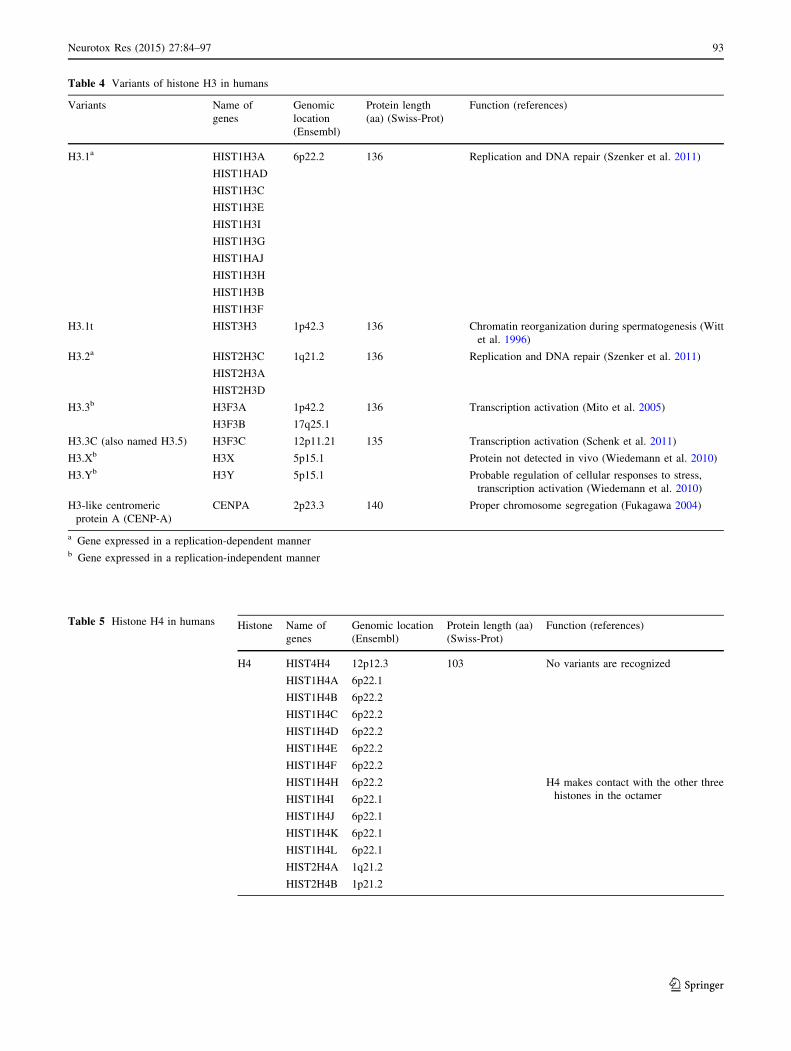
Table 4 Variants of histone H3 in humans
Variants Name of
genes
Genomic
location
(Ensembl)
Protein length
(aa) (Swiss-Prot)
Function (references)
H3.1a HIST1H3A 6p22.2 136 Replication and DNA repair (Szenker et al. 2011)
HIST1HAD
HIST1H3C
HIST1H3E
HIST1H3I
HIST1H3G
HIST1HAJ
HIST1H3H
HIST1H3B
HIST1H3F
H3.1t HIST3H3 1p42.3 136 Chromatin reorganization during spermatogenesis (Witt
et al. 1996)
H3.2a HIST2H3C 1q21.2 136 Replication and DNA repair (Szenker et al. 2011)
HIST2H3A
HIST2H3D
H3.3b H3F3A 1p42.2 136 Transcription activation (Mito et al. 2005)
H3F3B 17q25.1
H3.3C (also named H3.5) H3F3C 12p11.21 135 Transcription activation (Schenk et al. 2011)
H3.Xb H3X 5p15.1 Protein not detected in vivo (Wiedemann et al. 2010)
H3.Yb H3Y 5p15.1 Probable regulation of cellular responses to stress,
transcription activation (Wiedemann et al. 2010)
H3-like centromeric
protein A (CENP-A)
CENPA 2p23.3 140 Proper chromosome segregation (Fukagawa 2004)
a Gene expressed in a replication-dependent mannerb Gene expressed in a replication-independent manner
Table 5 Histone H4 in humans Histone Name of
genes
Genomic location
(Ensembl)
Protein length (aa)
(Swiss-Prot)
Function (references)
H4 HIST4H4 12p12.3 103 No variants are recognized
HIST1H4A 6p22.1
HIST1H4B 6p22.2
HIST1H4C 6p22.2
HIST1H4D 6p22.2
HIST1H4E 6p22.2
HIST1H4F 6p22.2
HIST1H4H 6p22.2 H4 makes contact with the other three
histones in the octamerHIST1H4I 6p22.1
HIST1H4J 6p22.1
HIST1H4K 6p22.1
HIST1H4L 6p22.1
HIST2H4A 1q21.2
HIST2H4B 1p21.2
Neurotox Res (2015) 27:84–97 93
123

Conclusions
DNA methylation is considered to be a relatively stable
epigenetic modification. Recent genome-wide analyses of
the DNA methylation in mammalian cells suggest that
some enzymes are capable of erasing or modifying existing
methylation patterns. Although DNA cytosine methylation
is well-characterized, little is known about the role of
cytosine derivatives in gene expression regulation. In the
future, high-resolution sequencing technologies should
enable creation of quantitative maps of 5hmC, 5fC, and
5caC in different cell types. Understanding the dynamics of
these modifications can help to explain their role in phys-
iological or pathological conditions. Interestingly, due to
subtle sequence divergences, incorporation of histone
variants may influence the stability of nucleosome and
change the potential of specific histone modifications.
Histone variant composition is a key player in shaping
chromatin structure; this also should be considered as one
of the epigenetic regulation elements. It is well known that
epigenetic disturbance may lead to different phenotypes
and monogenic or complex diseases as well as oncogenic
transformation. We strongly believe that rapidly growing
understanding of epigenetic phenomena could bring a
breakthrough in the diagnosis and treatment of many dis-
orders. Moreover, better knowledge about the epigenetic
etiology of the diseases provides an opportunity to develop
innovative new epigenetic drugs.
Acknowledgments This work was supported by the Grant from the
National Science Centre no. 2012/06/A/NZ3/00022 and the statutory
funds of the Laboratory of Drug Addiction Pharmacology, Institute of
Pharmacology PAS.
Conflict of interest The authors declare that they have no conflict
of interests.
Open Access This article is distributed under the terms of the
Creative Commons Attribution License which permits any use, dis-
tribution, and reproduction in any medium, provided the original
author(s) and the source are credited.
References
Adams IR, Meehan RR (2013) From paramutation to paradigm.
PLOS Genet 9(5):e1003537
Aguilera O, Fernandez AF, Munoz A, Fraga MF (2010) Epigenetics
and environment: a complex relationship. J Appl Physiol
109:243–251
Alioui A, Wheldon LM, Abakir A, Ferjentsik Z, Johnson AD, Ruzov
A (2012) 5-carboxylcytosine is localized to euchromatic regions
in the nuclei of follicular cells in axolotl ovary. Nucleus
3(6):565–569
Ausio J (2006) Histone variants—the structure behind the function.
Brief Funct Genom Proteom 5(3):228–243
Bestor TH (2000) The DNA methyltransferase of mammals. Hum
Mol Genet 9(16):2395–2402
Bird A (2002) DNA methylation patterns and epigenetic memory.
Genes Dev 16:6–21
Bonisch C, Hake SB (2012) Histone H2A variants in nucleosomes
and chromatin: more or less stable? Nucleic Acids Res
40(21):10719–10741
Branco MR, Ficz G, Reik W (2011) Uncovering the role of
5-hydroxymethylcytosine in the epigenome. Nat Rev Genet
13(1):7–13
Cetica V, Genitori L, Giunti L, Sanzo M, Bernini G, Massimino M,
Sardi I (2009) Pediatric brain tumors: mutations of two
dioxygenases (hABH2 and hABH3) that directly repair alkyl-
ation damage. J Neurooncol 94(2):195–201
Chen H, Dzitoyeva S, Manev H (2012) Effect of aging on
5-hydroxymethylcytosine in the mouse hippocampus. Restor
Neurol Neurosci 30(3):237–245
Chestnut BA, Chang Q, Price A, Lesuisse C, Wong M, Martin LM
(2011) Epigenetic regulation of motor neuron cell death through
DNA methylation. J Neurosci 31(46):16619–16636
Choi SW, Friso S (2010) Epigenetics: A new bridge between nutrition
and health. Adv Nutr 1:8–16
Clausell J, Happel N, Hale TK, Doenecke D, Beato M (2009) Histone
H1 subtypes differentially modulate chromatin condensation
without preventing ATP-Dependent remodeling by SWI/SNF or
NURF. PLoS ONE 4(10):e0007243
Conerly ML, Teves SS, Diolaiti D, Ulrich M, Eisenman RN, Henikoff
S (2010) Changes in H2A.Z occupancy and DNA methylation
during B-cell lymphomagenesis. Genome Res 20:1383–1390
Cooper GM (2000) The cell: a molecular approach 2nd edn.
Chapter 4, the organization of cellular genomes. Sinauer
Associates, Sunderland
Crider KS, Yang TP, Berry RJ, Bailey LB (2012) Folate and DNA
methylation: a review of molecular mechanisms and the
evidence for folate’s role. Adv Nutr 3:1–38
Dango S, Mosammaparast N, Sowa ME, Xiong LJ, Wu F, Park K,
Rubin M, Gygi S, Harper KW, Shi Y (2011) DNA unwinding by
ASCC3 helicase is coupled to ALKBH3-dependent DNA
alkylation repair and cancer proliferation. Mol Cell
44(3):373–384
Deaton AM, Bird A (2011) CpG islands and the regulation of
transcription. Genes and Dev 25:1010–1022
Dobrovic A (2010) Methods for analysis DNA methylation. In:
Coleman WB, Tsongalis GJ (eds) Molecular diagnostics for the
clinical laboratorian, 2nd edn. Humana Press, New York,
pp 149–160
Egger G, Liang G, Aparicio A, Jones PA (2004) Epigenetics in human
disease and prospects for epigenetic therapy. Nature
429(6990):457–463
Egger G, Jeong S, Escobar SG, Cortez CC, Li TW, Saito Y, Yoo CB,
Jones PA, Liang G (2006) Identification of DNMT1 (DNA
methyltransferase 1) hypomorphs in somatic knockouts suggests
an essential role for DNMT1 in cell survival. Proc Natl Acad Sci
USA 103(38):14080–14085
Estecio MRH, Issa JPJ (2011) Dissecting DNA hypermethylation in
cancer. FEBS Lett 585:2078–2086
Fan JY, Rangasamy D, Luger K, Tremethick DJ (2004) H2AZ alters
the nucleosome surface to promote HP1alpha–mediated chro-
matin fibre folding. Mol Cell 16(4):655–661
Fukagawa T (2004) Centromere DNA proteins and kinetochore
assembly in vertebrate cells. Chromosome Res 12(6):557–567
Fuks F, Hurd PJ, Deplus R, Kouzarides T (2003) The DNA
methyltransferases associate with HP1 and the SUV39H1
histone methyltransferase. Nucleic Acids Res 31(9):2305–2312
Globisch D, Munzel M, Muller M, Michalakis S, Wagner M, Koch S,
Bruckl T, Biel M, Carell T (2010) Tissue distribution of
5-hydroxymethylcytosine and search for active demethylation
intermediates. PLoS ONE 5(12):e15367
94 Neurotox Res (2015) 27:84–97
123

Goll MG, Kirpekar F, Maggert KA, Yoder JA, Hsieh CL, Zhang X,
Golic KG, Jacobsen SE, Bestor TH (2006) Methylation of tRNA
Asp by the DNA methyltransferase homolog Dnmt2. Science
311(5759):395–398
Gu TP, Guo F, Yang H, Wu HP, Xu GF, Liu W, Xie ZG, Shi L, He X,
Jin S, Iqbal K, Shi YG, Deng Z, Szabo PE, Pfeifer GP, Li J, Xu
GL (2011) The role of Tet3 DNA dioxygenase in epigenetic
reprogramming by oocytes. Nature 477(7366):606–610
Guillemette B, Bataille AR, Gevry N, Adam M, Blanchette M, Robert
F, Gaudreau L (2005) Variant histone H2AZ is globally
localized to the promoters of inactive yeast genes and regulates
nucleosome positioning. PLoS Biol 3(12):e384
Guo JU, Su Y, Zhong C, Ming GL, Song H (2011) Hydroxylation of
5-methylcytosine by TET1 promotes active DNA demethylation
in the adult brain. Cell 145(3):423–434
Hackett JA, Surani MA (2013) DNA methylation dynamics during the
mammalian life cycle. Phil Trans R Soc B 368:20110328
Hansen JC (2002) Conformational dynamics of the chromatin fiber in
solution: determinants mechanisms and functions. Ann Rev
Biophys Biomol Struct 31:361–392
He YF, Li BZ, Li Z, Liu P, Wang Y, Tang Q, Ding J, Jia Y, Chen Z, Li L,
Sun Y, Li X, Dai Q, Song CX, Zhang K, He C, Xu GL (2011) Tet-
mediated formation of 5-carboxylcytosine and its excision by
TDG in mammalian DNA. Science 333(6047):1303–1307
Hernandez DG, Singleton AB (2012) Using DNA methylation to
understand biological consequences of genetic variability. Neu-
rodegener Dis 9(2):53–59
Hernandez DG, Nalls MA, Gibbs JR, Arepalli S, van der Brug M,
Chong S, Moore M, Longo DL, Cookson MR, Traynor BJ,
Singleton AB (2011) Distinct DNA methylation changes highly
correlated with chronological age in the human brain. Hum Mol
Genet 20(6):1164–1172
Huh I, Zeng J, Park T, Yi SV (2011) DNA methylation and
transcriptional noise. Epigenet Chromatin 6:9
Inoue A, Shen L, Dai Q, He C, Zhang Y (2011) Generation and
replication-dependent dilution of 5fC and 5caC during mouse
preimplantation development. Cell Res 21(12):1670–1676
Ito S, D’Alessio AC, Taranova OV, Hong K, Lawrence C, Sowers L,
Zhang Y (2010) Role of Tet proteins in 5mC to 5hmC
conversion ES-cell self-renewal and inner cell mass specifica-
tion. Nature 466(7310):1129–1133
Ito S, Shen L, Dai Q, Wu SC, Collins LB, Swenberg JA, He C, Zhang Y
(2011) Tet proteins can convert 5-methylcytosine to 5-formylcy-
tosine and 5-carboxylcytosine. Science 333(6047):1300–1303
Iyer LM, Tahiliani M, Rao A, Aravind L (2009) Prediction of novel
families of enzymes involved in oxidative and other complex
modifications of bases in nucleic acids. Cell Cycle
8(11):1698–1710
Izzo A, Kamieniarz-Gdula K, Ramırez F, Noureen N, Kind J, Manke
T, van Steensel B, Schneider R (2013) The genomic landscape of
the somatic linker histone subtypes H1.1 to H1.5 in human cells.
Cell Rep 3:2142–2154
Javierre BM, Hernando H, Ballestar E (2011) Environmental triggers
and epigenetic deregulation in autoimmune disease. Discov Med.
12(67):535–545
Jin SG, Kadam S, Pfeifer GP (2010) Examination of the specificity of
DNA methylation profiling techniques towards 5-methylcytosine
and 5-hydroxymethylcytosine. Nucleic Acids Res 38(11):e125
Jin B, Li Y, Robertson KD (2011) DNA Methylation: superior or
subordinate in the epigenetic hierarchy? Genes Cancer
2(6):607–617
Jones PA (2012) Functions of DNA methylation: islands, start sites,
gene bodies and beyond. Nat Rev Genet 13(7):484–492
Kamakaka RT, Biggins S (2005) Histone variants: deviants? Genes
Dev 19(3):295–310
Kiani J, Grandjean V, Liebers R, Tuorto F, Ghanbarian H, Lyko F,
Cuzin F, Rassoulzadegan M (2013) RNA–mediated epigenetic
heredity requires the cytosine methyltransferase Dnmt2. PLOS
Gene 9(5):e1003498
Koh KP, Yabuuchi A, Rao S, Huang Y, Cunniff K, Nardone J, Laiho
A, Tahiliani M, Sommer CA, Mostoslavsky G, Lahesmaa L,
Orkin SH, Rodig SJ, Daley GQ (2011) Rao A Tet1 and Tet2
regulate 5-hydroxymethylcytosine production and cell lineage
specification in mouse embryonic stem cells. Cell Stem Cell
8(2):200–213
Kohli RM, Zhang Y (2013) TET enzymes, TDG and dynamics of
DNA demethylation. Nature 502(7472):472–479
Koivisto P, Robins P, Lindahl T, Sedgwick B (2004) Demethylation
of 3-methylthymine in DNA by bacterial and human DNA
dioxygenases. J Biol Chem 279(39):40470–40474
Kouzarides T (2007) Chromatin modifications and their function. Cell
128(4):693–705
Kriaucionis S, Heintz N (2009) The nuclear DNA base 5-hydroxym-
ethylcytosine is present in brain and enriched in Purkinje
neurons. Science 324(5929):929–930
Laurent L, Wong E, Li G, Huynh T, Tsirigos A, Ong CT, Low HM,
Sung KWK, Rigoutsos I, Loring J, Wei CL (2010) Dynamic
changes in the human methylome during differentiation.
Genome Res 20(3):320–331
Li E (2002) Chromatin modification and epigenetic reprogramming in
mammalian development. Nat Rev Genet 3(9):662–673
Lim U, Song MA (2012) Dietary and lifestyle factors of DNA
methylation. Methods Mol Biol 863:359–376
Lister R, Pelizzola M, Dowen RH, Hawkins RD, Hon G, Tonti-
Filippini J, Nery JR, Lee L, Ye Z, Ngo QM, Edsall L,
Antosiewicz-Bourget J, Stewart R, Ruotti V, Millar AH,
Thomson JA, Ren B, Ecker JR (2009) Human DNA methylomes
at base resolution show widespread epigenomic differences.
Nature 462:315–322
Luger K (2001) Nucleosomes: structure and function. In: Encyclo-
pedia of life sciences. Nature Publishing Group, New York,
pp 1–8
Malik HS, Henikoff S (2003) Phylogenomics of the nucleosome. Nat
Struct Biol 10:882–891
Matarese F, Carrillo-de Santa Pau E, Stunnenberg HG (2011)
5-hydroxymethylcytosine: a new kid on the epigenetic block?
Mol Syst Biol 7:562
McKay JA, Mathers JC (2011) Diet induced epigenetic changes and
their implications for health. Acta Physiol 202:103–118
Millan-Arino L, Islam ABMMK, Izquierdo-Bouldstridge A, Mayor R,
Terme JM, Luque N, Sancho M, Lopez-Bigaq N, Jordan A
(2014) Mapping of six somatic linker histone H1 variants in
human breast cancer cells uncovers specific features of H1.2.
Nucleic Acid Res 42(7):4474–4493
Mito Y, Henikoff JG, Henikoff S (2005) Genome-scale profiling of
histone H3.3 replacement patterns. Nat Genet 37:1090–1097
Moore LD, Le T, Fan G (2013) DNA methylation and its basic
function. Neuropsychopharmacology Reviews 38:23–38
Munzel M, Globisch D, Bruckl T, Wagner M, Welzmiller V,
Michalakis S, Muller M, Biel M, Carell T (2010) Quantification
of the sixth DNA base hydroxymethylcytosine in the brain.
Angew Chem Int Ed Engl 49(31):5375–5377
Nestor CE, Ottaviano R, Reddington J, Sproul D, Reinhardt D,
Dunican D, Katz E, Dixon JM, Harrison DJ, Meehan RR (2012)
Tissue type is a major modifier of the 5–hydroxymethylcytosine
content of human genes. Genome Res 22(3):467–477
Niesen MI, Osborne AR, Yang H, Rastogi S, Chellappan S, Cheng
JQ, Boss JM, Blanck G (2005) Activation of a methylated
promoter mediated by a sequence-specific DNA-binding protein,
RFX. J Biol Chem 280:38914–38922
Neurotox Res (2015) 27:84–97 95
123

Ooi SK, Bestor TH (2008) The colorful history of active DNA
demethylation. Cell 133(7):1145–1148
Penn NW, Suwalski R, O’Riley C, Bojanowski K, Yura R (1972) The
presence of 5-hydroxymethylcytosine in animal deoxyribonu-
cleic acid. Biochem J 126(4):781–790
Pfaffeneder T, Hackner B, Truss M, Munzel M, Muller M, Deiml CA,
Hagemeier C, Carell T (2011) The discovery of 5-formylcyto-
sine in embryonic stem cell. Angew Chem Int Ed Engl
50(31):7146–7150
Probst AV, Dunleavy E, Almouzni G (2009) Epigenetic inheritance
during the cell cycle. Nat Rev Mol Cell Biol 10(3):192–206
Pusarla RH, Bhargava P (2005) Histones in functional diversification
core histone variants. FEBS J 272(20):5149–5168
Raiber E, Beraldi D, Ficz G, Burgess HE, Branco MR, Murat P,
Oxley P, Booth MJ, Reik W, Balasubramanian S (2012)
Genome-wide distribution of 5-formylcytosine in embryonic
stem cells is associated with transcription and depends on
thymine DNA glycosylase. Genome Biol 13(8):R69
Raisner RM, Hartley PD, Meneghini MD, Bao MZ, Liu CL, Schreiber
SL, Rando OJ, Madhani HD (2005) Histone variant H2AZ marks
the 5‘ ends of both active and inactive genes in euchromatin.
Cell 123(2):233–248
Riggs AD, Xiong Z (2004) Methylation and epigenetic fidelity. Proc
Natl Acad Sci USA 101(1):4–5
Robertson KD (2001) DNA methylation methyltransferases and
cancer. Oncogene 20(24):3139–3155
Saitou M, Kagiwada S, Kurimoto K (2012) Epigenetic reprogram-
ming in mouse pre-implantation development and primordial
germ cells. Development 139(1):15–31
Sancho M, Diani E, Beato M, Jordan A (2008) Depletion of human
H1 variants uncovers specific roles in gene expression and cell
growth. PLoS Genet 4(10):e1000227
Santos-Rebouc CB, Pimentel MMG (2007) Implication of abnormal
epigenetic patterns for human diseases. Eur J Hum Genet
15:10–17
Schaefer M, Pollex T, Hanna K, Tuorto F, Meusburger M, Helm M,
Lyko F (2010) RNA methylation by Dnmt2 protects transfer
RNAs against stress-induced cleavage. Genes Dev
24(15):1590–1595
Schenk R, Jenke A, Zilbauer M, Wirth S, Postberg J (2011) H3.5 is a
novel hominid-specific histone H3 variant that is specifically
expressed in the seminiferous tubules of human testes. Chro-
mosoma 120(3):275–285
Shen L, Song CX, He C, Zhang Y (2014) Mechanism and function of
oxidative reversal of DNA and RNA methylation. Annu Rev
Biochem 83:585–614
Shukla MS, Syed SH, Goutte-Gattat D, Richard JL, Montel F,
Hamiche A, Travers A, Faivre-Moskalenko C, Bednar J, Hayes
JJ, Angelov D, Dimitrov S (2011) The docking domain of
histone H2A is required for H1 binding and RSC-mediated
nucleosome remodeling. Nucleic Acids Res 39(7):2559–2570
Smallwood A, Esteve PO, Pradhan S, Carey M (2007) Functional
cooperation between HP1 and DNMT1 mediates gene silencing.
Genes Dev 21(10):1169–1178
Song CX, Szulwach KE, Fu Y, Dai Q, Yi C, Li X, Li Y, Chen CH,
Zhang W, Jian X, Wang J, Zhang L, Looney TJ, Zhang B,
Godley LA, Hicks LM, Lahn BT, Jin P, He C (2010) Selective
chemical labeling reveals the genome-wide distribution of
5-hydroxymethylcytosine. Nat Biotechnol 29(1):68–72
Song J, Rechkoblit O, Bestor TH, Patel DJ (2011) Structure of
DNMT1-DNA complex reveals a role for autoinhibition in
maintenance DNA methylation. Science 331(6020):1036–1040
Stadler MB, Murr R, Burger L, Ivanek R, Lienert F, Scholer A, van
Nimwegen E, Wirbelauer C, Oakeley EJ, Gaidatzis D, Tiwari
VK, Schubeler D (2011) DNA-binding factors shape the mouse
methylome at distal regulatory regions. Nature 480(7378):
490–495
Stroud H, Feng S, Kinney SM, Pradhan S, Jacobsen SE (2011)
5-Hydroxymethylcytosine is associated with enhancers and gene
bodies in human embryonic stem cells. Genome Biol
12(6):R5433
Szenker E, Ray-Gallet D, Almouzni G (2011) The double face of the
histone variant H33. Cell Res 21(3):421–434
Szulwach KE, Li X, Li Y, Song CX, Wu H, Dai Q, Irier H, Upadhyay
AK, Gearing M, Levey AI, Vasanthakumar A, Godley LA,
Chang Q, Cheng X, He C, Jin P (2011) 5-hmC-mediated
epigenetic dynamics during postnatal neurodevelopment and
aging. Nat Neurosci 14(12):1607–1616
Tahiliani M, Koh KP, Shen Y, Pastor WA, Bandukwala H, Brudno Y,
Agarwal S, Iyer LM, Liu DR, Aravind L, Rao A (2009)
Conversion of 5-methylcytosine to 5-hydroxymethylcytosine in
mammalian DNA by MLL partner TET1. Science 324(5929):
930–935
Tolstorukov MY, Goldman JA, Gilbert C, Ogryzko V, Kingston RE,
Park PJ (2012) Histone variant H2ABbd is associated with active
transcription and mRNA processing in human cells. Mol Cell
47(4):596–607
Valinluck V, Sowers LC (2007) Endogenous cytosine damage
products alter the site selectivity of human DNA maintenance
methyltransferase DNMT1. Cancer Res 67(3):946–950
Valinluck V, Tsai HH, Rogstad DK, Burdzy A, Bird A, Sowers LC
(2004) Oxidative damage to methyl-CpG sequences inhibits the
binding of the methyl-CpG binding domain (MBD) of methyl-
CpG binding protein 2 (MeCP2). Nucleic Acids Res
32(14):4100–4108
Vardabasso C, Hasson D, Ratnakumar K, Chung CY, Duarte LF,
Bernstein E (2014) Histone variants: emerging players in cancer
biology. Cell Mol Life Sci 71(3):379–404
Varriale A (2014) DNA methylation, epigenetics, and evolution in
vertebrates: facts and challenges. Int J Evol Biol 475981:7
Vavouri T, Lehner B (2012) Human genes with CpG island promoters
have a distinct transcription–associated chromatin organization.
Genome Biol 13(11):R110
Vogler C, Huber C, Waldmann T, Ettig R, Braun L, Izzo A, Daujat S,
Chassignet I, Lopez-Contreras AJ, Fernandez-Capetillo O,
Dundr M, Rippe K, Langst G, Schneider R (2010) Histone
H2A C-terminus regulates chromatin dynamics remodeling and
histone H1 binding. PLoS Genet 6(12):e1001234
Weber CM, Henikoff S (2014) Histone variants: dynamic punctuation
in transcription. Genes Dev 28:672–682
Wiedemann SM, Mildner SN, Bonisch C, Israel L, Maiser A,
Matheisl S, Straub T, Merkl R, Leonhardt H, Kremmer E,
Schermelleh L, Hake SB (2010) Identification and characteriza-
tion of two novel primate-specific histone H3 variants H3X and
H3Y. J Cell Biol 190(5):777–791
Witt O, Albig W, Doenecke D (1996) Testis-specific expression of a
novel human H3 histone gene. Exp Cell Res 229(2):301–306
Wu SC, Zhang Y (2010) Active DNA demethylation: many roads
lead to Rome. Nat Rev Mol Cell Biol 11(9):607–620
Wu H, D’Alessio AC, Ito S, Wang Z, Cui K, Zhao K, Sun YE, Zhang
Y (2011a) Genome-wide analysis of 5-hydroxymethylcytosine
distribution reveals its dual function in transcriptional regulation
in mouse embryonic stem cells. Genes Dev 25(7):679–684
Wu SS, Xu W, Liu S, Chen B, Wang X, Wang Y, Liu S, Wu J (2011b)
Down-regulation of ALKBH2 increases cisplatin sensitivity in
H1299. Acta Pharmacol Sin 32(3):393–398
Wyatt GR, Cohen SS (1952) A new pyrimidine base from bacterio-
phage nucleic acids. Nature 170:1072–1073
96 Neurotox Res (2015) 27:84–97
123

Yang X, Lay F, Han H, Jones PA (2010) Targeting DNA methylation
for epigenetic therapy. Trends Pharmacol Sci 31(11):536–546
Yi C, Chen B, Qi B, Zhang W, Jia G, Zhang L, Li CJ, Dinner AR,
Yang CG, He C (2012) Duplex interrogation by a direct DNA
repair protein in search of base damage. Nat Struct Mol Biol
19(7):671–676
Zhang L, Lu X, Lu J, Liang H, Dai Q, Xu GL, Luo C, Jiang H, He C
(2012) Thymine DNA glycosylase specifically recognizes
5-carboxylcytosine-modified DNA. Nat Chem Biol 8:328–330
Zhu JK (2009) Active DNA demethylation mediated by DNA
glycosylases. Annu Rev Genet 43:143–166
Neurotox Res (2015) 27:84–97 97
123