a contribution to the knowledge of ryszardia decipiens (crotch
TRANSCRIPT

Instructions for use
Title A contribution to the knowledge of Ryszardia decipiens (Crotch), with descriptions of three related speciesfrom Indonesia (Coleoptera, Coccinellidae)
Author(s) Katakura, Haruo; Kahono, Sih; Katakura, Haruo; Kahono, Sih
Citation Insecta matsumurana. New series : journal of the Faculty of Agriculture Hokkaido University, seriesentomology, 72: 17-31
Issue Date 2016-09
Doc URL http://hdl.handle.net/2115/62822
Type bulletin (article)
File Information 2 Katakura.pdf
Hokkaido University Collection of Scholarly and Academic Papers : HUSCAP
17
INSECTA MATSUMURANA NEW SERIES 72: 17–31 SEPTEMBER 2016
A CONTRIBUTION TO THE KNOWLEDGE OF RYSZARDIA DECIPIENS (CROTCH), WITH DESCRIPTIONS OF
THREE RELATED SPECIES FROM INDONESIA (COLEOPTERA, COCCINELLIDAE)
By HARUO KATAKURA and SIH KAHONO
Abstract
KATAKURA, H. and KAHONO, S. 2016. A contribution to the knowledge of Ryszardia decipiens (Crotch), with descriptions of three related species from Indonesia (Coleoptera, Coccinellidae). Ins. matsum. n. s. Current knowledge of the phytophagous ladybird beetle Ryszardia decipiens (Crotch) was reviewed. It occurs on the mountainous areas of Java and Bali, Indonesia, feeding on leaves of Clematis lechenaultiana Clematis species. Three related species, Ryszardia sumatraedecipiens Katakura, sp. nov., R. paradecipiens Katakura, sp. nov. and R. clematophila Katakura et Kahono, sp. nov., were described from Sumatra, Sulawesi and Java, respectively, on Clematis spp. and/or related Naravelia sp.
Authors' address. Katakura, H.: The Hokkaido University Museum, Hokkaido University, Sapporo 060-0810, Japan (corresponding author: [email protected]). Kahono, S.: Zoology Division (Museum Zoologicum Bogoriense), Research Center for Biology, Indonesian Institute of Sciences (LIPI), Cibinong 16911, Indonesia.

18
INTRODUCTION
Ladybird beetles (Coccinellidae ) are species rich and ecologically diverse, with predaceous, fungivorous and phytophagous species. They are currently placed in the superfamily Coccinelloidea (Coleoptera, Cucujiformia) (Robertson et al. 2015). Until recently, the family Coccinellidae has been classified into six subfamilies. However, recent synthetic analysis of morphology and molecular data uncovered their phylogenetic relationship, resulting in the recognition of two subfamilies, Microweisinae and Coccinellinae (Seago et albeetles formerly treated as a subfamily is shown to be monophyletic, and is now treated as the tribe Epilachnini in the subfamily Coccinellinae along with many other tribes belonging to the four former subfamilies (Coccidulinae, Scymninae, Sticholotidinae and Ortaliinae) (Seago et al. 2011). As in other tropical and subtropical regions of the world, Indonesia harbors abundant phytophagous ladybird beetles of the tribe Epilachnini, including serious crop pests (Kalshoven 1981; Katakura et al. 1988) and some species for which intensive evolutionary studies have been made (Matsubayashi et al. 2011, 2013; Fujiyama et al. 2013). However, the taxonomic identity of epilachnine "species" recorded from this region is often not clear, and, moreover, there exists many undescribed species. For example, Katakura et al. (2001) recorded a total of 26 species on Sumatra and Java, of which 12 species were left undetermined. A number of additional species have been found on these and other Indonesian islands in the course of our faunistic and evolutionary studies of epilachnine beetles conducted since 1990's. On the basis of the specimens obtained by these surveys, we herewith review our current knowledge of Ryszardia decipiens (Crotch, 1874), a poorly known species with previous reliable records only on Java, and describe three new species that are related to this species (Fig. 1).
MATERIAL AND METHODS
(2010). Some terms followed Dieke (1947). The tergite X (proctiger) of female of the four species is bent over, and the true apical margin points frontward. In this paper we use "hind margin" to denote the posterior margin of tergite X, and "true apical margin" as above. Observation was made under a dissecting microscope. Genitalia and some body parts were cleared in KOH solution, rinsed in tap water and examined and photographed in ethanol or glycerol. Photos were taken using digital cameras. Composite images of the whole body were produced using a built-in function of a digital camera (Olympus OMD EM-1), and those of some parts of genitalia, and final plates were produced using Adobe Photoshop CC 2014Ⓡ. The following body parts were measured using an ocular micrometer attached to the dissecting microscope for specimens with adequate conditions (i.e., excluding specimens with distorted body and/or open elytra; the number of measured specimens are shown in paretheses): pronotum width (PRW), head width (HW), interocular distance (IOD), body length (BL), and body width (BW). Altitudes of sampling sites were shown in parentheses in the specimens list when available. Half of the specimens including the holotypes will be deposited in Museum Zoologicum Bogoriense (MZB), Indonesia, and the rest in the Systematic Entomology Section, the Hokkaido University Museum, Sapporo, Japan (SEHU).

19
TAXONOMY
et al. (2015) who revised the genera of world Epilachnini based on phylogenetic relationships inferred from morphological and DNA data, and established a number of new genera. Adopting their new system, we placed all the four species treated in this paper in the genus Ryszardia
et al., 2015, that was established for a part of Asian members of the genus Epilachna Chevrolat in Dejean, 1837 (sensuspecies Epilachna decipiens Crotch. The diagnostic characters of this genus shown by
et al. (2015) are: mid and hind coxae with small tubercles on hind margin, metaventral postcoxal lines separated on metaventral process, epipleuron with foveae, and mid and hind tibiae with oblique carina near apex. These conditions were met in all
et al. (2015) further noted that the inner edge of metanepisternum in this and a related genus Diekeana
et al., 2015, is serrate. However, serration was inconspicuous in the present specimens although the inner edge was not smooth.
Ryszardia decipiens (Crotch, 1874) (Fig. 2)
Epilachna decipiens
: Bielawski 1961: 389–392.Ryszardia decipiens et al. 2015: 563.
Redescription. Body (Fig. 2A) short oval, strongly convex above. Dorsum reddish brown. Pronotum with dark rhombus-shaped spot medially. Elytron with six black spots arranged as in Fig. 2A. Spot 1 (postscutellar spot) and spot 5 (post-median spot) reaching suture, spot 4 (lateral spot) reaching margin. In some specimens, spot 1 and spot 2
1000 km
0°
5°S
10°S
110°E100°E 120°E
●●
▼
▼
▼
●
▼
R. decipiensR. sumatraedecipiensR. paradecipiensR. clematophila★★
◆◆●
◆
★
●
Mt. Kerinci
Mt. Rasam
Mt. Gede
Mt. Patuha
Mt. Slamat
Bali
Rantepao
Manado
Tengger
Bantimurung
Fig. 1. Distribution map of the four species of Ryszardia treated in this study.

20
m
h
pcl
pm
pg
A B
C
J
KL
M
N
OP
tgX
cx
Q
D
E
G
H
I
oc
F
Fig. 2. Ryszardia decipiens (Crotch). A, habitus. B, epipleuron; arrows indicate foveae for the reception of middle (m) and hind (h) femora. C, postcoxal line (pcl). D, mandible (left). E, prosternal process. F, metaventral postcoxal line (part); postcoxal lines are separated by the distance shown by the white bar. G, metanepisternum (left). H, posterior part of left hind coxa from inside. I, left hind tibia and tarsus; oc, oblique cavity. J, ventrite 6 (male). K, ventrite 6 (female). L, male terminalia. M, tegmen, side view; pg, penis guide; pm, paramere. N, tegmen, from below. O, penis. P, penial apex. Q, female terminalia and genitalia (part); tgX, tergite X; cx, coxites. Scale bar for A, 5mm; for D to O, Q, 0.5mm.

21
(humeral spot) fused to form a basal fascia. Three spots on the middle level of elytron (spots 5-3-4) often fused to form a transverse fascia running from margin to suture; in such case constriction between spots obvious. Subapical spot always separate. In one specimen from Mt. Slamat, anterior half of elytral margin lined in black. Underside, metaventrite and anterior part of abdomen black, other parts including legs reddish brown. Interocullar distance ca. 2/3 head width. Antenna shorter than head width; composed of 11 antennomeres. Mandible tetradentate, incisor edge without tooth, surfaces without tubercles. Prosternal process with lateral carinae (Fig. 2E). Inner edge of metanepisternum inconspicously serrate (Fig. 2G). Scutellum subtriangular or bell-shaped, longer than wide. Elytral epipleuron with foveae for reception of middle and hind femora (Fig. 2B); inner margin with bordering line nearly complete. Metaventral postcoxal lines widely separated on metaventral process (Fig. 2F). Fore and mid trochanters roundly produced. Mid and hind coxae with small tubercles on hind margin to posterior part of inner surface (Fig. 2H). Tibial spurs formula 1-2-2. Mid tibia and hind tibia with oblique carina on outer margin near apex (Fig. 2I). Tarsal claws long and
posterior margin of ventrite 1, incomplete. Ventrite 6 deeply emarginate in male (Fig. 2J), rounded and equipped with low and short longitudinal ridge medially in the dorsal side in female (Fig. 2K). Male terminalia and genitalia: Terminalia as in Fig. 2L. Apophysis single, stout. Tegminal basal piece without protrusions on inner margin near base of tegminal strut. Penis guide (median lobe) (Figs 2M, N) tubular, glabrous, gently curved down with nearly constant thickness toward apex, and curved upward near apex to form a sharp and pointed end; seen from below, split lengthwise in the middle. Parameres as long as or slightly longer than penis guide, lacking apical thorn (Figs 2M, N). Penis (sipho) (Fig. 2O) gently curved, suddenly narrowed at about 3/4 from base, and bent downward with right angle near apex; apex (Fig. 2P) widened and with minute dentitions. Female terminalia and genitalia (Fig. 3Q): Tergite X, hind margin subtruncate, true apical margin subtruncate (Mt. Gede) or weakly convex (Mt. Slamat); coxites subtriangular. Size: Male, HW 1.6–1.9 mm, IOD 1.0–1.3 mm, PRW 3.6–4.4 mm (n = 5); BL 7.3–8.0 mm, BW 5.7–7.1 mm (n = 3). Female, HW 1.7–2.1 mm, IOD 1.1–1.4 mm, PRW 3.8–4.8 mm (n = 8); BL 8.0–8.7 mm, BW 6.7–7.5 mm (n = 6). Distribution (Fig. 1): Java (Mt. Gede, Mt. Slamet, Tengger), Bali. Host plants: Clematis lechenaultiana DC., Clematis sp. (Ranunculaceae).
Material examined
Kebun Raya Bali (1350m), Bali, 1 Dec. 2005 (H. Katakura). Diagnosis and remarks: Crotch (1874) described this species from Java. Later, Bielawski (1961) figured the elytral pattern and described and figured the genitalia
of the female specimen was "Java" and that of the male specimen was described as "Tengger, 1800m," which is the name of a mountain in East Java. The genitalia of both
22
specimens. The elytral pattern figured by Bielawski also coincides with some of the present specimens. This species inhabits mountain regions of Java and Bali (Fig. 1). Javanese and Balinese specimens are very much alike in genitalia of both sexes. They are also similar in their external appearance with well developed elytral spots, in which basal two spots (spots 1-2) and three spots on the middle level (spots 5-3-4) are respectively very frequently coalescent as shown in Fig. 2A. In Mt. Gede, West Java, this species was collected on Clematis lechenaultiana and another unidentified Clematis species. In Mt. Slamat in Central Java and Kebun Raya Bali (Bali Botanic Garden), this species was found on a Clematis species having very similar leaf morphology to Clematis lechenaultiana. Korschefsky (1933) and Miwa and Yoshida (1935) reported this species
Ryszardia decipiens is very similar to Epilachna paramagna(probably belonging to Ryszardia), from Yunnan, China, in the detailed structure of male
et al. 2009). Moreover, the pattern of elytral spots of R. decipiens has some similarities with that of E. paramagna in Ren et al. (2009). However, there is difference among literatures in the elytral pattern of E. paramagna. The shape and position of elytral spots in E. paramagna1979) was quite different from the photo of the same species in Ren et al. (2009), and the elytral pattern of R. decipiens resembles that of E. paramagna in Ren et al. (2009) but is
much more information about their morphology, biology and molecular data become available. It is because there is inconsistency in the elytral pattern of E. paramagna in the literature, and there are some cases in epilachnines in which morphologically nearly identical forms are shown to be different biological species based on biological (i.e., host plant) and molecular evidence (cf. Katakura 1997; Kobayashi et al. 2000). Unfortunately the host plant of R. paramagna is unknown. Ryszardia decipiens is also similar to Epilachna magna (Dieke, 1947) (also probably belonging to Ryszardia) from southern
from the latter by the thicker penis guide. Furthermore, E. magnaelytron according to Dieke (1947), although it has six spots according to Pang and Mao (1979) and Ren et al. (2009). The host plants of E. magna are the eggplant (Solanaceae)
et al. 2009), both of which are distinctly different from the host plants of R. decipiens, i.e., Clematis spp. (Ranunculaceae). The female internal reproductive organ of R. decipiens was treated in Katakura et al. (1994). The phylogenetic relationships of R. decipiens with some Asian epilachnines
et al. (1994) and Kobayashi et al. (2009).
Ryszardia sumatraedecipiens Katakura sp. nov. (Fig. 3)
Description. Body (Fig. 3A) nearly round, strongly convex above. Dorsum reddish

23
brown. Pronotum spotless or with a faint or small black spot medially. Elytron with 6 black spots arranged as in Fig. 3A. Elytral spots always separate from each other except postscutellar spot that sometimes attached to the counterpart on suture. Middle lateral spot usually reaching margin. Underside, metaventrite and anterior part of abdomen black, other parts including legs reddish brown. Interocullar distance ca. 2/3 head width. Antenna shorter than head width; composed of 11 antennomeres. Mandible tetradentate, incisor edge without tooth, surfaces without tubercles. Prosternal process with lateral carinae. Inner edge of metanepisternum serrate. Scutellum subtriangular or bell-shaped, longer than wide. Elytral epipleuron with foveae for reception of middle and hind femora (Fig. 3B); inner margin with bordering line nearly complete. Metaventral postcoxal lines widely separated on metaventral process. Fore and mid trochanters roundly produced. Mid and hind coxae with small tubercles on hind margin to posterior part of inner surface. Tibial spurs formula 1-2-2. Mid and hind
basal tooth. Abdominal postcoxal line (Fig. 3C), reaching posterior margin of ventrite 1,
KA B
C
D E
F
G
H
IJFig. 3. Ryszardia sumatraedecipiens Katakura sp. nov. A, habitus. B, epipleuron; arrows indicate
the depressions for the reception of middle and hind femora. C, abdominal postcoxal line. D, ventrite 6 (male). E, ventrite 6 (female). F, male terminalia. G, tegmen, side view. H, tegmen, from below. I, penis. J, penial apex. K, female terminalia and genitalia (part). Scale bar for A, 5mm; for D to I, K, 0.5mm.

24
incomplete laterally. Ventrite 6 deeply emarginate in male (Fig. 3D), rounded in female with a tiny medial ridge inside as in R. decipiens (Fig. 3E). Male terminalia and genitalia: Terminalia as in Fig. 3F. Apophysis single, stout. Tegminal basal piece without protrusions on inner margin near base of tegminal strut. Penis guide (Figs 3G, H) tubular, glabrous, seen from side nearly straight, narrowed in the middle and then thickened again; apex pointed and bent upward; seen from below, split lengthwise in the middle. Parameres lacking apical thorn, slightly shorter than penis guide. Penis (Fig. 3I) gently curved, narrowed at about 3/4 from the base, and bent downward with right angle near apex; apex (Fig. 3J) widened, equipped with minute dentitions. Female terminalia and genitalia (Fig. 3K): Tergite X with hind margin weakly concave and true apical margin subtruncate; coxites subtriangular. Size: Male, HW 1.8–2.1 mm, IOD 1.2–1.3 mm, PRW 3.8–4.3 mm (n = 7); BL 7.5–8.3 mm, BW6.7–7.2 mm (n = 3). Female, HW 1.8–1.9 mm, IOD 1.2 mm, PRW 3.8–4.0 mm (n = 2); BL 7.9 mm, BW 6.8 mm (n = 1). Distribution: Sumatra (Mt. Rasam, Mt. Kerinci) (Fig. 1). Host plants: Clematis spp. (Ranunculaceae). Etymology: The species epithet is derived from the distribution range, Sumatra, and the suggested close relationship with R. decipiens.
Material examined
lost), Mt. Kerinci (2080m), Jambi, Sumatra, 30 Jan. 2007 (N. Kamata). Diagnosis and remarks: Katakura et al. (2001) reported this species as Epilachna sp. M. Ryszardia sumatraedecipiens is very similar to R. decipiens, E. magna and E. paramagna in morphology of the genitalia of both sexes. However, in R. sumatraedecipiens, the penis guide is narrowest at the middle seen laterally, and the apical bended part of the penis guide is shorter than those of other species (Fig. 3G). From R. decipiens, R. sumatraedecipiens is further discernible by the hind margin of female tergite X being weakly rounded (subtruncate in R. decipiens; Figs 2K vs. 3K). Moreover, R. sumatraedecipiens has round spots that are always separate from each other except for spot 1 that meets with the counterpart on the suture (Fig. 3A), and the body of R. sumatraedecipiens is nearly round while that of R. decipiens is short oval (Figs 2A vs. 3A). This species has been known only in the Barisan Mountains in Sumatra (Fig. 1). In Mt. Kerinci, Jambi, this species was found on a Clematis species having very similar leaf morphology to Clematis lechenaultiana. In Mt. Rasam, West Sumatra, R. sumatraedecipiens feeds on another Clematis
Ryszardia paradecipiens Katakura sp. nov. (Fig. 4)
Description. Body (Fig. 4A) nearly round. Strongly convex above. Dorsum yellowish brown. Pronotum spotless. Elytron with 5 black spots arranged as 2-2-1; elytral spots separate from each other, and not reaching margin or suture except spot 4 (outer spot of the second row) that reaches margin. Underside, metaventrite and anterior

25
part of abdomen black, other parts including legs reddish brown. Interocullar distance ca. 2/3 head width. Antenna shorter than head width; composed of 11 antennomeres. Mandible tetradentate, incisor edge without tooth, surfaces without tubercles. Prosternal process with lateral carinae. Inner edge of metanepisternum obscurely serrate. Scutellum subtrianglular or bell-shaped, longer than wide. Elytral epipleuron with foveae for reception of middle and hind femora (Fig. 4B); inner margin with bordering line nearly complete. Metaventral postcoxal lines widely separated on metaventral process. Fore trochanter roundly, and mid trochanter angulately produced. Mid coxa with tubercles on hind margin. Hind coxa with small tubercles on hind margin to posterior part of inner surface. Tibial spurs formula 1-2-2. Mid and hind tibiae with oblique carina on outer margin near apex. Tarsal claws long and bifid, without basal tooth. Abdominal postcoxal line (Fig. 4C), reaching posterior margin of ventrite 1, incomplete laterally. Ventrite 6 deeply emarginate in male (Fig. 4D), rounded in female (4E). Male terminalia and genitalia: Terminalia as in Fig. 4F. Apophysis single, stout.
A B
C
D E
F
G
H
IJ
K
Fig. 4. Ryszardia paradecipiens Katakura sp. nov. A, habitus. B, epipleuron; arrows indicate the depressions for the reception of middle and hind femora. C, abdominal postcoxal line. D, ventrite 6 (male). E, ventrite 6 (female, left side broken). F, male terminalia. G, tegmen, side view. H, tegmen, from below. I, penis. J, penial apex. K, female terminalia and genitalia (part). Scale bar for A, 5mm; for D to I, K, 0.5mm.

26
Tegminal basal piece without protrusions on inner margin near base of tegminal strut.
seen from side nearly straight from base to ca. 3/5 of length, and then weakly curved down; near apex suddenly bent upward, forming rather long and sharply pointed apex. Parameres lacking apical thorn, distinctly shorter than penis guide. Penis (Fig. 4I) gradually narrowed and bent down near apex; apex blunt and multi-denticulate (Fig. 4J). Female terminalia and genitalia(Fig. 4K): Tergite X with hind margin subtruncate
Size: Male, HW 1.6–1.7 mm, IOD 1.0–1.1 mm, PRW 3.6–3.8 mm, BL 7.0–7.3mm; BW 6.1–6.5mm (n = 3). Female, HW 1.6–1.8 mm, IOD 1.0–1.1 mm, PRW 3.8–4.1 mm, BL 7.1–7.9mm, BW 6.3–6.8mm (n = 2). Distribution: Sulawesi (northern and southern parts; Fig. 1). Host plants: Clematis smilacifolia Wall., Naravelia sp. (?laurifolia Wall.) (Ranunculaceae). Etymology: Based on the suggested close relationship with R. decipiens.
Material examined
Pinaras (641m), Tomohon, near Manado, North Sulawesi, 18 Nov. 2005 (H. Katakura, N. Fujiyama
Diagnosis and remarks: This species has an essentially same design of male genitalia with Ryszardia decipiens, R. sumatraedecipiens, E. magna and E. paramagna, but is separable from these four species by the penis guide with underside widely open and long and sharply pointed apical part (Figs 4G, H), the parameres distinctly shorter than penis guide (Fig. 4), and by the blunt penial apex equipped with denticles (Fig. 4I). In female genitalia, the coxites of R. paradecipiensbeing different from rather subtriangular ones in R. decipiens and R. sumatraedecipiens. Furthermore, R. paradecipiens is easily discernible from R. decipiens , R. sumatraedecipiens and E. paramagna(six in the latter three species; for the elytral pattern of E. magna, see "Remarks" of R. decipiens). This species seemed widespread in Sulawesi (Fig. 1) feeding on Clematis and Naravelia, although only 6 individuals were available for us.
Ryszardia clematophila Katakura et Kahono sp. nov. (Fig. 5)
Description. Body oval (Fig. 5A), convex above. Dorsum reddish brown. Pronotum spotless or with a faint small dark spot medially. Elytra with two transverse fasciae and a pair of apical spots. Basal fascia not reaching base, margin, or suture. Second fascia touching margins, and may or may not be united on suture. Subapical spot transverse. In one female specimen, two separate spots present on each elytron instead of second
and anterior part of abdomen black, other parts including legs reddish brown. Interocullar distance ca. 2/3 head width. Antenna shorter than head width; composed
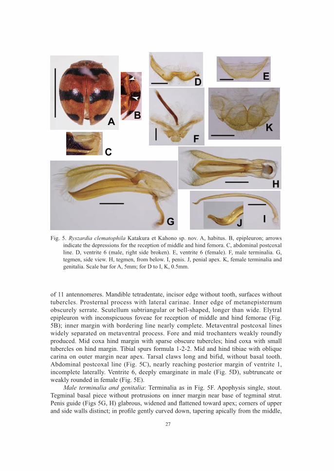
27
of 11 antennomeres. Mandible tetradentate, incisor edge without tooth, surfaces without tubercles. Prosternal process with lateral carinae. Inner edge of metanepisternum obscurely serrate. Scutellum subtriangular or bell-shaped, longer than wide. Elytral epipleuron with inconspicuous foveae for reception of middle and hind femorae (Fig. 5B); inner margin with bordering line nearly complete. Metaventral postcoxal lines widely separated on metaventral process. Fore and mid trochanters weakly roundly produced. Mid coxa hind margin with sparse obscure tubercles; hind coxa with small tubercles on hind margin. Tibial spurs formula 1-2-2. Mid and hind tibiae with oblique carina on outer margin near apex. Tarsal claws long and bifid, without basal tooth. Abdominal postcoxal line (Fig. 5C), nearly reaching posterior margin of ventrite 1, incomplete laterally. Ventrite 6, deeply emarginate in male (Fig. 5D), subtruncate or weakly rounded in female (Fig. 5E). Male terminalia and genitalia: Terminalia as in Fig. 5F. Apophysis single, stout. Tegminal basal piece without protrusions on inner margin near base of tegminal strut.
B
C
D E
F
G
H
IJ
KA
Fig. 5. Ryszardia clematophila Katakura et Kahono sp. nov. A, habitus. B, epipleuron; arrows indicate the depressions for the reception of middle and hind femora. C, abdominal postcoxal line. D, ventrite 6 (male, right side broken). E, ventrite 6 (female). F, male terminalia. G, tegmen, side view. H, tegmen, from below. I, penis. J, penial apex. K, female terminalia and genitalia. Scale bar for A, 5mm; for D to I, K, 0.5mm.

28
and curved upward near the apex to form a dull pointed end; seen from below, widely split lengthwise in the middle. Parameres as long as penis guide, lacking apical thorn. Penis (Fig. 5I) tapering apically near apex; apex (Fig. 5J) pointed, bent down and then
Female terminalia and genitalia (Fig. 5K): Tergite X with hind margin and true apical margin gently rounded; coxites subtriangular. Size: Male, HW 1.6–1.7 mm, IOD 0.9–1.0 mm, PRW 3.1–3.4 mm (n = 4); BL 6.3–6.8 mm, BW 4.9–5.7 mm (n = 4). Female, HW 1.5–1.7 mm, IOD 1.0–1.1 mm, PRW 3.1–3.5 mm (n = 7); BL 6.9– 7.3 mm, BW 5.6–6.1 mm (n = 2). Distribution: Java (Mt. Gede, Mt. Patuha) (Fig. 1). Host plant: Clematis lechenaultiana DC. (Ranunculaceae). Etymology: The species epithet is derived from the genus name of the host plant.
Material examined
Diagnosis and remarks: This species was referred to as Epilachna sp. G in Katakura et al. (1994, 2001), Kobayashi et al. (2009) and Katoh et al. (2014). In this species, the conditions of mid and hind legs (tubercles on hind margin of coxae and apically oblique outer margin of tibiae), which were considered very important as the characters of the genus Ryszardia et al. 2015), are detectable, but less obvious compared with the other three species treated here. In the structure of both male and female genitalia, this species evidently belongs to the group that includes R. decipiens, R. sumatraedecipiens and R. paradecipiens (and some continental species such as E. magna, E. paramagna, etc.). Katakura et al. (1994) and Kobayashi et al. (2009) showed a close relationship of R. clematophila and R. decipiens, based on morphological and molecular evidence, respectively. The present species is discernible from R. decipiens and two other species described here by the elytral pattern (Fig. 5A),
male; shape of tergite X in female) (Figs 5G, H, I, K). Ryszardia clematophila somewhat resembles Epilachna subacuta
et al. 2009) and female tergite X (Dieke 1947), but the two species are separable by the distinctly different patterns of elytral
et al. 2009), and the host plant (Schisandra, Schisandraceae, in E. subacuta Ryszardia clematophila is also similar to Ryszardia dorotae (Bielawski, 1979) from Bhutan (Bielawski 1979) in the shape of penis. However, the two species are quite different
dorotae is much larger, about 8.5–9.0 mm in length, and has entirely dark body with six separate spots on each elytron); tegmen and female's tergite X also differ. Katakura et al. (2001) mentioned that Epilachna sp. G (= R. clematophila) did not have cavities on elytral epipleura for the reception of middle and hind femora, but actually this species has inconspicuous foveae on the epipleura (see "Additional notes" below). Ryszardia clematophila has been known only from mountain areas in West Java, feeding on Clematis lechenaultiana. In Mt. Gede, this species co-occurred with R. decipiens on the same host plant. In Mt. Gede, this species composes a putative mimicry

29
complex together with Afissa orthofasciata Dieke, 1947, feeding on Tetrastigma papillosum Planch. (Vitaceae) and Henosepilachna bifasciata (Fabricius, 1781) feeding
other despite their rather remote relationships (Katakura et al. 2001; Kobayashi et al. 2009). The female internal reproductive system of R. clematophila was treated in Katakura et al. (1994), its oviposition pattern was given in Nakano et al. (2001), and phylogenetic relationships with other groups of epilachnines were treated in Katakura et al. (1994), Kobayashi et al. (2009) and Kotoh et al. (2014) under the name Epilachna sp. G.
ADDITIONAL NOTES
szechuana group and the chapini group in the species of the genus (later treated as Epilachna sensuet al. 2015 revived this name) with "tergite X of the female having its apical part folded down and over so that the true apical margin pointing frontward." According to him, the szechuana group lacked "cavities" in elytral epipleura for the reception of middle and hind femora, which the chapiniin his two specimens of E. decipiens. However, Crotch (1874) mentioned in the original description of E. decipiens that "epipleuræ of the elytra foveolate." We also found certain foveae on elytral epipleura that would function as the receptors of femora in all the four species treated in this paper (Figs 2B, 3B, 4B, 5B). The four species should be placed in the chapini group, provided that these foveae correspond to Dieke's "cavities." However, the depth of foveae was variable among species. In R. clematophila, the foveae, especially that for middle femur, were shallow. Dieke (1947) also noticed the difference in the depth of "cavities" between two species of his chapini group, namely, Epilachna chapini (Dieke, 1947) and E. magna. These facts pose a question about the validity of the szechuana group and the chapiniThey may be better treated as a single group sharing the common characteristic feature
et al. (2015) suggested that the two species of the szechuana group (szechuana and subacuta) might also be included in Ryszardia. Molecular phylogenetic analyses showed that R. clematophila form a clade with R. decipiens within a clade comprising Asian species of the former Epilachna (Kobayashi et al. 2009; Katoh et al. 2014). However, no phylogenetic analysis has been done for R. sumatraedecipiens and R. paradecipiens, although the overall resemblance in morphology and host plants of the three allopatric species, R. decipiens in Java and Bali, R. sumatraedecipiens in Sumatra, and R. paradecipiens in Sulawesi, strongly suggests that they are closely related to each other and probably speciated through vicariance events. It is notable that the distribution range of this group covers the areas separated by the Wallace line (i.e., Sulawesi vs. other islands), though no species related to these species has been known in more eastern parts of Indonesia, including the Lesser Sunda Islands (excluding Bali) and Papua. A detailed molecular phylogenetic analysis of the three species, as well as those involving the Asian continental relatives such as E. magna and other species in the szechuana + chapini group may contribute to our better
Peninsula to the Greater Sunda Islands.
30
ACKNOWLEDGMENTS
We thank I. Abbas, S. Nakano, Asril, Gyanto, Sarino, N. Fujiyama, N. Kamata and M.
permission of Research Center for Biology, Indonesian Institute of Science (LIPI) (permit numbers: 5669/SK/1990 to 6452/SU/KS/2005), and was supported by MEXT/JSPS KAKENHI Grant Numbers 02041033, 05041086, 11691161, 14204081 and 18207005.
REFERENCES
Annales Zoologici 19, 383–415.
Bielawski R (1979) Ergebnisse der Bhutan-Expedition 1972 des Naturhistorischen Museums in Basel. Coleoptera: Fam. Coccinellidae. Entomologica Basiliensia 4, 83–125.
Crotch GR (1874) A Revision of the Coleopterous Family Coccinellidae. E.W. Jason, London, XVI + 311 pp.
Dejean PMFA (1837) Catalogue des Coléoptères de la Collection de M. le Comte Dejean. Troisième édition, revue, corrigée et augmentée. Méquignon-Marvis Pères et Fils, Paris, XVI + 311 pp.
Dieke GH (1947) Ladybird beetles of the genus Epilachna (sens. lat.) in Asia, Europe, and Australia. Smithsonian miscellaneous Collections, Washington D.C., 106, 1–183.
Fabricius JC (1781) Species Insectorum exhibentes eorum differentias specificas, synonyma auctorum, loca natalia, metamorphosin adiectis obervationibus, descriptionibus. Tom. I. Impensis Carol. Ernest. Bohnii, Hamburgi et Kilonii. VIII + 552 pp.
Fujiyama N, Ueno H, Kahono S, Hartini S, Matsubayashi KW, Kobayashi N, Katakura H (2013) Distribution and differentiation of the herbivorous ladybird beetle Henosepilachna diekei on two host plant species across Java, Indonesia. Annals of the Entomological Society of America 106, 741–752.
World Catalogue of Coccinellidae, Part I–Epilachninae.
Kalshoven, LGE (1981) Pests of Crops in Indonesia (PA van der Laan, rev. and transl.). 701pp. P.T. Ichtiar Baru-Van Hoeve, Jakarta.
Katakura H (1997) Species of Epilachna ladybird beetles. Zoological Science 14, 869–881.
Katakura H, Abbas I, Nakamura K, Sasaji H (1988) Records of epilachnine crop pests in Sumatera Barat, Sumatra, Indonesia. Kontyu, Tokyo 56, 281–297.
Katakura H, Nakano S, Hosogai T, Kahono S (1994) Female internal reproductive organs, modes of sperm transfer, and phylogeny of Asian Epilachninae (Coleoptera: Coccinellidae). Journal of Natural History 28, 577–583.
Katakura H, Nakano S, Kahono S, Abbas I, Nakamura K (2001) Epilachnine ladybird beetles (Coleoptera, Coccinellidae) of Sumatra and Java. Tropics 10, 325–352.
Katoh T, Koji S, Ishida TA, Matsubayashi KW, Kahono S, Kobayashi N, Furukawa K, Viet BT, Vasconcellos-Neto J, Nakano S, Katakura H. (2014) Phylogeny of Epilachna, Henosepilachna, and some minor genera of phytophagous ladybird beetles (Coleoptera: Coccinellidae: Coccinellinae: Epilachnini), with an analysis of
Zoological Science 31, 820–830.Kobayashi N, Ohta Y, Katoh T, Kahono S, Hartini S, Katakura H (2009) Molecular

31
phylogenetic analysis of three groups of Asian epilachnine ladybird beetles
Journal of Natural History 43, 1637–1649.Kobayashi N, Shirai Y, Tsurusaki N, Tamura K, Aotsuka T, Katakura H (2000) Two
cryptic "species" of the phytophagous ladybird beetle Epilachna vigintioctopunctata detected by the analyses of mitochondrial DNA and karyotypes, and crossing experiments. Zoological Science 17, 1159–1166.
Korschefsky R (1933) Bemerkungen über Coccinelliden von Formosa. Trans. nat. Hist. Soc. Formosa, Taihoku 23, 299–303.
Li CS, Cook EF (1961) The Epilachninae of Taiwan (Coleoptera: Coccinellidae). Insects 3, 31–91.
as the critical driving force in speciation between populations of a phytophagous ladybird beetle. Journal of Evolutionary Biology 24, 1421–1432.
Matsubayashi KW, Kahono S, Katakura H (2013) Divergent host plant preference causes assortative mating between sympatric host races of the ladybird beetle, Henosepilachna diekei. Biological Journal of the Linnean Society 110, 606–614
Miwa Y, Yoshida T (1935) Catalogue of Japanese Insects. Fasc. IX. Col. Coccinellidae. Ent. World, Tokyo 3, 31–53. (In Japanese.)
Nakano S, Katakura H, Abbas I, Kahono S, Nakamura K (2001) Oviposition patterns of Asian phytophagous ladybird beetles (Coleoptera, Coccinellidae, Epilachninae). Tropics 10, 353–362.
Pang XF, Mao JL (1979) Economic Insects of China, 14, Coleoptera-Coccinellidae, II. 170 pp, 16 pls, Science Press, Beijing. (In Chinese.)
Epilachna Chevrolat with descriptions of new species (Coleoptera: Coccinellidae: Epilachnini). Zootaxa 3420, 1–7.
Ren SX, Wang XM, Pang H, Peng ZQ, Zeng T. (2009) Colored Pictorial Handbook of Ladybird Beetles in China. Science Press, Beijing. (In Chinese.)
Coccinelloidea (Coleoptera: Cucujiformia). Systematic Entomology 40, 745–778E
of ladybird beetles (Coleoptera: Coccinellidae) based on simultaneous analysis of molecular and morphological data. Molecular Phylogenetics and Evolution 60, 137–151.
Beutel RG, Lawrence, JF (eds) Handbook of Zoology. Vol. 2, Coleoptera, 454–472.
and evolution of phytophagous ladybird beetles (Coleoptera: Coccinellidae: Epilachnini), with recognition of new genera. Systematic Entomology 40, 547–569. First published online: 28 Jan. 2015, DOI: 10.1111/syen.12121.