a direct interaction between nqo1 and a chemotherapeutic ... · a direct interaction between nqo1...
TRANSCRIPT
RESEARCH ARTICLE Open Access
A direct interaction between NQO1 and achemotherapeutic dimeric naphthoquinoneLakshmi Swarna Mukhi Pidugu1,2,3, J.C. Emmanuel Mbimba3, Muqeet Ahmad3, Edwin Pozharski1,2,3,Edward A. Sausville2, Ashkan Emadi2 and Eric A. Toth1,2,3*
Abstract
Background: Multimeric naphthoquinones are redox-active compounds that exhibit antineoplastic, antiprotozoal,and antiviral activities. Due to their multimodal effect on perturbation of cellular oxidative state, these compoundshold great potential as therapeutic agents against highly proliferative neoplastic cells. In our previous work, wedeveloped a series of novel dimeric naphthoquinones and showed that they were selectively cytotoxic to humanacute myeloid leukemia (AML), breast and prostate cancer cell lines. We subsequently identified the oxidoreductaseNAD(P)H dehydrogenase, quinone 1 (NQO1) as the major target of dimeric naphthoquinones and proposed amechanism of action that entailed induction of a futile redox cycling.
Results: Here, for the first time, we describe a direct physical interaction between the bromohydroxy dimericnaphthoquinone E6a and NQO1. Moreover, our studies reveal an extensive binding interface between E6a and theisoalloxazine ring of the flavin adenine dinucleotide (FAD) cofactor of NQO1 in addition to interactions with proteinside chains in the active site. We also present biochemical evidence that dimeric naphthoquinones affect the redoxstate of the FAD cofactor of NQO1. Comparison of the mode of binding of E6a with those of other chemotherapeuticsreveals unique characteristics of the interaction that can be leveraged in future drug optimization efforts.
Conclusion: The first structure of a dimeric naphthoquinone-NQO1 complex was reported, which can be used fordesign and synthesis of more potent next generation dimeric naphthoquinones to target NQO1 with higher affinityand specificity.
Keywords: NQO1, Dimeric naphthoquinone, Oxidative stress, Anti-cancer agents
BackgroundMultimeric naphthoquinones are reduction/oxidation(redox)-active compounds that possess a wide array oftherapeutic activities. In particular, these compoundshave exhibited well tolerated antibacterial, antifungal,antiviral, and antithrombotic activities [1]. One of themost notable members of this class of compounds isconocurvone, a naturally-occurring trimeric naphthoqui-none with a potent anti-HIV activity [2]. Synthetic andnatural naphthoquinones have demonstrated significantantineoplastic activity against hematologic and solidmalignant cells [3–5]. In an effort to regiospecifically
synthesize conocurvone, we previously developed aseries of novel dimeric naphthoquinones and showedthat they were selectively cytotoxic to human acute mye-loid leukemia (AML), breast and prostate cancer celllines and in particular those cell lines that rely on oxida-tive phosphorylation [6–8]. To better understand themechanism of action of these agents, we performed achemical genetic screen in yeast and identified the yeastoxidoreductase Nde1 as the major target of dimericnaphthoquinones [6, 9]. The human homologue of Nde1is NAD(P)H quinone oxidoreductase 1 (E.C. 1.6.99.2,hereon referred to as NQO1, also known as DT-diaphorase and NAD(P)H dehydrogenase, quinone 1).NQO1 is a quinone detoxifying flavoenzyme that cata-
lyzes the two-electron reduction of quinones to hydroqui-nones. For dimeric naphthoquinones, the resultinghydroquinone is highly unstable and spontaneously giveselectrons to oxygen and reverts to the oxidized form of
* Correspondence: [email protected] of Biochemistry and Molecular Biology, University of MarylandSchool of Medicine, Baltimore, MD 21201, USA2Marlene and Stewart Greenebaum Cancer Center, University of MarylandSchool of Medicine, Baltimore, MD 21201, USAFull list of author information is available at the end of the article
© 2016 Pidugu et al. Open Access This article is distributed under the terms of the Creative Commons Attribution 4.0International License (http://creativecommons.org/licenses/by/4.0/), which permits unrestricted use, distribution, andreproduction in any medium, provided you give appropriate credit to the original author(s) and the source, provide a link tothe Creative Commons license, and indicate if changes were made. The Creative Commons Public Domain Dedication waiver(http://creativecommons.org/publicdomain/zero/1.0/) applies to the data made available in this article, unless otherwise stated.
Pidugu et al. BMC Structural Biology (2016) 16:1 DOI 10.1186/s12900-016-0052-x

quinone, producing two moles of superoxide per one moleof NAD(P)H [10]. The ultimate outcome is a futile redoxcycle in NQO1-overexpressing cells, such as many cancercells, which can culminate in formation of substantialreactive oxygen species (ROS), oxidative damage to DNAand single- and double-strand DNA breaks. NQO1 existsas a homodimer with two tightly-associated flavin adeninedinucleotide (FAD) cofactors that reside at the deepestpoint of each active site of two monomers of 274 residues[11]. The two active sites reside at opposite ends of thedimer and incorporate residues from each monomer. Thenormal biological function of NQO1 is to protect cellsfrom the mutagenic, cytotoxic, and carcinogenic effects ofnatural and synthetic quinones [12]. The obligate twoelectron reduction performed by NQO1 averts one-electron reduction of quinones by other flavoproteins suchas cytochrome P450, which produces highly reactiveradical semiquinone.The role of NQO1 in cancer varies due to its role in
redox biology. NQO1 exhibits tumor suppressor proper-ties by modulating the stability of p53 [13, 14] and partici-pating in suppression of the inflammatory response [15].Conversely, increased expression of NQO1 can confer agrowth advantage in some cancers such as melanoma,pancreatic adenocarcinoma, non-small cell lung cancer,and prostate cancer [16, 17]. The association between theNQO1 C609T polymorphism and increased risk of AMLand acute lymphoblastic leukemia (ALL) has also beenreported [18, 19]. Exploitation of NQO1 as a target forcancer therapy typically entails two strategies. In somecases, inhibition of NQO1 can suppress cancer cell growthand potentiate chemotherapeutic cytotoxicity [20]. Inother cases, NQO1 can be used to activate particularquinone-based chemotherapeutics via its redox activity[21, 22]. For dimeric naphthoquinones, we have proposedthat their unique chemical structures undergo NQO1-dependent redox cycling that produces an insurmountableamount of ROS that ultimately lead to mitochondrialdysfunction, DNA damage and cell death [9].In the present study, we have determined the crystal
structure of the novel dimeric naphthoquinone, 3-bromo-3′-hydroxy-2,2′-binaphthalenyl-1,4,1′,4′-tetraone(E6a [23], Additional file 1: Figure S1) bound to NQO1.This structure represents the first evidence of a directinteraction between a dimeric naphthoquinone andNQO1. Moreover, we present biochemical evidence thatthis interaction affects the redox state of the FAD cofac-tor. Our structure reveals an extensive binding interfacebetween E6a and the isoalloxazine ring of the FADcofactor of NQO1 in addition to interactions withprotein side chains in the active site. This structure canbe used as a starting point to design and synthesizemore potent dimeric naphthoquinones tailored to targetNQO1 with high affinity and specificity.
Results and discussionOverall quality of crystal structureCrystals of hNQO1-FAD (holo-hNQO1) belong to spacegroup P21 and diffract to 2.0 Å resolution. The asym-metric unit contains two dimers. The crystal structureshows excellent stereochemistry with 96 % of residues inthe most favored region of Ramachandran plot. Eachphysiological dimer [11, 24–27] is made up of two sub-units that are related by a non-crystallographic two foldaxis. The overall structure of the dimer is almost identicalto that of a previously-determined holo-NQO1 structure(PDB accession code: 1D4A [26]) with a root-mean-square deviation (rmsd) of 0.1 Å. We observe unexplainedFo-Fc density in the active site which does not belong toany of the possible ingredients of protein purification and/or crystallization. When we try to model benzoic acid inthis density, it refines to partial occupancy (0.5). Thecrystal structure of the hNQO1-FAD-E6a complex wasdetermined to a resolution of 2.9 Å with excellent stereo-chemistry (Table 1) as 93 % of the residues reside in themost favored region of Ramachandran plot. The overallstructure shows interpretable electron density for most ofthe polypeptide chain and five bound E6a molecules. Theasymmetric unit of this P212121 crystal form containsseven physiological dimers. Each physiological dimer isformed by two subunits related by non-crystallographicsymmetry. Residues 1–273 (of 274 for the full-lengthprotein) are visible in each monomer. The overall struc-ture of each of the seven dimers is nearly identical toholo-hNQO1 except for a few changes in residues at theactive site region interacting with E6a and the isoalloxa-zine ring of FAD. The Cα superposition of seven dimersonto those of the holo-hNQO1 (above) and to that of1D4A resulted in rmsd ranging from 0.26 to 0.27 Å. In
Table 1 Data collection and refinement statistics
hNQO1 Apo hNQO1+E6a
Space Group P21 P212121
Unit Cell (Å) a = 56.93 a = 95.60
b = 107.16 b = 210.77
c = 99.76 c = 228.08
β = 100.68
Resolution (Å) 2.01 2.9
Unique Reflections 72707 102839
Multiplicity (Last shell) 2.7 (2.5) 11.7 (10.4)
Completeness (Last shell) 96.3 (92.7) 100 (99.9)
Rpim 0.09 0.116
Refinement
Rwork 18.0 18.3
Rfree 21.6 22.0
Rms Bond/angle 0.01/1.7 0.01/1.1
Pidugu et al. BMC Structural Biology (2016) 16:1 Page 2 of 10

this crystal form, a considerable amount of surface area isburied by the inter-dimer interactions (>1000 Å2 for allbut one pair) with neighboring dimers within the asym-metric unit as well as those related by crystallographicsymmetry.Each subunit in the physiological dimer contains a
catalytic domain (1–220) and a C-terminal domain(221–273). Each dimer of hNQO1 has two active sitesformed at opposite ends of the dimer interface (Fig. 1).Each catalytic domain has a bound FAD molecule withits adenine ring interacting mainly with the catalytic do-main and isoalloxazine ring present at the dimer inter-face. The flavin of the FAD forms the floor of thecatalytic site while the residues Trp105, Phe106, Gly149,Gly150, Tyr155, His161 from one subunit contribute tohydrophobic walls and Tyr126′, Tyr128′ and Phe178′from the other subunit make up the roof. The noticeabledifferences at the active site of holo-hNQO1, whencompared to that of 1D4A, include differences in theside chain orientations of Phe106, Tyr128′, Phe178′ andPhe232′ (Additional file 2: Figure S2). There are nosignificant changes in the overall structure upon E6abinding with the exception that the loop containing ac-tive site residues (main chain of residues 127–130)moves about 1.2–1.5 Å (measured at Cα of Tyr128′)towards the active site contributing to the slight shift inthe position of Tyr128′. Residues Phe232 and Gln233from a loop (230–236) face away from the dimer andinteract with the active site of neighboring dimer(Additional file 3: Figure S3). As this loop competes forspace with that of the active site loop containingTyr128′, only the side chain of either Tyr128′ or
Gln233″ (i.e. not both) are ordered in each active site.Tyr128′ from subunit K and corresponding Gln233″ arewell-ordered in the active site containing E6a. Hence thisactive site was used for the analysis and making figures.There are no significant changes in the overall structuresof these complexes when compared to the holo formexcept for the active site residues Tyr126′ Tyr128′ andPhe178′ (Fig. 2). FAD binding is identical to that of theholo form. This is consistent with previously reportedcrystal structures, which show that the substratesNADH, chemotherapeutic quinones and coumarin-based inhibitors bind at this active site with minor differ-ences in active site architecture [11, 28–30].
E6a interactions with the NQO1 active siteE6a binds in the active site at the same site as the nico-tinamide ring of NADH as observed in a previous study[29]. Five out of 14 active sites show clear density formost atoms of the dimeric naphthoquinone (Fig. 3a).The E6a was modelled into the electron density at theactive site by positioning its Br atom in the Fo-Fc mapcontoured at 5σ. Two more active sites show clear densityfor only one ring, i.e., the brominated naphthoquinonering. Partial density is visible for the hydroxyl-containingnaphthoquinone ring. Approximately 116.8 Å2 of surfacearea of FAD is buried upon E6a binding. This is almosttwo fifths of the surface area buried by protein atoms inthe interaction (299.0 Å2). This indicates that interactionswith FAD contribute substantial favorable energy to theinteraction with E6a. E6a binds with its halogenatednaphthoquinone ring (Ring A) stacking against the isoal-loxazine ring of FAD with the remainder of the compound
Fig. 1 The biological dimer of hNQO1 with two active sites, one at each end of the dimer interface. One monomer is colored magenta while theother monomer is colored blue. Two FAD molecules present at each active site are shown orange and an E6a molecule is shown in green. Theinset shows the surface area buried upon FAD-E6a interaction
Pidugu et al. BMC Structural Biology (2016) 16:1 Page 3 of 10

Fig. 2 Conformational changes at the active site of holo (orange) and E6a bound (blue) hNQO1
Fig. 3 a 2Fo-Fc electron density for E6a contoured at 1σ. b Interactions of E6a with the active site residues of hNQO1. The residues from onesubunit are represented in blue while the second subunit is shown in magenta. E233 from neighboring dimer is shown in light pink. The hydrogenbonds are shown in gray and weak electrostatic interactions in pale blue
Pidugu et al. BMC Structural Biology (2016) 16:1 Page 4 of 10
interacting with the residues from both subunits of thedimer. The distance between the isoalloxazine ring andthe brominated ring of E6a is approximately 3.7 Å. In thisposition the quinone core of E6a interacts mostly with Aand B rings of the flavin. Most of the interactions ofbrominated ring of E6a with FAD are hydrophobic exceptfor a hydrogen bond between O19 of E6a and hydrogenattached to N10 of the central ring of the isoalloxazinemoiety and a weak electrostatic interaction of 3.5 Åbetween O41 of E6a and O3′ of FAD (Fig. 3b). The bromi-nated ring of E6a sits in a hydrophobic pocket lined byTrp105, Phe106, Phe178′, Tyr126′ and Tyr128′. Of theseresidues, Tyr128′ participates in a hydrogen bond withO20 of E6a while Tyr126′engages in a weak electrostaticinteraction (3.5 Å) with O19 of E6a (Fig. 3b). The bromineis held by van der Waals and weak electrostatic interac-tions ranging from 3.5 to 3.8 Å with the main chain ofGly149 and Gly150. Interestingly, the loop 230–236 froma neighboring dimer, specifically the main chain atoms ofPhe232″ and Glu233″, forms the majority of interactionswith the other naphthoquinone ring, which contains ahydroxyl group in place of the bromine. Van der Waalsinteractions with Gln66′, Ala67′, Pro68′ and Val 72′ alsohelp hold this ring in position. To verify this positioningof the second naphthoquinone ring is not an artifact ofcrystal packing, we co-crystallized hNQO1 with E6a.These crystals show poor diffraction to 3.5 Å and belongto the space group P41212. This crystal form has a differ-ent packing where we do not see the loop 230–236 of theneighboring molecules interacting with the active site(data not shown). This crystal form also shows a similarbinding mode for E6a, confirming that binding mode andorientations of the two naphthoquinone rings are uniqueand independent of the crystal form. Movement of themain chain of loop 230–236 and side chain of Phe232 is toaccommodate the substrates and is mentioned in previ-ously reported structures of NQO1 complexed with dicou-marol (2F1O), duroquinone (1QRD) and ES936 (1KBQ)[28, 29, 31]. It was also shown that a specific conformationof Tyr128′ and Phe232′ is important for NQO1 inter-action with p53 and the movement in these residues uponthe binding to dicoumarol changes the surface propertiesof hNQO1, rendering it incapable of binding to its clientproteins like p53 and p73β [11].
Increase in FAD fluorescence upon E6a bindingFAD, the co-factor of hNQO1, is naturally fluorescent.Fluorescence of FAD bound to several proteins like fla-vodoxin, lactate oxidase, etc. in its oxidized state exhibitsmuch higher fluorescence intensity than that of itsreduced state [32, 33]. In the case of cholesterol oxidase,on and off states of FAD fluorescence were observed asthe redox state of flavin toggles between oxidized (FAD)and reduced states (FADH2) [34]. Interestingly, the
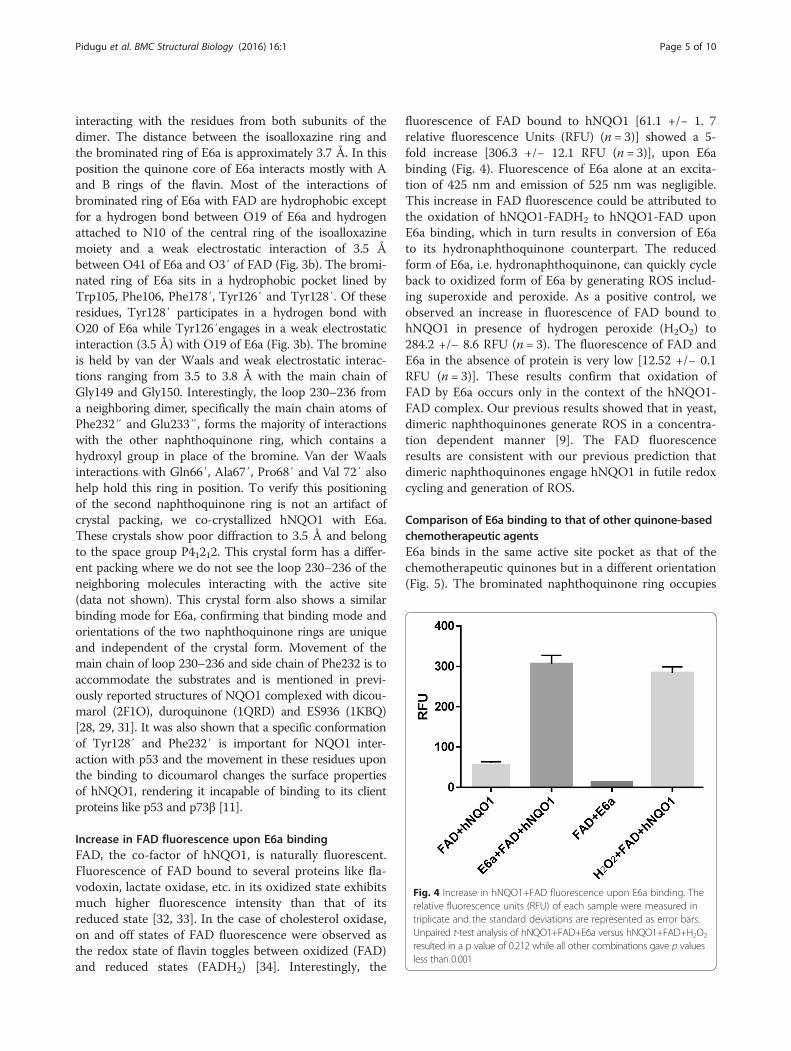
fluorescence of FAD bound to hNQO1 [61.1 +/− 1. 7relative fluorescence Units (RFU) (n = 3)] showed a 5-fold increase [306.3 +/− 12.1 RFU (n = 3)], upon E6abinding (Fig. 4). Fluorescence of E6a alone at an excita-tion of 425 nm and emission of 525 nm was negligible.This increase in FAD fluorescence could be attributed tothe oxidation of hNQO1-FADH2 to hNQO1-FAD uponE6a binding, which in turn results in conversion of E6ato its hydronaphthoquinone counterpart. The reducedform of E6a, i.e. hydronaphthoquinone, can quickly cycleback to oxidized form of E6a by generating ROS includ-ing superoxide and peroxide. As a positive control, weobserved an increase in fluorescence of FAD bound tohNQO1 in presence of hydrogen peroxide (H2O2) to284.2 +/− 8.6 RFU (n = 3). The fluorescence of FAD andE6a in the absence of protein is very low [12.52 +/− 0.1RFU (n = 3)]. These results confirm that oxidation ofFAD by E6a occurs only in the context of the hNQO1-FAD complex. Our previous results showed that in yeast,dimeric naphthoquinones generate ROS in a concentra-tion dependent manner [9]. The FAD fluorescenceresults are consistent with our previous prediction thatdimeric naphthoquinones engage hNQO1 in futile redoxcycling and generation of ROS.
Comparison of E6a binding to that of other quinone-basedchemotherapeutic agentsE6a binds in the same active site pocket as that of thechemotherapeutic quinones but in a different orientation(Fig. 5). The brominated naphthoquinone ring occupies
Fig. 4 Increase in hNQO1+FAD fluorescence upon E6a binding. Therelative fluorescence units (RFU) of each sample were measured intriplicate and the standard deviations are represented as error bars.Unpaired t-test analysis of hNQO1+FAD+E6a versus hNQO1+FAD+H2O2
resulted in a p value of 0.212 while all other combinations gave p valuesless than 0.001
Pidugu et al. BMC Structural Biology (2016) 16:1 Page 5 of 10
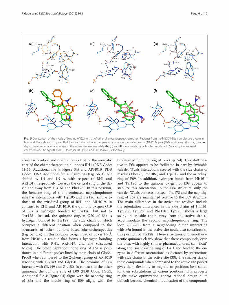
a similar position and orientation as that of the aromaticcore of the chemotherapeutic quinones RH1 (PDB Code:1H66, Additional file 4: Figure S4) and ARH019 (PDBCode: 1H69, Additional file 4: Figure S4) (Fig. 5b, f ), butshifted by 1.4 and 1.9 Å, with respect to RH1 andARH019, respectively, towards the central ring of the fla-vin and away from His161 and Phe178′. In this position,the benzene ring of the brominated naphthoquinonering has interactions with Trp105 and Tyr126′ similar tothose of the aziridinyl group of RH1 and ARH019. Incontrast to RH1 and ARH019, the quinone oxygen O19of E6a is hydrogen bonded to Tyr126′ but not toTyr128′. Instead, the quinone oxygen O20 of E6a ishydrogen bonded to Tyr128′, the side chain of whichoccupies a different position when compared to thestructures of other quinone-based chemotherapeutics(Fig. 5a, c, e). In this position, oxygen O20 of E6a is 4.5 Åfrom His161, a residue that forms a hydrogen bondinginteraction with RH1, ARH019, and E09 (discussedbelow). The other naphthoquinone ring of E6a is posi-tioned in a different pocket lined by main chain of Gln66-Pro68 when compared to the 2-phenyl group of ARH019stacking with Gly149 and Gly150. The bromine of E6ainteracts with Gly149 and Gly150. In contrast to the otherquinones, the quinone ring of E09 (PDB Code: 1GG5,Additional file 4: Figure S4) aligns with the naphthyl ringof E6a and the indole ring of E09 aligns with the
brominated quinone ring of E6a (Fig. 5d). This shift rela-tive to E6a appears to be facilitated in part by favorablevan der Waals interactions created with the side chains ofresidues Phe178, Phe106′, and Trp105′ and the aziridinylring of E09. In addition, hydrogen bonds from His161′and Tyr126 to the quinone oxygen of E09 appear tostabilize this orientation. In the E6a structure, only thevan der Waals contacts between Phe178 and the naphthylring of E6a are maintained relative to the E09 structure.The main differences in the active site residues includethe orientation differences in the side chains of His161,Tyr126′, Tyr128′ and Phe178′. Tyr128′ shows a largeswing in its side chain away from the active site toaccommodate the second naphthoquinone ring. Theloop 230–236 from a neighboring dimer interactingwith E6a bound in the active site could also contribute tothis position of Tyr128′. These structures of chemothera-peutic quinones clearly show that these compounds, eventhe ones with highly similar pharmacophores, can “float”along the isoalloxazine ring of FAD and bind to the en-zyme in different orientations as dictated by interactionswith side chains in the active site [30]. The smaller size ofthese compounds when compared to the active site pocketgives them flexibility to migrate to positions best suitedfor their substitutions at various positions. This propertymight make optimization and/or rational design quitedifficult because chemical modification of the compounds
Fig. 5 Comparison of the mode of binding of E6a to that of other chemotherapeutic quinones. Residues from the hNQO1-E6a complex are shown inblue and E6a is shown in green. Residues from the quinone complex structures are shown in orange (ARH019), pink (E09), and brown (RH1). a, c and edepict the conformational changes in the active site residues while (b), (d) and (f) show variations of binding modes of E6a and quinone-basedchemotherapeutic agents ARH019 (orange), E09 (pink) and RH1 (brown), respectively
Pidugu et al. BMC Structural Biology (2016) 16:1 Page 6 of 10

could change their binding orientation, thereby thwartingthe intention of the design. On the other hand, thedimeric naphthoquinones are bulky, occupy most of theactive site, and appear to have little room for changingorientations. This opens up the possibility for a design inwhich the halogenated naphthoquinone ring is anchoredat the FAD and the other naphthoquinone ring can beoptimized to improve efficacy.
Comparison of E6a binding to coumarin-based inhibitorsof NQO1In contrast to the other therapeutic quinones, the positionand orientation of bound E6a differs markedly whencompared to those of coumarin-based inhibitors of NQO1including dicoumarol (PDB Code: 2F1O) and AS1 (PDBCode: 3JSX, Additional file 4: Figure S4) (Fig. 6). Thecoumarin ring of dicoumarol that stacks against the isoal-lozaxine ring of FAD is stabilized by the hydrogen bondsbetween O5 and Tyr128′ and O17 and NE2 of His161.The other coumarin ring is stacked against Tryr128′ andis held in position by hydrogen bonds between O38-His161 and O32-Gly149 main chain. The crystal structureof hNQO1-AS1 complex shows that the coumarin ring ofAS1 that stacks against the flavin ring binds in thesame position but in a different orientation to that ofdicoumarol (Fig. 6b), with its hydroxyl group at position 4
interacting with Tyr128′ while O2 and O7 are hydrogenbonded to His161 (Fig. 6a). This is clearly due to the me-thyl substitutions at positions C1 and C6 of this coumarinring. The naphthyl group of AS1 occupies similar positionto that of the second coumarin ring of dicoumarol but ina perpendicular direction interacting mainly with Gly149,Gly150, Met154, Phe232′ and Tyr128′ (Fig. 6d). In thiscrystal structure, the naphthyl ring interacts with Phe232″and Glu233″ of the neighboring dimers due to crystalpacking, with the distances ranging from 3.5 to 5.8 Å ineight monomers present in the asymmetric unit. Theoverall binding pocket but not orientation of the bromi-nated naphthoquinone ring of E6a matches that of thecoumarin ring of dicoumarol and AS1 that stacks againstthe flavin, mirroring the van der Waals interactions withTyr126′, Phe178 and Phe106. However the hydrogenbonding pattern differs in the case of E6a. The strikingdifference in E6a binding compared to that of thecoumarin-based derivatives is the positioning of thesecond naphthoquinone ring in a pocket lined by the mainchains of Gln66-Pro68, phosphates of FAD. In the secondring of dicoumarol and AS1, both occupy a differentpocket which resides 8.0 Å away from the pocket occupiedby the corresponding ring on E6a. Other differences in theactive site include the conformation of side chains ofHis161, Phe106, His 194, Tyr128′ and Phe232′ (Fig. 6a, c).
Fig. 6 Comparison of E6a and coumarin-based NQO1 inhibitors binding to the active site of hNQO1. Residues from the hNQO1-E6a complex areshown in blue and E6a is shown in green. Residues from the coumarin complexes are shown in gray (dicoumarol) and brown (AS1). a and c showthe structural differences in the active site residues of hNQO1-dicoumarol and hNQO1-AS1 with reference to the hNQO1-E6a complex. b and dshow the differences in binding orientations of dicoumarol (gray) and AS1 (brown) compared to that of E6a
Pidugu et al. BMC Structural Biology (2016) 16:1 Page 7 of 10

The mechanism of dicoumarol inhibition involved increas-ing superoxide levels via inhibition of NQO1 [35–37].However, other coumarin-based inhibitors show minimalor no superoxide generation though they show efficient in-hibition of hNQO1. Dimeric naphthoquinones on theother hand showed a concentration dependent ROS gener-ation in yeast [9]. Our earlier studies on MDA-453 andPC-3 cancer cell lines suggested that generation of reactiveoxygen species leading to oxidative stress and mitochon-drial dysfunction are the anti-cancer mechanisms of thisclass of compounds [6, 7].
ConclusionsThe crystal structure of hNQO1 complexed with FADand E6a presented here is the first evidence of directinteraction of the dimeric naphthoquinones with NQO1at the active site. This is a valuable starting point forbetter understanding of the mode of binding of dimericnaphthoquinones to NQO1. Such data are required inorder to establish structure activity relationships thatsupport further structure-based optimization to improvethe anti-neoplastic efficacy of this novel class of chemo-therapeutics. High resolution crystal structures of hNQO1with more dimeric naphthoquinones would help to under-stand the complete mechanism of activation of theseagents by hNQO1.
MethodsExpression and purificationDNA for hNQO1 was codon optimized for expression inEscherichia coli and subcloned into a modified pET19bvector containing N-terminal 10XHis tag and PreScis-sion protease cleavage site preceding the insert. The ex-pression plasmid was transformed into BL21 (DE3) cells.Cells were grown at 37 °C to an OD600 of 0.6 and in-duced with 0.3 mM Isopropyl β-D-1-thiogalactopyrano-side (IPTG) at 18 °C overnight. The cells were harvestedat 4000 rpm for 20 min and resuspended in lysis buffercontaining, 50 mM Tris pH 8.0, 500 mM NaCl and2 mM beta-mercaptoethanol. The cells were lysed bysonication and soluble proteins were separated by centri-fugation at 15000 rpm for 45 min. The clarified lysatewas first purified using Ni-affinity chromatography. Thehistidine tag was removed from the purified hNQO1 bypreScission protease cleavage, combined with a dialysisagainst 50 mM Tris pH 8.0, 150 mM NaCl and 1 mMDTT. Cleaved hNQO1 was further subjected to a finalpurification step using size exclusion chromatography.
Crystallization and structure determinationhNQO1 stored at a concentration of approximately18–20 mg/ml in 50 mM Tris pH 8.0, 50 mM NaCl and5 μM FAD was used for crystallization screening. Initialscreening with JCSG+, Classics suite I and II from Qiagen
resulted in initial hits in more than 20 conditions. Nativedata up to 2.0 Å resolution were collected using P21 crys-tals obtained in 20 % (w/v) PEG3350 and 0.2 M Ammo-nium Citrate. Another crystal form in the space groupP212121 were obtained using 20 % PEG3350 and 0.2 MPotassium Sodium Tartarate. The complex betweenNQO1 and E6a was obtained by soaking the P212121 na-tive crystals in mother liquor containing 1 mM E6a. X-raydiffraction data were collected in house and at beam line5.0.3 of the Advanced Light source at Lawrence BerkeleyNational Laboratory for the holo and E6a-bound hNQO1crystals respectively. The data were reduced usingiMosflm [38] and Aimless [39] from the CCP4 programsuite. Initial phases for both holo and E6a-bound hNQO1were determined by molecular replacement using the pro-gram Phaser [40–43] using the coordinates of an hNQO1monomer from a previously reported holo-structure (PDBaccession code: 1D4A) [26]. The initial molecular replace-ment solution for the hNQO1-E6a complex containedonly 8 of the 14 monomers in the asymmetric unit. Usingthese 8 monomers as a fixed solution, the remainingmonomers were placed iteratively using a combination ofPhaser [40–43] and Molrep [44]. The structure wasrefined using Refmac5 [45–49] from CCP4 programsuite. Iterative cycles of model building using COOT[50–53] and refinement by refmac5 and TLS- andNCS- restrained [54] refinement using buster [55]yielded final structures with Rwork/Rfree of 18.0/21.6for native and 18.3/22.0 for the E6a complex. FinalStructures were deposited in PDB (Accession Codes:holo–hNQO1: 5EA2, hNQO1-E6a: 5EAI).
Fluorescence measurementsAll fluorescence measurements were done using Spectra-Max M5 plate reader at room temperature. An absorbancescan for 100 μM FAD in 50 mM Tris pH 8.0 and 50 mMNaCl resulted in a peak at 440 nm. The excitation wave-length was chosen to be 425 nm for FAD to avoid overlapof excitation and emission peaks. An emission scan with anexcitation wavelength at 425 nm resulted in a peak at525 nm. These wavelengths were confirmed by performingsimilar scans with FAD bound to hNQO1 in the same buf-fer and thus were used for all of the fluorescence experi-ments. Approximately 100 μl of each of the followingsamples in 50 mM Tris pH 8.0, 50 mM NaCl and 5 μMFAD were added in the wells of a costar Black/clear bottom96 well plate. Fluorescence of hNQO1 alone in the abovebuffer was initially recorded. Next, NQO1 supplementedwith 1 mM E6a in the above buffer was analyzed. Amixture of 5 μM FAD and 1 mM E6a mixture in the abovebuffer was used as a negative control. Approximately100 μM hydrogen peroxide was added to the NQO1sample and used as a positive control for the oxidized FAD.These data were analyzed using softMax Pro software.
Pidugu et al. BMC Structural Biology (2016) 16:1 Page 8 of 10

Availability of supporting dataThe atomic coordinates and structure factor amplitudesare available in the Protein Data Bank repository (PDB),Accession Codes 5ea2 (holo-NQO1) and 5eai (NQO1-E6acomplex).
Additional files
Additional file 1: Figure S1. The chemical structure of E6a (3-bromo-3′-hydroxy-2,2′-binaphthalenyl-1,4,1′,4′-tetraone). (PNG 40 kb)
Additional file 2: Figure S2. Superposition of active site residues inholo-hNQO1 structures from the current study (Cyan) and previouslyreported structure 1D4A (gray). (PNG 1167 kb)
Additional file 3: Figure S3. Two dimers of E6a bound hNQO1 structureshowing the loop 230–236 interacting with the active site of neighboringdimer. The FAD molecules are shown in stick representation in each activesite. The E6a molecule is shown in ball-and-stick representation. (PNG 2799 kb)
Additional file 4: Figure S4. The chemical structures of other knowninhibitors of NQO1. (PNG 79 kb)
AbbreviationsALL: acute lymphoblastic leukemia; AML: acute myeloid leukemia; ARH019: 3-hydroxymethyl-5-(2-methylaziridin-1-yl)-1-methyl-2-phenylindole-4,7-dione;AS1: 4-Hydroxy-6,7-dimethyl-3-(1-naphthylmethyl)-2H-chromen-2-one; E09: 3-hydroxymethyl-5-aziridinyl-1-methyl-2-(H-indole-4,7-indione)-propenol; E6a: 3-bromo-3′-hydroxy-2,2′-binaphthalenyl-1,4,1′,4′-tetraone; FAD: flavin adeninedinucleotide; NAD: nicotinamide adenine dinucleotide; RFU: relativefluorescence units; RH1: 2,5-diaziridinyl-3-hydroxyl-6-methyl-1,4-benzoquinone;ROS: reactive oxygen species.
Competing interestsAshkan Emadi shares a patent on drugs used in this study.
Authors’ contributionsEAT, AE, and EAS conceived and designed the study. LSMP executed theprotein expression, purification, crystallization, holo-NQO1 data collectionand structure determination. EP collected the E6a crystallographic data andassisted with data analysis. JCEM and MA assisted with protein expression,purification and crystallization. LSMP, EAT, AE, and EAS analyzed the data andwrote the manuscript. All authors read and approved the final manuscript.
AcknowledgementsThe X-ray data for holo-hNQO1 structure were collected at W.M. Keck/NISTX-ray Crystallography Core Facility at the Institute for Bioscience and Biotech-nology Research and for hNQO1-E6a complex were collected at beam line5.0.3 of the Advanced Light source at Lawrence Berkeley National Laboratory.We thank Dr. Daniel Nelson and Sara Linden for letting us use their Spectra-Max M5 for the fluorescence experiments and useful discussions in analyzingthe data. This work was supported by institutional funds from the Center forBiomolecular Therapeutics.
Author details1Department of Biochemistry and Molecular Biology, University of MarylandSchool of Medicine, Baltimore, MD 21201, USA. 2Marlene and StewartGreenebaum Cancer Center, University of Maryland School of Medicine,Baltimore, MD 21201, USA. 3Institute for Bioscience and BiotechnologyResearch, and Center for Biomolecular Therapeutics, 9600 Gudelsky Drive,Rockville, MD 20850, USA.
Received: 5 November 2015 Accepted: 19 January 2016
References1. O’Brien PJ. Molecular mechanisms of quinone cytotoxicity. Chem Biol
Interact. 1991;80(1):1–41.2. Decosterd LA, Parsons IC, Gustafson KR, Cardellina II JH, McMahon JB, Cragg GM,
et al. Structure, absolute stereochemistry, and synthesis of conocurvone, a
potent, novel HIV-inhibitory naphthoquinone trimer from a Conospermum sp.J Amer Chem Soc. 1993;115:6673–9.
3. Kayser O, Kiderlen AF, Laatsch H, Croft SL. In vitro leishmanicidal activity ofmonomeric and dimeric naphthoquinones. Acta Trop. 2000;77(3):307–14.
4. Lim ES, Rhee YH, Park MK, Shim BS, Ahn KS, Kang H, et al. DMNQ S-64induces apoptosis via caspase activation and cyclooxygenase-2 inhibition inhuman nonsmall lung cancer cells. Ann N Y Acad Sci. 2007;1095:7–18.
5. Wellington KW. Understanding cancer and the anticancer activities ofnaphthoquinones – a review. RSC Adv. 2015;5:20309–38.
6. Ross AE, Emadi A, Marchionni L, Hurley PJ, Simons BW, Schaeffer EM, et al.Dimeric naphthoquinones, a novel class of compounds with prostatecancer cytotoxicity. BJU Int. 2011;108(3):447–54.
7. Emadi A, Le A, Harwood CJ, Stagliano KW, Kamangar F, Ross AE, et al.Metabolic and electrochemical mechanisms of dimeric naphthoquinonescytotoxicity in breast cancer cells. Bioorg Med Chem. 2011;19(23):7057–62.
8. Emadi A, Sadowska M, Carter-Cooper B, Wonodi O, Muvarak N, Rassool F, etal. Dimeric Naphthoquinones: Novel Anti-Leukemic Agents ModulatingCellular Redox Status. In: American Society of Hematology (ASH) 55thAnnual Meeting. New Orleans, LA, USA; 2013.
9. Emadi A, Ross AE, Cowan KM, Fortenberry YM, Vuica-Ross M. A chemicalgenetic screen for modulators of asymmetrical 2,2′-dimericnaphthoquinones cytotoxicity in yeast. PLoS One. 2010;5(5):e10846.
10. Bey EA, Bentle MS, Reinicke KE, Dong Y, Yang CR, Girard L, et al. An NQO1- andPARP-1-mediated cell death pathway induced in non-small-cell lung cancercells by beta-lapachone. Proc Natl Acad Sci U S A. 2007;104(28):11832–7.
11. Asher G, Dym O, Tsvetkov P, Adler J, Shaul Y. The crystal structure ofNAD(P)H quinone oxidoreductase 1 in complex with its potent inhibitordicoumarol. Biochemistry. 2006;45(20):6372–8.
12. Ernster L. DT Diaphorase: a historical review. Chem Scr. 1987;27A:1–13.13. Anwar A, Dehn D, Siegel D, Kepa JK, Tang LJ, Pietenpol JA, et al. Interaction
of human NAD(P)H:quinone oxidoreductase 1 (NQO1) with the tumorsuppressor protein p53 in cells and cell-free systems. J Biol Chem.2003;278(12):10368–73.
14. Asher G, Lotem J, Kama R, Sachs L, Shaul Y. NQO1 stabilizes p53 through adistinct pathway. Proc Natl Acad Sci U S A. 2002;99(5):3099–104.
15. Chen XL, Varner SE, Rao AS, Grey JY, Thomas S, Cook CK, et al. Laminar flowinduction of antioxidant response element-mediated genes in endothelialcells. A novel anti-inflammatory mechanism. J Biol Chem. 2003;278(2):703–11.
16. Garate M, Wani AA, Li G. The NAD(P)H:Quinone Oxidoreductase 1 inducescell cycle progression and proliferation of melanoma cells. Free Radic BiolMed. 2010;48(12):1601–9.
17. Chakrabarti G, Gerber DE, Boothman DA. Expanding antitumor therapeuticwindows by targeting cancer-specific nicotinamide adenine dinucleotidephosphate-biogenesis pathways. Clin Pharmacol. 2015;7:57–68.
18. Li C, Liu Y, Wei S, Zhou Y. A meta-analysis of the association between NQO1C609T variation and acute myeloid leukemia risk. Pediatr Blood Cancer.2014;61(5):771–7.
19. Li C, Zhou Y. Association between NQO1 C609T polymorphism and acutelymphoblastic leukemia risk: evidence from an updated meta-analysis basedon 17 case-control studies. J Cancer Res Clin Oncol. 2014;140(6):873–81.
20. Zeekpudsa P, Kukongviriyapan V, Senggunprai L, Sripa B, Prawan A.Suppression of NAD(P)H-quinone oxidoreductase 1 enhanced thesusceptibility of cholangiocarcinoma cells to chemotherapeutic agents.J Exp Clin Cancer Res. 2014;33:11.
21. Siegel D, Gibson NW, Preusch PC, Ross D. Metabolism of diaziquone byNAD(P)H:(quinone acceptor) oxidoreductase (DT-diaphorase): role indiaziquone-induced DNA damage and cytotoxicity in human coloncarcinoma cells. Cancer Res. 1990;50(22):7293–300.
22. Siegel D, Gibson NW, Preusch PC, Ross D. Metabolism of mitomycin C byDT-diaphorase: role in mitomycin C-induced DNA damage and cytotoxicityin human colon carcinoma cells. Cancer Res. 1990;50(23):7483–9.
23. Emadi A, Harwood JS, Kohanim S, Stagliano KW. Regiocontrolled synthesisof the trimeric quinone framework of conocurvone. Org Lett. 2002;4(4):521–4.
24. Adams MA, Jia Z. Modulator of drug activity B from Escherichia coli: crystalstructure of a prokaryotic homologue of DT-diaphorase. J Mol Biol.2006;359(2):455–65.
25. Buryanovskyy L, Fu Y, Boyd M, Ma Y, Hsieh TC, Wu JM, et al. Crystal structureof quinone reductase 2 in complex with resveratrol. Biochemistry.2004;43(36):11417–26.
26. Faig M, Bianchet MA, Talalay P, Chen S, Winski S, Ross D, et al. Structures ofrecombinant human and mouse NAD(P)H:quinone oxidoreductases: species
Pidugu et al. BMC Structural Biology (2016) 16:1 Page 9 of 10

comparison and structural changes with substrate binding and release.Proc Natl Acad Sci U S A. 2000;97(7):3177–82.
27. Pey AL, Megarity CF, Timson DJ. FAD binding overcomes defects in activityand stability displayed by cancer-associated variants of human NQO1.Biochim Biophys Acta. 2014;1842(11):2163–73.
28. Nolan KA, Doncaster JR, Dunstan MS, Scott KA, Frenkel AD, Siegel D, et al.Synthesis and biological evaluation of coumarin-based inhibitors of NAD(P)H:quinone oxidoreductase-1 (NQO1). J Med Chem. 2009;52(22):7142–56.
29. Li R, Bianchet MA, Talalay P, Amzel LM. The three-dimensional structure ofNAD(P)H:quinone reductase, a flavoprotein involved in cancerchemoprotection and chemotherapy: mechanism of the two-electronreduction. Proc Natl Acad Sci U S A. 1995;92(19):8846–50.
30. Faig M, Bianchet MA, Winski S, Hargreaves R, Moody CJ, Hudnott AR, et al.Structure-based development of anticancer drugs: complexes of NAD(P)H:quinone oxidoreductase 1 with chemotherapeutic quinones. Structure.2001;9(8):659–67.
31. Winski SL, Faig M, Bianchet MA, Siegel D, Swann E, Fung K, et al.Characterization of a mechanism-based inhibitor of NAD(P)H:quinoneoxidoreductase 1 by biochemical, X-ray crystallographic, and massspectrometric approaches. Biochemistry. 2001;40(50):15135–42.
32. Ghisla S, Massey V, Lhoste JM, Mayhew SG. Fluorescence and opticalcharacteristics of reduced flavines and flavoproteins. Biochemistry.1974;13(3):589–97.
33. Sun M, Moore TA, Song PS. Molecular luminescence studies of flavins. I. Theexcited states of flavins. J Am Chem Soc. 1972;94(5):1730–40.
34. Sevcik J, Solovicova A, Hostinova E, Gasperik J, Wilson KS, Dauter Z.Structure of glucoamylase from Saccharomycopsis fibuligera at 1.7 Aresolution. Acta Crystallogr D Biol Crystallogr. 1998;54(Pt 5):854–66.
35. Cullen JJ, Hinkhouse MM, Grady M, Gaut AW, Liu J, Zhang YP, et al.Dicumarol inhibition of NADPH:quinone oxidoreductase induces growthinhibition of pancreatic cancer via a superoxide-mediated mechanism.Cancer Res. 2003;63(17):5513–20.
36. Lewis A, Du J, Liu J, Ritchie JM, Oberley LW, Cullen JJ. Metastaticprogression of pancreatic cancer: changes in antioxidant enzymes and cellgrowth. Clin Exp Metastasis. 2005;22(7):523–32.
37. Lewis A, Ough M, Li L, Hinkhouse MM, Ritchie JM, Spitz DR, et al. Treatmentof pancreatic cancer cells with dicumarol induces cytotoxicity and oxidativestress. Clin Cancer Res. 2004;10(13):4550–8.
38. Battye TG, Kontogiannis L, Johnson O, Powell HR, Leslie AG. iMOSFLM: anew graphical interface for diffraction-image processing with MOSFLM.Acta Crystallogr D Biol Crystallogr. 2011;67(Pt 4):271–81.
39. Evans PR, Murshudov GN. How good are my data and what is theresolution? Acta Crystallogr D Biol Crystallogr. 2013;69(Pt 7):1204–14.
40. Storoni LC, McCoy AJ, Read RJ. Likelihood-enhanced fast rotation functions.Acta Crystallogr D Biol Crystallogr. 2004;60(Pt 3):432–8.
41. McCoy AJ, Grosse-Kunstleve RW, Storoni LC, Read RJ. Likelihood-enhanced fasttranslation functions. Acta Crystallogr D Biol Crystallogr. 2005;61(Pt 4):458–64.
42. McCoy AJ. Solving structures of protein complexes by molecular replacementwith Phaser. Acta Crystallogr D Biol Crystallogr. 2007;63(Pt 1):32–41.
43. McCoy AJ, Grosse-Kunstleve RW, Adams PD, Winn MD, Storoni LC, Read RJ.Phaser crystallographic software. J Appl Crystallogr. 2007;40(Pt 4):658–74.
44. Vagin A, Teplyakov A. Molecular replacement with MOLREP. Acta CrystallogrD Biol Crystallogr. 2010;66(Pt 1):22–5.
45. Murshudov GN, Vagin AA, Dodson EJ. Refinement of macromolecularstructures by the maximum-likelihood method. Acta Crystallogr D BiolCrystallogr. 1997;53(Pt 3):240–55.
46. Murshudov GN, Vagin AA, Lebedev A, Wilson KS, Dodson EJ. Efficientanisotropic refinement of macromolecular structures using FFT. ActaCrystallogr D Biol Crystallogr. 1999;55(Pt 1):247–55.
47. Winn MD, Isupov MN, Murshudov GN. Use of TLS parameters to modelanisotropic displacements in macromolecular refinement. Acta Crystallogr DBiol Crystallogr. 2001;57(Pt 1):122–33.
48. Winn MD, Murshudov GN, Papiz MZ. Macromolecular TLS refinement inREFMAC at moderate resolutions. Methods Enzymol. 2003;374:300–21.
49. Murshudov GN, Skubak P, Lebedev AA, Pannu NS, Steiner RA, Nicholls RA, etal. REFMAC5 for the refinement of macromolecular crystal structures. ActaCrystallogr D Biol Crystallogr. 2011;67(Pt 4):355–67.
50. Brown A, Long F, Nicholls RA, Toots J, Emsley P, Murshudov G. Tools formacromolecular model building and refinement into electroncryo-microscopy reconstructions. Acta Crystallogr D Biol Crystallogr.2015;71(Pt 1):136–53.
51. Debreczeni JE, Emsley P. Handling ligands with Coot. Acta Crystallogr D BiolCrystallogr. 2012;68(Pt 4):425–30.
52. Emsley P, Lohkamp B, Scott WG, Cowtan K. Features and development ofCoot. Acta Crystallogr D Biol Crystallogr. 2010;66(Pt 4):486–501.
53. Emsley P, Cowtan K. Coot: model-building tools for molecular graphics. ActaCrystallogr D Biol Crystallogr. 2004;60(Pt 12 Pt 1):2126–32.
54. Smart OS, Womack TO, Flensburg C, Keller P, Paciorek W, Sharff A, et al.Exploiting structure similarity in refinement: automated NCS andtarget-structure restraints in BUSTER. Acta Crystallogr D Biol Crystallogr.2012;68(Pt 4):368–80.
55. Bricogne G BE, Brandl M, Flensburg C, Keller P, Paciorek W, Roversi P, et al.BUSTER In., version 2.10.2 edn. Cambridge: Global Phasing Ltd.; 2011.
• We accept pre-submission inquiries
• Our selector tool helps you to find the most relevant journal
• We provide round the clock customer support
• Convenient online submission
• Thorough peer review
• Inclusion in PubMed and all major indexing services
• Maximum visibility for your research
Submit your manuscript atwww.biomedcentral.com/submit
Submit your next manuscript to BioMed Central and we will help you at every step:
Pidugu et al. BMC Structural Biology (2016) 16:1 Page 10 of 10