a dissertation onrepository-tnmgrmu.ac.in/9132/1/220100718arunkumar.pdf · introduction diabetes is...
TRANSCRIPT

A Dissertation on
‘’A STUDY OF PATHOPHYSIOLOGY, MANAGEMENT AND
FACTORS INFLUENCING DIABETIC FOOT ULCERS AMONG
DIABETIC PATIENTS’’
Dissertation Submitted to
THE TAMIL NADU Dr.M.G.R. MEDICAL UNIVERSITY
CHENNAI- 600032
with partial fulfillment of the regulations
for the award of the degree of
M.S. GENERAL SURGERY
(BRANCH 1)
COIMBATORE MEDICAL COLLEGE,
COIMBATORE
MAY 2018


CERTIFICATE
Certified that this is the Bonafide Dissertation in ‘’A STUDY OF
PATHOPHYSIOLOGY, MANAGEMENT AND FACTORS
INFLUENCING DIABETIC FOOT ULCERS AMONG DIABETIC
PATIENTS’’ was a work done by Dr.ARUNKUMAR.N.R. and
submitted in partial fulfillment of the requirements for the Degree of
M.S.General Surgery, Branch I of The Tamil nadu Dr.M.G.R Medical
University, Chennai.
Date: Professor and Unit Chief
Department of General Surgery
Coimbatore Medical College.
Date: Professor and HOD
Department of General Surgery
Coimbatore Medical College.
Date: The DEAN
Coimbatore Medical College

DECLARATION
I Solemnly declare that the Dissertation titled ‘’A STUDY OF
PATHOPHYSIOLOGY, MANAGEMENT AND FACTORS
INFLUENCING DIABETIC FOOT ULCERS AMONG DIABETIC
PATIENTS’’ was done by me at Coimbatore Medical College during the
academic year July 2016 – June 2017 under the guidance of
Prof. Dr.V.Elango, M.S. this Dissertation is submitted to the Tamilnadu
Dr.M.G.R Medical University towards the fulfillment of the requirement
for the award of M.S. Degree in General Surgery (Branch ).
PLACE: Dr.ARUNKUMAR.N.R
DATE:

ACKNOWLEDGEMENT
I express my gratitude to Dr.B.Asokan, M.S, MCh, the Dean,
Coimbatore Medical College Hospital for providing facilities to carry out
my project work successfully.
I sincerely thank Prof. Dr. V.Elango, M.S, Chief and HOD, Department
of General Surgery for his constant guidance and encouragement
throughout the period of my study.
I would like to express my gratitude to my guides
Prof. Dr. Ranganathan M.S, Prof. Dr. Natarajan M.S,
Prof. Dr. Shanthi M.S and Prof. Dr. Nirmala M.S, for their valuable
guidance and encouragement throughout the period of my study.
I would like to give special thanks to Dr. Narayanamoorthy M.S, Dr.
Sumitra M.S, DGO and Dr. Jayalakshmi M.S, Assistant Professors,
Department of General Surgery for their voluntary and useful guidance
and support.
And also my special thanks to Dr Veena for completing my study.
PLACE: Dr. ARUNKUMAR.N.R

CERTIFICATE – II
This is to certify that this dissertation work titled ‘’A STUDY OF
PATHOPHYSIOLOGY, MANAGEMENT AND FACTORS
INFLUENCING DIABETIC FOOT ULCERS AMONG DIABETIC
PATIENTS’’ of the candidate DR.ARUNKUMAR.N.R with
registration Number 221511303 for the award of M.S in the branch of
General Surgery,I personally verified the urkund.com website for the
purpose of plagiarism Check. I found that the uploaded thesis file
contains 96 pages from introduction to conclusion and the result shows
0% (Zero) percentage of plagiarism in the dissertation.
Guide & Supervisor sign with Seal.


ABBREVIATIONS
CMCH - Coimbatore Medical College Hospital
IMV - Infectious Mononucleosis
HLA - Human Leukocyte Antigen
HDL - High Density Lipoprotein
LDL - Low Density Lipoprotein
DNA - Deoxyribonucleic Acid
DM - Diabetes Mellitus
IV - Intravenous
OGT - Oral Glucose Tolerance Test
GTT - Glucose Tolerance Test
CBP - Complete Blood Picture
SPP - Systemic Pulse Pressure
SVR - Systemic Venous Resistance
DKA - Diabetic Ketoacidosis
ABI - Ankle Brachial Index
VAC - Vacuum Assisted Closure
LA - Local Anaesthetic
AKA - Above Knee Amputation
BKA - Below Knee Amputation
SSG - Split Skin Graft
PUFA - Polyunsaturated Fatty Acid
I&D - Incision & Drainage
RBS - Random Blood Sugar
FBS - Fasting Blood Sugar
PPBS - Post prandial Blood Sugar

LIST OF FIGURES
S. No.
Figure Page
No.
1. Bones of Foot 12
2. Gangrene Foot 19
3. Debridement 49
4. Neuropathic Ulcer involving heel 50
5. Granulation Tissue 51
6. Split Skin Grafting 52
7. Vacuum Assisted Closure 58

INDEX
SR.NO CONTENT PAGE NO.
I INTRODUCTION 1
II AIMS AND OBJECTIVES 4
III REVIEW OF LITERATURE 6
IV MATERIALS AND METHODS 62
V OBSERVATIONS AND
RESULTS
66
VI DISCUSSION 82
VII SUMMARY 94
VIII CONCLUSION 95
IX BIBLIOGRAPHY 97
X
ANNEXURES
PROFORMA
CONSENT FORM
MASTER CHART

INTRODUCTION
Diabetes is an endocrine disorder that has reached epidemic
proportions worldwide. Overall 15 % of individuals with diabetes
mellitus will have foot ulcer in their life time and the annual incidence of
2-5%. Diabetic foot is becoming a major concern of diabetic patients and
those who treat them from quality of life, social and economic stand
point.
The word “Diabetic foot” means that the pathophysiological
process of diabetes mellitus do something to foot, that puts at increased
risk of tissue damage resulting to ulcer formation. (Payne & Florkowski,
1998).
Foot damages such as ulceration, infection and gangrene are one of
the important causes of hospital admission in patients with diabetic
mellitus.(1)
Natural history of diabetic foot.
Evidence that the pathological process of Diabetes have put the
foot for more risk for tissue damage has happened and foot is at risk for
end stage complications (amputation). Of all complications of diabetes,
those occurring in the foot are considered most avoidable.

Epidemiology: (2)
3-5% of those with diabetes have a foot ulcer.
15% of all those with diabetes will, during their lifetime develops an
ulcer.
4-5% of foot ulcers are increased by external trauma.
Up to 20% undergo same side amputation within 12 months.
Up to 50% undergo opposite side amputation within 1-3 yrs; 75% within
5 years.
3years increased death rate after complication is 20-50%.
Most important risk factors are:
Loss of sensations.
Longer duration of diabetes.
High foot pressure.
In 2000, The International Diabetic Federation endorsed the
International Working on the Diabetic foot as a Consultative Section on
the Diabetic foot. Organizations altogether established goals for the
future of diabetic care worldwide (3).
Goals
To inform the people about the diabetic foot problems worldwide.

To increase alertness of the diabetic foot among those at threat and
those in place to act.
To influence healthcare decision makers that action is both
achievable and reasonably priced.
To caution health care decision makers of the problems of not
taking action.
To inform people with diabetes of the actions they can avoid foot
complications.
Multidisciplinary Management
The plan in supervising diabetic foot is always to keep the patient
at as low a phase possible. At each of the diabetic foot, it is essential to
interfere early and to manage the foot to prevent further progression. No
one person can manage the diabetic foot. Members of the team will
include physician, general surgeon, orthopedic surgeon, radiologist,
expert nurse and podiatrist. (4)
It is helpful if the team works very much mutually, within the focal
point of the diabetic foot clinic and also meets frequently to carry out
ward rounds and x-rays conferences. Each team member should be
available hastily in an emergency.

AIMS AND OBJECTIVES
AIM
1) The aim of this study is to study the current trends concerning the
pathology, complications and treatment of diabetic foot ulcers.
2) To study the co-relation between atherosclerotic changes in the
blood vessels of the lower limb & diabetic ulcers.
3) To study the bacterial flora & evolution of the ulcer with the
relation to rigorousness of diabetes.
OBJECTIVES
1. PRIMARY
To assess the prevalence of diabetic foot ulcer and relative distribution
according to age, sex, occupation and other factors among diabetic
patients in patients attending CMCH.
2. SECONDARY
a. To study the mode of presentation and appearance of diabetic foot
ulcers
b. To emphasize and enhance the knowledge of diabetic patients
regarding self-care and regular diabetic foot evaluation.

c. To understand the pathology of diabetic foot ulcer and early
recognition of complications of peripheral neuropathy and ischemia and
using a multidisciplinary approach.
d. To avert the various complications due to diabetic foot ulcers and
early management of its complications.
e. To study the different treatment modalities in management of
diabetic foot ulcers.

REVIEW OF LITERATURE
There is considerable geographic variation in the incidence of
both type 1 and type 2 Diabetes mellitus. The variability is likely due to
genetic, behavioural, and environmental factor. Diabetes mellitus
prevalence also varies among different ethnic populations within a given
country (5).
Diabetes mellitus
Diabetes mellitus is the most common endocrine disorder 6
characterized by metabolic abnormalities and by long term complications
involving eye, kidney, nerves and blood vessels.
W.H.O CLASSIFICATION OF DIABETES MELLITUS ( 6)
Primary
Type 1 – Insulin dependent diabetes mellitus (IDDM).
Type 2 – Non insulin dependent diabetes mellitus (NIDDM).
Secondary
Diabetes caused by pancreatic disorder.
Diabetes caused by hormonal abnormalities.
Chemical induced diabetes mellitus.
Diabetes caused by insulin receptor abnormalities.
Diabetes associated with genetic syndromes.
Diabetes of other causes.

ENVIRONMENTAL EVENT
The fact that monozygotic twin of IDDM patient may remain
asymptomatic proves that there are other factors other than genome in the
pathology of IDDM. Environmental factors also play a role. In many
cases it is a viral infection of beta cells. Mumps, Hepatitis, IMN,
Congenital Rubella. Coxsackie virus may trigger the disease. It is also
postulated that exposure to cow s milk products early in life predispose to
autoimmune diabetes. The proposed mechanism is bovine albumin acting
through mechanism of molecular mimicry.
NIDDM is 7-8 times more common than IDDM (7).
PATHOGENESIS OF IDDM (8)
Pathology of IDDM is due to destruction of beta cells of
pancreas. Pathogenesis starts with genetic susceptibility to disease, some
environmental factors also play in the pathogenesis. Viral infection
usually triggers the destruction. During pathogenesis the patient is
initially non – diabetic. Progressively beta cells are infiltrated and
destroyed by macrophages and T lymphocytes, this stage is called
insulinitis or isleititis. During this period reserve of insulin progressively
diminishes and when it is insufficient to maintain blood glucose level
within normal the patient develops diabetes. Genetic susceptibility to

IDDM probably involves more than one gene. Candidate loci are
proposed in chromosomes 2, 6, 11 and 15. It is HLA associated.
PATHOGENESIS OF NIDDM
Aetiopathogenesis include both insulin resistance and beta cell defects.
Major environmental factor is obesity.
Genetics: Maturity onset diabetes of the young shows autosomal
dominant penitence. Other varieties of NIDDM are polygenic.
PATHOPHYSIOLOGY
Patient with Type 2 NIDDM have physiological defects.
Abnormal insulin secretion and insulin resistance.
CLINICAL FEATURES OF DIABETES MELLITUS
IDDM: It usually begins below 40 years. Peak incidence is within 14
years. Onset of symptoms is often abrupt with thirst, excessive urination
or increased appetite.
NIDDM: Usually begins in middle life or later. Patient is often obese.
Symptoms begin gradually, frequently diagnosed in asymptomatic patient
during blood sugar measurements. Plasma insulin level is normal to high
but there is relative insulin deficiency in relation to high blood sugar.

General characteristics OF NIDDM AND IDDM are given below (9):
Characteristics IDDM NIDDM
General factors Chromosome 6 Unknown
Age of onset Below 40 More than 40
Body habitués Normal or wasted Obese
Plasma insulin Low or absent Normal or high
Acute complication Ketoacidosis Hyperosmolar coma
Insulin therapy Responsive Responsive to resistant
Response to Sulfonyl
urea therapy
Unresponsive Responsive
SURGICAL ANATOMY OF FOOT (10)
SKIN AND NAILS
The skin of dorsum of foot is thin and highly flexible,
containing hair follicles; sweat glands and scanty sebaceous gland. Hairs
are sparse and thin. It is less than 2mm thick and few fibrous septa
penetrate to deeper facial structures. The plantar skin is 5mm thick
especially over those points which bear weight viz. Heel, ball of big toe
and lateral margins of the sole. It has no hair follicles or sebaceous glands
but glands are numerous.

NERVES
Cutaneous nerves are arranged in the following way. The medial
plantar nerve supplies the three and half digits on the big side of the foot.
The lateral plantar nerve supplies one and half digits. The medial
calcaneal branches of the posterior tibial nerve supply the skin under the
heel.
The motor and sensory components of the sciatic nerve supply
the foot. The innervations to the sole is from medial calcaneal branch of
tibial nerve. The dorsum of the toes is supplied by the digital branches of
these nerves except the saphenous on the terminal phalanges the supply is
from the plantar nerves.
VASCULATURE OF FOOT
All the arterial supply of the foot is derived from popliteal
artery. The anterior tibial artery enters the extensor compartment of the
leg and becoming in the foot the dorsalis pedis artery. The posterior tibial
artery divides into medial and lateral plantar arteries. The medial plantar
artery runs forward on the medial side of the medial plantar nerve. The
artery supplies medial side of foot and its digital supply is restricted
practically to the big toe. Lateral plantar artery crosses the sole obliquely
on the marginal side of the nerve, just deep to the first layer of the sole

towards the base of the fifth metatarsal bone. The plantar arch curves
convexly forward across the base of fourth, third and second metatarsals
and is joined in the proximal part of the first inter-metatarsal space by the
dorsalis pedis artery from the convexity of the plantar arch, plantar
metatarsal arteries run forward and bifurcate to supply the four web
spaces and digits. The veins accompaning perforating arteries take most
of the blood from the sole and from the interosseous muscles to the dorsal
venous arch.
MUSCLES OF THE FOOT
The muscles in the extensor group are located anteriorly in the
leg, they include tibialis anterior, extensor hallucis longus, laterally are
the peroneal muscles. The flexors are in the posterior compartment of the
leg. The deep fascia encloses the muscles in the leg. The gastrocnemus
arises on the distal posterior femur. In the sole of the foot the plantar
aponeurosis is the most superficial layer, the fibers of plantar fascia is
divided into five processes beneath fascia the fascia. The muscles in the
sole of the foot are categorized into four layers only the muscles of first
layer cover the whole extent of the foot muscles in the first layer include
flexor digitorum brevis, abductor hallucis and abductor digiti minimi.
In the second layer are tendon of flexor hallucis longus, flexor
digitorum accessories (quadrates plantae) and the lumbricals. In the third

layer are flexor hallucis brevis, abductor hallucis and flexor digiti
minimum brevis. In the fourth layer is peroneous longus tendon, of the
tibialis posterior, four dorsal interossei and three plantar interossei.
PERIOSTEUM
Periosteum is a fibrous membrane investing the bones except at
their articular surfaces. It is adherent to the bone and varies in different
places. In adult bone it is firmly adherent and especially so at the
insertion of tendons and ligaments where more periosteal, fibers penetrate
into bone as the perforating fibers of Sharpey.
Periosteum consists of two layers. The outer layer is composed
of coarse fibrous connective tissue containing few cells but as numerous
blood vessels and nerves. The inner layer is less vascular but more
cellular and contain many elastic fibers.
BONES OF THE FOOT

The bones of the foot are the tarsal bones, metatarsals and the
phalanges. The tarsal bones are the calcaneum, the talus, the navicular,
the cuboid and the three cuneiform bones. Calcaneum is the largest bone
of the foot and forms the prominence of the heel it articulates with talus
above and cuboid in front. Talus carries the whole body weight. It lies on
weight bearing calcaneum below the tibia and communicates thrust from
one to the other. Navicular bone can be seen and felt on the medial border
of the foot. Cuboid bone is rather wedge shaped narrowest at the lateral
margin and broadest medially, where it articulates with lateral cuneiform.
Cuneiform bones, all three are wedge shaped. Medial is largest
and the edge lies upwards. Intermediate is smallest. All the three
articulates posteriorly with navicular and anteriorly with metatarsal
bones. This completes medial longitudinal arch.
METATARSAL BONES AND PHALANGES
The metatarsal bones and phalanges resemble metacarpals and
phalanges of hand. Each has a head distally, a shaft and a base
proximally. There are five metatarsals and they are numbered from
medial to lateral side. The first metatarsal bone is large and strong and it
plays an important role in supporting the weight of the body. Second to
fifth metatarsals have slender shafts. Each toe has three phalanges except
the big toe, which posses only two.

PATHOPHYSIOLOGY OF DIABETIC FOOT
It has been recognized that persons with diabetes are prone to foot
problems.
Recent advances in molecular biology have added substantial
insight into the pathophysiology of the disease and opened new avenues
for treatment (11).
The predisposing factors to pathologic changes in the foot of a diabetic
are:
1. Metabolic factors – hyperglycemia
2. Vascular changes
3. Neuropathy
4. Infection
Polyol pathway.
Glucose → sorbitol → accumulation in nerves, retina, kidney.
Hyperglycemia results in increased levels of sorbitol in the cell, which
acts like an osmolyte a competitive inhibitor of myoinositol uptake. This
preferential shunting of glucose through the sorbitol pathway results in
decreased mitochondrial pyruvate utilization and decreased energy
production. This process is termed hyperglycemia induced
pseudohypoxia.

Glucose + protein amino group
↓
Early glycosylation products (poorly irreversible)
↓
Advanced glycosylation products (completely irreversible)
↓ ↓ ↓
Endothelium Macrophages Extracellular matrix
protein
↑Procoagulant ↑ chemotaxis ↑ cross linking of collagen
Activity GF synthesis Trapping of serum
proteins
↑Permeability Monokinins Susceptibility to
enzymatic
Secretion degradation
↑Activation of NF-KB
Risk factors: (12)
Hypertriglyceridemia (VLDL)
Low levels of HDL
Increase in cholesterol : lecithin ratio
Pathogenesis : Enhanced non – enzymatic glycosylation of lipoprotein
has been shown to impair the binding of glycosylated LDL to the LDL
receptor (13). Glycosylated LDL enhances the formation of cholesteryl
ester and accumulation human macrophages – formation of foam cells
characteristic of early atheromatous lesion (14).
It is also noted that, vascular smooth muscle cells exhibit
increased growth on exposure to high glucose in vitro.
Endothelium Proliferation (15)
Polyol pathway DNA changes
Advanced glycation products Matrix protein synthesis
Diacyl glycerol protein kinase Permeability
Pathway Coagulation
Microangiopathy:
Hyperglycemia causes thickening of basement membrane of
small vessels and capillaries due to incorporation of carbohydrates into
basement membrane by induction of enzymes such as glycosyl, gactosyl
transferase. The chemical changes in the basement membrane are:
Increased hydroxylysine and glucose disaccharide content
Decrease in proteoglycan and heparin sulfate

Increase in collagen type 4
Decrease in lysine
Decrease in laminin
Thickening interferes with transfer of oxygen and nutrients to
the tissues migration of leucocytes to area of sepsis, there by delaying
wound healing (16).
Hematological Changes:
The haemotological abnormalities are increased plasma and blood
viscosity such as alteration in the plasma protein profile and disturbance
in erythrocyte behavior. Erythrocytes are prone to increased aggregation
and also showed reduced deformability (17).
Haemostatic imbalances originate from acquired coagulation defects. The
abnormalities of haemostatic system in DM are:
Endothelium - ↓ Prostacyclin
Platelets - ↓ Tissue factor production
↓ Hypersensitivity to agonists
↓ Aggregation
↓ Membrane fluidity

↓ Platelet volume
Coagulation abnormalities are :
Coagulation factors :
↑ Fibrinogen
↑ Factor 7 and 8
↑ Von willebrand factor
Coagulation inhibitors:
↓ Antithrombin III activity
↓ Heparin cofactor II activity
↓ Thrombin – anti thrombin complex levels
↓Protein C levels
Fibrinolysis abnormalities:
↑ Plasminogen activator inhibitor
Megakaryocyte platelet system is activated in diabetes mellitus.
Gangrene
Type I- Patchy gangrene
Type II- Extensive gangrene

Gangrene Foot
Neuropathy in the diabetic foot
Peripheral neuropathies are found in 55% of diabetes. The incidence
of neuropathies increases with duration of disease and episodes of
neuropathies increases with duration of disease and episodes of
hyperglycemia. Peripheral neuropathy clearly renders the patient to
unrecognized injury, which potentiates the risk of bacterial invasion and
infection (18).
DEFINITION OF DIABETIC NEUROPATHY: Is controversial,
however the presently accepted definitions is demonstrable (clinical or
sub clinical) disorder of somatic or autonomic parts of peripheral nervous
system occurring in patients with DM (19).

Signs & Symptoms- Paraesthesia, Hyperaesthesia,
Hypoaesthesia, Radicular pain, Loss of deep tendon reflexes, Loss of
vibratory and position sensation, Anhydrosis, Heavy callus formation
over pressure points, Infection complication of trophic ulcers, Foot drop,
changes in bones and joints (20).
Radiographic changes- Demineralization, Osteolysis, Charcot joint
Staging system (21)
Stage 0 : No neuropathy ( no symptoms and fewer than 2
abnormalities on testing)
Stage 1 : No symptoms, but 2 or more abnormalities of functional
testing
Stage 2 : Symptoms of lesser degree than stage III along with 2 or
more functional abnormalities
Stage 3 : (Disabling neuropathy) Disabling symptoms and 2 or more
functional abnormalities.
The functional tests done are- Nerve conduction, Neurologic
examination, Quantitative nerve testing of muscle strength, Threshold of
vibratory ,cooling or warming sensation and Autonomic function.
CLASSIFICATION:

1) Symmetrical polyneuropathies
Sensory or sensimotor polyneuropathy
Symmetrical proximal lower limb motor neuropathy
Acute or sub acute distal motor neuropathy
Autonomic neuropathy
2) Focal or multifocal neuropathies
Cranial neuropathy
Traumatic and limb mononeuropathy
Asymmetrical lower limb motor neuropathy
Mixed forms
Risk factors- Duration of diabetes and neuropathy, Male and tall
individuals, Elderly diabetics, Excessive consumption of alcohol in
diabetes, Smoking tobacco and Diabetes with lower limb ischemia caused
by peripheral vascular disease.
Treatment of diabetic neuropathy:
a) Aldose reductase inhibitors
Glucose aldose sorbitol
Reductase
Drugs used are:- Alrestatin, Epalrestal, Sorbinil, Tolrestate, Ponatrestal.

b) Gangliosides: results in some degree of recovery following
nerve injury by stimulating sprouting mechanisms, activation of Na+ -
K+ ATPase and promotion of production of nerve growth
factors by Schwann cells.(22)
c) The attainment and maintenance of normal blood glucose &
lipids, in conjugation with ideal body weight remain the corner
stone of improvement of DM.
Symptomatic treatment for painful diabetic neuropathy. Simple
reassurance is the best treatment. Simple analgesics for mild cases
Drugs used are-
a. Tricyclic antidepressents - Amitryptaline, Despiramine, Fluphenazine
b. Anticonvulsants – Carbamazepine and Phenytoin
c. Baclofen
d. Clonidine
e. Lidocaine, Mexiletine
f. Transcutaneous electrical nerve stimulator
Infections: The patients with DM are prone to infection than normal
individual. In a normal individual the flora of the lower leg and foot are
restricted because of following reasons:
1. Skin temperature is much lower than optimum for many human
pathogens
2. Metabolic products of skin have antimicrobial chemical effect
3. Acid surface of dorsum of foot and lower leg, making cervical
dependent on the ability of various microbes to resist drying.
4. Thick stratum corneum.
Of all the infections seen in diabetic patient, bacterial and fungal
infections of the skin are most common.
Predisposing factors- Vascular deficiency, Neuropathy, Resistance to
infection could be due to leucocyte mobilization, Defective chemotaxis,
Neutrophil bactericidal defects, Defect in formation of reactive oxygen
metabolites (23)
Arterial insufficiency locally tissue pressure& metabolism
↓ ↓
Increased tissue demand for oxygen
↓
Increased extra vascular tension
↓
Local production of tissue destructive enzymes
↓
Local thrombosis and small vessel occlusion
↓
INFECTION

Common organisms are: aerobes/anaerobes
Aerobes:
1. Gram negative bacilli - P.mirabilis, E.coli, P. Aeroginosa,
E.aerogenes
2. Gram positive bacilli - Enterococcus, S.Aureus, Group B
streptococcus
Anaerobes:
1. Gram negative bacilli - B.fragilis, B.ovatus, B.ureolyticus
2. Gram positive bacilli - P.magnus, P.anaerobes, C.bifirmentans
The infections are polymicrobial in DM.
Laboratory Diagnosis:
Gram stains, aerobic & anaerobic bacterial culture should be obtained
from all suspected foot infections. Materials collected by curettage of the
case of the ulcer are best specimens.
Anaerobic samples should be taken using a syringe, injecting the sample
through the diaphragm in the tube cap and transported in oxygen free
medium.
Treatment:

Initial broad spectrum I.V antibiotics coverage for all of these bacteria is
indicated on admission. Appropriate changes are made according to
bacterial sensitivity studies.
Commonly used antimicrobial agents are- Ciprofloxacin, Ampicillin
clavulanate, Ampicillin sulbactum, Ceftazidime, Ceftriaxone,
Cefoperazone, Piperacillin tazobactum, Imipenam cilastatin, Clindamycin
and Metronidazole.
Aggressive nutrition support, Complete rest of injured part, Extensive
debridement, adequate dependent drainage, appropriate arterial
reconstruction and well chosen, conservative amputation are mandatory.
Development and complication of diabetic foot ulcers:
Neuropathy and ischemia are the 2 important predisposing factors for the
formation of diabetic ulcer. Physical or mechanical stress is required for
an ulcer to develop.
Predisposing factors :
1. Motor neuropathy: motor involvement results in weakness of
intrinsic muscles of foot. This causes imbalance between long
flexors and extensor tendons, which results in typical cavus or high
arched foot along with clawing of toes. These factors lead to

increase in pressure under metatarsal heads & heal, ultimately
leading to ulceration (1).
2. Sensory neuropathy: The major factor in neuropathy is loss of
protective pain sensation and repetitive injuries to an insensitive
extremity.
3. Autonomic neuropathy: cause central complications such as
postural hypotension.
Effect on neuropathy on circulation:
Loss of sympathetic tone, Peripheral flow, loss of postural
vasoconstriction, Pressure in capillaries, Basement membrane thickening,
AV shunting.
Sensory component is assessed by “Bio Thesiometer”. This measures
vibration perception threshold.
The readings are taken at medial malleous & pulp of the great toe to
produce mean score. Inability to feel vibration at 35 V or more is
indicative of peripheral neuropathy.
This can also be studied by Semmes – Weinstein hairs which consists of
nylon filaments of equal length but different diameters, that buckle at a
constant force.

EXAMINATION OF FOOT IN DIABETIC PATIENTS
Clinical examination Objective testing
Shape & deformities Toe deformities, prominent
metatarsal heads, hallux valgus,
callus, Charcot’s deformity
Radiograph of foot
Foot pressure studies
Sensory function Vibration, thermal
proprioception, Semmes
Weinstein filament
Biothesiometry
thermal threshold
testing
Motor function Wasting weakness
Ankle reflexes
Electrophysiological
tests
Autonomic function Reduced sweating, callus,
warm foot, distended dorsal
foot veins
Quantitative sweat
test Thermograph of
skin temperature
Vascular status
Foot pulses, pallor cold feet,
oedema
Non- invasive
Doppler studies
TcPO2
Ischemia : These ulcers are relatively uncommon. Common over 1st and
5th
metatarsal heads. In the lower limb, vessels most commonly affected
are the distal superficial femoral, tibial and peroneal arteries.
Initiation of ulceration:
Ischemic ulcers are developed by physical or mechanical stress.

Wagner diabetic foot lesion grading system.
Wagner (1983) grades lesions of diabetic foot from 0-5 by depth and
extent.
Grade Description
O No ulcer but high risk foot
1 Superficial ulcer (commonest site is head of 1st
Metatarsal)
2 Deep ulcer with no bony involvement
3 Abscess with bony involvement
4 Localised gangrene
5 Gangrene of whole foot
Liverpool classification system
Classification Description
Primary Neuropathic
Ischemic
Combined
Secondary Uncomplicated & Complicated
Complications in diabetic ulcers- Cellulitis, Abscess,
Osteomyelitis, Septicemia, Necrosis, Charcot foot.

DIABETIC FOOT COMPLICATIONS
Osteomyelitis is a common problem in the diabetic foot. This
compromises blood supply and decreased sensation in the diabetic foot
makes diagnosis and treatment of osteomyelitis difficult.
Stage 1 - It is a simple infection with no permanent anatomic damage.
This is medullary osteomyelitis in a bone, acute septic arthritis of a joint
or cellulitis of soft tissue.
Stage 2 - Is superficial periosteal or cortical osteomyelitis, chondrolysis,
sub acute septic remains arthritis, or ulcerated soft tissue.
Stage 3 - Infection is deeper but remains localized. It involves both the
cortex and medullary canal for osteomyelitis, bone about the the joint for
septic arthritis, or an abscess in soft tissue.
Stage 4 - Diffuse infection, diffuse osteomyelitis (nonunion), end stage
septic arthritis (unstable point), or a permeating necrotizing infection (gas
gangrene, necrotizing fasciitis).

The UTMB Staging system for Musculoskeletal infections
Anatomic type Bone OM Septic arthritis Soft tissue Cellulitis
I Simple Medullary OM Simple SA Cellulitis
II Superficial Superficial OM SA with
chondrolysis
Ulcer
III Localized Local OM SA with
localized OM
Abscess
IV Diffused Diffuse OM Unstable joint Permeative
Physiologic class
A Host Good immune system and delivery
B Host Compromised locally (B1) or Systematically (Bs)
C Host Requires suppressive or no treatment, minimal
Disability, treatment worse than disease; not a Surgical candidate
Clinical Stage
Type + class = Clinical stage
Example : Stage IV (Bs) Osteomyelitis = a diffuse lesion in a
Systematically compromised host.
Abbrevations : OM – Osteomyelitis, SA – Septic arthritis
Permeative soft tissue infections includes gangrene, necrotizing fascitis,
etc

Causes for Charcot’s joint
Charcot s joint is a type neurogenic arthropathy.
1. Diabetic neuropathy
2. Autonomic nervous system dysfunction
3. Sepsis of the part
4. Ischemia of the part
INVESTIGATIONS
The following investigations are to be done for the diagnosis and
treatment of diabetic foot.
To demonstrate the extent and severity of the disease process.
To screen diabetic patients for peripheral vascular insufficiency.
To confirm and control the intercurrent diseases interfering with the
healing process.
URINE EXAMINATION
ALBUMIN SUGAR- Microalbuminuria > 300mg/dl indicates future risk
for Diabetic Nephropathy.
GLYCOSURIA : Glucose in urine in concentration less than 0.1% to be
considered normal. Benedict s qualitative and quantitative test, enzymatic
test, and clinical tests are used. The most specific method available is

glucose oxidase test which oxidase glucose to gluconic acid and liberates
hydrogen peroxide, which is measured. This test applies either to blood or
urine.
KETONURIA : If glucose is present in urine ketone bodies should also
be determined can be detected by Rothera’s acetone test. It is the first
sign to be recognized in ketosis.
BLOOD EXAMINATION
FASTING BLOOD SUGAR : Hyperglycemia is most decisive indication
of diabetes. It is estimated by folin Wu or Somgy’s nelson method.
Fasting blood sugar more than 120 mg% is indicative of diabetes.
POST PRANDIAL BLOOD SUGAR: After overnight fast, the patient is
given breakfast of 100gm of carbohydrates or 100gm of glucose load then
after venous blood is checked for glucose level every half hour for two
hours, if it exceeds 180mg% is indicative of diabetes meliitus.
ORAL GLUCOSE TOLERANCE TEST: Sample of blood and urine are
taken prior to the test 100gm of glucose in water are administered orally
to an overnight fasting patient. Once again venous blood and urine
samples are taken half hourly intervals. For about two to three hours
blood samples are examined quantitatively and urine sample
quantitatively for glucose. This gives glucose tolerance curve.

In normal subject fasting blood sugar is 80- 120 mg% and peak of
the curve is not above 180gm%. The blood sugar value returns to normal
fasting level or slightly lower at the end of two hours and there is no
sugar in any sample.
INTRAVENOUS GLUCOSE TOLERANCE TEST: Intravenous GTT is
indicated in certain conditions where there is adequate absorption of
glucose from intestine as in steatorrhea, pancreatic islet cell tumors,
Addison’s disease, hypopituitary states or post gastrectomy syndrome.
A sterile glucose solution of 20 – 30 gm(20%w/v) over one to
three minutes period in an amount of 0.5gm/kg bodyweight is
administered intravenously.
Blood samples were taken thereafter and estimated for sugar. In
normal individuals, the fasting level is regained in about 1 hour where as
in diabetes; the blood glucose level remains high as in OGT.
CORTISONE GLUCOSE TOLERANCE TEST: This test may reveal
prediabetic patients especially in relatives of known diabetics. Cortisone
promotes intolerance in latent or mild diabetes. After performing a initial
glucose tolerance test a standard dose of cortisone 50mg for adults is
given parentarally eight and half hours and again two hours before a
glucose tolerance test. A positive test shows a blood glucose

concentration of 140mg% or higher with 2 hour specimen. Follow up
studies are necessary for such individuals.
In diabetes the blood glucose may rise to a high peak value of
300mg% or even more subsequently very slow fall sets in, so that may
hours may relapses before the fasting blood sugar levels is recognized is
regained and urine samples contain sugar. Cholesterol and triglycerides
usually raised, can be assessed chemically or by electrophoresis.
BLOOD UREA:
Indicates the renal function but may vary with the hydration of the
patient.
SERUM CREATININE
This is more sensitive indicator of the renal function.
COMPLETE BLOOD PICTURE:
It is essential to have it as a baseline investigation. It may provide
information about concomitant anemia requiring correction by blood
transfusion. Repeated assessment of CBP is important when uncontrolled
infection is suspected, as leucocytosis is invariably present in such cases.
LIPID PROFILE:
These help in detecting the familial links in this disease and also
reflected the severity of vascular disease which might be present.

BLEEDING AND CLOTTING TIME:
Not only as a routine, these investigations have specific relevance with
regard to platelet function and response to the vessels and blood
corpuscles. They may require correction when contemplating may
surgery on the patient.
CULTURE AND SENSITIVITY TESTS:
Pus from infected area is cultured for microorganisms and their
sensitivity to various antibiotics is tested so that appropriate antibiotic can
be administered to control the infection.
X-RAY
X – ray of the foot should be taken if there is any suspicious
infection deep to the foot, e.g: abscess or osteomyelitis. The sign, which
suggests the presence of osteomyelitis, is destruction of bone commonly
seen at metatarsophalangeal joint or in the interphalangeal joint of the
great toe.
NON-INVASIVE EVALUATION
1) Toe pressure
They provide a highly accurate method for determining the success in the
healing of an ulcer or in minor amputation. A toe pressure of 20-30
mmHg below which healing is doubtful.

2) Duplex scanning with ultrasound analysis (Doppler study)
The recorded Doppler signal is used in two ways:
To measure segmental systolic pressure
To provide flow velocity wave form pattern for analysis.
This combines B- mode anatomic capabilities of revealing the location
and amount of vessel lumen and stenosis can be recorded with Doppler
derived velocity recordings.
3) Others
PHOTOPLETHYSMOGRAPHY
SEGMENTAL PRESSURE
WAVEFORM EVALUATION
INVASIVE TECHNIQUES
1. Angiography
Percutaneous femoral angiography
Anatomic evaluation of the vascular supply to the leg and foot require
arteriography. In young patients with vascular insufficiency diagnosis of
obstruction can be made when arteriogram show severe diffuse
atherosclerotic disease involving the tibial and peroneal arteries. The
possibility of large vessel stenosis are occlusion superimposing on distal
diabetic vascular disease is most important indication for angiography.

Complications- Local bleeding and hematoma, Thrombosis, Peripheral
embolization, High doses of contrast will cause reduced renal function,
Idiosyncratic or allergic reaction to contrast mainly Nausea, vomiting and
itching, Hypertension, laryngeal edema and Bronchospasm
2. Digital subtraction angiography
The term digital subtraction angiography refers to visualization of vessels
using digital fluoroscopic techniques for image enhancement.
Advantages:
Digital subtraction angiography has the following advantages:
a) Digital subtraction angiography accomplishes significantly better
contrast resolution; it does with a sacrifice of spatial resolution.
b) Firstly, the highly sensitive screening technique for diagnosing for
carotids and lower limb vessels.
c) When compared to conventional angiography cost is less.
d) Digital subtraction angiography can be performed routinely on out-
patient basis.
e) IV digital subtraction angiography is probably less dangerous
than conventional angiographic procedures.
f) Digital subtraction angiography may demonstrate small
reconstituted vessels distal to an obstruction not seen on a catheter
cut film study.

Disadvantages
a) By comparison to conventional angiography digital subtraction
angiography has poor spatial resolution.
b) With digital subtraction angiography biplane capability is harder to
achieve.
c) Overlapping vessels have proven to be a significant problem.
d) Patient motion is also problem with digital subtraction
angiography.
3. Radionucleotide bone Scintigraphy:
Bone scanning using Technitium 99m phosphonates is useful in
identifying early osteomyelitis.
Gallium accumulates in areas of active inflammation
Sequential gallium scan are useful in monitoring the response to
treatment for chronic osteomyelitis.
4. Computed tomography
Well suited for imaging complex articulations and numerous soft
tissue structure.
Can identify and characterize the extent of soft tissue infection.
5. Magnetic resonance imaging
Detects and display bone marrow alteration in osteomyelitis

Displays the contrast between soft tissue, medullary tissue and cortex
with clarity
Chance of Ischemic rest pain
Ankle pressure Unlikely Probable Likely
Non diabetic More than 55 35-55 Less than 35
Diabetic More than 80 55- 80 Less than 55
Prediction of healing of ulcer
Ankle pressure Likely Probable Unlikely
Non-diabetic More than 65 55-65 Less than 55
Diabetic More than 90 80-90 Less than 80
Chance below knee amputation healing
Diabetics Likely Probable Unlikely
Calf pressure More than 65 More than 65 Less than 65
Ankle pressure More than SO More than SO Less than SO

CHART FOR MANAGEMENT OF DIABETIC FOOT
Diabetic foot
↓
Measure SPP, SVR
↓
Healing likely
↙ ↘
Yes No
Arterial surgery not required Angiography
↓ ↓
Rest, antibiotics, Arterial recontrucion
Drainage of abscess ↙ ↘
Yes No
Arterial bypass Primary amputation
MANAGEMENT OF DIABETIC FOOT
MEDICAL MANAGEMENT
CONTROL OF DIABETES
Patient education
Dietary modification
Pharmacological therapy
Treatment of DKA
Patient education :
Patient education goes a long way in the accurate control of diabetic
status.
Dietary modification:
Composition and timing of meals are particularly important for these
patients. Caloric goal should be supply roughly about 35 Kcal/kg/day.
Regarding food composition, it is prudent to maintain a balance of
carbohydrates, proteins and fat, i.e
Carbohydrates : 60-65%
Protein : 10-20%
Fats : 25-35%

Pharmacologic therapy:
INSULIN REQUIREMENTS AND ADMINISTRATION:
May rise to 1 or 2 units/kg daily acute illness (basal=0.5 or 0.6
units/kg/day). Adjust the insulin dosage in the dextrose insulin infusion
based on blood glucose monitoring. An additional single bolus of regular
insulin may be given intravenously or subcutaneously for rapid lowering
of the blood glucose level to below 300mg/dl.
For total parenteral nutrition, add regular insulin 15-40 units to each litre
of solution infused over 8 hour period.
Blood glucose monitoring, four to six time per 24hours, is needed to
provide guidance for insulin dosage in all systems of insulin delivery
during total parenteral nutrition or insulin resistant stages.
Separate infusions of glucose and insulin are used in a piggy back system
(5 to 7.5g of glucose/hr and 1-3 units /hr of insulin by pump) for severe
problems or for emergency surgery. Increments of insulin are made when
urine is positive: regular blood and urine sugar measurements must be
done even after the patient is stabilized.
Split normal dose regimen:
1) Determine fasting blood glucose level

2) Administer insulin, half the usual morning dose is given
subcutaneously as NPH
3) Start an infusion of 10 dl 5% dextrose normal saline solution at 1
dl/hr.
4) During the operation, a separate intravenous line for IV fluids
may be needed.
5) Monitor blood glucose levels intra operatively during long
operations, in recovery room, and four times daily thereafter.
6) If the blood glucose level greater than 250mg/dl give 10% to
15% of the total daily insulin requirements as regular insulin
subcutaneously.
7) Give a second dose of NPH half the usual dose, 8-12 hours later.
8) Continue administrating IV dextrose 5% solution with 20 meq of
potassium chloride. Total daily dextrose intake is 150 g or more.
Potassium chloride additions are based on potassium monitoring.
9) A split dose of NPH should be continued daily after surgery.
Add regular insulin if the blood glucose level is elevated.
10) For unstable patients, change to the dextrose insulin infusions
method or to separate iv insulin infusion.
11) Resume the usual insulin regimen when oral feeding has started.

GLUCOSE INSULIN INFUSION METHOD FOR INSULIN
DEPENDENT DIABETICS:
Schedule surgery in the morning, if possible obtain plasma and
electrolyte determinations. Start an infusion of 10 dl 5% dextrose in 0.9%
sodium chloride (DNS) at 1 dl /hr with regular insulin added as follows.
Daily insulin requirement
units/24hr
Insulin (units/1 D5
NS)
Infusion rate units/hr
40 or less 10 1.0
40 – 80 15 1.5
Over 80 20 2.0
The infusion rate may be increased during the operation if the
fluid requirements are elevated.
Check the blood glucose level in the recovery room and four
times daily thereafter. Adjust the insulin dose based on the blood glucose
levels follows:
Blood glucose levels Adjust insulin dose
˂ 90 mg/dl Reduce dose by 5 units
90- 199 Continue same dose
200- 280 Add 5 units to infusion
˃280 Add 10 units to infusion and give 5
units subcutaneously or as IV bolus

Add potassium chloride , 20 meq to infusion post operatively .If serum
potassium level ˂3.5, increase potassium chloride to 40 – 80 meq/L; if
serum potassium level ˃5, no potassium chloride is added.
Post operative management: Administer dextrose insulin potassium
infusion at rate to maintain hydration usually 3L daily in half normal
saline solution; Add multivitamin preparation. Adjust insulin and
potassium chloride based on daily monitoring data.
Glucose – insulin infusion for non- insulin dependent diabetics:
Monitor the plasma glucose level in recovery room and four times daily
thereafter.
Blood glucose
level
Adjust insulin dose
˂ 90 mg/dl No insulin
90 – 199 Continue same dose
200- 280 Add 5 units to infusion
˃280 10 units infusion and give 5 units subcutaneous
Add potassium chloride (20 meq) to the dextrose – insulin infusion post
operatively.
Continue the dextrose – insulin – potassium infusion until oral feeding is
resumed.

Adjust regular insulin and potassium chloride additions based on
monitoring data.
At discharge from hospital, when we contemplate on reinstituting dietary
therapy and or oral hypoglycemic drugs for diabetic control.
TREATMENT OF DIABETIC KETOACIDOSIS:
This should include-
Restoration if volume with appropriate fluids and electrolytes as fluid
deficit of 4-5 litres.
Reversal of acidosis and ketosis.
Control of plasma glucose
Stabilization of patients in shock and management of coma should
proceed without delay.
1) INSULIN REPLACEMENT
Initially started with a loading dose of 0.3units /kg as a bolus to prime
tissue insulin receptors followed by 0.1 unit/kg/hr; either continuously
infused or given intramuscularly doesn’t improve.
2) CONTROL OF INFECTION:
In most instances, the initial choice of antimicrobial agents is made prior
to knowledge of microbiologic culture results. The decision will be based
on the most likely probability; the severity of the illness may also play a

role in the choice of antimicrobial therapy. The antibiotic can be changed
based on culture reports.
Mild infections: (minimal cellulitis, mild purulence in a pre-existent
ulcer, or an infected blister)
If there are no clinical manifestations of sepsis, monoantibiotic therapy
may be instituted while awaiting culture and sensitivity reports. In the
absence of necrotic tissue, foul smelling discharge and frank gangrene, it
is more common to isolate single microorganisms and anaerobes are
relatively uncommon (24). In this gram + aerobic cocci are usually
dominant organisms.
These include - Staphylococcus aureus, Coagulase negative
staphylococci, Nongroup D streptococci, Enterococci..
GENERAL SUPPORTIVE CARE:
a) To increase local tissue perfusion by administrating tab
Pentoxyphylline 400mg/PO/TID for 8-10weeks.
b) General improvement in nutrition by replacing vitamins orally or
parenterally along with a balanced caloric diet.
c) Correction of anemia by blood transfusion (preferable instead of oral iron)

SURGICAL MANAGEMENT:
1. TREATMENT OF CORNS AND CALLUSES
Corns calluses are due to friction and pressure, most often from
improperly fitting shoes and stockings. Wear proper fitting shoes. Do not
tear it off. Do not cut corns or calluses unless there is underlying
infection.
Excision and drooling can be done and granulation scraped if there is
infection.
2. TREATMENT OF FISSURES:
Fissures are more frequently caused by dry skin especially in the area of
the heel around calluses or in the presence of chronic fungal infections.
They must be treated with regular applications of moisturizing and/or
antifungal creams like Eucerin, hydrated lanoline, antifungal like
Tolnaftate.
3. TREATMENT OF BLISTERS:
Blisters must be opened or unroofed to relieve the pressure on the
underlying tissue and to permit culture of the exudates.
4 . LOCAL WOUND CARE:
This is directed at protecting the wound from further trauma and
contamination, to facilitate wound drainage and to provide antibacterial

coverage that also serves to clean and debride the ulcer. The primary goal
in the treatment of diabetic foot is to obtain wound closure.
MINOR ULCERS
For a minor ulcer topical medication covered with a dry gauge pad
changed once or twice daily is adequate. Topical medications frequently
used are 5 % Povidine iodine solution, 1 % silver Sulphadiazine cream,
Gentamicin cream, Bacitracin ointment, Hydrogen peroxide, EUSOL.
MAJOR ULCERS
Infected lesions need hospitalization and adequate debridement.
METHODS:
Superficial debridement
Incision and drainage of abscess
Debridement of deep infection and necrosis

Debridement
These Principles to be followed during a debridement.
i. All areas of dead tissue should be removed.
ii. The margins of the ulcer should be trimmed up to the point, where it
bleeds freely.
iii. An open wound particularly near a joint should be probed to see for
communication.
iv. Radical debridement including the dorsal skin should be performed if
skin of the dorsal surface following cellulitis get necrosed.
v. Palmar surface drainage with amputation of the necrotic toes in case of
central palmar space abscess.
vi. Tissues left after debridement should not be damaged by forceps or any
other crushing instruments.
vii. If pus or fluid is present it should be sent for culture and sensitivity.
TREATMENT OF NEUROPATHIC ULCERS:

Neuropathic ulcer involving Heel
The main objective in the treatment of neuropathic ulcers is to
reduce the mechanical stress of the insensitive feet is by giving rest,
maintaining ambulation, elevation of foot and relief of pressure.
The cast is applied in such a way that it spread the excess
pressure throughout foot and lower leg and allowing the ulcer to heal
quickly. It also protects the foot from further damage, reduce edema. It
should be reapplied weekly.
The main disadvantages of using a total contact cast are joint
stiffness and muscle atrophy on long term use. An improperly applied
cast lead to abrasions and skin ulcerations. They cannot be used in
presence of active infection, foot swelling, obesity and ABI ˂0.45.

Granulation Tissue
SOFT TISSUE TECHNIQUES TO SALVAGE DIABETIC FOOT
1. Skin grafts are 2 types
a) Split skin graft- epidermis & portion of dermis only
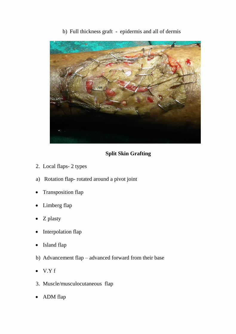
b) Full thickness graft - epidermis and all of dermis
Split Skin Grafting
2. Local flaps- 2 types
a) Rotation flap- rotated around a pivot joint
Transposition flap
Limberg flap
Z plasty
Interpolation flap
Island flap
b) Advancement flap – advanced forward from their base
V.Y f
3. Muscle/musculocutaneous flap
ADM flap

Abductor hallucis brevis flap
FOB flap
4. Faciocutaneous flap
Lateral Calcaneal artery FC flap
Plantar flap
RECONSTRUCTIVE ARTERIAL SURGERY:
It should be considered only after measuring the ankle systolic pressure
by Doppler flow meter. If an ankle pressure is ˂ 2/3 of the arm pressure,
there will be large vessel disease where reconstructive arterial surgery for
limb salvage is worth trying.
The surgery carried out will be depending on the extent of arterial
occlusion which can be find out by arteriogram.
INDICATIONS OF PERIPHERAL VASCULAR SURGERY IN THE
DIABETIC FOOT- Nocturnal pain, Rest pain, Foot ulcers not responding
to treatment, Infections not responding to treatment, Incipient gangrene,
Severe disabling intermittent claudication.
In a patient with arterial insufficiency arterial surgery should be
considered in the following- Claudication which significantly interferes
with work, Rest pain and an area of gangrene which doesn’t heal or can t
be removed surgically, treated with success by a local foot operation.

NON-OPERATIVE CORRECTION OF SELECTED ARTERIAL
BLOCKAGES: ANGIOGRAPHY
Transluminal balloon dilatation is done by means of a small
balloon catheter which is carefully introduced into site of stenosis and
distended under fluoroscopy, thereby relieving the obstruction.
MINOR AMPUTATIONS:
The principles regarding local amputations are:
1. Infected areas should be widely opened
2. Tourniquet should be avoided.
3. No place for small incisions, as if the infections usually will be deep
which requires wide incisions and excision of devitalized tissues.
4. The other foot must be taken care of because this may be the only one the
patient will have.
VARIOUS TYPES OF LOCAL AMPUTATION
1. Transphalangeal amputation of a toe
2. Amputation of a single toe and Ray amputation
3. Transmetatarsal amputation
4. Syme’s amputation
TRANSPHALANGEAL AMPUTATION OF A TOE:

This is the most commonly performed amputation. If there is
evidence of arterial insufficiency, adequate collateral circulation as
shown by a venous filling time of 20 sec or less is necessary. The lesion
must be in the distal 1/3 of the toe, leaving reasonably healthy skin at the
time of incision.There should be no dependent redness of the proximal
part of the toe and all cellulitis and lymphangitis should have cleared
prior to operation.
RAY AMPUTATION
This operation removes one toe and distal half metatarsal shaft
including the head through a racquet shaped incision, this leaves a
residual space which cannot be surgically closed and healed by secondary
intention with scar formation which eventually pulls the adjacent
metatarsal together. The adjacent joint capsules are avascular structures
which further delay healing, the operation is therefore reserved for those
patients with quite good collateral circulation. This procedure is more
useful in the neuropathic rather than the ischemic foot and hence mainly
done for ulcers under the first or fifth metatarsal heads or beneath one of
the other metatarsal head
TRANS METATARSAL AMPUTATION

Transmetatarsal amputation may have to be done if more than
one toe is involved or there is persistent recurrent plantar ulcer. In this,
one removes all of the toes and metatarsal heads, using a plantar flap
resembling a Turkish slipper for closure.
Infection must be controlled and the skin of the dorsum and the
sole of the foot must be healthy. It may take upto 3 weeks of care in the
hospital to prepare the patient for the operation. Pulses need not to be
present in the foot but the collateral with a venous filling time of less than
25 seconds and absence of dependent redness at the level of incision.
SYME ˈS AMPUTATION
This is indicated where more than one toe is involved, sepsis is
confined to distal half of the foot and the large vessel disease is absent.
The role of this amputation is diabetic foot however still remains
controversial. The main reason for its advocacy is that the patient will be
able to walk on his own leg (feeling of earth) and without shortening of
his limb.
BELOW KNEE AMPUTATION:
We select the below knee level if the patient is expected to use a
prosthesis, if the area of gangrene is below the ankle with a demarcation

at or below the ankle level. The amputation is usually accomplished
slightly below the mid lower leg level using short and equal anterior or
posterior flaps, dividing the fibula one inch higher that the tibia, and the
tibia at a suitable level to permit easy closure
ABOVE KNEE AMPUTATION
The operations most commonly performed are:
1. Through knee disarticulation
2. Supracondylar articulation
3. Mid – thigh amputation
The first two operations are likely to heal well due to presence of
collateral vessels in the area of the knee joint and the subcutaneous tissue.
Both operations involve division of only small amount of muscle tissues
which has the practical advantage of reduced anaerobic sepsis. Both have
a disadvantage of difficulty in fitting a proper prosthesis. Of the three the
supracondylar amputation offers the ideal stump.
RECENT TRENDS
Vacuum assisted closure

Clean, non- healing deep cavity wounds may respond to repeated
treatments by application of negative pressure under an occlusive wound
dressing (VAC).
Vaccum assisted closure
Hydrotherapy
Intractable, infected, cavity wounds so metimes improve with
hydrotherapy using saline pulse lavage under pressure.
Skin grafts
The autologous skin graft is the standard criterion for viable
coverage of the partial thickness wound. The graft could be harvested
under LA as an outpatient procedure. Meshing the graft allows wider
coverage and promotes drainage of serum and blood.

Tissue cultured skin substitutes
Dermagraft: is a cryopreserved human fibroblast- derived
dermal substitute produced by seeding neonatal foreskin fibroblasts onto
a bioabsorbable polyglactin mesh scaffold. It is used for managing full
thickness chronic diabetic foot ulcers, which is not appropriate for
infected ulcers, those involving bone or tendon or those having sinus
tracts.
Apligraf (Organogenesis): is a living, bilayered human skin
substitute. It is not used for infected ulcers, with involvement of tendon or
bone or those having sinus tracts.
Surgical wound closure
Delayed primary closure of a chronic wound requires well-
vascularized clean tissues and tension free apposition. It usually requires
undermining and mobilization of adjacent tissue planes by creation of
skin flaps or myocutaneous flaps.
Hyperbaric oxygen treatment:
This is rarely used, not a substitute for revascularization. In the
presence of intractable wound and associated non correctible ischemic
arterial disease, the hyperbaric oxygen therapy may be beneficial
REHABILITATION

The following are done in our study:
a) Protective shoes – protect from further trauma and helps in effective
weight bearing.
b) Crutches – after amputation, gait training is given with this
c) Prosthesis – Absolute minimum prosthetic requirement for the AKA is
a wheel chair or a set of walking crutches.
FOLLOW-UP
Health education regarding the foot care and diabetic control were given
at the time of discharge. All the patients were asked to review after 1
week during the follow up, diabetic status was checked and detailed
general and local examination was done.
PREVENTION (DIABETIC FOOT CARE)
Care of foot takes place at three level:
1. The patient must take routine measures to take care of his foot
2. Early lesions require expert care either from a podiatrist or from an
experienced doctor in the care of the patient.
3. Advanced lesions require specialized care.
The best way is to make use of multidisciplinary professionals who
are committed to limb salvage. Team members involved are- Physician,

Nurse, Endocrinologist, Podiatrist, Neurologist, Vascular surgeon,
Orthopedist, Physiotherapist, Social workers, Home care nurse.
Patient education is important in case of diabetic foot.
The advice given to patient include (25)
Do not smoke
Inspect the feet daily for blisters, cuts and scratches. With the use of a
mirror can see the bottom of feet, also check in between the toes.
Wash feet daily, dry carefully, especially between the toes.
Avoid extreme temperature. Test water with hand, elbow or
thermometer before bathing
If feet feel cold at night wear socks. Do not apply hot bottle water or
heating pads.
Do not walk on hot surfaces.
Do not walk barefoot or soak feet.
Do not use chemical agents for removal of corns, calluses
Inspect the feet daily for foreign objects, nail points, rough areas
Do not wear sandals with thongs between the toes
In winter season, wear wool socks and protective gear such as fleece
lined boots.

MATERIALS AND METHODS
TITLE OF THE STUDY: A STUDY OF PATHOPHYSIOLOGY,
MANAGEMENT AND FACTORS INFLUENCING DIABETIC FOOT
ULCERS AMONG DIABETIC PATIENTS.
STUDY DESIGN : Observational study
METHODOLOGY
Sample size –
n = (Zα)2pq / d
2
Zα2 =1.96
Where p- prevalence and q = 100 – p;
d = maximum allowable limit of error over prevalence; i.e. (20% of p)
p = 29 % ; q =71% and d = 20% of p = 5.8
Hence sample size n = 250
A observational study was conducted on 250 diabetic foot ulcer patients
attending surgical op/ emergency department (1 B) in CMCH over a
period of 1 year, from July 2016- July 2017.

STUDY POPULATION
INCLUSION CRITERIA
250 patients with age > 13 years presenting to surgical op/ emergency
department (1 B) with diabetic foot ulcer
EXCLUSION CRITERIA
Patients below 13 years
Pregnant females
Psychiatric patients.
Diabetic patients who have ulcer other than diabetic ulcer for example
traumatic ulcer, venous ulcer.
METHODS OF COLLECTION OF DATA
After registration and admission , detailed clinical history of patient
taken, followed by a detailed clinical examination.
The name, age, sex, address, profession of the patient is noted. The
symptoms and clinical features are recorded in chronological order.
Clinical history includes the following points- Known case of diabetic or
not, Duration of diabetes, regarding the treatment received if any, Family
history of diabetes, any history of injury, Local symptoms such as

swelling, pain, wound, discoloration andPersonal habits such as smoking
and alcoholism.
General examination of the patient includes all vitals and other system
examination.
Clinical features of neuropathic foot are- Warm with intact pulses,
Diminished sensations, callus, Ulceration, Sepsis, Local necrosis, Edema,
Charcot’s joints.
Clinical features of ischaemic or neuro-ishaemic foot are Cold with
absent pulse, diminished sensations, Ulceration and Necrosis or gangrene
In the examination of the feet, the following points are to be noted- Types
of lesion and extent, evidence of any predisposing factors, any changes
suggestive of neuropathy or vascular involvement
The neurological status of the lower limb assessed to rule out diabetic
neuropathy. All the sensations, power, reflexes, and neurological deficit
were noted.
Vasculopathy of the limb was found by assessing Colour of limb: normal,
pale, purpule, black, local temperature: normal or cold and the pulsations
of the lower limb: dorsalis pedis, posterior tibial, popliteal and femoral
artery.

OUTCOME
Enhancing the importance of patient’s knowledge in self-care practice
and regular diabetic foot evaluation.
Reduce the morbidity of patients with diabetic foot ulcer and enhance
early healing of ulcer.
Early rehabilitation of the patients with diabetic foot ulcer.

OBSERVATIONS AND RESULTS
An analysis of 250 cases of diabetic foot was done. These cases were
treated in different surgical units in the Department of Surgery,
Coimbatore Medical College Hospital from July 2016 to June 2017.
In our study 250 patients of diabetic foot lesions were studied. In 35
patients only incision and drainage and fasciotomy was done, healing of
wound occurred without complications. In 125 patients debridement was
done as the definite treatment, as a groundwork to amputation. Skin
grafting was well thought-out in 40 patients once the wound was clean
and granulating.
In our study amputation rate was 20%. Out of these, patients underwent
Above Knee or Below Knee Amputations or minor amputations. In 40
patients major amputations like Below Knee and Above Knee
amputations done. 15 Patients had wound infection and suturing gaping.
For these patients with wound gaping secondary suturing was done. In 10
patients, Above Knee amputations was done, 5 patients stump was closed
primarily and in 5 patients Gullettine amputation was carried out. A total
of 10 patients died because of various complications of diabetes during
the line of treatment.

1.Age distribution
Table :-1 Showing the Age distribution
Age (years) No. of patients Percentage (%)
0-10 - -
11-20 2 1
21-30 5 2
31-40 25 10
41-50 55 22
51-60 80 32
61-70 66 26
71-80 15 6
81-90 2 1
Total 250 100
Of 250 cases studied, youngest patient was 19 years and oldest was 84
years of age. Highest number of cases was found in the age group 51-60
years (31%) followed by 61- 70 years (27%). Maximum number of
diabetic foot i.e 80% are between the age group of 41-70 years.
Age distribution
0-10
11-20
21-30
31-40
41-50
51-60
61-70

2. Sex
Table:- 2 Showing the sex distribution
Sex No. of cases studied Percentage(%)
Male 160 64
Female 90 36
Of the 250 cases studied in this series, 160 (64%) cases were male and
90(36%)
Showing the Sex distribution
MALE
FEMALE

3.Occupation
Table 3:- Occupation
Sl.No. Profession of patient No. of cases Percentage
1 Farmers 135 54
2 Business 25 10
3 Office Employee 10 4
4 Manual Labourer 20 8
5 Drivers 10 4
6 Housewife 50 20
Total 250 100
In this study maximum patients were farmers (54%) and minimum
patients were drivers and office employee (4%).
Occupation
Farmers
Business
Office Employee
Manual Labourer
Driver
House Wife

4.Type of diabetes
Table:- 4- Type of diabetes
Type of diabetes No. of patients %
I 35 14
II 215 86
In this study 35 patients had type I diabetes, remaining 215 patients had
type II diabetes.
Type of diabetes
Type I
Type II

5.Incidence of Trauma
Chart 5:- Incidence of Trauma
In this study 64 cases exposed a history of some kind of trauma
before the onset of lesion.
6. Mode of presentation
Table 6 :- Showing incidence of various types of lesions.
Mode of presentation No. of cases %
Ulcer 120 48
Gangrene 50 20
Abscess 15 6
Cellulitis 65 26
Showing the incidence of trauma
YES
NO

The different types of lesions seen including ulcer, cellulitis, abscess and
gangrene. Most of the patients present with more than one lesions. Only
major lesion is considered here. Ulcer was the major lesion seen and is
present in 120 patients. 15 patients presents as a abscess was the least
common lesion.
In above patients x-ray of 30 cases showed changes of Osteomyelitis. 15
patients present with Charcot’s joint.
Doppler studied in 10 patient showed atherosclerotic changes with low
volume flow in anterior and posterior tibial arteries.
7. Duration of diabetes
Duration is not accurately known, as few patients were unaware of
being diabetics and were newly diagnosed as suffering from diabetes on
admission with complaints of non- healing ulcer or other complications of
Diabetes.
Mode of presentation
ULCER
GANGRENE
ABSCESS
CELLULITIS

Table 7 :- Showing duration of Diabetes
Duration of diabetes in years No. of patients %
0-1 45 18
2-5 30 12
6-10 80 32
11-15 40 16
16-20 35 14
˃20 20 8
Total 250 100
Showing duration of diabetes:-
In our study 18% presented with duration less than or equal to 1 year.
Most of these patients were diagnosed post admission. Only 20 patients
had diabetes of more than 20 years. Maximum 80 patients in our study
were diabetes of 6- 10 years.
In the present series 10 patients were detected as a diabetic at the time of
admission.
0
10
20
30
40

8.Culture and Sensitivity
Table 8 :- Culture and Sensitivity
% Bacterial No. of cases
40 Staphylococcal aureus 100
12 Pseudomonas 30
18 Klebsiella 45
14 E- coli 35
10 Proteus 25
6 Non-hemolytic streptococci 15
In our study majority of septic lesions yielded Staphylococcus aureus in
about 40% on culture of pus. Other organisms were isolated are
Pseudomonas 12%, Klebsiella 18%, E-coli 14%, Proteus 10%.
Bacterial
Staph aureus
Pseudomonas
Klebsiella
E-coli
Proteus
NH Strep.

9. Sensitivity report
Table 9:- Showing Antibiotic Sensitivity according to Culture.
Antibiotics % of patients
Imipenam 45
Ceftazidime 25
Amoxycillin and Clavulanic acid 45
Amikacin 25
Cefotaxime 10
Ciprofloxacin 25
Ampicillin 5
Vancomycin 30
Imipenem and Amoxicillin & Clavulanic acid were sensitive against most
of the organisms as they cover a wide range of organisms.
Sensitivity report
Imipenam
Ceftazidime
Amoxy & Clavulanic
Amikacin
Cefotaxime
Ciprofloxacin
Ampiciliin

10. Neuropathic lesions
Table 10:- Neuropathic lesions
Neuropathic lesions No. of cases %
Yes 130 52
No 120 48
In the present study 130 cases were found to have neuropathy. Patients
with neuropathy varied from 35-80 years. Majority had history of
diabetes more than 5 years. This shows that peripheral neuropathy is
common in long standing diabetic patients. 50 patients had gangrene.
Neuropathic lesion
YES
NO
11. Duration of hospitalization
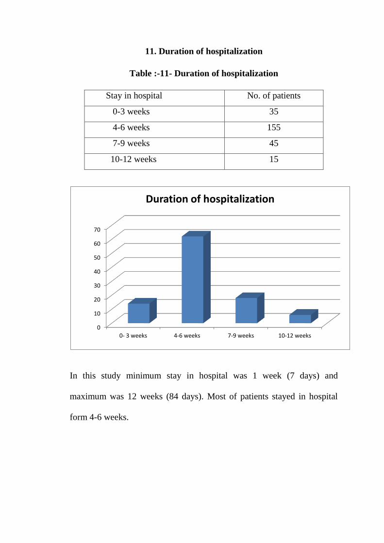
Table :-11- Duration of hospitalization
Stay in hospital No. of patients
0-3 weeks 35
4-6 weeks 155
7-9 weeks 45
10-12 weeks 15
In this study minimum stay in hospital was 1 week (7 days) and
maximum was 12 weeks (84 days). Most of patients stayed in hospital
form 4-6 weeks.
0
10
20
30
40
50
60
70
0- 3 weeks 4-6 weeks 7-9 weeks 10-12 weeks
Duration of hospitalization

12. Investigations
Table 12:- Showing blood sugar level at the time of admission
Blood sugar
Random blood sugar Fasting blood sugar
Normal range ˃normal range Normal range ˃normal range
75 175 95 155
In this study at the time of admission 175 patients had RBS more than
normal and 75 patients had RBS within normal range. While FBS at the
time of admission in the same age group more than normal in 155
patients and within normal range in 95 patients.
Glycaemic Control
Normal range
˃Normal range
Normal range
˃Normal range

13.Treatment
Table 13:- Treatment
Treatment No. of cases %
Wound debridement, slough exicision, regular dressing 125 50
SSG 40 16
I & D, Fasciotomy 35 14
Minor amputation 10 4
BKA 30 12
AKA 10 4
In this series 125 cases were managed by daily dressing and wound
debridement, and slough excision. 40 patients were treated with SSG, 35
patients under went Incision & Drainage for abscess and some of them
fasciotomy. Minor amputation was done in 10 cases. BKA was done in
30 cases and AKA in 10 cases. In most of the cases, limb was salvaged
by conservative treatment and minor amputation.
Treatment Wound debridement, slough exicision, regular dressing
SSG
I & D, Fasciotomy
Minor amputation
BKA
AKA

14. Outcome
Table 14:- Outcome
Result No. of cases %
Recovery 240 96
Death 10 4
In the present study 240 patients recovered from their lesion after
treatment while remaining 10 patients died due to various complications.
15. Atherosclerotic changes.
Table 15:- Atherosclerotic changes.
Result No. of cases %
Atherosclerotic changes present 160 64
No atherosclerotic changes 90 36
Outcome
RECOVERY
DEATH

Out of 250 patients with diabetic ulcers 160 patients showed
atherosclerotic changes in major arteries of lower limb which was proven
categorically by an arterial Doppler color examination.
Atherosclerotic changes
Present
Absent

DISCUSSION
Diabetes is a common problem. It affects about 16 million people
of all ages and is a major cause of end stage renal disease, blindness and
peripheral neuropathies. The disease is chronic and affects the
metabolism of carbohydrate, protein, fat, water and electrolytes.
Many diabetic ulcers are ignored because they may produce few
symptoms and their importance is not valued by patients and secondly it
falls between specialities. Not being entirely in the surgeon or physician
domain. Reduction in the occurrence and prompt treatment of foot
ulceration would ultimately lead to drop in the amputation rate in diabetic
patients.
Without doubt, the problem of foot infection in diabetic patient
population is costly to both the patient and society. As a result, health
care expenditure and medical hard work must be directed education and
prevention, early detection and prompt therapy of food infection.
Total 250 cases of diabetic foot were included in this study.
Following is the result of my study conducted at our hospital from
( duration of study)
The analysis of this study is as follows.

AGE-
Prevalence of diabetes is greater in persons over the age of 50 years.
Pedal infection is a devastating and severe complication of diabetes seen
often is elderly patients.
In our study of 250 patients age was ranging from 19 years to 84
years. It was found that age group 51-60 years, had the highest number of
80 patients.
SEX
Most of the diseases have the male predominance, this is same with
diabetic foot also. In our series 64% patients were males and 36%
patients were females.
The following may be the reason for the male predominance are exposed
to the external environment more than females hence they are likely
candidates for injury leading to ulcer formation. Alcoholism and smoking
habits are more common in males and this may be an important factor.
Barefoot walking is more common in India this also contributes to the
foot ulceration.
OCCUPATION
As seen in our study, farmers had more incidence of diabetic foot
lesions because of unawareness of having Diabetes , lack of foot care and

poor management of diabetes. It is evident that the surgical complications
of patients whose profession exposes them to the risk of trauma and
injuries making the vulnerable to the complications of diabetic foot.
TYPE OF DIABETES
As per literature foot ulcers have been more in the patient with NIDDM
(86%) as per our study. NIDDM age group is being elder and foot
complications are more in these patients.
History of trauma
64 cases in this series had history of trauma, before the onset of foot
lesion.
In majority of the cases of surgical complications of diabetes, some kind
of trauma is the beginning of the problem. This is because of three
factors, they are ischemia:- due to ischemia the parts, which are
traumatized will be underperfused, thus impairing the healing process.
Neuropathy :- Result in loss of sensation and the patient will be unaware
of the injury and neglects it.
Hyperglycemia :- acts as a perfect medium for the multiplying organisms.
Neuropathy which is seen more than 50% of diabetes of long standing
duration is considered at to be the single most major course of ulceration.

In present study neuropathy changes seen in 52 cases, ischemic
complications was noted in 20 cases and infective complication of foot
noted in all cases.
TYPE OF LESION:-
In our study of foot lesions are Cellulitis, Abscess, Ulcer and Gangrene.
In some patients there was more than one lesion. Ulcer (48%), Cellulitis
(26%), Gangrene (20%), Abscess (6%). Ulcer was the commonest
presentation.
DIABETIC STATUS:-
In our study 96% patients were known diabetics. Other 10 patients were
diagnosed after hospital admission to hospital.
DURATION OF DIABETES:-
In our study mean duration of diabetes mellitus is 12 years.
CULTURE & SENSITIVITY:-
In our study pus was sent for culture and sensitivity in 120 patients. In
most of patientˈs more than one organism were grown on culture.
In our study staphylococcus aureus (40%), E Coli (14%), Klebsiella
(18%), Proteus (10%), Pseudomonas species (12%) were commonly
isolated organisms.

Imipenem, Meropenem and Cefepime were the most effective agents
against gram negative organisms. Vancomycin was the most efficient
against gram positive organisms.
In our study 30 patients had osteomyelitis, most of the patients had
osteomyelitis of phalanx and metatarsals, most of the osteomyelitis of
foot begin as chronic perforating ulcers. If the ulcer is ignored or
inappropriately cared bacterial invasion may progress at the base of the
lesion by spreading along the fascial planes or by perforating.
Neuropathy
Neuropathy foot has got three main complications neuropathic ulcer,
neuropathic joint (Charcot’s ) and neuropathic edema.
Neuropathy consists of three main components – sensory, motor and
autonomic.
In our series 52% patients had neuropathy. It was diagnosed by weakness
of small muscles, foot deformity, loss of sensation, loss of sweating,
formation of new pressure points, callosities and eventually ulcer.
In autonomic neuropathy skin becomes dry as a result of loss of sweating.
There will be cracking of skin and impaired defense against infection. In
case of sensory neuropathy there will be loss of sensation which
predispose to the trauma and ulcer formation.

SPREAD OF INFECTION
After trauma there will be devitilisation of the tissue, if the infection
supervenes there is rapid formation of inflammatory exudates, especially
in the deeper underlying tissue which leads increased pressure in the
compartment resulting in obstruction to the blood supply, eventually
resulting in gangrene and uncontrolled infection.
Due to vasculopathy and neuropathy, foot infections are deserted, there is
spread of the infection from distal to the proximal part.
Factors which are responsible for impaired wound healing in diabetes are
Decreased vascularity
Reduced resistance to infection
Retained devitilised tissue due to vasculopathy and neuropathy
Oedma due to inflammation
Poor nourishment
Repeated trauma
Co- relation of atherosclerosis and diabetic ulcers
As seen in various studies. Diabetes leads to microangiopathy
and macroangiopathy with resultant atherosclerosis of blood vessels
leading to decreased vascularity to the distal lower limb causing
ulcerations, gangrenes etc.
Our study also corroborates these findings by showing high incidence
and atherosclerosis in the patients with diabetic ulcers which can be
considered as one of the causes.
DURATION OF HOSPITALIZATION
In this study minimum duration in hospital was 7 days (1week) and
maximum was 84 days (12 weeks). Most of patients stay in hospital for 4-
6 week.
This long duration of hospitalization can be explained by the refractory to
patients diminished resistance of body due to uncontrolled sugar level,
resistance of the organism to the antibiotic therapy, poor nutrition,
advanced age.
INVESTIGATIONS
The study indicated that at the time of admission in hospital, RBS level
of 70 patients were above the normal level and FBS level of patients done
that showed 155 patients had more than normal range. It is recommended
that diabetic patients present with diabetic foot lesions in uncontrollable
blood sugar level. 75 patients showing normal blood sugar level because
they are regularly taking anti-diabetic treatment.

TREATMENT
In our study diabetic foot management is a difficult task because of
Illiteracy and ignorance
Walking barefoot and poor socioeconomic status.
Taking treatment in earlier stages by quacks because of fear of
amputation.
Careless removal of nails and unhygienic foot care.
Most of the cases in our study were referred to us in a difficult stage with
serious infection and associated complications.
Before starting treatment every patient must be assessed to decide the
relative importance of predisposing factors – neuropathy or vascular
disease. In absence of severe ischemia, conservative medical and surgical
management give good results. In presence of ischemia unless the limb is
salvaged by direct arterial surgery, we may have to consider major
amputations.
MEDICAL MANAGEMENT
For successful management it is important to assess the diabetic status,
severity of infection and general nourishment of the patient.
Control of Diabetes mellitus by:-
Diabetic diet

Oral hypoglycemic drugs
Insulin therapy
All our patients were advised diabetic diet with low cholesterol and high
PUFA.
All patients were shifted to crystalline insulin therapy. According to the
blood sugar level dosage of the insulin was adjusted. In case of
Ketoacidosis, planned aggressive treatment was necessary because this is
life threatening complication, where severity of infection is more and
there is defective neutrophil function.
Principles of the treatment are:
Correction of fluid and electrolyte imbalance
Reversal of acidosis and ketosis with crystalline insulin therapy.
Dosage of insulin was required more in cases of ketoacidosis, severe
infection. After correction of ketoacidosis and control of infection, the
dosage of insulin required was reduced.
At the time of discharge 100 patients were shifted to oral hypoglycemic,
115 patients to Lente insulin and the rest were continued with the
crystalline insulin.
Control of infection:-

Control of infection was done by careful debridement and antibiotic
therapy. The role of antibiotics in foot infection is to limit the spread of
infection.
Indications of antibiotic therapy were:-
Limiting Cellilitis and spreading infection
Prevent secondary infection
Prophylactic therapy before surgery.
In our study most of the patients were treated with multidrug regime.
In our study we have used higher antibiotics because it is known to grow
resistant organisms in diabetic foot infection.
Staphylococcus was the commonest organism grown, which was
sensitive to Cefotaxime, Ampicillin and Gentamycin in 80-90% of cases.
We have used combination antibiotic therapy. Combination antibiotic
therapy is advised by Brodsky J.W.et al & L.Bhasker Reddy et al .
Brodsky J.W. Advised the following combinations:-
-Ticarcillin + Clavulanic acid
-Vancomycin + Metronidazole
-Third generation cephalosporin + Metronidazole
L. Bhasker Reddy et al advised the following combination:-
Cefotaxime + Metronidazole
According to the availability of drugs we could use broad spectrum
antibiotics like Ciprofloxacin, Cefotaxime, in combination with
Metronidazole and Gentamycin.
Ampicillin, penicillin, erythromycin, tetracycline were used in some of
the patients.
In the present series, 125 cases were treated by slough excision, 40 with
skin graft, 35 by fasciotomy and I&D. Below Knee Amputation was done
in 30 patients and Above Knee
Amputation were done in 10 cases. Minor amputations are done in 10
cases. Proper control of diabetes is very important in diabetic foot
management. Fasting and PPBS estimations were well under control.
Urine sugar estimation was done thrice daily.
Infection was treated with broad spectrum antibiotics. Patients were
educated about care of foot and Pentoxyphylline was administered to in
patients with ischemic lesions.
In our study amputation rate is 20%, that is lower when compared to
standard studies. This could be due to better education of patient, better
glycemic control, proper care of foot, proper use of antibiotics, extensive

debridement and regular dressing. After amputation, wound healed well.
The patients were referred to rehabilitation center for prosthesis.
RESULTS
Out of 250 patients studied, 240 recovered and discharged from hospital
and 10 patients died during the course of treatment due to various
diabetes related complications like septicemia. Ketoacidosis, uremia and
respiratory symptoms.

SUMMARY
Foot ulcers are one of the major complications of diabetes; they have a
poor tendency to heal, which may result in long stay in hospital for
treatment. The foot in diabetic patient is the cross road of several of
several pathological processes, in which almost all components of lower
extremity are involved- skin, subcutaneous tissue, muscles, bones, joints,
blood vessels and nerves. An understanding of the development of the
complications and application of preventive and management strategies
will reduce the complications of diabetic foot. Patients who are diabetic
should take proper care of their feet so that they will not suffer from the
consequences. They should take proper medical care at the earliest once
any trauma occurs, so that the chain of events could be halted.
With proper care by the attending surgeon which includes regular
dressing, wound debridement, proper antibiotics the outcome can be
favourable and avoid the unwanted surgeries like amputations.
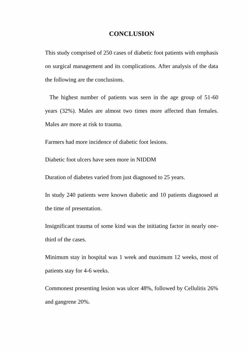
CONCLUSION
This study comprised of 250 cases of diabetic foot patients with emphasis
on surgical management and its complications. After analysis of the data
the following are the conclusions.
The highest number of patients was seen in the age group of 51-60
years (32%). Males are almost two times more affected than females.
Males are more at risk to trauma.
Farmers had more incidence of diabetic foot lesions.
Diabetic foot ulcers have seen more in NIDDM
Duration of diabetes varied from just diagnosed to 25 years.
In study 240 patients were known diabetic and 10 patients diagnosed at
the time of presentation.
Insignificant trauma of some kind was the initiating factor in nearly one-
third of the cases.
Minimum stay in hospital was 1 week and maximum 12 weeks, most of
patients stay for 4-6 weeks.
Commonest presenting lesion was ulcer 48%, followed by Cellulitis 26%
and gangrene 20%.

After 5 to 6 years of diabetes most of patients present with neuropathic
lesions and they are in 35-80 years age group some of them develops
gangrene.
Staphylococcus aureus is a commonest organism causing infection.
Atherosclerotic changes may lead to formation of ulceration of foot in
diabetic patients.
Conservative treatment consisting of control of diabetes with plain /
Lente insulin along with appropriate oral /IV antibiotics was effective in
some cases.
Wound debridement, slough excision followed by dressing with Povidine
iodine/ Magnesium sulphate/ Metrogyl/ Collagenase dressing resulted in
healing in some cases. Split skin graft, disarticulations, Transmetatarsal
amputation, Below Knee Amputation and Above Knee Amputation were
other modes of treatment.
240 patients cured and 10 patients died during course of treatment.

BIBLIOGRAPHY
1. The History of Diabetes. Article from Canadian Diabetes Association
(Google research).
2. Boulton A.J The Diabetic foot; A Global view
3. Micheal E Edmonds and Alethea VM Foster, Managing the Diabetic
foot, 2nd
edition. Page 23.
4. Harrisonˈs Principles of Internal Medicine, 19th edition.
5. Reiber G.E; Epidemiology of foot ulcers and Amputations in Diabetic
foot , 6th edition.
6. Peter C Williams, Roger Warwick. Grayˈs Anatomy, 36th
edition.
7. Gayle R Benjamin AL, Gary NG. The burden of diabetic foot ulcers. The
American Journal of Surgery 2016
8. Tooke JE, Microvascular hemodynamics in Diabetes mellitus, Clin sci
1986;70: 119-125.
9. Mac Rury SM, Lowe GDO, Blood rheology in DM. Diabet med
1990;7:285-291.
10. Pyorala K, Lasko M, Diabetes and atherosclerosis, An epidemiologic
view, Diabet Methob rev 1987; 3:463-524.
11. Thomas P.K metabolic Neuropathy, London: JR Coll Phy 1973; 7:154.
12. Harati Diabetic peripheral neuropathies. Ann Inter Med 1987;107:546.

13. Delbring L, Ctectelco G, Fowler C. The etiology of Diabetic neuropathy
ulceration, Br J Surgery 1985; 72:1-6
14. Harati. Y frequently asked questions about peripheral neuropathies.
Neuropathic Clinic 1992 Aug; 10(3): 783-801.
15. Sigimura K, Duck PJ, Multifocal fibre loss in proximal sciatic nerve in
symmetrical distal diabetic neuropathy, Neurol Sci 1982; 53:501.
16. Farad JV, Sotalo J. Low serum levels of nerve growth factor in diabetic
neuropathy. Acta Neurol Scand 1990; 81:402-406.
17. Shah SV, Wallen JD, Eilen SD. Chemiluminescene and superoxide anion
production by leukocyte from diabetic patients. J Clin endocrine metab
1983;57:402-409.
18. Salvapandian AJ , Infections of the foot. In: Disorders of the foot,Iahss
MH Philadelphia; WB Saunders 1982;401.
19. Donovan DL, File TM, team approach In the management of diabetic
foot infections. Journals of foot sugery
20. George WL, Other infection of skin, soft tissue and muscle. In: anaerobic
infections in humans, Gold SM, San Diego; Academic press 1989.
21. Siebert WT, Dewans , William JW Jr. Puedomonas puncture wound
osteomyelitis in adult. Amaerican journal medical sciences.
22. Bessman AN, Sapico FL, Tobatabai MF, Montgomerie JZ.Persistence of
polymicrobial abscess in poorly controlled diabetic host. Diabetes 1986;
36:448.

23. Leslie CA, Sapico FL Ginunas VJ, Adkins RH. Randomized clinical trial
of tropical hyperbaric oxygen for treatment of diabetic foot ulcers.
Diabetic care 1988;32:717
24. Moore RD, Smith CR, Lipsky JJ. Risk factors for nephrotoxicity in
patient treated with aminoglycosides. Ann internal medicine
1984;100:352.
25. Bessman AN,Kasim S, Managing foot infections in the older diabetic
patients Geriatrics 1985;40:54.
26. Rolfe RD, Finegold SM. Comparative in vitro activity of new beta-lactum
antibiotics against anaerobic bacteria. Antimicrob Ag chemother
1981;20:600.
27. Lipsky BA. Osteomyelitis of the foot in the diabetic patients. Clin infect
dis 1997; 25: 1318-26.
28. Cavanagh PR, Ulbrechte JS,Caputo GM. New developments in the
biomechanics of the diabetic foot. Diab metab Res Rev 2000; 90:57-65.
29. Lipsky BA, Pecoraro RE Wheat LJ. The diabetic foot. Soft tissue and
bone infection. Infect Dis Clin North Am 1990;4:409-32.
30. ABC of Diabetes mellitus , BMJ 2003; 326,977-979.
31. Levin ME:Pathogenesis and general management of foot lesions, 6th
edition,2001,pp 219-260.
32. Ramani,A Kundaje G.N: Etiology of diabetic foot ulceration, JAPI 1990;
Feb:38:843-845.

33. Caballero e, Frykberg RG. Diabetic foot infections. J foot Ankle Surg
1998;37:248-55.
34. Quari FA, Akbar D, Diabetic foot presentation and treatment, Saudi med
J. 2000 May ;21(5):443-6.
35. Stone HH, Martin J.D. Synergistic Necrotizing cellulitis . Ann.Surg
1972:175;702-711

ANNEXURE
PROFORMA
Case No:-
Name:- Age:- Sex:- M/F
Occupation:-
IP No:- DOA:- DOD:-
Address:-
Socio economic status:-
Chief complaints:-
History of present illness:-
Polyuria
Polydypsia, Polyphagia
Weakness
Burning sensation of palms and sole
Tingling /numbness
Loss of appetite
Loss of weight
Ulceration
Gangrene
Neuropathy
Poor wound healing

Recurrent infections
Fever
Claudication
Duration
Distance
Intermittent pain, Rest pain
Any other associated history
Diabetic history:
Type of diabetes:- NIDDM/IDDM
Duration of Diabetes
Controlled/ uncontrolled
Duration of control and type of control
-Good
-Poor
-Bad
Method of treatment /control
-Diet
- Oral Hypoglycemic Agent: Dose
- Insulin: Dose
-Operative : Incision/Drainage/Excision/Amputation

Past history
- Visual disturbance
- Septic lesions - Ulcerations / Gangrene / Carbuncle / Furuncle / Abscess
- CVS - Angina / Myocardial infarction
- CNS: TIA / Peripheral neuropathy
- Recurrent Systemic infections
- H/O previous hospital admissions
Number
Duration
Treatment
Cured/ Not cured
Family history :- Family members suffering from DM- Y/N
Personal history:-
- Diet: Veg/Non veg
- Appetite: Normal/Increased/Decreased
- Micturition: Normal/Polyuria
- bowel: Regular/Irregular
- Smoker: Y/N
- Alcoholic consumption :Y/N
- History of prolong intake of drug: Y/N: Duration

Menstrual history:- in Females
GENERAL PHYSICAL EXAMINATION:-
- Built : Well / Moderate / Poor
- Nourishment: Well / Moderate / Poor
- Pallor / Icterus / Clubbing / Cyanosis / Oedema / Lymphadenopathy
- Vitals :-
Pulse: /min BP: RR: Temp:
SYSTEMIC EXAMINATION
Nervous system
Central:
Peripheral:
-Motor
- Sensory
- Reflexes
Respiratory system
Cardiovascular system
Abdomen examination

LOCAL EXAMINATION
Site
Toe, Sole, Heel, Web space
Other foot lesions- Dorsum/ Lateral borders
Mode of presentation:
- Ulcer
- Cellulitis
- Blister
- Abscess
- Gangrene
Local temperature
Tenderness: Y/N
Peripheral Neuropathy:- Y/N
Sensation:- Touch / Vibration / Joint
Skin/ Nail changes
Presence/ Absence of callosities – Dry / Wet
Peripherl Pulses:
-Dorsalis pedis artery - Present / Weak / Absent
- Posterior tibial artery – Present / Weak / Absent

INVESTIGATIONS
Urine - Albumin / Sugar / Microscopy / Ketone bodies
Blood – Hb% / Total count / Differential count
FBS- PPBS-
Blood urea: Serum Creatinine:
Lipid profile
Serum electrolytes
X ray – Chest/Foot
ECG
Wound Swab for C&S
SPECIAL INVESTIGATIONS: Arterial Doppler
TREATMENT
- Diet
- Oral Hypoglycemic Agents: Dose
- Insulin: Dose
- Antibiotics
- Dressing : Glycerine Magnesium Sulphate/ Soframycin/ Betadine
- Supplementary treatment
- Preoperative treatment
- Anaesthesia used

- Surgical:-
I&D
Slough excision
Fascitomy
SSG
Amputation / RA / TMA
- Post operative treatment
- Discharge notes
- Date of discharge
- Condition of patient
- Nature of operation
- Advise given
Diet, Treatment, Change of Occupation, other advice.
FOLLOW UP:- General condition/ local condition/ Complications/
Recurrence of fresh lesions.

xg;g[jy; gotk;
vdJ rh;f;fiu nehahy; fhypy; g[z; ,Ug;gij kUj;Jth; \yk;
mwpe;J bfhz;nld;. me;j rh;f;fiu neha; g[z;zpy; cs;s
fpUkpia gw;wp mwpat[k; mjw;fhd itj;jpaj;ij gw;wpa[k;/ me;j
g[z;zhy; Vw;gLk; ghjpg;g[ gw;wpa[k; kUj;Jth; tpsf;fpdhh;. me;j
g[z;iz guhkhpg;g[ bra;tij gw;wpa[k; kUj;Jth; tpsf;fpdhh;. me;j
g[z;iz Rj;jk; bra;tJ gw;wpa[k; kUj;Jth; tpsf;fpdhh;. ,it
midj;ij gw;wpa[k; Muha;r;rp bra;tij gw;wp kUj;Jth;
tpsf;fpdhh;. ,it midj;Jk; mwpe;J KG kdJld; ,jw;F
rk;kjk; mspf;fpnwd;.
,g;gof;F

MASTER CHART
SL.
NO
IP/OP
NO
AGE SEX OCCUPATION STAY IN
HOSPITAL
(DAYS)
TYPE
OF
DM
DURATION OF
DM (YEARS)
MODE OF
PRESENTATION
NEUROPATHY ATHEROSCLEROTIC CHANGES
CULTURE RBS FBS TREATMENT OUTCOME
1 112871 43 M Farmer 65 II 1 U N Y S N N D Recover
2 22187 39 M M L 23 II 2 U N N P >N N D Recover
3 119181 53 F M L 40 II 8 C Y Y S >N >N SE Recover
4 133215 61 F H W 38 II 5 U Y Y K N N SSG Recover
5 144578 52 M Business 14 II 10 A N N S >N >N I&D Recover
6 146781 69 M Farmer 46 II 14 U Y Y Pr >N >N D Recover
7 29186 47 F M L 28 II 9 Month C N N S >N >N FASC Recover
8 30897 33 F Farmer 30 I 8 U Y Y K N N SSG Recover
9 143571 53 M Farmer 36 II 4 U N N S >N >N D Recover
10 31059 63 M Farmer 55 II 11 G Y Y P >N >N TMA Recover
11 28958 73 M Farmer 40 II >20 U N N E N N SE Recover
12 141098 43 M O E 36 II 1 G Y N S >N >N RA Recover
13 22154 64 F H W 25 II 12 C N Y K >N N D Death
14 140981 66 F H W 20 II >20 U Y N S >N >N D Recover
15 143450 49 M Farmer 28 II Ne D C N Y NHS >N >N D Recover
16 31456 57 M Farmer 62 II 9 U Y N E N N SSG Recover
17 31876 69 M Farmer 72 II 10 C Y Y P >N >N SE Recover
18 31487 61 M Farmer 27 II 10 G Y Y K >N >N AKA Recover
19 30958 58 M Business 12 II 8 U Y N S >N N D Recover
20 138901 19 M M L 32 I 1 U N N P N N D Recover
21 39341 64 F H W 46 II 8 G Y Y Pr >N >N BKA Recover
22 131408 53 F Farmer 42 II 11 U N Y S N N D Recover
23 130938 44 M Farmer 36 II 4 U N N P N N D Recover
24 31786 65 M Farmer 18 II 12 U Y Y E >N >N SE Recover
25 31945 67 M Farmer 29 II 18 C Y Y S N N FASC Recover
26 36098 64 M Farmer 52 II 16 U Y Y E >N >N SSG Recover
27 35798 79 F H W 25 II 19 A Y N S >N >N I&D Recover
28 110958 42 M Driver 22 I 3 Month A N Y Pr >N >N I&D Recover
29 101893 34 M Farmer 7 I 9 U N Y P >N N D Recover
30 33946 52 M Farmer 36 II Ne D G Y Y S >N >N BKA Recover
31 33914 75 F H W 56 II >20 U N N K >N >N SSG Recover

32 33046 62 F H W 42 II 15 C Y Y S N N SE Recover
33 109815 41 F Farmer 40 II 4 U N N NHS >N >N D Recover
34 108956 55 M Business 17 II 7 U Y Y K N N D Recover
35 33785 66 M Farmer 62 II 14 C Y Y S N N FASC Recover
36 109089 63 F H W 28 II 16 U Y Y Pr N N D Recover
37 30186 59 F Farmer 30 II 9 U Y Y E >N >N SSG Recover
38 118764 44 M M L 26 II 9 Month C N Y S >N N D Recover
39 34596 69 M Business 48 II 14 U Y N E >N >N SSG Recover
40 139085 84 M Farmer 24 II >20 U Y Y K N N D Recover
41 133569 51 M O E 19 II 8 A N N S >N >N I&D Recover
42 38641 47 M Driver 73 II 7 C N N NHS >N >N FASC Recover
43 41509 66 M Farmer 37 II 17 U N Y S >N >N SE Recover
44 45096 22 F Farmer 26 I 4 G Y Y K >N >N BKA Recover
45 21258 53 M Farmer 50 II 7 U Y Y NHS >N >N D Death
46 34081 62 F H W 38 II 19 U N Y K >N >N SSG Recover
47 11096 46 F Farmer 40 II 9Month U Y Y S N N SE Recover
48 131809 54 F Business 52 II 8 U N Y P >N >N D Recover
49 131560 35 M Farmer 34 II 5 C Y Y E N N D Recover
50 35085 58 M Farmer 38 II 8 G Y Y Pr >N >N AKA Recover
51 45096 67 M Farmer 20 II 17 U N Y S >N >N SSG Recover
52 136801 43 M Farmer 40 II 7Month C N N P N N D Recover
53 48174 54 M Farmer 60 II 6 C Y Y K >N N FASC Recover
54 31267 62 M Farmer 28 II 13 U Y Y S >N >N BKA Death
55 136578 50 F M L 13 II 7 G Y Y E >N >N D Recover
56 139083 32 F Farmer 37 I 9 U Y Y S N N D Recover
57 136045 33 M Farmer 45 I 8 C Y N S N N D Recover
58 19817 59 M Farmer 80 II 10 U Y Y K >N N SSG Recover
59 15908 60 M Farmer 26 II 1 G Y Y S >N >N SE Recover
60 34843 60 M Farmer 24 II 10 G Y Y Pr >N >N BKA Recover
61 34957 76 F Business 12 II 6 C N N S N N SSG Recover
62 134768 41 M H W 42 II >20 U Y N E >N >N D Recover
63 134096 57 F Farmer 56 II 4 G Y Y P >N >N D Recover
64 35793 42 F H W 23 II 8Month G Y Y S >N >N BKA Recover
65 38093 62 M M L 28 I 9 U N Y K >N >N SE Recover
66 136098 54 M Farmer 30 II 16 C N N E N N D Recover
67 10281 49 M Farmer 32 II 11 U Y Y S >N >N D Death
68 21198 53 M Driver 36 II 5 G Y Y E >N >N BKA Recover
69 111903 61 F H W 60 II 6 U N Y S N N SE Recover
70 110895 28 F H W 40 II 15 U N Y S >N >N D Recover
71 101095 52 M Farmer 34 I 12 U N N S >N >N D Recover

72 119807 59 F Business 16 II 6 G Y Y K >N >N TMA Recover
73 104575 37 M Farmer 36 II 10 U N Y S >N >N S Recover
74 36098 60 F Farmer 30 II 1 G Y Y Pr >N >N BKA Recover
75 101834 67 M Business 32 II 16 U N Y P N N D Recover
76 146786 45 M Farmer 84 II >20 U Y N S >N >N D Recover
77 134876 51 F Farmer 40 II 6 C N N NHS >N N SE Recover
78 31908 66 M H W 17 II 5 G Y N S >N >N BKA Recover
79 34675 48 M Farmer 30 II 18 U N Y K N N SSG Recover
80 156093 55 M M L 48 II 4Month C N Y S >N >N D Recover
81 109814 59 F Farmer 22 II 9 A N N K >N >N I&D Recover
82 118905 43 F H W 24 I 14 U Y Y S >N >N D Recover
83 46186 60 M Business 30 II 19 U N Y E N N D Recover
84 45098 39 F Farmer 11 I 3 C N N S N N FASC Recover
85 54190 32 M H W 28 I 15 G Y Y P >N >N AKA Recover
86 55409 51 M Driver 62 II 1 U N Y K N N SSG Recover
87 54318 42 F Farmer 36 II 11 G Y Y S >N >N BKA Recover
88 146789 72 M H W 32 II 7 U Y Y Pr >N >N D Recover
89 60189 58 F Farmer 26 II >20 G Y Y S >N >N TPA Recover
90 176589 64 M H W 48 II Ne D U N N K N N SE Recover
91 146036 38 F Farmer 37 II 9 C N Y P >N >N D Recover
92 141809 57 M O E 32 II 2 U N N S >N >N D Recover
93 16617 46 M Farmer 39 II 18 C N N Pr >N >N FASC Recover
94 146087 63 F Business 15 II 9Month C N Y S >N >N D Recover
95 18908 55 M H W 40 II 10 U N N E N N SSG Recover
96 19080 42 M Farmer 32 I 2 G Y Y NHS >N >N BKA Recover
97 54190 67 F Farmer 24 I 15 A N N K >N >N I&D Recover
98 19015 60 M H W 26 II >20 C N N S N N S Recover
99 145068 60 M Farmer 70 II 9 C N N E >N >N D Recover
100 14576 78 M Farmer 44 II 19 G Y N Pr >N >N BKA Recover
101 143164 43 M Farmer 65 II 1 U N Y S N N D Recover
102 140183 39 M M L 23 II 2 U N N P >N N D Recover
103 35906 53 F M L 40 II 8 C Y Y S >N >N SE Recover
104 22906 61 F H W 38 II 5 U Y Y K N N SSG Recover
105 135068 52 M Business 14 II 10 A N N S >N >N I&D Recover
106 13906 69 M Farmer 46 II 14 U Y Y Pr >N >N D Recover
107 41807 47 F M L 28 II 9 Month C N N S >N >N FASC Recover
108 31765 33 F Farmer 30 I 8 U Y Y K N N SSG Recover
109 138041 53 M Farmer 36 II 4 U N N S >N >N D Recover
110 19199 63 M Farmer 55 II 11 G Y Y P >N >N TMA Recover
111 110065 73 M Farmer 40 II >20 U N N E N N SE Recover

112 19931 43 M O E 36 II 1 G Y N S >N >N RA Recover
113 10985 64 F H W 25 II 12 C N Y K >N N D Death
114 110083 66 F H W 20 II >20 U Y N S >N >N D Recover
115 104583 49 M Farmer 28 II Ne D C N Y NHS >N >N D Recover
116 41586 57 M Farmer 62 II 9 U Y N E N N SSG Recover
117 45386 69 M Farmer 72 II 10 C Y Y P >N >N SE Recover
118 47809 61 M Farmer 27 II 10 G Y Y K >N >N AKA Recover
119 110981 58 M Business 12 II 8 U Y N S >N N D Recover
120 143156 19 M M L 32 I 1 U N N P N N D Recover
121 19803 64 F H W 46 II 8 G Y Y Pr >N >N BKA Recover
122 178601 53 F Farmer 42 II 11 U N Y S N N D Recover
123 177098 44 M Farmer 36 II 4 U N N P N N D Recover
124 18096 65 M Farmer 18 II 12 U Y Y E >N >N SE Recover
125 22096 67 M Farmer 29 II 18 C Y Y S N N FASC Recover
126 22305 64 M Farmer 52 II 16 U Y Y E >N >N SSG Recover
127 100186 79 F H W 25 II 19 A Y N S >N >N I&D Recover
128 100786 42 M Driver 22 I 3 Month A N Y Pr >N >N I&D Recover
129 103147 34 M Farmer 7 I 9 U N Y P >N N D Recover
130 15698 52 M Farmer 36 II Ne D G Y Y S >N >N BKA Recover
131 14908 75 F H W 56 II >20 U N N K >N >N SSG Recover
132 16098 62 F H W 42 II 15 C Y Y S N N SE Recover
133 130981 41 F Farmer 40 II 4 U N N NHS >N >N D Recover
134 141096 55 M Business 17 II 7 U Y Y K N N D Recover
135 18073 66 M Farmer 62 II 14 C Y Y S N N FASC Recover
136 141096 63 F H W 28 II 16 U Y Y Pr N N D Recover
137 17068 59 F Farmer 30 II 9 U Y Y E >N >N SSG Recover
138 140387 44 M M L 26 II 9 Month C N Y S >N N D Recover
139 17809 69 M Business 48 II 14 U Y N E >N >N SSG Recover
140 110981 84 M Farmer 24 II >20 U Y Y K N N D Recover
141 17065 51 M O E 19 II 8 A N N S >N >N I&D Recover
142 17408 47 M Driver 73 II 7 C N N NHS >N >N FASC Recover
143 17046 66 M Farmer 37 II 17 U N Y S >N >N SE Recover
144 17487 22 F Farmer 26 I 4 G Y Y K >N >N BKA Recover
145 11358 53 M Farmer 50 II 7 U Y Y NHS >N >N D Death
146 18065 62 F H W 38 II 19 U N Y K >N >N SSG Recover
147 11567 46 F Farmer 40 II 9Month U Y Y S N N SE Recover
148 110080 54 F Business 52 II 8 U N Y P >N >N D Recover
149 110908 35 M Farmer 34 II 5 C Y Y E N N D Recover
150 31341 58 M Farmer 38 II 8 G Y Y Pr >N >N AKA Recover
151 34765 67 M Farmer 20 II 17 U N Y S >N >N SSG Recover

152 43187 43 M Farmer 40 II 7Month C N N P N N D Recover
153 18046 54 M Farmer 60 II 6 C Y Y K >N N FASC Recover
154 13576 62 M Farmer 28 II 13 U Y Y S >N >N BKA Death
155 144096 50 F M L 13 II 7 G Y Y E >N >N D Recover
156 180916 32 F Farmer 37 I 9 U Y Y S N N D Recover
157 156781 33 M Farmer 45 I 8 C Y N S N N D Recover
158 15890 59 M Farmer 80 II 10 U Y Y K >N N SSG Recover
159 14509 60 M Farmer 26 II 1 G Y Y S >N >N SE Recover
160 13099 60 M Farmer 24 II 10 G Y Y Pr >N >N BKA Recover
161 18098 76 F Business 12 II 6 C N N S N N SSG Recover
162 154196 41 M H W 42 II >20 U Y N E >N >N D Recover
163 155091 57 F Farmer 56 II 4 G Y Y P >N >N D Recover
164 18801 42 F H W 23 II 8Month G Y Y S >N >N BKA Recover
165 15544 62 M M L 28 I 9 U N Y K >N >N SE Recover
166 155578 54 M Farmer 30 II 16 C N N E N N D Recover
167 17854 49 M Farmer 32 II 11 U Y Y S >N >N D Death
168 11096 53 M Driver 36 II 5 G Y Y E >N >N BKA Recover
169 14851 61 F H W 60 II 6 U N Y S N N SE Recover
170 154187 28 F H W 40 II 15 U N Y S >N >N D Recover
171 151308 52 M Farmer 34 I 12 U N N S >N >N D Recover
172 17303 59 F Business 16 II 6 G Y Y K >N >N TMA Recover
173 18011 37 M Farmer 36 II 10 U N Y S >N >N S Recover
174 19901 60 F Farmer 30 II 1 G Y Y Pr >N >N BKA Recover
175 153409 67 M Business 32 II 16 U N Y P N N D Recover
176 159081 45 M Farmer 84 II >20 U Y N S >N >N D Recover
177 15081 51 F Farmer 40 II 6 C N N NHS >N N SE Recover
178 14083 66 M H W 17 II 5 G Y N S >N >N BKA Recover
179 15305 48 M Farmer 30 II 18 U N Y K N N SSG Recover
180 157809 55 M M L 48 II 4Month C N Y S >N >N D Recover
181 151605 59 F Farmer 22 II 9 A N N K >N >N I&D Recover
182 18756 43 F H W 24 I 14 U Y Y S >N >N D Recover
183 189017 60 M Business 30 II 19 U N Y E N N D Recover
184 19435 39 F Farmer 11 I 3 C N N S N N FASC Recover
185 19683 32 M H W 28 I 15 G Y Y P >N >N AKA Recover
186 17058 51 M Driver 62 II 1 U N Y K N N SSG Recover
187 15078 42 F Farmer 36 II 11 G Y Y S >N >N BKA Recover
188 180710 72 M H W 32 II 7 U Y Y Pr >N >N D Recover
189 37865 58 F Farmer 26 II >20 G Y Y S >N >N TMA Recover
190 109873 64 M H W 48 II Ne D U N N K N N SE Recover
191 108786 38 F Farmer 37 II 9 C N Y P >N >N D Recover

192 109457 57 M O E 32 II 2 U N N S >N >N D Recover
193 37890 46 M Farmer 39 II 18 C N N Pr >N >N FASC Recover
194 106793 63 F Business 15 II 9Month C N Y S >N >N D Recover
195 37908 55 M H W 40 II 10 U N N E N N SSG Recover
196 37935 42 M Farmer 32 I 2 G Y Y NHS >N >N BKA Recover
197 108954 67 F Farmer 24 I 15 A N N K >N >N I&D Recover
198 110954 60 M H W 26 II >20 C N N S N N S Recover
199 107434 60 M Farmer 70 II 9 C N N E >N >N D Recover
200 37978 78 M Farmer 44 II 19 G Y N Pr >N >N BKA Recover
201 108573 43 M Farmer 65 II 1 U N Y S N N D Recover
202 105793 39 M M L 23 II 2 U N N P >N N D Recover
203 104809 53 F M L 40 II 8 C Y Y S >N >N SE Recover
204 100393 61 F H W 38 II 5 U Y Y K N N SSG Recover
205 37987 52 M Business 14 II 10 A N N S >N >N I&D Recover
206 93476 69 M Farmer 46 II 14 U Y Y Pr >N >N D Recover
207 37998 47 F M L 28 II 9 Month C N N S >N >N FASC Recover
208 38008 33 F Farmer 30 I 8 U Y Y K N N SSG Recover
209 90854 53 M Farmer 36 II 4 U N N S >N >N D Recover
210 38045 63 M Farmer 55 II 11 G Y Y P >N >N TMA Recover
211 104876 73 M Farmer 40 II >20 U N N E N N SE Recover
212 38101 43 M O E 36 II 1 G Y N S >N >N RA Recover
213 19182 64 F H W 25 II 12 C N Y K >N N D Death
214 98765 66 F H W 20 II >20 U Y N S >N >N D Recover
215 113546 49 M Farmer 28 II Ne D C N Y NHS >N >N D Recover
216 38931 57 M Farmer 62 II 9 U Y N E N N SSG Recover
217 113765 69 M Farmer 72 II 10 C Y Y P >N >N SE Recover
218 38945 61 M Farmer 27 II 10 G Y Y K >N >N AKA Recover
219 119678 58 M Business 12 II 8 U Y N S >N N D Recover
220 143789 19 M M L 32 I 1 U N N P N N D Recover
221 38654 64 F H W 46 II 8 G Y Y Pr >N >N BKA Recover
222 133098 53 F Farmer 42 II 11 U N Y S N N D Recover
223 109876 44 M Farmer 36 II 4 U N N P N N D Recover
224 138765 65 M Farmer 18 II 12 U Y Y E >N >N SE Recover
225 38765 67 M Farmer 29 II 18 C Y Y S N N FASC Recover
226 38674 64 M Farmer 52 II 16 U Y Y E >N >N SSG Recover
227 119087 79 F H W 25 II 19 A Y N S >N >N I&D Recover
228 101001 42 M Driver 22 I 3 Month A N Y Pr >N >N I&D Recover
229 109845 34 M Farmer 7 I 9 U N Y P >N N D Recover
230 38706 52 M Farmer 36 II Ne D G Y Y S >N >N BKA Recover
231 38810 75 F H W 56 II >20 U N N K >N >N SSG Recover

232 111987 62 F H W 42 II 15 C Y Y S N N SE Recover
233 135876 41 F Farmer 40 II 4 U N N NHS >N >N D Recover
234 131864 55 M Business 17 II 7 U Y Y K N N D Recover
235 38901 66 M Farmer 62 II 14 C Y Y S N N FASC Recover
236 104576 63 F H W 28 II 16 U Y Y Pr N N D Recover
237 38911 59 F Farmer 30 II 9 U Y Y E >N >N SSG Recover
238 118976 44 M M L 26 II 9 Month C N Y S >N N D Recover
239 167851 69 M Business 48 II 14 U Y N E >N >N SSG Recover
240 190186 84 M Farmer 24 II >20 U Y Y K N N D Recover
241 135436 51 M O E 19 II 8 A N N S >N >N I&D Recover
242 190690 47 M Driver 73 II 7 C N N NHS >N >N FASC Recover
243 110987 66 M Farmer 37 II 17 U N Y S >N >N SE Recover
244 38976 22 F Farmer 26 I 4 G Y Y K >N >N BKA Recover
245 21394 53 M Farmer 50 II 7 U Y Y NHS >N >N D Death
246 39786 62 F H W 38 II 19 U N Y K >N >N SSG Recover
247 117654 46 F Farmer 40 II 9Month U Y Y S N N SE Recover
248 39891 54 F Business 52 II 8 U N Y P >N >N D Recover
249 39899 35 M Farmer 34 II 5 C Y Y E N N D Recover
250 40181 58 M Farmer 38 II 8 G Y Y Pr >N >N AKA Recover
ABBREVIATIONS
Ne D – Newly Diagnosed E- E.coli C- Cellulitis FASC- Fasciotomy O E- Office Employee
S- Staphylococcus aureus Pr- Proteus D- Debridement AKA- Above Knee Amputation H W- Housewife
NHS- Non- Hemolytic Streptococci U- Ulcer SE- Slough Excision BKA- Below Knee Amputation M L- Manual Labour
P- Pseudomonas A- Abscess SSG- Split Skin Grafting TMA- Trans- Metarsal Amputation
K-Klebsiella G- Gangrene I&D- Incision & Drainage RA- Ray Amputation