a functional kv1.2-herg chimaeric channel expressed … · a functional kv1.2-herg chimaeric...
TRANSCRIPT

A functional Kv1.2-hERG chimaericchannel expressed in Pichia pastorisMandeep S. Dhillon1, Christopher J. Cockcroft1, Tim Munsey1, Kathrine J. Smith3, Andrew J. Powell3,Paul Carter3, David C. Wrighton1, Hong-lin Rong1, Shahnaz P. Yusaf3 & Asipu Sivaprasadarao1,2
1School of Biomedical Sciences, Faculty of Biological Sciences, 2Multidisciplinary Cardiovascular Research Centre, University ofLeeds, LS2 9JT, Leeds, U.K, 3GlaxoSmithKline, Gunnels Wood Road, Stevenage, Hertfordshire, SG1 2NY, UK.
Members of the six-transmembrane segment family of ion channels share a common structural design.However, there are sequence differences between the members that confer distinct biophysical properties onindividual channels. Currently, we do not have 3D structures for all members of the family to help explainthe molecular basis for the differences in their biophysical properties and pharmacology. This is due tolow-level expression of many members in native or heterologous systems. One exception is rat Kv1.2 whichhas been overexpressed in Pichia pastoris and crystallised. Here, we tested chimaeras of rat Kv1.2 with thehERG channel for function in Xenopus oocytes and for overexpression in Pichia. Chimaera containing theS1–S6 transmembrane region of HERG showed functional and pharmacological properties similar to hERGand could be overexpressed and purified from Pichia. Our results demonstrate that rat Kv1.2 could serve as asurrogate to express difficult-to-overexpress members of the six-transmembrane segment channel family.
Ion channels represent a major family of membrane proteins, each evolved to perform a specific, often unique,physiological task1. Knowledge of high resolution structure for each member of the family would not only helpunderstand the molecular basis for their unique function, but would facilitate structure-guided drug develop-
ment. Although in recent years, there have been significant breakthroughs in the structural biology of ionchannels2–4, structures are available only for a handful of channels and the majority of these structures are notfor human proteins. The reason for the frustratingly slow progress is that these channels occur in low abundanceand many of them are difficult to over-express in heterologous systems in quantities suitable for X-ray crystal-lographic studies.
One potential approach to circumvent this problem is to generate chimaeric constructs between a channel (wename this ‘host’) that has been successfully over-expressed and crystallised and the desired target channel (wename this ‘guest’). An ideal chimaera would include parts of the host channel that enable over-expression offunctionally important and ‘druggable’ regions of the ‘guest’ channel. Such approaches are already yieldingstructural information on key functional elements of several ion channels5,6, although they are not of humanin origin.
A major subfamily of ion channels is the six-transmembrane (6-TM) segment channels. They are tetramers,each subunit comprising 6 membrane spanning domains with cytoplasmic N- and C-termini. With over 140members, the family comprises voltage-gated potassium (Kv) channels, cyclic nucleotide gated (CNG) channels,hyperpolarisation activated (HCN) channels and TRP (transient receptor potential) channels1. They contributeto a wide range of important physiological roles in the body, including neuronal excitability, muscle contraction,regulation of hormone secretion, vascular tone and several specialised functions1. As such, they are beingincreasingly recognised as novel drug targets for a variety of diseases. The fact that they all share a commonmembrane topology, and presumably follow a similar folding paradigm, allows design of functional chimaerasbetween a member whose structure has been solved and a member whose structure is important to solve.
Here we chose the rat Kv1.2 channel as a ‘host’ because it is closer to human channels than bacterial channels,has been over-expressed in Pichia pastoris7 and subsequently crystallised to produce a high resolution structure8.We chose the ether-a-go-go related (hERG) potassium channel as ‘guest’ because this is one of the most sought-after ion channels when it comes to structure. It is of interest to channel biologists because it displays uniquebiophysical properties and plays a crucial role in cardiac rhythm2–4, and to the pharmaceutical industry because itpresents a significant hurdle in the way of production of safe drugs (see later9).
OPEN
SUBJECT AREAS:PROTEINS
CARDIOVASCULAR DISEASES
MEMBRANE STRUCTURE ANDASSEMBLY
POTASSIUM CHANNELS
Received9 December 2013
Accepted7 February 2014
Published26 February 2014
Correspondence andrequests for materials
should be addressed toA.S. (a.
SCIENTIFIC REPORTS | 4 : 4201 | DOI: 10.1038/srep04201 1
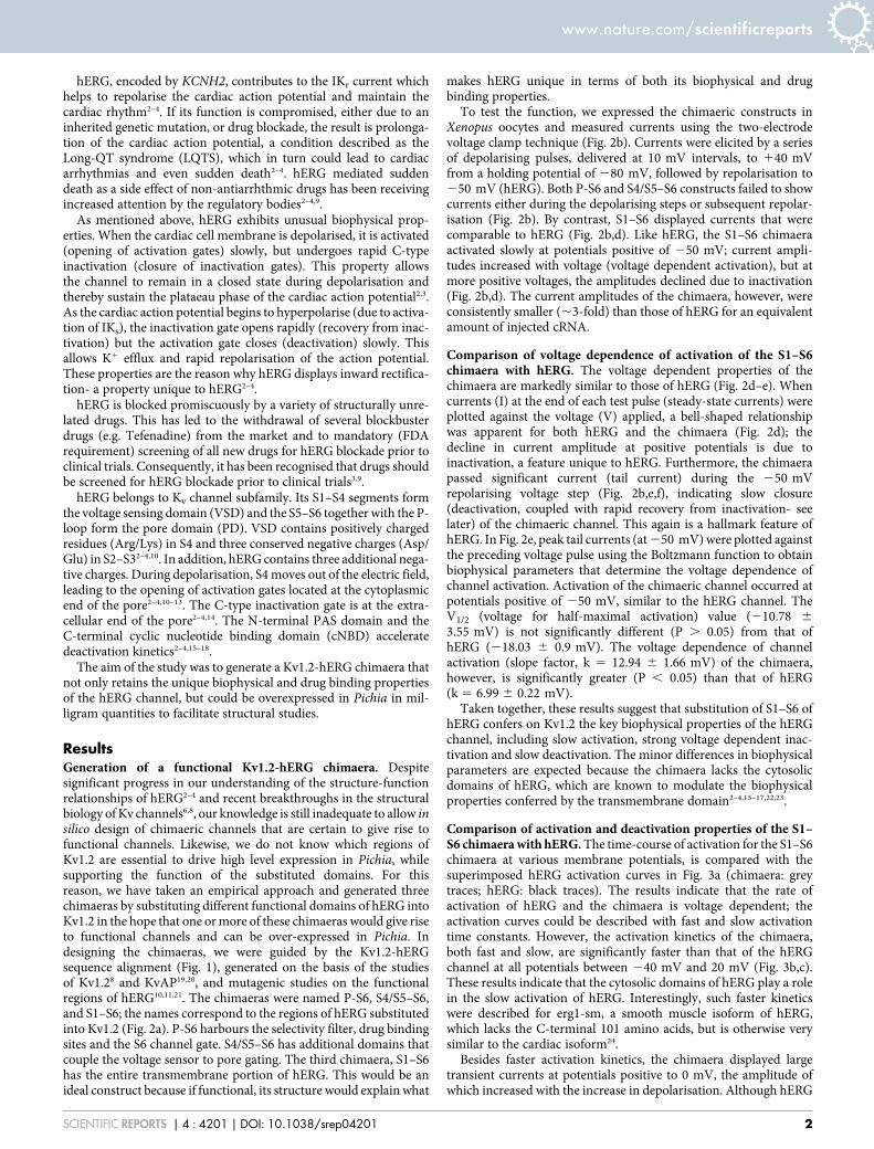
hERG, encoded by KCNH2, contributes to the IKr current whichhelps to repolarise the cardiac action potential and maintain thecardiac rhythm2–4. If its function is compromised, either due to aninherited genetic mutation, or drug blockade, the result is prolonga-tion of the cardiac action potential, a condition described as theLong-QT syndrome (LQTS), which in turn could lead to cardiacarrhythmias and even sudden death2–4. hERG mediated suddendeath as a side effect of non-antiarrhthmic drugs has been receivingincreased attention by the regulatory bodies2–4,9.
As mentioned above, hERG exhibits unusual biophysical prop-erties. When the cardiac cell membrane is depolarised, it is activated(opening of activation gates) slowly, but undergoes rapid C-typeinactivation (closure of inactivation gates). This property allowsthe channel to remain in a closed state during depolarisation andthereby sustain the plataeau phase of the cardiac action potential2,3.As the cardiac action potential begins to hyperpolarise (due to activa-tion of IKs), the inactivation gate opens rapidly (recovery from inac-tivation) but the activation gate closes (deactivation) slowly. Thisallows K1 efflux and rapid repolarisation of the action potential.These properties are the reason why hERG displays inward rectifica-tion- a property unique to hERG2–4.
hERG is blocked promiscuously by a variety of structurally unre-lated drugs. This has led to the withdrawal of several blockbusterdrugs (e.g. Tefenadine) from the market and to mandatory (FDArequirement) screening of all new drugs for hERG blockade prior toclinical trials. Consequently, it has been recognised that drugs shouldbe screened for hERG blockade prior to clinical trials3,9.
hERG belongs to Kv channel subfamily. Its S1–S4 segments formthe voltage sensing domain (VSD) and the S5–S6 together with the P-loop form the pore domain (PD). VSD contains positively chargedresidues (Arg/Lys) in S4 and three conserved negative charges (Asp/Glu) in S2–S32–4,10. In addition, hERG contains three additional nega-tive charges. During depolarisation, S4 moves out of the electric field,leading to the opening of activation gates located at the cytoplasmicend of the pore2–4,10–13. The C-type inactivation gate is at the extra-cellular end of the pore2–4,14. The N-terminal PAS domain and theC-terminal cyclic nucleotide binding domain (cNBD) acceleratedeactivation kinetics2–4,15–18.
The aim of the study was to generate a Kv1.2-hERG chimaera thatnot only retains the unique biophysical and drug binding propertiesof the hERG channel, but could be overexpressed in Pichia in mil-ligram quantities to facilitate structural studies.
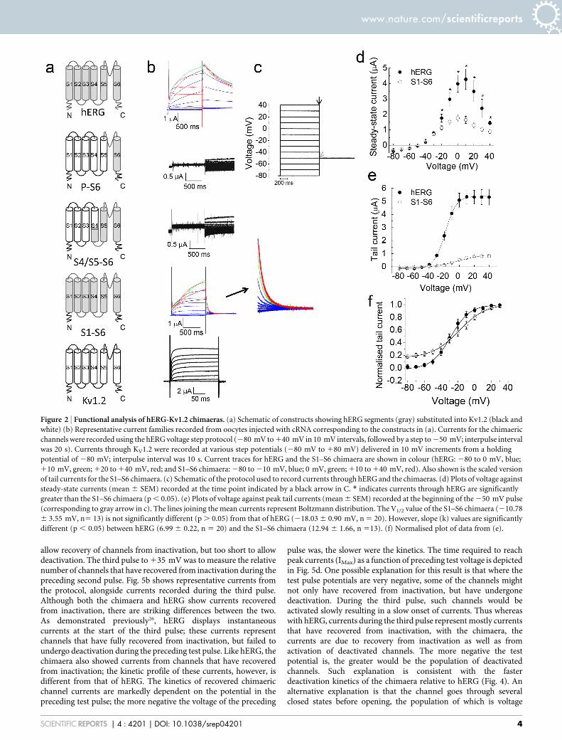
ResultsGeneration of a functional Kv1.2-hERG chimaera. Despitesignificant progress in our understanding of the structure-functionrelationships of hERG2–4 and recent breakthroughs in the structuralbiology of Kv channels6,8, our knowledge is still inadequate to allow insilico design of chimaeric channels that are certain to give rise tofunctional channels. Likewise, we do not know which regions ofKv1.2 are essential to drive high level expression in Pichia, whilesupporting the function of the substituted domains. For thisreason, we have taken an empirical approach and generated threechimaeras by substituting different functional domains of hERG intoKv1.2 in the hope that one or more of these chimaeras would give riseto functional channels and can be over-expressed in Pichia. Indesigning the chimaeras, we were guided by the Kv1.2-hERGsequence alignment (Fig. 1), generated on the basis of the studiesof Kv1.28 and KvAP19,20, and mutagenic studies on the functionalregions of hERG10,11,21. The chimaeras were named P-S6, S4/S5–S6,and S1–S6; the names correspond to the regions of hERG substitutedinto Kv1.2 (Fig. 2a). P-S6 harbours the selectivity filter, drug bindingsites and the S6 channel gate. S4/S5–S6 has additional domains thatcouple the voltage sensor to pore gating. The third chimaera, S1–S6has the entire transmembrane portion of hERG. This would be anideal construct because if functional, its structure would explain what
makes hERG unique in terms of both its biophysical and drugbinding properties.
To test the function, we expressed the chimaeric constructs inXenopus oocytes and measured currents using the two-electrodevoltage clamp technique (Fig. 2b). Currents were elicited by a seriesof depolarising pulses, delivered at 10 mV intervals, to 140 mVfrom a holding potential of 280 mV, followed by repolarisation to250 mV (hERG). Both P-S6 and S4/S5–S6 constructs failed to showcurrents either during the depolarising steps or subsequent repolar-isation (Fig. 2b). By contrast, S1–S6 displayed currents that werecomparable to hERG (Fig. 2b,d). Like hERG, the S1–S6 chimaeraactivated slowly at potentials positive of 250 mV; current ampli-tudes increased with voltage (voltage dependent activation), but atmore positive voltages, the amplitudes declined due to inactivation(Fig. 2b,d). The current amplitudes of the chimaera, however, wereconsistently smaller (,3-fold) than those of hERG for an equivalentamount of injected cRNA.
Comparison of voltage dependence of activation of the S1–S6chimaera with hERG. The voltage dependent properties of thechimaera are markedly similar to those of hERG (Fig. 2d–e). Whencurrents (I) at the end of each test pulse (steady-state currents) wereplotted against the voltage (V) applied, a bell-shaped relationshipwas apparent for both hERG and the chimaera (Fig. 2d); thedecline in current amplitude at positive potentials is due toinactivation, a feature unique to hERG. Furthermore, the chimaerapassed significant current (tail current) during the 250 mVrepolarising voltage step (Fig. 2b,e,f), indicating slow closure(deactivation, coupled with rapid recovery from inactivation- seelater) of the chimaeric channel. This again is a hallmark feature ofhERG. In Fig. 2e, peak tail currents (at 250 mV) were plotted againstthe preceding voltage pulse using the Boltzmann function to obtainbiophysical parameters that determine the voltage dependence ofchannel activation. Activation of the chimaeric channel occurred atpotentials positive of 250 mV, similar to the hERG channel. TheV1/2 (voltage for half-maximal activation) value (210.78 6
3.55 mV) is not significantly different (P . 0.05) from that ofhERG (218.03 6 0.9 mV). The voltage dependence of channelactivation (slope factor, k 5 12.94 6 1.66 mV) of the chimaera,however, is significantly greater (P , 0.05) than that of hERG(k 5 6.99 6 0.22 mV).
Taken together, these results suggest that substitution of S1–S6 ofhERG confers on Kv1.2 the key biophysical properties of the hERGchannel, including slow activation, strong voltage dependent inac-tivation and slow deactivation. The minor differences in biophysicalparameters are expected because the chimaera lacks the cytosolicdomains of hERG, which are known to modulate the biophysicalproperties conferred by the transmembrane domain2–4,15–17,22,23.
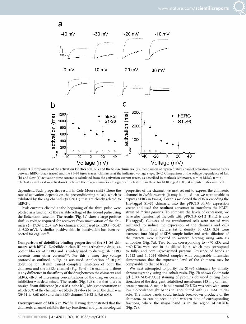
Comparison of activation and deactivation properties of the S1–S6 chimaera with hERG. The time-course of activation for the S1–S6chimaera at various membrane potentials, is compared with thesuperimposed hERG activation curves in Fig. 3a (chimaera: greytraces; hERG: black traces). The results indicate that the rate ofactivation of hERG and the chimaera is voltage dependent; theactivation curves could be described with fast and slow activationtime constants. However, the activation kinetics of the chimaera,both fast and slow, are significantly faster than that of the hERGchannel at all potentials between 240 mV and 20 mV (Fig. 3b,c).These results indicate that the cytosolic domains of hERG play a rolein the slow activation of hERG. Interestingly, such faster kineticswere described for erg1-sm, a smooth muscle isoform of hERG,which lacks the C-terminal 101 amino acids, but is otherwise verysimilar to the cardiac isoform24.
Besides faster activation kinetics, the chimaera displayed largetransient currents at potentials positive to 0 mV, the amplitude ofwhich increased with the increase in depolarisation. Although hERG
www.nature.com/scientificreports
SCIENTIFIC REPORTS | 4 : 4201 | DOI: 10.1038/srep04201 2

current traces show occasional transients, they are considerablysmaller, being detectable only at strong depolarising potentials($30 mV). In this respect, again, the chimaera is similar to erg1-sm which showed large transient currents24. Such large transients arealso a characteristic feature of ELK channels25, which are closelyrelated to hERG.
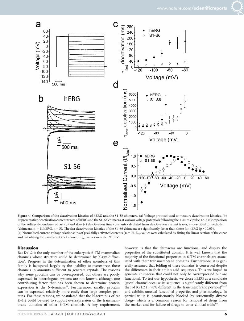
Deactivation kinetics were determined from current tracesrecorded at various repolarisation potentials (2120 to 150 mV)following a strong depolarising pulse (140 mV; Fig. 4a). Fig. 4bshows representative deactivation current traces during repolarisa-tion for hERG and the S1–S6 chimaera. The traces could be describedwith slow and fast components, from which time constants for deac-tivation were calculated for each voltage. Fig. 4c–d shows plots ofrepolarising voltage against fast and slow deactivation time con-stants. The results show that deactivation kinetics of the S1–S6 chi-maera for the fast component are significantly faster than those ofhERG. At 290 mV, for example, the mean tfast (time constant forfast deactivation) for the chimaera was 24.5 6 8.1 ms, and that forhERG was 96.4 6 18.0 ms. Time constants (tslow) for the slow(minor) component of deactivation showed no significant differencebetween hERG and the chimaera in the voltage region (2120 to260 mV) where they could be reliably determined. In this respect,the chimaera again shows similarity to erg1-sm, which deactivatesabout four fold faster than hERG24.
Comparison of inward rectification properties of the S1–S6chimaera with hERG. hERG is unique among Kv channels in thatit shows inward rectification. The mechanism of inward rectificationin hERG, however, is mechanistically different from that of inwardlyrectifying potassium (Kir) channel family2–4. In hERG, inward recti-fication is brought about by fast inactivation at depolarising poten-tials followed by the rapid recovery from inactivation, coupled withslow deactivation, at hyperpolarised potentials. Fig. 4e shows a plot ofpeak current amplitudes during the repolarising pulses againstvoltage for hERG and the S1–S6 chimaera (protocol Fig. 4a). BothhERG and the chimaera display bell-shaped I-V relationships, withreversal potentials (,290 mV) expected for a potassium selectivechannel. However, there is a rightward shift for peak currentamplitudes by ,20 mV (from 240 mV for hERG to 220 mV forthe chimaera). This shift appears to result from a positive shift inactivation (Fig. 2e) and even greater shift in inactivation (Fig. 5c, seelater).
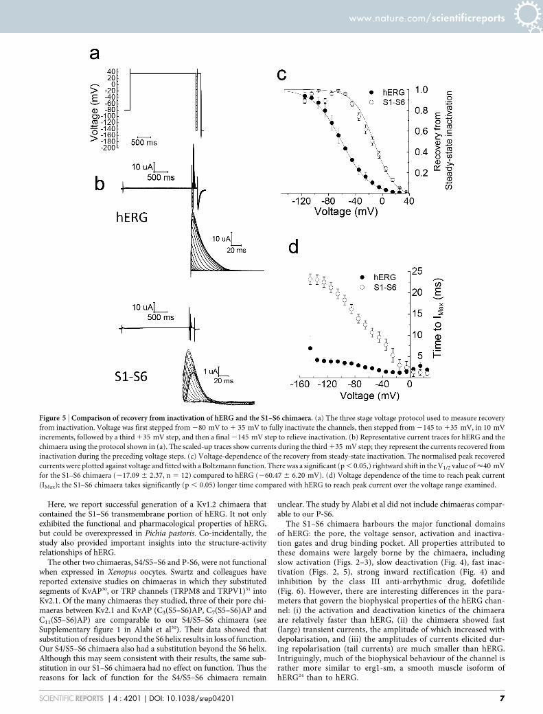
Comparison of inactivation properties of the S1–S6 chimaera withhERG. A standard three pulse protocol was used to determine thevoltage dependence of inactivation/channel availability (Fig. 5a). A1 s pulse to 135 mV was first applied in order to attain steady stateinactivation. This was followed by a series of test pulses to potentialsranging from 2145 to 135 mV for 20 ms which is long enough to
Figure 1 | Sequence alignment of Kv1.2 and hERG showing sites of substitutions. Potential transmembrane regions (highlighted in yellow) and other
functional elements (S4–S5 linker and pore-helix) are labelled. Arrows indicate sites where different chimaeras were joined: Black, S1–S6;
red, S4/S5–S6; blue, P-S6.
www.nature.com/scientificreports
SCIENTIFIC REPORTS | 4 : 4201 | DOI: 10.1038/srep04201 3
allow recovery of channels from inactivation, but too short to allowdeactivation. The third pulse to 135 mV was to measure the relativenumber of channels that have recovered from inactivation during thepreceding second pulse. Fig. 5b shows representative currents fromthe protocol, alongside currents recorded during the third pulse.Although both the chimaera and hERG show currents recoveredfrom inactivation, there are striking differences between the two.As demonstrated previously26, hERG displays instantaneouscurrents at the start of the third pulse; these currents representchannels that have fully recovered from inactivation, but failed toundergo deactivation during the preceding test pulse. Like hERG, thechimaera also showed currents from channels that have recoveredfrom inactivation; the kinetic profile of these currents, however, isdifferent from that of hERG. The kinetics of recovered chimaericchannel currents are markedly dependent on the potential in thepreceding test pulse; the more negative the voltage of the preceding
pulse was, the slower were the kinetics. The time required to reachpeak currents (IMax) as a function of preceding test voltage is depictedin Fig. 5d. One possible explanation for this result is that where thetest pulse potentials are very negative, some of the channels mightnot only have recovered from inactivation, but have undergonedeactivation. During the third pulse, such channels would beactivated slowly resulting in a slow onset of currents. Thus whereaswith hERG, currents during the third pulse represent mostly currentsthat have recovered from inactivation, with the chimaera, thecurrents are due to recovery from inactivation as well as fromactivation of deactivated channels. The more negative the testpotential is, the greater would be the population of deactivatedchannels. Such explanation is consistent with the fasterdeactivation kinetics of the chimaera relative to hERG (Fig. 4). Analternative explanation is that the channel goes through severalclosed states before opening, the population of which is voltage
Figure 2 | Functional analysis of hERG-Kv1.2 chimaeras. (a) Schematic of constructs showing hERG segments (gray) substituted into Kv1.2 (black and
white) (b) Representative current families recorded from oocytes injected with cRNA corresponding to the constructs in (a). Currents for the chimaeric
channels were recorded using the hERG voltage step protocol (280 mV to 140 mV in 10 mV intervals, followed by a step to 250 mV; interpulse interval
was 20 s). Currents through KV1.2 were recorded at various step potentials (280 mV to 180 mV) delivered in 10 mV increments from a holding
potential of 280 mV; interpulse interval was 10 s. Current traces for hERG and the S1–S6 chimaera are shown in colour (hERG: 280 to 0 mV, blue;
110 mV, green; 120 to 140 mV, red; and S1–S6 chimaera: 280 to 210 mV, blue; 0 mV, green; 110 to 140 mV, red). Also shown is the scaled version
of tail currents for the S1–S6 chimaera. (c) Schematic of the protocol used to record currents through hERG and the chimaeras. (d) Plots of voltage against
steady-state currents (mean 6 SEM) recorded at the time point indicated by a black arrow in C. * indicates currents through hERG are significantly
greater than the S1–S6 chimaera (p , 0.05). (e) Plots of voltage against peak tail currents (mean 6 SEM) recorded at the beginning of the 250 mV pulse
(corresponding to gray arrow in c). The lines joining the mean currents represent Boltzmann distribution. The V1/2 value of the S1–S6 chimaera (210.78
6 3.55 mV, n5 13) is not significantly different (p . 0.05) from that of hERG (218.03 6 0.90 mV, n 5 20). However, slope (k) values are significantly
different (p , 0.05) between hERG (6.99 6 0.22, n 5 20) and the S1–S6 chimaera (12.94 6 1.66, n 513). (f) Normalised plot of data from (e).
www.nature.com/scientificreports
SCIENTIFIC REPORTS | 4 : 4201 | DOI: 10.1038/srep04201 4
dependent. Such properties results in Cole-Moore shift (where therate of activation depends on the preconditioning pulse), which isexhibited by the eag channels (KCNH1) that are closely related tohERG27.
Peak currents elicited at the beginning of the third pulse wereplotted as a function of the variable voltage of the second pulse usingthe Boltzmann function. The results (Fig. 5c) show a large positiveshift in voltage required for recovery from inactivation of the chi-maera (217.09 6 2.37 mV for chimaera, compared to hERG 260.476 6.20 mV). A similar positive shift in inactivation has been re-ported for erg1-sm24.
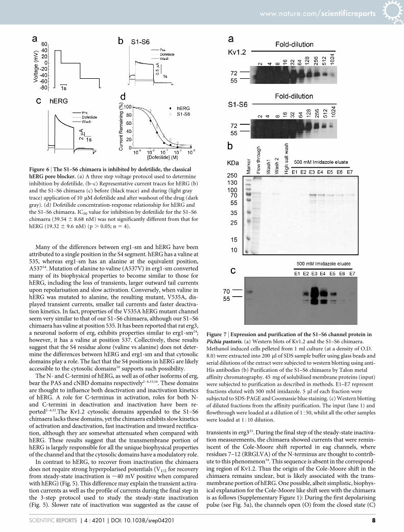
Comparison of dofetilide binding properties of the S1–S6 chi-maera with hERG. Dofetilide, a class III anti-arrhythmic drug is apotent blocker of hERG and is widely used to distinguish hERGcurrents from other currents9,28. For this a three step voltageprotocol as outlined in Fig. 6a was used. Application of 10 mMdofetilide for 10 min caused complete inhibition of both thechimaera and the hERG channel (Fig. 6b–d). To examine if thereis any difference in the affinity of the drug between the chimaera andhERG, effect of increasing concentrations of the drug on currentinhibition was determined. The results (Fig. 6d) show that there isno significant difference (p . 0.05) in the IC50 (drug concentration atwhich 50% of the channels are blocked) values between the chimaera(39.54 6 8.68 nM) and the hERG channel (19.32 6 9.6 nM).
Overexpression of hERG in Pichia. Having demonstrated that thechimaeric channel exhibits the key functional and pharmacological
properties of the channel, we next set out to express the chimaericchannel in Pichia pastoris (it may be noted that we were unable toexpress hERG in Pichia). For this we cloned the cDNA encoding theHis-tagged S1–S6 chimaera into the pPIC3.5 Pichia expressionvector and used the resultant construct to transform the KM71strain of Pichia pastoris. To compare the levels of expression, wehave also transformed the cells with pPIC3.5-Kv1.2 (Kv1.2 is alsoHis-tagged). Cultures of the transformed cells were treated withmethanol to induce the expression of the channels and cellspelleted from 1 ml culture (at a density of O.D. 8.0) wereextracted into 200 ml of SDS sample buffer and serial dilutions ofthe extracts were subjected to western blotting using anti-Hisantibodies (Fig. 7a). Two bands, corresponding to ,70 KDa and,60 KDa, were seen in the diluted lanes, which may correspondto fully- and core -glycosylated proteins. Presence of bands at15512 and 151024 diluted samples with comparable intensitiesdemonstrates that the expression level of the chimaera may becomparable to that of Kv1.2.
We next attempted to purify the S1–S6 chimaera by affinitychromatography using the cobalt resin. Fig. 7b shows Coomassiegel (10% SDS-PAGE) staining of proteins obtained during frac-tionation of the detergent solubilised membranes (45 mg of mem-brane protein). A major band around 70 KDa was seen with somelow molecular weight bands in lanes eluted with 500 mM imida-zole. The minor bands could include breakdown products of thechimaera, as can be seen in the western blot of correspondingfractions, where the major band is in the region of 70 kDa(Fig. 7c).
Figure 3 | Comparison of the activation kinetics of hERG and the S1–S6 chimaera. (a) Comparison of representative channel activation current traces
between hERG (black traces) and the S1–S6 (grey traces) chimaeras at the indicated voltage steps. (b–c) Comparison of the voltage dependence of fast
(b) and slow (c) activation time constants calculated from the activation current traces, as described in methods (chimaera, n 5 8; hERG, n 5 5).
The fast as well as slow activation kinetics of the S1–S6 chimaera are significantly faster than those for hERG (p , 0.05) at all potentials examined.
www.nature.com/scientificreports
SCIENTIFIC REPORTS | 4 : 4201 | DOI: 10.1038/srep04201 5
DiscussionRat Kv1.2 is the only member of the eukaryotic 6-TM mammalianchannels whose structure could be determined by X-ray diffrac-tion8. Progress in the determination of other members of thisfamily is hampered largely by the inability to overexpress thesechannels in amounts sufficient to generate crystals. The reasonswhy some proteins can be overexpressed, but others are poorlyexpressed in heterologous systems are not known, although onecontributing factor that has been shown to determine proteinexpression is the N-terminus29. Furthermore, smaller proteinscan be expressed relatively more easily than large complex pro-teins. For these reasons, we postulated that the N-terminus of ratKv1.2 could be used to support overexpression of the transmem-brane domains of other 6-TM channels. A key requirement,
however, is that the chimaeras are functional and display theproperties of the substituted domain. It is well known that themajority of the functional properties in 6-TM channels are assoc-iated with their transmembrane domains. Furthermore, it is gen-erally assumed that folding of these domains is conserved despitethe differences in their amino acid sequences. Thus we hoped togenerate chimaeras that could not only be overexpressed but arefunctional. To test our hypothesis, we chose hERG as a candidate‘guest’ channel because its sequence is significantly different fromthat of Kv1.2 (,90% different in the transmembrane portion)15–17
and exhibits unusual functional properties and pharmacology. Inparticular, it is promiscuously blocked by structurally diversedrugs- which is a common reason for removal of drugs fromthe market and for failure of drugs to enter clinical trials3,9.
Figure 4 | Comparison of the deactivation kinetics of hERG and the S1–S6 chimaera. (a) Voltage protocol used to measure deactivation kinetics. (b)
Representative deactivation current traces of hERG and the S1–S6 chimaera at various voltage potentials following the 140 mV pulse. (c–d) Comparison
of the voltage dependence of fast (b) and slow (c) deactivation time constants calculated from deactivation current traces, as described in methods
(chimaera, n 5 8; hERG, n5 5). The fast deactivation kinetics of the S1–S6 chimaera are significantly faster than those for hERG (p , 0.05).
(e) Normalised current-voltage relationships of peak fully activated currents (n 5 7). Erev values were calculated by fitting the linear section of the curve
and calculating the x-intercept (not shown). Erev values were <290 mV.
www.nature.com/scientificreports
SCIENTIFIC REPORTS | 4 : 4201 | DOI: 10.1038/srep04201 6
Here, we report successful generation of a Kv1.2 chimaera thatcontained the S1–S6 transmembrane portion of hERG. It not onlyexhibited the functional and pharmacological properties of hERG,but could be overexpressed in Pichia pastoris. Co-incidentally, thestudy also provided important insights into the structure-activityrelationships of hERG.
The other two chimaeras, S4/S5–S6 and P-S6, were not functionalwhen expressed in Xenopus oocytes. Swartz and colleagues havereported extensive studies on chimaeras in which they substitutedsegments of KvAP30, or TRP channels (TRPM8 and TRPV1)31 intoKv2.1. Of the many chimaeras they studied, three of their pore chi-maeras between Kv2.1 and KvAP (C3(S5–S6)AP, C7(S5–S6)AP andC11(S5–S6)AP) are comparable to our S4/S5–S6 chimaera (seeSupplementary figure 1 in Alabi et al30). Their data showed thatsubstitution of residues beyond the S6 helix results in loss of function.Our S4/S5–S6 chimaera also had a substitution beyond the S6 helix.Although this may seem consistent with their results, the same sub-stitution in our S1–S6 chimaera had no effect on function. Thus thereasons for lack of function for the S4/S5–S6 chimaera remain
unclear. The study by Alabi et al did not include chimaeras compar-able to our P-S6.
The S1–S6 chimaera harbours the major functional domainsof hERG: the pore, the voltage sensor, activation and inactiva-tion gates and drug binding pocket. All properties attributed tothese domains were largely borne by the chimaera, includingslow activation (Figs. 2–3), slow deactivation (Fig. 4), fast inac-tivation (Figs. 2, 5), strong inward rectification (Fig. 4) andinhibition by the class III anti-arrhythmic drug, dofetilide(Fig. 6). However, there are interesting differences in the para-meters that govern the biophysical properties of the hERG chan-nel: (i) the activation and deactivation kinetics of the chimaeraare relatively faster than hERG, (ii) the chimaera showed fast(large) transient currents, the amplitude of which increased withdepolarisation, and (iii) the amplitudes of currents elicited dur-ing repolarisation (tail currents) are much smaller than hERG.Intriguingly, much of the biophysical behaviour of the channel israther more similar to erg1-sm, a smooth muscle isoform ofhERG24 than to hERG.
Figure 5 | Comparison of recovery from inactivation of hERG and the S1–S6 chimaera. (a) The three stage voltage protocol used to measure recovery
from inactivation. Voltage was first stepped from 280 mV to 1 35 mV to fully inactivate the channels, then stepped from 2145 to 135 mV, in 10 mV
increments, followed by a third 135 mV step, and then a final 2145 mV step to relieve inactivation. (b) Representative current traces for hERG and the
chimaera using the protocol shown in (a). The scaled-up traces show currents during the third 135 mV step; they represent the currents recovered from
inactivation during the preceding voltage steps. (c) Voltage-dependence of the recovery from steady-state inactivation. The normalised peak recovered
currents were plotted against voltage and fitted with a Boltzmann function. There was a significant (p , 0.05,) rightward shift in the V1/2 value of <40 mV
for the S1–S6 chimaera (217.09 6 2.37, n 5 12) compared to hERG (260.47 6 6.20 mV). (d) Voltage dependence of the time to reach peak current
(IMax); the S1–S6 chimaera takes significantly (p , 0.05) longer time compared with hERG to reach peak current over the voltage range examined.
www.nature.com/scientificreports
SCIENTIFIC REPORTS | 4 : 4201 | DOI: 10.1038/srep04201 7
Many of the differences between erg1-sm and hERG have beenattributed to a single position in the S4 segment. hERG has a valine at535, whereas erg1-sm has an alanine at the equivalent position,A53724. Mutation of alanine to valine (A537V) in erg1-sm convertedmany of its biophysical properties to become similar to those forhERG, including the loss of transients, larger outward tail currentsupon repolarisation and slow activation. Conversely, when valine inhERG was mutated to alanine, the resulting mutant, V535A, dis-played transient currents, smaller tail currents and faster deactiva-tion kinetics. In fact, properties of the V535A hERG mutant channelseem very similar to that of our S1–S6 chimaera, although our S1–S6chimaera has valine at position 535. It has been reported that rat erg3,a neuronal isoform of erg, exhibits properties similar to erg1-sm24;however, it has a valine at position 537. Collectively, these resultssuggest that the S4 residue alone (valine vs alanine) does not deter-mine the differences between hERG and erg1-sm and that cytosolicdomains play a role. The fact that the S4 positions in hERG are likelyaccessible to the cytosolic domains10 supports such possibility.
The N- and C-termini of hERG, as well as of other isoforms of erg,bear the PAS and cNBD domains respectively2–4,15,16. These domainsare thought to influence both deactivation and inactivation kineticsof hERG. A role for C-terminus in activation, roles for both N-and C-termini in deactivation and inactivation have been re-ported2–4,32.The Kv1.2 cytosolic domains appended to the S1–S6chimaera lacks these domains, yet the chimaera exhibits slow kineticsof activation and deactivation, fast inactivation and inward rectifica-tion, although they are somewhat attenuated when compared withhERG. These results suggest that the transmembrane portion ofhERG is largely responsible for all the unique biophysical propertiesof the channel and that the cytosolic domains have a modulatory role.
In contrast to hERG, to recover from inactivation the chimaeradoes not require strong hyperpolarised potentials (V1/2 for recoveryfrom steady-state inactivation is ,40 mV positive when comparedwith hERG) (Fig. 5). This difference may explain the transient activa-tion currents as well as the profile of currents during the final step inthe 3-step protocol used to study the steady-state inactivation(Fig. 5). Slower rate of inactivation was suggested as the cause of
transients in erg333. During the final step of the steady-state inactiva-tion measurements, the chimaera showed currents that were remin-iscent of the Cole-Moore shift reported in eag channels, whereresidues 7–12 (RRGLVA) of the N-terminus are thought to contrib-ute to this phenomenon34. This sequence is absent in the correspond-ing region of Kv1.2. Thus the origin of the Cole-Moore shift in thechimaera remains unclear, but is likely associated with the trans-membrane portion of hERG. One possible, albeit simplistic, biophys-ical explanation for the Cole-Moore like shift seen with the chimaerais as follows (Supplementary Figure 1): During the first depolarisingpulse (see Fig. 5a), the channels open (O) from the closed state (C)
Figure 6 | The S1–S6 chimaera is inhibited by dofetilide, the classicalhERG pore blocker. (a) A three step voltage protocol used to determine
inhibition by dofetilide. (b–c) Representative current traces for hERG (b)
and the S1–S6 chimaera (c) before (black trace) and during (light gray
trace) application of 10 mM dofetilide and after washout of the drug (dark
gray). (d) Dofetilide concentration-response relationship for hERG and
the S1–S6 chimaera. IC50 value for inhibition by dofetilide for the S1–S6
chimaera (39.54 6 8.68 nM) was not significantly different from that for
hERG (19.32 6 9.6 nM) (p . 0.05; n 5 4).
Figure 7 | Expression and purification of the S1–S6 channel protein inPichia pastoris. (a) Western blots of Kv1.2 and the S1–S6 chimaera.
Methanol induced cells pelleted from 1 ml culture (at a density of O.D.
8.0) were extracted into 200 ml of SDS sample buffer using glass beads and
serial dilutions of the extract were subjected to western blotting using anti-
His antibodies (b) Purification of the S1–S6 chimaera by Talon metal
affinity chromatography. 45 mg of solubilised membrane proteins (input)
were subjected to purification as described in methods. E1–E7 represent
fractions eluted with 500 mM imidazole. 5 ml of each fraction were
subjected to SDS-PAGE and Coomassie blue staining. (c) Western blotting
of diluted fractions from the affinity purification. The input (lane 1) and
flowthrough were loaded at a dilution of 1550, whilst all the other samples
were loaded at 1510 dilution.
www.nature.com/scientificreports
SCIENTIFIC REPORTS | 4 : 4201 | DOI: 10.1038/srep04201 8

and undergo inactivation (I) (C R O R I); during the followingsecond hyperpolarising pulse, the channels recover from inactivation(I R O), but a significant proportion of the open channels alsoundergo deactivation (I R O R C); during the third depolarisingtest pulse, open channels are expected to elicit instantaneous outwardcurrents, but the closed channels open slowly (C R O) due to slowactivation kinetics. The net result of these events would be appear-ance of currents that resemble the Cole-Moore shift. In the case ofhERG, deactivation is relatively slow; thus the occurrence of I R O RC transition during the second pulse is expected to be limited; for thisreason, hERG shows mostly instantaneous currents.
There is very little sequence similarity between the cytosolicdomains of hERG and Kv1.2. The sequence homology within thetransmembrane regions of the two proteins, likewise, is also low,compared to other members of the Kv channels2. Thus the fact thatwe were able to generate functional chimaeras of Kv1.2 that repro-duced almost all properties of hERG is highly significant because itsuggests that the transmembrane portion of hERG and cytosolicdomains of Kv1.2 could fold into independent functional domains,and folding and function are not dependent on each other. Thisobservation suggests that the approach could be used as a generalstrategy to generate functional chimaeric channels with other 6-TMchannels, including those that are distantly related to Kv1.2.
The S1–S6 chimaera showed expression levels that were compar-able to those of rat Kv.2 when examined by western blotting of theextracts of induced Pichia cells diluted serially (Fig. 7). Westernblotting of the purified fraction showed two bands that presumablycorrespond to the core and glycosylated versions of the protein. Thechimaera showed double bands of similar size when expressed inHEK293 cells and showed clear expression at the plasma membrane,indicating that the chimaera likely attains native conformation(Supplementary Figure 2). However, the functional integrity of thepurified chimaera remains to be determined.
In summary, our study demonstrated that the transmembraneregion harbouring the key functional elements of hERG can be sub-stituted into Kv1.2 to generate chimaeras that were able to recapitu-late almost all of the properties of hERG. Thus Kv1.2 could provide aperfect framework for functional substitution of distantly related Kvand other 6-TM channels. Since Kv1.2 is also one of the eukaryotic6-TM proteins that could exceptionally be overexpressed and crystal-lised, the present study presents a promising strategy for overpro-duction of other 6-TM channels that are difficult to overexpress andcrystallise.
Experimental proceduresMaterials. pPIC3.5-rat Kv1.2 containing the His9 tag at the N-terminus7 and hERGcDNA in pSP64 were kindly provided Dr. D. Parcej and Prof. S.A. Goldsteinrespectively. KM71 Pichia pastoris cells were from Invitrogen. Enzymes for molecularbiology were purchased from New England Biolabs, Promega or Stratagene. Allgeneral chemicals were obtained from Sigma Chemicals Co. Mouse anti-His tag, goatanti-mouse HRP were purchased from Novagen and BioRad laboratoriesrespectively.
MethodsPreparation of DNA constructs. Overlap extension or substitution PCR was used togenerate the desired chimaeric constructs depicted in Fig. 2a. The constructs weremade from His9-Kv1.2 and hERG cDNA templates, such that all constructs containedthe N-terminal His9 tag. Three chimaeric constructs were generated using the Kv1.2and hERG sequence alignment (based on the X-ray structure of Kv1.2 and structure-function studies of hERG) as a guide (Fig. 1). The chimaeras have the following aminoacid sequences (hERG in bold; the numbers correspond to the primary sequences ofrat Kv1.2 (GI:1235594) and hERG (GI:60391379).
P-S6 1–363/614–667/412–499S4/S5–S6 1–328/539–675/425–499S1–S6 1–164/395–675/426–499The chimaeras were subcloned into the pKS-Globin (Kv1.2, P-S6 and S4/5-S6
constructs) or pBF (hERG and S1–S6) oocyte expression vectors for electrophysio-logical studies in Xenopus oocytes or pPIC3.5 for overexpression in Pichia pastoris.All constructs were fully sequenced to confirm the identity.
Preparation of cRNA and current recordings. cRNA was prepared using the T7/SP6 mMessage machine synthesis kit (Ambion). cRNA was injected into the stageV or VI oocytes, isolated from Xenopus toads (euthanised by cervical dislocationafter anaesthetization with MS-222) as described previously11. Injected oocyteswere incubated in ND96 solution (NaCl 96 mM, KCl 2 mM, MgCl2 1 mM,HEPES 5 mM, CaCl2 1.8 mM, pH 7.5, 50 mg/ml G418) at 18uC, for 1–3 days,prior to current recordings. Currents were recorded from oocytes by two-electrodevoltage clamp. The recording chamber was perfused with Ringer’s extracellularsolution (NaCl 115 mM, KCl 2.5 mM, CaCl2 1.8 mM, HEPES 10 mM, pH 7.2) ata rate of ,1 ml/min. Voltage clamp was established with two thin-walledborosilicate glass (GC100F-15, Harvard Apparatus Ltd) microelectrodes filled with3 M KCl that had a resistance of 0.5 to 5 MV. Membrane potential was controlledusing a GeneClamp 500 amplifier (Molecular Devices), digitised using a NI USB-6211 (National Instruments) and recorded using WinWCP (V 4.0.5) software.Recordings were filtered at 2 kHz and sampled at 4 kHz. A holding potential of280 mV was used unless otherwise stated. Protocols used varied depending onthe objective of the experiment (see figures).
Currents were elicited by pulsing to various test potentials (280 mV to140 mV) for 1 s from a holding potential of 280 mV (Fig. 2c) before stepping to250 mV (1 s). Currents at the end of the test pulse (steady-state) and at thebeginning of the 250 mV step (tail current) were plotted against the test poten-tials. Activation time constants were determined from the rising phase of currentsfor each test potential. To determine voltage dependence of fully activated chan-nels, a 140 mV pulse (1 s) was delivered from a holding potential of 280 mVbefore stepping to various test potentials (2120 to 150 mV) (Fig. 4a). Currents atthe beginning of each test potential were plotted against voltage. Deactivation timeconstants were determined from the decay of currents at each test potential. Todetermine recovery from steady-state inactivation, a three pulse voltage protocolwas used (Fig. 5a). The first 135 mV pulse (1 s) was delivered from a holdingvoltage of 280 mV to fully inactivate the channels; the second pulse to various testpotentials in 10 mV increments (2145 mV to 135 mV) was brief (20 ms) toallow voltage dependent recovery from inactivation; and the third pulse was to135 mV for 20 s, which allowed measurement of currents recovered during thesecond pulse. Peak currents during the third pulse were plotted against voltage todetermine the voltage dependence of recovery from steady-state inactivation. Alldata are collected from n $ 3.
Electrophysiological data analysis. Raw data were analysed using Clampfit 9.2(Molecular Devices) and then compiled in Excel 2007 (Microsoft) and OriginPro 7.5(Originlab). Mean and SEM values were calculated for current recordings and thenon-paired two-tailed Students T-test was used to determine statistical significancewhere appropriate. Tail currents and recovery from steady state inactivation currentswere fitted with unconstrained Boltzmann function (equation 1).
G~Gmax{G0
1zeV{V1
2
� �k
zG0 ð1Þ
Where G0 is the initial conductance, V1/2 is the half-activation voltage and k is ameasure of the voltage dependence.
Activation time courses were fitted in WinWCP software with a two exponentialequation (equation 2).
It~Afast| exp t=tfastð ÞzAslow| exp t=tslowð Þ ð2Þ
where tfast and tslow are the time constants for the fast and slow components ofactivation, and Afast and Aslow are the current amplitudes of each component.
Deactivation time courses were fitted in WinWCP software with two decayingexponentials (equation 3).
Itail tð Þ~Afast| exp {t=tfastð ÞzAslow| exp {t=tslowð ÞzC ð3Þ
where tfast and tslow are the time constants for the fast and slow components ofdeactivation, Afast and Aslow are the current amplitudes of each component, and C is aconstant.
Percentage inhibition of current by dofetilide (I%) was calculated by normalisationof current in the presence of the drug (I) to current in the presence of vehicle (I[B]0)(equation 4)
I%~I
I B½ �0
� �� 100 ð4Þ
Data from dose dependent drug inhibition of currents were fitted individually to thenormalised data for each oocyte with maxima asymptoted to 100% with equation 5.
I~I B½ �0
1zB½ �
IC50
� �H� � ð5Þ
where H is the Hill coefficient, [B] is inhibitor concentration and I[B]0 is the currentin the absence of inhibitor. IC50 values were collated and averaged (6SEM) todetermine final IC50 values for each construct.
www.nature.com/scientificreports
SCIENTIFIC REPORTS | 4 : 4201 | DOI: 10.1038/srep04201 9
Expression and purification of the S1–S6 chimaera. Transformation of P. pastorisKM71, cell culture and methanol induction of protein expression were carried out asdescribed previously7. Colonies resistant to 1 mg/ml G418 were used for proteinexpression. Cells were grown in 2 l flaks to sufficient density in MGYH (1.34% yeastnitrogen base, 1% glycerol, 0.00004% biotin, 0.004% histidine) medium at 27uC in ashaking incubator (200 r.p.m.) Protein expression was induced by replacing theMGYH medium with MMH (1.34% yeast nitrogen base, 0.5% methanol, 0.00004%biotin, 0.004% histidine) medium and growing cells for 3 days at 27uC in a shakingincubator at 200 r.p.m. 0.5% methanol was supplemented every 24 hr. Cells wereharvested by centrifugation and the pellets were suspended in lysis buffer (50 mMphosphate buffer, pH 8.0, 5% (v/v) glycerol, 1 mM EDTA, 34 mg/ml PMSF, 13
EDTA-free protease inhibitor cocktail (Roche)). The suspension was homogenisedwith 0.5 mm acid washed glass beads in a BeadBeater (BioSpec Products: Bartlesville,USA), following the instructions of the manufacturer. Unbroken cells and glass beadswere collected by centrifugation at 4000 g. The supernatant was then centrifuged at100000 3 g to pellet the membrane fraction. The membrane pellet was washed byresuspending in lysis buffer and re-centrifugation at 100000 3 g.
Membranes (50–100 mg) were solubilised in 1% NP-40, 50 mM Tris, 150 mMKCl, 10 mM 2-mercaptoethanol, pH 7.5 at room temperature for 3 hr, using arotating mixer (detergent to protein ratio was kept at 551). The suspension wascentrifuged at 100,000 3 g for 1 hr to remove insoluble material. The supernatant wasloaded onto TalonH metal affinity resin (Clontech) equilibrated with equilibrationbuffer (0.5% NP-40, 50 mM Tris, 150 mM KCl, 10 mM 2-mercaptoethanol, 20 mMimidazole, pH 7.5). After washing with the equilibration buffer and a high salt washbuffer (1 M NaCl in equilibration buffer), the bound protein was eluted with 500 mMimidazole made up in the equilibration buffer. Aliquots of each fraction were sub-jected to SDS-PAGE and stained with Coomassie Brilliant Blue.
Western blotting. Expression of the S1–S6 chimaera in Pichia was detected by westernblotting using anti-His-tag antibody (1520000), goat anti-mouse-HRP conjugatedIgG (1540000), and SuperSignal West Femto Maximum Sensitivity ECL Substrate(Pierce).
1. Yu, F. H., Yarov-Yarovoy, V., Gutman, G. A. & Catterall, W. A. Overview ofmolecular relationships in the voltage-gated ion channel superfamily. Pharmacol.Rev. 57, 387–395 (2005).
2. Vandenberg, J. I. et al. hERG K1 channels: structure, function, and clinicalsignificance. Physiol.Rev. 92, 1393–1478 (2012).
3. Sanguinetti, M. C. & Tristani-Firouzi, M. hERG potassium channels and cardiacarrhythmia. Nature 440, 463–469 (2006).
4. Cheng, Y. M. & Claydon, T. W. Voltage-dependent gating of HERG potassiumchannels. Front. Pharmacol. 3, 83 (2012).
5. Nishida, M., Cadene, M., Chait, B. T. & MacKinnon, R. Crystal structure of aKir3.1-prokaryotic Kir channel chimera. EMBO. J. 26, 4005–4015, 7601828(2007).
6. Long, S. B., Tao, X., Campbell, E. B. & MacKinnon, R. Atomic structure of avoltage-dependent K1 channel in a lipid membrane-like environment. Nature450, 376–382 (2007).
7. Parcej, D. N. & Eckhardt-Strelau, L. Structural characterisation of neuronalvoltage-sensitive K1 channels heterologously expressed in Pichia pastoris. J. Mol.Biol. 333, 103–116 (2003).
8. Long, S. B., Campbell, E. B. & Mackinnon, R. Crystal structure of a mammalianvoltage-dependent Shaker family K1 channel. Science 309, 897–903 (2005).
9. Perry, M., Sanguinetti, M. & Mitcheson, J. Revealing the structural basis of actionof hERG potassium channel activators and blockers. J.Physiol. 588, 3157–3167(2010).
10. Zhang, M., Liu, J. & Tseng, G. N. Gating charges in the activation and inactivationprocesses of the HERG channel. J. Gen. Physiol. 124, 703–718 (2004).
11. Elliott, D. J., Dondas, N. Y., Munsey, T. S. & Sivaprasadarao, A. Movement of theS4 segment in the hERG potassium channel during membrane depolarization.Mol. Memb.Biol. 26, 435–447 (2009).
12. Smith, P. L. & Yellen, G. Fast and slow voltage sensor movements in HERGpotassium channels. J.Gen.Physiol. 119, 275–293 (2002).
13. Piper, D. R., Varghese, A., Sanguinetti, M. C. & Tristani-Firouzi, M. Gatingcurrents associated with intramembrane charge displacement in HERGpotassium channels. Proc.Natl.Acad.Sci.U.S.A 100, 10534–10539 (2003).
14. Smith, P. L., Baukrowitz, T. & Yellen, G. The inward rectification mechanism ofthe HERG cardiac potassium channel. Nature 379, 833–836 (1996).
15. Morais Cabral, J. H. et al. Crystal structure and functional analysis of the HERGpotassium channel N terminus: a eukaryotic PAS domain. Cell 95, 649–655(1998).
16. Gustina, A. S. & Trudeau, M. C. hERG potassium channel gating is mediated by N-and C-terminal region interactions. J. Gen. Physiol. 137, 315–325 (2011).
17. Ng, C. A. et al. The N-terminal tail of hERG contains an amphipathic alpha-helixthat regulates channel deactivation. PloS one 6, e16191 (2011).
18. Wang, J., Trudeau, M. C., Zappia, A. M. & Robertson, G. A. Regulation ofdeactivation by an amino terminal domain in human ether-a-go-go-related genepotassium channels. J.Gen.Physiol. 112, 637–647 (1998).
19. Jiang, Y. et al. X-ray structure of a voltage-dependent K1 channel. Nature 423,33–41 (2003).
20. Neale, E. J., Rong, H., Cockcroft, C. J. & Sivaprasadarao, A. Mapping themembrane-aqueous border for the voltage-sensing domain of a potassiumchannel. J. Biol. Chem. 282, 37597–37604 (2007).
21. Ng, C. A. et al. The S4–S5 linker acts as a signal integrator for HERG K1 channelactivation and deactivation gating. PloS one 7, e31640 (2012).
22. Spector, P. S., Curran, M. E., Zou, A., Keating, M. T. & Sanguinetti, M. C. Fastinactivation causes rectification of the IKr channel. J.Gen.Physiol. 107, 611–619(1996).
23. Schonherr, R. & Heinemann, S. H. Molecular determinants for activation andinactivation of HERG, a human inward rectifier potassium channel. J.Physiol.493, 635–642 (1996).
24. Shoeb, F., Malykhina, A. P. & Akbarali, H. I. Cloning and functionalcharacterization of the smooth muscle ether-a-go-go-related gene K1 channel.Potential role of a conserved amino acid substitution in the S4 region. J.Biol.Chem.278, 2503–2514 (2003).
25. Trudeau, M. C., Titus, S. A., Branchaw, J. L., Ganetzky, B. & Robertson, G. A.Functional analysis of a mouse brain Elk-type K1 channel. J.Neurosci. 19,2906–2918 (1999).
26. Zou, A., Xu, Q. P. & Sanguinetti, M. C. A mutation in the pore region of HERG K1
channels expressed in Xenopus oocytes reduces rectification by shifting thevoltage dependence of inactivation. J.Physiol. 509, 129–137 (1998).
27. Shi, W. et al. Identification of two nervous system-specific members of the ergpotassium channel gene family. J.Neurosci. 17, 9423–9432 (1997).
28. Ficker, E., Jarolimek, W., Kiehn, J., Baumann, A. & Brown, A. M. Moleculardeterminants of dofetilide block of HERG K1 channels. Circ.Res. 82, 386–395(1998).
29. Grisshammer, R. Understanding recombinant expression of membrane proteins.Curr. Opin. Biotech. 17, 337–340 (2006).
30. Alabi, A. A., Bahamonde, M. I., Jung, H. J., Kim, J. I. & Swartz, K. J. Portability ofpaddle motif function and pharmacology in voltage sensors. Nature 450, 370–375(2007).
31. Kalia, J. & Swartz, K. J. Exploring structure-function relationships between TRPand Kv channels. Sci. Rep. 3, 1523 (2013).
32. de la Pena, P., Machin, A., Fernandez-Trillo, J., Dominguez, P. & Barros, F.Mapping of interactions between the N- and C-termini and the channel core inHERG K1 channels. Biochem. J. 451, 463–474 (2013).
33. Shi, W. et al. Identification of two nervous system-specific members of the ergpotassium channel gene family. J. Neurosci. 17, 9423–9432 (1997).
34. Terlau, H., Heinemann, S. H., Stuhmer, W., Pongs, O. & Ludwig, J. Aminoterminal-dependent gating of the potassium channel rat eag is compensated by amutation in the S4 segment. J.Physiol. 502, 537–543 (1997).
AcknowledgmentsThe research was funded by BBSRC-GSK case studentships.
Author contributionsM.D., C.J.C., P.C., D.C.W., H.L. and T.M. performed the experiments; K.J.S., A.J.P., S.P.Y.and A.S. designed the experiments; A.S. wrote the manuscript.
Additional informationSupplementary information accompanies this paper at http://www.nature.com/scientificreports
Competing financial interests: The authors declare no competing financial interests.
How to cite this article: Dhillon, M.S. et al. A functional Kv1.2-hERG chimaeric channelexpressed in Pichia pastoris. Sci. Rep. 4, 4201; DOI:10.1038/srep04201 (2014).
This work is licensed under a Creative Commons Attribution 3.0 Unported license.To view a copy of this license, visit http://creativecommons.org/licenses/by/3.0
www.nature.com/scientificreports
SCIENTIFIC REPORTS | 4 : 4201 | DOI: 10.1038/srep04201 10