a gradient of molecules in avian retina with dorsoventral ... · against cells cultured for 2 days...
TRANSCRIPT

A Gradient of Molecules in Avian Retina with Dorsoventral Polarity
G. DAVID TRISLER, MICHAEL D. SCHNEIDER, JOSEPH R. MOSKAL, and MARSHALL NIRENBERG Laboratory of Biochemical Genetics National Heart, Lung, and Blood Institute National Institutes of Health Bethesda, Maryland 20205
l Mechanisms that impart positional information for the assembly of the developing nervous system and the establishment of specific synaptic connections have been studied in model systems. The retina is a highly ordered laminar structure due to segregation of different classes of neuronal cell bodies and synapses into separate strata (Karten, this volume). Synaptic connections between retina ganglion neurons and tectum neurons preserve the topographic relations of ganglion neurons in the retina, resulting in a point-to-point retino- tectal map. However, the mechanisms underlying these phenomena have not been defined. Sperry (1963) postulated that two orthogonal gradients of molecules on retina ganglion neurons and corresponding gradients of complementary molecules in the optic tectum might determine the specificity of connections between retina and tectum neurons. Other mechanisms that have been proposed include adhe- sive interactions between migrating retina neurites, myelination of bundles of retina axons, and formation of extracellular channels by glia to guide retina axons (Silver and Sidman 1980).
Antisera have been widely used to study retinal structure and function (Goldschneider and Moscona 1972; Thiery et al. 1977; Haus- man and Moscona 1979). Clonal neural retina hybrid cell lines de- rived from single retina cells [as a homogeneous immunogen) have been used to produce rabbit antiserum demonstrating an antigen of the rodent neural retina that was expressed in a restricted topograph- ic domain (Trisler et al. 1979). Hybridoma cell lines were derived by fusing mouse myeloma cells with spleen cells from mice immunized with retina cells to produce monoclonal antibodies to retina neurons or Miiller cells (Eisenbarth et al. 1979; Barnstable 1980).
231

232 Monoclonal Antibodies to Neural Antigens
To detect cell-surface molecules with topographic specificity in the retina, hybridoma antibodies, as candidates for neuronal recogni- tion molecules, were obtained by fusing P3-X63/Ag-8 mouse myeloma cells (Kohler and Milstein 1975) with spleen cells from mice immu- nized with small portions of dorsal or ventral $&day chick embryo neural retina or clonal retina hybrid cells. A hybridoma antibody was obtained that binds to cell-membrane molecules distributed in a dorsoposterior + ventroanterior gradient in retina (Trisler et al. 1981).
DETECTION OF ANTIBODIES WITH TOPOGRAPHIC SPECIFICITY
l The rationale for the experiments shown schematically in Figure 1 was to immunize mice with cells from dorsoposterior or ventral 14- day chick embryo left retina to obtain hybridoma cell lines synthesiz- ing antibody with specificity for the retina sector used for immuniza- tion. The choroid fissure was used as a landmark. Female BALBlc
MOUSE lMMUNlZATlON WITH CHICK RETINA SEGMENTS
1 A(,$)P AC-P
HYBRIDOMA ANTIBODY SPECIFICITY
A&;P 6 (i;) A@P
V NUMBER V NUMBER OF OF
HYBRIDOMA HYBRIDOMA COLONIES COLONIES
++++ + 1 + f 5 -t + 13 + + 3
- 54 - - 101
Figure 1 Strategy for detecting region-specific retina surface-membrane antigens. Lymphocyte hybridomas were derived from mice immunized with cells from dorsoposterior (left panels) or ventral (right panels) retina; hy- bridoma antibody binding to paired cultured-cell monolayers from the portions of I&day chick embryo retina shown in black in the lower panels was determined.

A Gradient of Molecules in Avian Retina 233
m ice were immunized on days 0, 7, and 14 intraperitoneally with 8 x lo6 mechanically dissociated retina cells and intravenously with 2 x lo6 cells from dorsoposterior or ventral retina. On the 17th day, I x 10’ spleen cells from an immunized mouse were fused with 2 x 10’ P3-X63/Ag-8 mouse myeloma cells as described by Galfre et al. (1977). After fusion, the cells were suspended with 5 x lo6 spleen ceils from a nonimmunized mouse in 50 m l of medium (Trisler et al. 1981), and 672 wells of 96-well m icroculture plates were inoculated with cells (74 pi/well). Additional medium was added (0.1 m l/well) on the 5th and 10th days after fusion.
To detect an antibody to a surface molecule enriched in one region of the retina, tissue culture supernatants from we& with hybridoma colonies were tested by indirect radioimmunoassay against cells cultured for 2 days (Schneider and Eisenbarth 1979);
TOPOGRAPHIC GRADIENT OF ANTIGEN
h A LEFT
2 RIGHT RETINA
P 0.
0 12345678
ANT 7 7
DORSAL
VENTRAL
LEFT RETINA
RETINA SECTION Figure 2 Topographic gradient of TOP antigen in (0) right or (A) left la-day chick embryo neural retina. Equivalent sectors from different retinas were pooled and assayed for [12SI]F(ab’), binding after incubation with (o-; A A) TOP antibody, (o---------o; A---------A) P3- X63lAg-8 antibody, or (o-, A A) buffer in the absence of antibody. The right half of the figure depicts, within each sector of the retina, the pmoles of specifically bound [‘2JI]F(ab’)2 per mg of retina protein (binding due to antibody to TOP m inus binding due to antibody synthesized by P3-X63/Ag-8 parental cells). Assay conditions are de- scribed in the legend to Fig. 3.

234 MonocIonal Antibodies to Neural Antigens
cells were taken from the one eighth of the retina originally used as immunogen or from the remaining seven eighths of the retina. Hybri- doma antibody binding to retina cells was detected with an lz51- labeled, affinity-purified F(ab’), fragment of rabbit anti -mouse IgG ([12511F(ab’),).
A TOPOGRAPHIC GRADIENT OF SURFACE-MEMBRANE MOLECULES
l One of 155 hybridoma cell lines examined synthesized antibody to a cell-surface antigen distributed preferentially in dorsoposterior ret- ina (Fig. 2), designated TOP, for toponymic antigen (i.e., a marker of position). Neural retinas from right or left eyes of IQ-day chick
0 pg RETINA PROTEIN pl ASCITES FLUID g pM l~l-F(ab’l2
n. P
Figure 3 (A] Effect of dorsal retina protein concentration (pooled sections 4 and 5 from M-day chick embryos as illustrated in Fig. Z) on [7’)F(ab’), binding due to: (A) antibody to TOP; (0) antibody synthesized by P3-X63/Ag-8; (0) specific binding. (B) Effect of monoclonal antibody concentration on (0, A) [“SIIF(ab’), specific binding due to antibody to TOP or (0, A) nonspecific binding due to P3-X631Ag-8 antibody, to cells from (0) dorsal retina sections 4 and 5 or (A) ventral retina sections 1 and 8. (C) Effect of concentration of [‘251jF(ab’), rabbit anti-mouse IgG. For all experiments each reaction mixture contained the following components, except where specified, in a final volume of 50 ~1: X50-200 pg of retina protein, 50 pg of gelatin, and 1 1~1 of hybridoma ascites fluid; the reaction mixtures were incubated for 30 min at 4°C and washed three times. The pellets were resuspended in 50 ~1 contain- ing 440 nM [‘251)F(ab’), (50,000 cpm), XI pg of gelatin, and 500 pg of bovine serum albumin in Dulbecco’s phosphate-buffered saline, incubated for 30 min at 4% washed three times, and counted.
A Gradient of Molecules in Avian Retina 235
embryos were cut into eight 45” sectors and assayed for TOP antigen. The highest concentration of antigen detected was in dorsoposterior retina and the lowest in ventroanterior retina. Bilaterally symmetric gradients were found in right and left eyes. Other hybridoma anti- bodies, including A2B5 (Eisenbarth et al. 1979), bound equally to cells from each region of retina.
The effects of varying concentrations of retina protein, hybri- doma antibody, and [‘2srJF(ab’)2 rabbit anti-mouse IgG are shown in Figure 3, A, B, and C, respectively. Assay conditions employed are summarized in the legend to Figure 3.
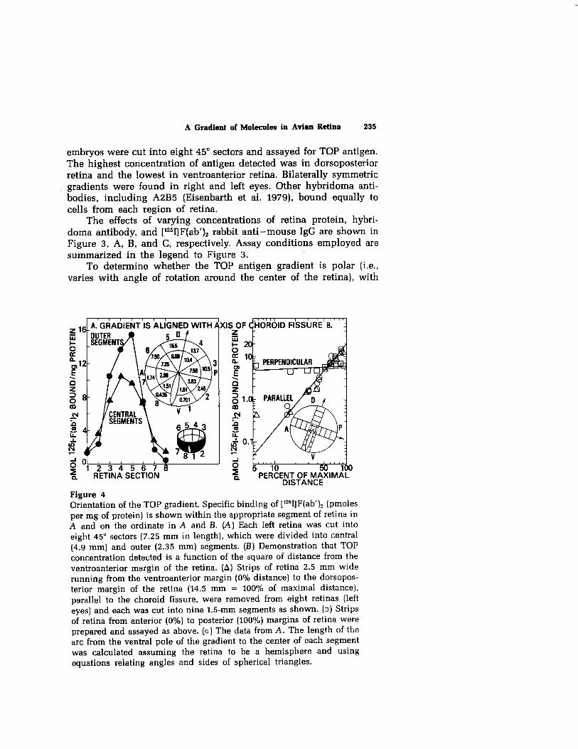
To determine whether the TOP antigen gradient is polar (i.e., varies with angle of rotation around the center of the retina), with
r IS ALIGNED WITH I
L P - PEdtENT OF M/i%IM/iL- DISTANCE
Figure 4 Orientation of the TOP gradient. Specific binding of [“511F(ab’)2 (pmoles per mg of protein) is shown within the appropriate segment of retina in A and on the ordinate in A and B. (A) Each left retina was cut into eight 45’ sectors (7.25 m m in length], which were divided into central (4.9 mm) and outer (2.35 mm) segments. (B) Demonstration that TOP concentration detected is a function of the square of distance from the ventroanterior margin of the retina. (A) Strips of retina 2.5 m m wide running from the ventroanterior margin (0% distance) to the dorsopos- terior margin of the retina (14.5 m m = 100% of maximal distance], parallel to the choroid fissure, were removed from eight retinas (left eyes) and each was cut into nine 1.5-mm segments as shown. (0) Strips of retina from anterior (0%) to posterior (100%) margins of retina were prepared and assayed as above. (0) The data from A. The length of the arc from the ventral pole of the gradient to the center of each segment was calculated assuming the retina to be a hemisphere and using equations relating angles and sides of spherical triangles.

236 Monoclonal Antibodies to Neural Antigens
uniform antigen concentration along any line of radius, or varies with distance from the dorsoposterior to the ventroanterior margins of the retina, left retinas of 14day chick embryos were divided into 16 sections as shown in Figure 4A and assayed for TOP. Specific [1z51]F(ab’)p binding varied from 0.35 pmoles per mg of protein at the ventroanterior margin to 15.5 pmoles per mg of protein at the dorso- posterior margin of the retina. Thus, a %-fold gradient of TOP antigen was detected, aligned with the dorsoposterior-ventroanterior axis of the eye (i.e., oriented parallel to the long axis of the choroid fissure).
Antigen concentration detected was a logarithmic function of distance from the dorsoposterior pole of the gradient to the ventroan- terior pole along the circumference of the retina (Fig. 4B). In contrast, little or no variation in TOP antigen concentration was detected along the axis perpendicular to the choroid fissure. The concentration of [1251]F(ab’)2*anti-TOP antibodyTOP antigen complex detected (F,) is a function of the square of the circumferential distance (D,) from the ventroanterior pole of the gradient toward the dorsoposterior pole. Thus, TOP molecules can be used as a marker of cell position along a ventroanterior-dorsoposterior axis of retina, i.e.,
Dx = Qn,, Fx~max)0~5~ where D,,, and F,,, are maximal values for retina at the dorsoposter- ior margin of the retina. Under the conditions used for the experi- ments shown in Figure 4, A and B, F,,, was 20 pmoles of [1251]F(ab’)2 bound specifically per mg of protein, and D,,, was 14.5 m m . Thus, the calculated mean position in retina of cells that bind 5 pmoles of [1251]F(ab’)2 specifically per mg of protein is 7.25 m m from the ven- troanterior pole of the gradient, which agrees well with the experi- mentally determined values.
EXPRESSION OF TOP ANTIGEN DURING DEVELOPMENT
l The antigen was detected in the optic cups of 48-hour chick em- bryos, and evidence for a gradient of TOP in retina was found with 4- day chick embryos, the earliest stage tested, through the adult (Fig. 5). Whereas the amount of TOP detected per mg of ventral retina protein remained constant throughout development, the concentra- tion of TOP antigen in dorsal retina increased threefold between the 4th and 12th days of embryonic development and then decreased somewhat in the adult. In summary, these results suggest that a gradient of TOP molecules is formed early in retina development, during active neuroblast proliferation and neuron genesis, and is maintained after neuron genesis ceases.

A Gradient of Molecules in Avian Retina 237
z iii G
I I I I 1 1 1 A. TOPOGRAPHIC GRADIENT VS. DEVELOPMENTAL AGE
RETINA SECTION 4 8 12 18 26 ADULT
DAYS AFTER FERTILIZATION
Figure 5 Expression of TOP antigen in chick retina during embryonic develop- ment and in the adult. [A) Symbols represent antigen concentration detected in each of eight radial sectors of retina (shown in Fig. 2) from chick embryos at: (0) day 8, (A) 10, (0) 12, ( 0 ) 14, (V) 16, ((3) 18, and (B) adult. The insert depicts antigen detected in dorsal or ventral halves of retina from 4- through 18-day embryos and adults. (B) (0) TOP antigen detected per retina; (A) protein per retina: (B) [“51]F(ab’), spe- cifically bound per mg of protein.
A chick embryo was found with a third eye in the m iddle of the forehead (Fig. 6); retinas from the right, m iddle, and left eyes each had a gradient of TOP molecules with normal symmetry, polarity, and orientation. Thus, a gradient of TOP with normal orientation was generated in the third eye despite the abnormal orientation of the eye in the embryo.
ANTIGEN DI!TIXIBUTION
l The antigen was detected by autoradiography (Fig. 7) or indirect immunofluorescence on most, if not all, cell types in la-day chick embryo dorsoposterior retina. More antigen was detected in the inner and outer synaptic regions than elsewhere in the retina. Little antigen was detected in ventroanterior retina. All mechanically dissociated cells examined from 8-day chick embryo dorsoposterior retina had pun&ate rim fluorescence; all cells examined from m iddle retina also

238 Mono&ma1 Antibodies to Neural Antigens
225 , , , ( , , , , E 2 v, 20- 9 6 e lS- 4 3 z lo-
2
g 5-
6 .
E O 1 I I I I I I I BEAKS 12345678
RETINA SECTION Figure 6 (A] TOP antigen gradients in retinas from the (0) right, (o) m iddle, and (A) left eyes of a 14-day chick embryo with three eyes. Total (U51)F(ab’)2 bound per retina section is shown. Reaction m ixtures contained 2.38 nM WlF(ab’),. (B) Frontal view of head of embryo. The third eye is situated on the forehead and oriented in a dorsoanterior direction, The embryo had two pairs of beaks, two brains in one head, and one body.
were fluorescent, but less intensely than dorsoposterior retina cells. No fluorescent cells were detected in ventroanterior retina.
The highest concentrations of TOP antigen were found in tissue derived from the forebrain (Table 1): retina > cerebrum > thalamus. Lower levels of antigen were found in optic nerve, cerebellum, dorsal and ventral retina pigment epithelium, and optic tectum. Little or no antigen was detected in heart, liver, kidney, or blood cells.
Gradients of TOP molecules with similar orientation and symme- try were detected in turkey, quail, and duck retina (Fig. 8), but the antigen was not detected in goldfish, Xenopus laevis, Rana pipiens, or postnatal Fisher rat retina.
PROPERTIES OF TOP ANTIGEN
l Incubation of la-day chick embryo retina cells at 100°C or incuba- tion at 37°C with 11 pM trypsin resulted in loss of antigenic activity from cells (Table 2). To study the possible involvement of carbohy- drate residues, hapten-competition experiments were performed (Table 2). Incubation of retina cells with wheat germ agglutinin (which binds to P-N-acetylglucosaminyl residues) at 4°C reduced

R OS
IN
IS
G A
R OS
IN
IS
G A
Figure 7 Autoradiographs of Id-day chick embryo retina. Frozen sections (16 pm) from outer halves of dorsal or ventral retina were treated with antibody to TOP, followed by [1251]F(ab’),, exposed 22 days, and stained with toluidine blue. [A) Dark-field and (B) phase-contrast views of dorsal retina. In A, some silver grains over cell soma in the inner nuclear layer appear dim due to staining of cells by toluidine blue. (C) Dark-field and (D) phase-contrast views of ventral retina. (R) photoreceptor layer; (OS) outer synaptic layer; (IN) inner nuclear layer; (IS) inner synaptic layer; (G) ganglion cell layer; (A) ganglion cell axon layer. Magnification, 517x.
239

Table 1 Distribution of TOP Antigen in Chicken Tissues
Tissue 14-Day embryo Adult
(pmoles [12”I]F(ab’)2 specifically boundlmg protein)
Dorsal neural retina Ventral neural retina Cerebrum Thalamus Optic nerve Optic tectum Cerebellum Dorsal retina pigment epithelium Ventral retina pigment epithelium Heart, liver, kidnev, or blood cells
12.0 1.08 3.30 2.13
0.15 0.23 0.26 0.25 0.002-
7.70 2.70 3.12
0.58
0.46 - -
-0.055
l2,, , , , ( , , ,
8
0 12345678
RETINA SECTION
Figure 8 TOP antigen gradients in retina from: (0) Japanese quail (IS-day embryo); (A) White Pekln duck (16-day em- bryo); (0) turkey (17-day embryo); the eggs hatch 17, 28, and 28 days after fertilization, respectively. [1251]F(ab’)2 concentrations and &i/pmole were 0.26 nh4 (0.90 &i/pmole), 0.074 nh4 (2.92 $Zilpmole), and 0.078 nM (2.38 @X/pmole), respectively.

A Gradient of Molecules in Avian Retina 241
Table 2 Effect of Trypsin, Heat, or Lectins on TOP Antigenicity
Treatment of retina cells cpm [“51)F(ab’)2 Exp. bound specifically no. O-30 m in 30-40 m in to retina cells o/o
1 control + trypsin inhibitor 1700 100 trypsin + trypsin inhibitor 93 6 trypsin + trypsin
inhibitor - 1803 106
2 4’C, 30 m in 1607 100 lOO”C, 30 m in 132 8
3 control 467 100 50 pg wheat germ
agglutinin 271 58 50 pg concanavalin A 951 204 50 c(g Ulex europaeus
agglutinin I 607 130
Properties of TOP antigen molecules in 14-day chick embryo dorsal retina. In experiment I, retina cells were incubated for 30 min at 37°C in either phosphate- buffered saline (PBS), PBS with 11 pM trypsin [crystallized three times; Worthington), or PBS with 11 FM trypsin inactivated with 12 pM soybean trypsin inhibitor (Worthing- ton], then for 10 min with soybean trypsin inhibitor. TOP ascites fluid was diluted 1966-fold; 1.86 IBM [‘Z51JF(ab’), (9.05 x lo-* &i/pmole) was used. In experiment 2, TOP and P3-X63/Ag-8 antibodies were diluted loo-fold; 1.82 IBM [V)F(ab’), (9.69 x lo-* &ilpmole) was used. In experiment 3, retina cells were incubated with lectins (56 pg per 6 x 10' cells] for 15 min at 4”C, washed to remove unbound lectin, and assayed for TOP. TOP and P3-X63/Ag-8 antibodies were diluted 566fold.
antibody binding to 58% of the control value, whereas Ulex euro- paeus agglutinin I (specific for L-fucosyl residues] had no effect, and concanavalin A (specific for a-D-mannosyl and a-D-ghCOSy1 residues) increased antibody binding twofold. Increased anti-TOP antibody binding was also observed after treatment with succinylated concana- valin A.
ANTIGEN CONTENT OF CULTURED RETINA CELLS
l Cells dissociated from 8-day chick embryo retina with 0.05% tryp- sin (crystallized three times], 0.003% collagenase, 2% chick serum, and 0.001% DNase contain little or no TOP antigen; however, cells cultured for 24 hours in vitro bound 50% as much antibody to TOP as did cells from intact retina in ovo. With cells dissociated with trypsin from dorsal or ventral 8-day embryo retina and cultured for 6 days, approximately the same amount of antigen per mg of protein was

Monoclonal Antibodies to Neural Antigens
ANTIGEN MF’RESSION IN COCULTURES OF DORSAL AND VENTRAL RETINA CELLS
D 14 DAYS
IN OVO
Figure 9 TOP antigen in monolayer cultures contain- ing varying proportions of cells dissociated with 0.05% trypsin and 0.005% DNase from dorsal or ventral a-day chick embryo retina and cultured for 6 days. Each loo-mm culture plate contained 8 x 10~ retina cells and 15 m l of medium (90% Eagle’s m inimal essential medium, 10% fetal bovine serum).
detected as with dorsal and ventral 14-day chick embryo retina in ovo (Fig. 9). With trypsinized cells from dorsal and ventral retina that were m ixed in different proportions and cocultured for 6 days, anti- gen levels detected were nearly additive (Fig. 9; linear correlation coefficient, 13, 0.98). No evidence of induction or suppression of antigen was obtained under the conditions tested. Dorsal, m iddle, and ventral 8-day chick embryo retina cells were dissociated with trypsin collagenase, chick serum, and DNase, then cultured for 4 days, dissociated a second time by the same method, and cultured for 6 additional days. The antigen contents detected were similar to those found with dorsal, m iddle, and ventral retina cells that were dissociated only once and then were cultured for 10 days (Fig. 10). Thus, a gradient of antigen can be reexpressed and maintained for at least 10 days in culture.

A Gradient of Molecules in Avian Retina 243
ANTIGEN GRADIENT IN 10 DAY CULTURES OF REl lNA CELLS
TRYFSN TRYFSIN DAY 0 DAYS 0,4
DMV DM V
REl lNA SECTION
Figure 10 TOP antigen in monolayer cultures con- taining cells dissociated with 0.05% trypsin, 0.003% collagenase, 2% chick serum, and 0.001% DNase from dorsal, m iddle, or ventral 8-day chick embryo retina, and cultured for 10 days as in Fig. 9; where indicated, cells were dis- sociated again as above on the 4th day of culture, recovered, and cultured in fresh 100~mm plates for 6 additional days.
MONOCLONAL ANTIBODIES TO RETINA HYBRID CELL LINES
l As an alternative strategy to identify surface membrane molecules localized to restricted topographic regions of the neural retina, mono- clonal antibodies were generated against clonal hybrid cell lines originated by fusing retina cells from rat, mouse, or Chinese hamster embryos with clonal mouse neuroblastoma N18TGZ or human fibro- blast VA-2-M7 cells. The retina hybrid cell lines used for immuniza- tion of m ice were: NCE-9AK (Chinese hamster embryo retina x N18TG-2), N18RE-103 (Fisher rat embryo retina X N18TG2), N18ME- 1 (C57/Bl mouse embryo retina x N18TG-2), and CHEM7A2al (Chi- nese hamster embryo retina x VA-2-M7). NCE-SAK, N18RE-103, and

244 Monoclonal Antibodies to Neural Antigens
Nl8ME-1 hybrid cells generate action potentials when stimulated electrically, possess tetrodotoxin-sensitive Na+ channels, extend long neurites, and resemble neurons in morphology. N18RE-103 cells syn- thesize dopamine, and N18ME-1 cells synthesize norepinephrine (E. Heldman et al., unpubl.). In contrast, neither catecholamine synthesis nor tyrosine hydroxylase activity was detected in parental N18TG-2 neuroblastoma cells. NCE-9AK cells possess large, dense-core vesicles and are more adhesive to lo-day chick embryo retina cells than to cerebral cortex cells or N18TG-2 cells (M. Delong and M . Nirenberg, unpubl.).
Spleen cells from immunized m ice were fused with P3-X63/Ag-8 mouse myeloma cells, and 1136 hybridoma cell lines were obtained: 133 hybridoma cell lines were obtained that synthesize antibodies that bind to the retina hybrid cells used for immunization (11.7%). Twenty-eight antibodies bound to retina hybrid cells but not to N18TG2 or VA-2-M7 parental cells or other nonneural cell lines tested from the same species as the retina parental cells.
CONCLUSIONS
l A monoclonal antibody was obtained that defines a cell-surface antigen distributed in a 36-fold concentration gradient in avian ret- ina. The concentration of antigen is highest at the dorsoposterior margin of the retina and lowest at the ventroanterior margin. The antigen was found on most or all cell types in chick retina, and the amount of antigen detected is related to cell position in the retina. The concentration of antigen found is a function of the square of circumferential distance from the ventroanterior pole of the gradient toward the dorsoposterior pole. The dorsoposterior portion of the retina differs from other portions of the retina in neuronal lineage and embryologic development, m igration of cells and axons across the m idline of the embryo, synapse specificity, adhesive specificity, and function. The function of TOP molecules has not been determined; however, our working hypothesis is that TOP molecules play a role in the encoding or decoding of positional information in the retina. The mechanism of generating and maintaining the highly ordered topo- graphic gradient also remains to be determined.
REFRRENcEs
Barnstable, C.J. 1980. Monoclonal antibodies which recognize different cell types in the rat retina. Nature 286:231.
Eisenbarth, G.S., F.S. Walsh, and M . Nirenberg. 1979. Monoclonal antibody to a plasma membrane antigen of neurons. Proc. Nat]. Acad. Sci. 76:4913.

A Gradient of Molecules in Avian Retina 245
Galfre, G., S.C. Howe, C. M ilstein, G.W. Butcher, and J.C. Howard. 1977. Antibodies to major histocompatibility antigens produced by hybrid cell lines. Nature 266:550.
Goldschneider, I. and A.A. Moscona. 1972. Tissue-specific cell-surface anti- gens in embryonic cells. I. Cell Biol. 53:435.
Hausman, R.E. and A.A. Moscona. 1979. Immunologic detection of retina cognin on the surface of embryonic cells. Exp. Cell Res. 119:191.
Kohler, G. and C. M ilstein. 1975. Continuous cultures of fused cells secreting antibody of predefined specificity. Nature 256:495.
Schneider, M .D. and G.S. Eisenbarth. 1979. Transfer plate radioassay using cell monolayers to detect anti-cell surface antibodies produced by lym- phocyte hybridomas. J. Immunol. Methods 29:331.
Silver, J. and R.L. Sidman. 1980. A mechanism for the guidance and topo- graphic patterning of retinal ganglion cell axons. J. Comp. Neurol. 189:lOl.
Sperry, R.W. 1963. Chemoaffinity in the orderly growth of nerve fiber pat- terns and connections. Proc. Nat]. Acad. Sci. 50:703.
Thiery, J.-P., R. Brackenbury, U. Rutishauser, and G.M. Edelman. 1977. Adhe- sion among neural cells of the chick embryo. II. Purification and char- acterization of a cell adhesion molecules from neural retina. J. Biol. Chem. 252:6841.
Trisler, G.D., M .D. Schneider, and M . Nirenberg. 1981. A topographic gradi- ent of molecules in retina can be used to identify neuron position Proc. Natl. Acad. Sci. 78:2145.
Trisler, G.D., M .A. Donlon, W.G. Shain, and H.G. Coon. 1979. Recognition of antigenic differences among neurons using antiserums to clonal neural retina hybrid cells. Fed. Proc. 38:2368.