a guide to breast imaging: the latest technology for ...€¦ · what is breast tomosynthesis? a...
TRANSCRIPT
1
A Guide to Breast Imaging: The Latest
Technology for Screening and Detecting
Breast Cancer
Sally Herschorn, MD
Associate Professor of Radiology
University of Vermont College of Medicine
Medical Director Breast Imaging Division
Department of Radiology, FAHC

2
What is breast imaging?
Any modality that can image the breast:
mammography, ultrasound, MRI, molecular
breast imaging (FDA approved)
Breast imagers are radiologists specializing in
the diagnosis and follow up of breast diseases

3
Definitions
Screening – asymptomatic; elective procedure
Diagnostic – symptom: lump, focal pain, swelling, or nipple discharge
OR an abnormal screening study
For diagnostic studies the clinical question must be addressed.
Patients are scheduled as quickly as possible.
Patient gets results on same day.

4
Normal breast anatomy and function
Glands that produce
milk (lobules)
Ducts that convey
milk to nipple
Supporting tissues
Fat

5
A Good Screening Test:
An acceptable test
Minimal risk, reasonable cost
Test is accurate
Test detects early disease
Evidence that early detection and treatment
improve outcomes
Mammography

6
ACS Screening Guidelines
General population - annual mammogram
beginning at age 40
Accelerated screening based on family or
personal history of breast cancer or high risk
markers
Annual MRI in addition to mammography for
those with a lifetime risk of over 20%

7
Risk of breast cancer
Lifetime risk of breast cancer for general population is 1/8 or 12-13%.
Patients with BRCA gene mutations have a very high risk 50-85%.
BRCA gene mutations are most common in patients of Askenazi Jewish descent but can occur in all ethnic backgrounds.
You can estimate your personal life time risk of breast cancer by finding a personal breast cancer risk estimator on the web or having your doctor calculate this for you based on your family history.
http://www.cancer.gov/bcrisktool/

8
Mammography disadvantages
Overlap of tissue
Sensitivity of cancer detection dependent on
breast density
Radiation
False positives
Compression needed to produce good quality
image and to decrease radiation
9
DMIST (digital mammography imaging screening trial)
For the general population of women, the study showed no difference between digital and film mammography in detecting breast cancer.
Digital mammography detected up to 28% more cancers than standard film mammography in:
1) women under age 50
2) premenopausal and perimenopausal women
3) women with dense breasts
In the DMIST trial, 65% of women were within at least one of these groups.

10
What is Breast Tomosynthesis?
A method of imaging the breast in three
dimensions (3D)
Image slices are 1 mm thick
Image slices high resolution: like mammograms

11
Tomosynthesis Acquisition
X-ray tube moves in an arc across the breast
Series of low dose images are acquired at different angles
Total dose similar to single view breast exam
Digital
detector
Compression
plate
Breast
X-ray tube
Reconstructed
planes
12
Mammography Limitations
As many as 20% of breast cancers will be missed by
mammography.
Approximately 10% of women are recalled for additional
workup
A significant portion of these 10% prove to have no
abnormality, resulting in unnecessary anxiety and cost:
(false positives).
13
Tomosynthesis was FDA approved because
Data from thousands of cases and hundreds of readers
showed it…
Detected more cancers
Called back fewer false positives
14
Ultrasound is used to assess
Palpable lumps
Mammographic abnormalities
Screening - as an adjunct to mammography
Biopsy planning
Assessment of axillary lymph nodes

15
Ultrasound advantages
Uses sound waves: No radiation!
Relatively inexpensive
No compression, no discomfort
16
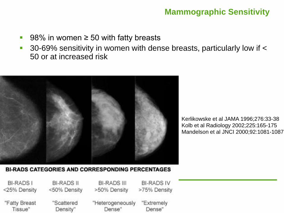
Mammographic Sensitivity
98% in women ≥ 50 with fatty breasts
30-69% sensitivity in women with dense breasts, particularly low if < 50 or at increased risk
Kerlikowske et al JAMA 1996;276:33-38 Kolb et al Radiology 2002;225:165-175 Mandelson et al JNCI 2000;92:1081-1087

17
Breast Density
Breast density contributes to
greater difficulty in detecting
cancer on mammography
Breast density is an independent
risk factor for cancer
Mammographic Density and the Risk and Detection of Breast Cancer
Boyd N et al. N Engl J Med 2007;356:227-236
18
Density notification legislation
Connecticut law since October 2009 requires women be
informed of their breast density and be offered supplementary screening if dense.
Illinois requires insurers to cover ultrasound screening if breasts are heterogeneously dense or extremely dense.
NY, Texas, California have passed legislation and many others are working on it.
Federal legislation for density notification is being considered.
U-systems sponsoring a large multi-institution study to determine screening benefit.

19
Despite legislation…
Lack of evidence (mortality benefit) that
supplementary screening is beneficial.
Breast density assessment by radiologists is still
subjective!
20
Screening US
Detects some cancers not seen on mammography
Especially in high risk and dense breasts
No long term studies on mortality to show benefit
Very operator dependent
Time consuming
Many false positives
Combined Screening With Ultrasound and Mammography vs. Mammography Alone in Women at Elevated Risk
of Breast Cancer Berg et al; JAMA. 2008;299(18):2151-2163
21
Whole breast ultrasound
Remove operator dependence
Improve reliability of exams
Facilitate measurements for follow up exams
Correlation with mammography, MRI
Quick whole breast scanning

22
Breast MRI
Anatomical detail and functional/physiologic information
Cancers require blood supply in order to grow.
Growing cancers recruit and develop new blood vessels in order to supply them.
These rapidly forming blood vessels are defective/immature.
When you give a dye (Gadolinium) that circulates through blood vessels, it will leak out of defective ones and appear on the image as bright areas.

23
High Risk Screen
Staging a known breast cancer
Assess Response to chemotherapy
Assess Implant Integrity
Carcinoma of unknown primary
Problem Solving
MRI Indications

24
MRI advantages / disadvantages
Not affected by breast density
No radiation
Gadolinium is taken up by abnormal tumor blood vessels
High sensitivity for invasive breast cancer and for DCIS
(higher than mammography)
Quality of exam and interpretation currently variable.
Biopsy capability not universally available. (compare with
mammography in early 90’s)
ACR accreditation for breast MRI is here and will be
mandatory beginning January 2012.
Very costly; follow ups may not be covered
Claustrophobia, discomfort
25
High risk screening with MRI:
Who needs it and who doesn’t?

26
Recommend Annual MRI Screening (Based on Evidence*)
BRCA mutation
First-degree relative of BRCA carrier, but untested
Lifetime risk 20–25% or greater, as defined by
BRCAPRO or other models that are largely dependent
on family history
*Evidence from nonrandomized screening trials and observational studies.
American Cancer Society Guidelines for Breast Screening with MRI as an Adjunct to Mammography Saslow et al; CA Cancer J Clin 2007; 57:75-89

27
Recommend Annual MRI Screening (Based on Expert
Consensus Opinion)
Radiation to chest between age 10 and 30 years
Li-Fraumeni syndrome and first-degree relatives
Cowden and Bannayan-Riley-Ruvalcaba
syndromes and first-degree relatives
American Cancer Society Guidelines for Breast Screening with MRI as an Adjunct to Mammography Saslow et al; CA Cancer J Clin 2007; 57:75-89
28
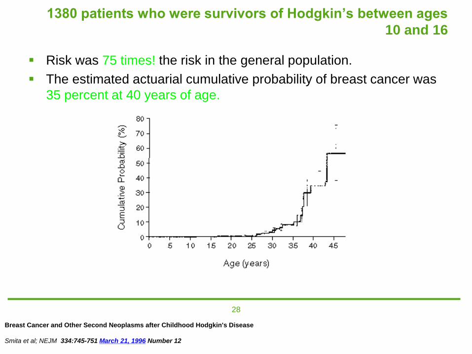
1380 patients who were survivors of Hodgkin’s between ages
10 and 16
Risk was 75 times! the risk in the general population.
The estimated actuarial cumulative probability of breast cancer was
35 percent at 40 years of age.
Breast Cancer and Other Second Neoplasms after Childhood Hodgkin's Disease
Smita et al; NEJM 334:745-751 March 21, 1996 Number 12

29
Insufficient Evidence to Recommend For or Against MRI Screening
Lifetime risk 15–20%, as defined by BRCAPRO or other models that are largely dependent on family history
Lobular carcinoma in situ (LCIS) or atypical lobular hyperplasia (ALH)
Atypical ductal hyperplasia (ADH)
Heterogeneously or extremely dense breasts on mammography
Women with a personal history of breast cancer, including ductal carcinoma in situ (DCIS)
American Cancer Society Guidelines for Breast Screening with MRI as an Adjunct to Mammography Saslow et al; CA Cancer J Clin 2007; 57:75-89
30
Recommend Against MRI Screening (Based on Expert
Consensus Opinion)
Women at <15% lifetime risk
American Cancer Society Guidelines for Breast Screening with MRI as an Adjunct to Mammography Saslow et al; CA Cancer J Clin 2007; 57:75-89

31
If you’re having annual MRI why do you still need a mammogram?
All studies have shown some cases of cancers
which were missed by MRI and were detected
with mammography (mostly low and
intermediate grade DCIS with calcifications).
32
MRI screening: advantages and disadvantages
Extremely sensitive for breast cancer
Variability in background parenchymal enhancement.
Hormonal issues. Ideal scanning is between days 7-14
of cycle.
Recall rate goes down in screeners having had one or
more prior exams.
Expensive and time consuming.
No data on mortality

33
Molecular breast imaging
Physiologic (vs. anatomic) imaging.
Metabolically active tissues will take up Tc99m Sestamibi. (Same isotope used for cardiac scanning.)
Uses detectors similar to mammography.
No compression. Breast is immobilized.
Exam takes 40 minutes (10 minutes per breast)
Current FDA approved techniques use 20 mCi.
Ongoing research on use of lower doses.
Current vendors: Dilon (BSGI) and Gamma Medica (LumaGEM MBI)

34
MBI advantages
Not impaired by breast density or presence of implants or free silicone.
High sensitivity and relatively good specificity.
Well tolerated.
Fewer false positives (vs. ultrasound and MRI).
Relatively less expensive (vs. MRI).

35
Radiation from MBI
Higher radiation dose to whole body (6.2 to 9.4 mSv = 2 to 3 years of natural background radiation exposure).
Compared with mammography (0.44 to 0.56 mSv, = 2 months of natural background radiation.)
The organs receiving the highest doses and therefore at greatest risk for cancer induction from radionuclide administration are the colon, lungs, and bladder. Not breast!
Potential impact for high risk women having frequent screening.

36
Dedicated Dual-Head Gamma Imaging for Breast Cancer Screening in Women with
Mammographically Dense Breasts
936 women with dense breasts and normal screening mammograms enrolled and followed for 12 months
Results: 11 cancers
Diagnostic yield:
mammo: 3.2/1000
MBI: 9.6/1000
combined MBI/mammo: 10.7/1000
Significant increased detection rate but at increased dose
Rhodes et al; Radiology January 2011 258:106-118

37
Recommended references
Mammography Saves Lives: information about the controversies in screening mammography and the recommended guidelines. http://www.mammographysaveslives.org/
American Cancer Society: general information about breast cancer, detection and treatment, as well as screening guidelines. http://www.cancer.org/Cancer/BreastCancer/DetailedGuide/index
American College of Radiology: information about individual tests and procedures. http://www.radiologyinfo.org/en/sitemap/system.cfm?sistem=breast
Information on breast density and supplemental screening: http://www.areyoudense.org/