a hand book of brachial plexus blcok - isakanyakumari
TRANSCRIPT
A HAND BOOK OF BRACHIAL PLEXUS BLCOK
From the author’s Desk
Dear Friends,
It gives me immense pleasure to bring out this booklet on brachial plexus block. My aim is to reach out mainly to postgraduate students and practitioners with this ready reckoner on brachial plexus block
Nowadays, the buzzword as far as regional anaesthesia goes in ultrasound guided blocks. But coming from the Indian subcontinent we have limited access to such equipment even in tertiary care centres. Hence, I have focused on how to perform these blocks with the cost effective equipment available to us viz- the peripheral nerve locator. It should hold a place in the armamentarium of any practicing anaesthesiologist with a serious interest in performing regional blocks.
This booklet covers brachial plexusw blocks in most of its forms. I hope it will be a useful guide.
Dr.R.Silamban

BRACHIAL PLEXUS BLOCK
INTRODUCTION:
Brachial plexus block is one of the most commonly used peripheral nerve blocks in clinical practice. Itcan be used as the sole anaesthetic tecyhnique or in combination with general anaesthesia for intraoperative and postoperative analgesia. Continuous catheterization of the brachial plexus is one of the best methods of postoperative analgesia.
The common sites of approach to the brachial plexus are:
a. Interscalene approachb. Supraclavicular approachc. Infraclavicular approachd. Axillary approache. Posterior approach
It is a must for the practicing anaesthesiologist to be familiar with all the above approaches as well as each one’s advantages and limitations.
HISTORY OF BRACHIAL PLEXUS BLOCKADE
The bock was first performed by William Steward Halsted in 1889. He directly exposed the brachial plexus in the neck to perform the block and used cocaine. Hirschel first described the percutaneous approach to the brachial plexus. Kulenkampff first described

the classicial supraclavicular rpproach to the brachial plexus. The subclavian perivascular block was first described by Winnie and Collins. This approach became popular as it was associated with less incidence of pneumothorax than the Kulenkampff approach. The infraclavicular approach was first developed by Raj, an anaesthesiologist of Indian origin practicing in USA. The axillary rpproach was first performed by Accardo and Adriano in 1949.
ANATOMY
Formation of the Plexus
Roots: The plexus is formed by the anterior primary rami of C5 to C8 nerves along with the bulk of T1 nerve. Occasionally there may be a contribution from C4 or T2 nerves leading to the formation of pre-fixed or post-fixed plexus. The roots emerge from the respective intervertebral foramina to enter the perivascular shealth.
Trunk: Sandwiched between the scalenus anterior and medius muscle, the roots combine to form the trunks. The C5 and C6 roots combine to form the upper trunk, C7 continues
as the middle trunk and C8 and T1 combines to form the lower trunk
Divisions: Behind the clavicle the trunks divide into a anterior and posterior divisions and stream into the axilla.
Cords: In the upper part of the axilla the six divisions combine to form lateral, medial and posterior cords.
-The lateral cord is formed by the union of anterior division of the upper and middle trunks.
-The medial cord is the continuation of the anterior division of the lower trunk.-The posterior cord is due to union of the posterior divisions of all the three trunks
Terminal branches: Lower down in the axilla the cords give rise to terminal branches namely the ulnar, median and radial nerves.

Relationship of the brachial plexus:
Roots: These lie between the scalenus anterior and medius muscles. It lies above the second part of the subclavian artery. The classical interscalene approach to the brachial plexus blocks is at the root level.
Trunks: These lie in close relationship to the subclavian artery above the clavicle. Here also they are sandwiched between the scalene muscles. The trunks extend upto the lateral border of the first rib. The subclavian perivascular approach blocks the plexus at this level.
Divisions: they start at thelateral border of the first rib and lie behind the clavicle. The rib hitching technique causes blockade at this level.
Cords: The divisions unite to form cords at the upper part of the axilla. They remain grouped around the axillary artery. The infraclavicular approach causes blockade at the junction of cords and divisions.
Terminal Branches: They are formed lower down in the axilla. The reorganization of the cords to form the terminal branches occurs at the lateral border of the pectoralis minor muscle. The axillary approach causes blockade at this level

ANATOMICAL CONSIDERATIONS:
The anatomic factors which determine the success and complications of the brachial plexus blockade are
-The perivascular sheath-The vertical arrangement of the cervical roots-The interconnections – This is due to combining, dividing, recombining and
redividing of the original five cervical roots.-The relationship of the site of needle entry to vital structures.
Perivascular Sheath – Its Importance
The perivascular sheath is a fibrous sheath covering the brachialPlexus in its entirety. It extends from the origin of the scalene muscles down to middle of upper arm. The potential space formed by this sheath can hold upto 80-100 ml of local anaesthetic.
This sheath gives a classical ‘Pop Off ‘ feeling when pierced by the needle.
This sheath is the single most important factor in determining the success of bracial plexus blockade. The plexus can be blocked by introducing a needle at any point along the sheath. But the site of needle entry determines which components arepreferentially blocked and which compenents are spared. However this can be overcome to a certain extent by increasing the volume of the local anaesthetic and by applying proximal or distal digital pressure.
The suggestion that the covering is discontinuous with septa subdividing the space into separate compartments that clinically prevent the spread of local anaesthetic
However these septal divisions are more prominent in the axilla than above. This probably is the reason for frequent sparing of the radial and musculocutaneous nerves during axillary blockade.As the septa are more prominent in the lower part, there may be more sparing in the infraclavicular approach than in the supraclavicular approach. The perivascular sheath may also be discontinuous leading to spillage of drug out side thesheath.
Vertical arrangement of roots:
This arrangement of the brachial plexus assumes significance in the classical interscalene approach. Here the needle is applied close to the C5 and C6 nerve roots. As the roots are vertically arranged the local anaesthetic will have to travel caudally to reach C8 and T1 level. If the caudal travel of the local anaesthetic is deficient then these roots may be spared leading to poor analgesia in the ulnar nerve distribution. In the infraclavicular approach sparing of the ulnar nerve is rarely seen.
The interconnections:
The interconnections between the original five cervical roots means that the cutaneous distribution of the individual nerve terrioratories differs from the myotomal and detematomal pattern and that the muscular and other deep structures do not underlie the sensory distribution of that nerve. For example blockade of the ulnar nerve at the elbow, produces sensory loss on the ulnar side of the hand but motor loss of the flexor muscles on the anterior aspect.
Site of needle entry and complications:
--If site of needle entry is at the C6 level, r=the chances of epidural and subarachnoid injections are more. The chances of vertebral artery puncture, phrenic and recurrent laryngeal nerve paralysis are also higher.
--If the site of needle entry is close to the clavicle the chances of pneumothorax and subclavian artery puncture are more.
--If the site of needle entry is below the clavicle as in the infraclavicular approach, complication rate is much less than in the above routes but the chances of incomplete blockade are more.
--In the axillary site apart from accidental artery puncture other complications are less but chances of incomplete blockade increases.
SUPRACLAVICULAR APPROACH
Introduction:
Cornerstone of upper extremity of regional anaesthesia. It is used extensively in clinical practice. Various techniques of reaching the brachial plexus from the supraclavicular site are described. In this approach the drug is delivered at the level of the trunks which is the most compact part of the brachial plexus.
In any supraclavicular technique the chance of pneumothorax will always have to be kept in mind. The dome of the pleura is in close relationship to the convacity of the first rib. If the needle is introduced close to the lateral border of the clavicular head of the sternocleidomastoid as usual, the chance of pleural damage is high. In this technique which we are going to describe, the needle is introduced between the two scalene muscle

which is a safe 1.5cm away from the dome of the pleura. It is always better to have a safe distance 1cm from the pleura.
Distrbution of consistent anaesthesia
It can be used for any upper extremity procedure from the shoulder to the fingers. Distal digital pressure makes the drug to rise up to the level of the roots and making surgery in the alteral half of the clavicle also possible.
Position:
Patient is supine, hands on the side, head turned to the opposite side.
Landmarks
Clavicle, lateral head of sternocleidomastoid, scalenus anterior and medius muscle, external jugular vein.
Site of needle entry
In the interscalene groove about 1.5 to 2 cm above the clavicle.

Technique
The patient is asked to lift his head up to make the lateral head of sternocleidomastoid prominent. After the lateral border of the sternocleidomastoid is identified, the fingers are slowly moved laterally to feel the anterior and medial scalene muscles and the groove between them is appreciated. A point 1.5 to 2cm above the clavicle is marked in the groove and a 5cm long needle with nerve locator attached is entered through the skin. The needle is directed towards the ipsilateral toe. The needle is advanced till a ‘pop off’ is felt signifying entry into the perivascular sheath. Now the nerve locator isswitched on at 1mA current. At this point muscle twitches are invariably seen. A current greater than 1mA may be uncomfortable to the patient. As the brachial plexus is in close proximity to the inferior surface of the scalene muscle, difficulty in nerve location does not occur. The current is slowly reduced -.2mA to get the desired response below 0.6mA. At this point the drug is given.
Motor response to nerve stimulation
The following motor responses are elicited and result in the same success rate.
-Pectoralis muscle twitch-Deltoid muscle twitch-Triceps muscle twitch-Biceps muscle twitch-Hand and forearm muscle twitch
Rhythmic movements of the shoulder, elbow, hands and fingers are seen. Form ideal results the twitches should be obtained at less than 0.6mA. Twitches of the neck muscles and movement of the scapula are not considered.
“The initial sign of supraclavicular blockade is loss of should abduction which occurs before sensory block’.
Advantages1. Drug is delivered at the level of the trunks which is the most compact part of the brachial plexues. So success rates are high.2. Rapid onset3. Dense anaesthesia4. Predictable result5. As the needle is entered about 1.5cm above the calvicle, chance of
phenumothraox are very rare.
INFRACLAVICULAR APPROACH
There are 2 approachesa) Raj infraclavicular blockb) Lateral infraclavicular block
Position: It is the same for both approaches
Patient supine, neck turned to opposite side, arm preferably in abduction. In cases where arm abduction is not possible it can be done in any position.
RAJINFRACLAVICULAR BLOCK
Landmarks:-Acromioclavicular joint-Sternoclavicular joint-Midpoint of clavicle-Brachial artery pulsations in the axilla
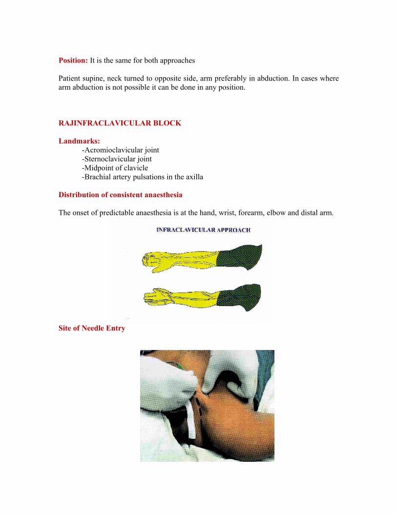
Distribution of consistent anaesthesia
The onset of predictable anaesthesia is at the hand, wrist, forearm, elbow and distal arm.
Site of Needle Entry

A point 2 to 2.5cm below the midpoint of the clavicle
Technique:
Mark acromioclavicular and stemoclavicular junction. The line joining the two points determines the length of the clavicle. Market the mid point of the line. Mark another point 2 to 2.5cm below the first point. This is the site of needle entry.
A7.5cm insulated needle connected to a nerve locator is preferred. After skin infiltration with 2% xylocaine, the insulated needle is inserted at 45 degree angle into the skin and directed towards the axillary artery pulsations in the axilla. This maneuver directs the needle laterally posteriorly and slightly caudally.
A initial current of 1.5 to 2mA is preferred. As the needle passes through the pectoralis muscle twitches can be seen due to direct stimulation of the muscle. Once the muscle is crossed the twitches cease. Now the current is reduced to 1mA and needle is advanced to get the nerve response. A lower current causes less patient discomfort and is preferred. The needle is directed in a cephalocaudal are to search for motor response. In thin individuals the response may be obtained at a depth of 3cm. In obese the response may be obtained at about 5-7.5cm.
Motor response:
Flexion of the wrist and the fingers due to nerve stimulation is preferred. Stimulation of the radial and ulnar nerves leading to any movement in the region of forearm, wrist and fingers can be taken as the end point. Contraction of the deltoid and biceps are not acceptable as the axillary and musculocutaneous nerve leave the plexus at a higher level. For a successful blockade, response at a current less than 0.4mA is required. Response to higher current leads to unsuccessful and incomplete blockade.
LATERAL INFRACLAVICULAR BLOCK
This block was developed to present a more constant landmark, the coracoid process.
Landmark - Coracoid process
Position - Similar to Raj technique

Site of needle entry - 1cm medial and caudal to corocoid process
Technique
The coracoid process of scapula is identified. Marked shrugging helps identify the coracoid process better. A point 1cm medial and 1cmcaudal to the medial aspect of coracoid process is marked. This is the point of needle entry. After skin infiltration with local anaesthetic the stimulation needle is inserted directly perpendicular to the skin. The usual depth of desired response is about 3cm in thin individuals but may vary upto 7.5cm in obese individuals. The needle size, motor response and eliciting parasthesia are similar to the Raj approach
Interpreting responses to nerve stimulation
There are some common responses that occur due to nerve stimulation. Then corrective measures will have to be taken to obtain the desired response.
Stimulation Motor responses Explanation Corrective actionPectrolis muscle direct stimulation
Arm abduction Shallow needle placement
Continue to advance the needle
Axillary nerve Deltoid muscle contraction
Needle placed inferiorly
Withdraw needle to the skin and reinsert with a superior angulation
Musculocutaneous nerve
Biceps twitch Needle placed superiorily
Withdrfaw the needle to skin and reinsert with caudal angulation
Advantage:-Decreased incidence of lung injury and pneumothorax-Decreased incidence of arterial injury-Can be performed, with the hand in any position-Ideal site for continuous catheter insertion-More patient comfort after catheterization-The chances of unilateral phrenic nerve blockade are nil. In the classical
approach it is almost 100% and about 30-50% in the supraclavicular approach.

Disadvantages:-The plexus is deeply placed at this site. The pectoralis major and minor muscles
will have to be pierced to reach the plexus. So, more painful to the patient-As the plexus is deeply placed, blind approach is not possible. Eliciting
paraesthesia is more difficult. So a nerve locator or ultrasound guidance becomes mandatory for performance of the block.
-Because of the above problem a more deep sedation may be required during performance of the block
-Surgeries on the shoulder and upper humerus cannot be done.
Potential Problems:
Complications are less when compared to classical and supraclavicular approaches. Pneumothorax and arterial puncture are possible and mostly related to a wrongly directed needle.
Despite adequate initial needle position, if the catheter is threaded too much it may be away from the plexus resulting the ineffective blockade.
Usage of a large volume of local of local anaesthetic may lead to systemic toxicity.
Torniquet application can be a problem as musculocutaneous nerve is not blocked.
CLASSICAL APPROACH
One of the first techniques of brachial plexues block to be established. This is not frequently practiced nowadays.
Landmarks
Clavicular head of sternocleidomastoidExtenarnal jugular veinChassaignac tubercle (C6)Cricoid cartilage

Position:Patient is supine, head partially turned to the opposite side to be blocked and arm by the side.
Distribution of consistent anaesthesia
This drug is usually consistent at the shoulder, arm and elbow. The forearm, wrist and fingers may sometimes be blocked but ideally requires more distal approach to the brachial plexus.
Technique:
A3.5 cm or 5cm long needle is used. The needle is introduced at the level of the Chassaignac’s tubercule with a caudal angulation. The needle should not be introduced perpendicularly or with a cephaloid direction as there is a chance of cervical cord injury. Usually motor response is obtained at a depth of 1-2cm.

Motor response to nerve stimulation:
Twitches of the shoulder and arm are seen. Administration of local anaesthesia to any of the above responses has the same success rate. The commonest response is the twitching of the deltoid muscle (c5,6)
Diaphragmatic movement due to stimulation of the phrenic nerve and scapular movement due to stimulation of the serratus anterior muscle or dorsal scapular nerve should not be considered..
AXILLARY RPPROACH
It is a very basic nerve block technique and very commonly employed. Here the durg is delivered within the perivascular sheath close to the axillary artery.
Distribution of consistent anaesthesia
This block provides anaesthesia of the lower part of arm, elbow, forearm, wrist and hand. As the musculocutaneous nerve leaves the plexus much before the site of needle entry, tourniquet pain is not abolished.
Position:
Patient is in supine position with head turned away from the side to be blocked. The arm is abducted and to form a 90 degree angle in the elbow. Excessive abduction makes palpation of axillary artery pulse difficult and also stretches and fixes the brachial plexus making it vulnerable to injury during needle placement.
Landmarks:Axillary artery pulsationCoracobrachalis musclesPectrolis major muscle

-The axildlary artery pulsation are best felt high in the axilla between the coracobrachalis and pectrolis major muscle.
-When location of the pulse is not immediately apparent, adduction of the arm decreases the resistance and makes the groove between the coracobrachalis and pectrolis major muscle prominent. This maneuver helps to identify the pulse better.
Technique
One finger is kept on the axillary pulse and a 5cm needle is advanced justg below the pulse. In most patients motor response obtained at 2cm depth at about 1mA current. The needle tip is adjusted to get a motor response below 0.4mA and drug is injected.
Motor response
Twitches in the region of the wrist and fingers are indicators of successful blockade. Biceps and triceps twitch should not be considered.

Interpreting responses to nerve stimulation
Local twitche of arm muscle
Direct stimulation of biceps and
triceps
Needle has been inserted for superior or inferiorly
Withdrawl the needle and direct
Needle contacts bone at 3cm
Needle stopped by humerus
Brachial plexus missed
Withdraw the needle to skin and reinsert at an angle of 15-30 degree superiorly or inferiorly
POSTERIOR APPROACH
A very rarely used approach. It is not performed in many centres. It does not some indication especially in a pain clinic setup.
Landmarks: C6 spineC7 spine
Position:
Can performed in sitting or lateral decubitus position
Distribution of consistent anaesthesia
Mainly around the shoulder upto the middle of the clavicle and upper arm. So this is useful in surgeries involving the clavicle, shoulder and upper arm.
Site of needle entry
A point 3cm lateral to the midpoint of C6 and C7 spine
Technique
In the sitting position C6 and C7 spine are identified. A point 3cm lateral to the midpoint of the spine is the site of needle entry. The needle entered perpendicular to the floor and directed slightly laterally. The depth at which stimulus is obtained is between 4-6cm. A nerve locator starting with a current strength of 3.5mA is connected to the needle. Response to nerve stimulation is mainly movement of the shoulder muscles. If response is obtained at a current of 0.4mA, drug can be given.
Indication-Mainly surgeries on the clavicle shoulder and upper arm-For relief of severe pain due to apical lung cancer inadvertently infiltrating the
brachial plexus.
Disadvantage
-As it is deeply placed it is a painful procedure-Surgeries on the forearm, wrist and fingers cannot be done-Chances of neuraxial catheter placement and blockade are high
CONTINUOUS CATHETRIZATION
Catheterization of the brachial plexus for postoperative pain relief is becoming increasing popular nowadays. It is one of the best modality of pain relief. Routinely catheters can be kept for upto 5 days in the postoperative period. Daily cleaning and dressing of the catheter entry site is a must.
These cathetes are similar to the epidural catheters with markings. They are about 30cm in length.
The catheters are placed about 3cms past needle tip. If placed more the chances of moving away from the plexus may arise.
The catheter can be usedfor intermittent or continuous drug administration. PCA pumps can also be attached.
PHARMACOLOGY
The drugs used for anaesthesia are1.Lignocaine 2% with adrenaline2. Bupivacaine 0.5%3.The addition of opioids to the local anaesthetic ensures a more intense pain
relief
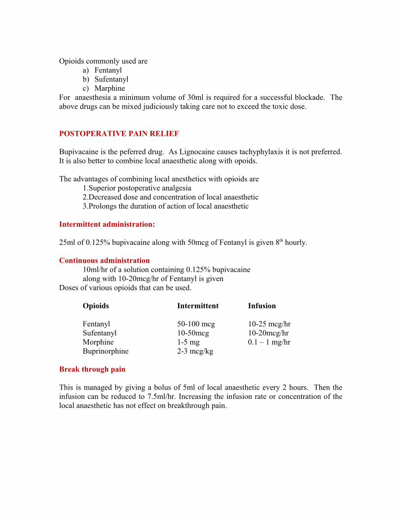
Opioids commonly used area) Fentanylb) Sufentanylc) Marphine
For anaesthesia a minimum volume of 30ml is required for a successful blockade. The above drugs can be mixed judiciously taking care not to exceed the toxic dose.
POSTOPERATIVE PAIN RELIEF
Bupivacaine is the peferred drug. As Lignocaine causes tachyphylaxis it is not preferred. It is also better to combine local anaesthetic along with opoids.
The advantages of combining local anesthetics with opioids are1.Superior postoperative analgesia2.Decreased dose and concentration of local anaesthetic 3.Prolongs the duration of action of local anaesthetic
Intermittent administration:
25ml of 0.125% bupivacaine along with 50mcg of Fentanyl is given 8th hourly.
Continuous administration10ml/hr of a solution containing 0.125% bupivacainealong with 10-20mcg/hr of Fentanyl is given
Doses of various opioids that can be used.
Opioids Intermittent Infusion
Fentanyl 50-100 mcg 10-25 mcg/hrSufentanyl 10-50mcg 10-20mcg/hrMorphine 1-5 mg 0.1 – 1 mg/hrBuprinorphine 2-3 mcg/kg
Break through pain
This is managed by giving a bolus of 5ml of local anaesthetic every 2 hours. Then the infusion can be reduced to 7.5ml/hr. Increasing the infusion rate or concentration of the local anaesthetic has not effect on breakthrough pain.

CONCLUSION:
In the days of frequent trauma to the upper limb especially with full stomach patients the routine use of brachial plexus block decreases complications and ensures patient comfort. A good learning under expert hands is a must to develop a good technical expertise. It is a must know regional technique for all anaesthesiologist.