a hand book on sickle cell diseasescic.cg.nic.in/pdf/sickle cell handbook_english.pdfa hand book on...
TRANSCRIPT

A Hand Book on Sickle Cell Disease
Sickle Cell Institute Chhattisgarh, Raipur (An Autonomous Institute of Government of Chhattisgarh)
Genetic lab, Department of Biochemistry, Pt. J.N.M. Medical College, Raipur-492001, Chhattisgarh, India
Phone & Fax: 91-(771) 2885505 Web: www.scic.co.in Toll free No: 155217

Please match following ‘Sickle Kundli’ before marriage
S. No. Father Mother
Possible Haemoglobin pattern of children
(This does not show the order of child births)
Advice related
to marriage
1
All Normal Get
married
2
50% Normal, 50% Carriers
Get married
3
50% Normal, 50% Carriers
Get married
4
All Carriers Get
married
5
All Carriers Get
married
6
25% Normal, 25% Patients, 50% Carriers
Think
7
50% Patients, 50% Carriers
Should not get married
8
50% Patients, 50% Carriers
Should not get married
9
All Patients Should not get married
Note: Men and women belonging to serial numbers 6, 7, 8 and 9, should not get married.

A Hand Book on
Sickle Cell Disease
Sickle Cell Institute Chhattisgarh, Raipur (An Autonomous Institute of Government of Chhattisgarh)
Genetic lab, Department of Biochemistry, Pt. J.N.M. Medical College, Raipur-492001, Chhattisgarh, India
Phone & Fax: 91-(771) 2885505 Web: www.scic.co.in, Toll free No: 155217Toll

Prologue
In India, the sickle cell gene is distributed across the country, predominantly in Chhattisgarh, Madhya Pradesh, Orissa, Jharkhand, Maharashtra, Gujarat, Andhra Pradesh, Kerala, Karnataka, Tamil Nadu and some Northeastern states. Initial screening for sickle cell allele across the state of Chhattisgarh has revealed that its prevalence as 10% in Chhattisgarh population. In some communities, the prevalence of the sickle cell allele is as high as 30%.
Although its prevalence is high, most of the individuals are unaware of basic scientific facts of this inherited genetic disorder. This condition poses a serious medical as well as socio-economic burden to the patients and their families. The social stigma associated with this disease further aggravates their suffering.
In purview of the suffering associated with this disease, the government of Chhattisgarh has established a dedicated Sickle Cell Institute in 2013 at Raipur to cater the needs of patients. The institute provides specialized treatment and counseling facilities to patients and their family members. Further, to maintain the availability of trained manpower to deal with patients of sickle cell disease, the institute provides education and training to healthcare professionals of the state.
Globally, India contributes significantly to the overall disease load, however a very little research work have been carried-out on highly diverse Indian populations. Being a disease prevalent in developing countries, much attention has not been paid by developed countries and requires special attention. Therefore, the ‘Sickle Cell Institute Chhattisgarh’ is dedicated to pursue cutting edge research to develop markers, drugs as well as specialized treatment modalities.
This Handbook is a sincere attempt to meet one of our goals of generating trained workforce of health professionals to fight with sickle cell disease. I hope this book will be helpful not only to health professionals and researchers but also to the general public to get acquainted with the current knowledge regarding SCD. Comments and suggestions are invited for further improvement of this handbook.
Prof. (Dr.) P. K. Patra Director General
Sickle Cell Institute Chhattisgarh Raipur, C.G.
A Handbook on Sickle Cell Disease
Published: First Edition, November 2014 Compiled and Prepared by: Sickle Cell Institute Chhattisgarh, Raipur Editorial Board: Dr. P.K. Patra, Director General, SCIC, Raipur Dr. P.K. Khodiar, Director-Medical, SCIC, Raipur Dr. L.V.K.S. Bhaskar, Senior Scientist, SCIC, Raipur Dr. Hrishikesh Mishra, Scientist (Bioinformatics), SCIC, Raipur Dr. Aditya Nath Jha, Scientist, SCIC, Raipur Authors & Contributors: Dr. L.V.K.S. Bhaskar, Senior Scientist, SCIC, Raipur Dr. Chandra Vikas Rathore, General Duty Medical Officer, SCIC, Raipur Dr. Radha Rani Sahu, Counselor, SCIC, Raipur Dr. Hrishikesh Mishra, Scientist (Bioinformatics), SCIC, Raipur Dr. Aditya Nath Jha, Scientist, SCIC, Raipur Dr. Ram K. Yadav, General Duty Medical Officer, SCIC, Raipur Ms. Jyoti Rathore, Training Officer, SCIC, Raipur Mr. Anand Deo Tamraker, Training Coordinator, SCIC, Raipur Design and Layout: Dr. Chandra Vikas Rathore, General Duty Medical Officer, SCIC, Raipur Mr. Anand Deo Tamraker, Training Coordinator, SCIC, Raipur Published by: © Sickle Cell Institute Chhattisgarh, Raipur

Table of Contents
Unit 1 Introduction to Sickle Cell Disease and Pathophysiology
1.1 Sickle Cell Disease/Anaemia 1
1.2 Inheritance 1
1.3 Situation 2
1.4 Pathophysiology 2
1.5 Sickle Cell Trait 3
Unit 2 Laboratory Diagnosis of Sickle Cell Disease
2.1 Haemoglobin Structure and Forms 5
2.2 Haemoglobin S 6
2.3 Rapid Screening of Sickle Haemoglobin 6
2.3.1 Solubility Test 7
2.3.2 Sickling Test 8
2.4 Identification of Normal and Abnormal Haemoglobin Types 10
2.4.1 Electrophoresis 10
2.4.2 Isoelectric Focusing 13
2.4.3 Capillary Electrophoresis 14
2.4.4 High Performance Liquid Chromatography 15
2.5 Newborn Screening 16
2.6 Molecular Diagnostics 17
2.6.1 Restriction Fragment Length Polymorphism 17
2.6.2 DNA Sequencing 18
2.7 The Future of SCD Diagnostics 18
Unit 3 Clinical Manifestations and Treatment of Sickle Cell Anaemia
3.1 General Health Management 21
3.2 Fever and Bacteremia 22
3.3 Dactylitis or Hand-Foot Syndrome 24
3.4 Aplastic Crisis 25
3.5 Splenic Sequestration 26
3.6 Pain 28
3.7 Priapism 32
3.8 Neurologic Complications 33
3.9 Excessive Iron Stores 36
3.10 Lung Disease 37
3.11 Leg Ulcer 39
3.12 Deep Jaundice 40
3.13 Loss of Vision 41
3.14 Pregnancy & Contraception 41
3.15 Newborn Screening 43
3.16 Models of Management 44
3.17 Outlook 45
3.18 Conclusions 46
Unit 4 Counseling
4.1 Spread 47
4.2 Identification of Patient 47
4.3 Age-wise Counseling 48
4.4 Counseling for Diet Management 50
4.5 Do’s & Don’ts 51
Bibliography 52

1 | A H a n d B o o k o n S i c k l e C e l l D i s e a s e
Unit-1
Introduction to Sickle Cell Disease and Pathophysiology
1.1 Sickle Cell Disease/Anaemia
Sickle cell disease (SCD) is a life threatening autosomal recessive genetic disorder
resulting from inheritance of abnormal genes from both parents. Normal red blood cells
(RBCs) are biconcave disc shaped and move smoothly through the blood capillaries. The
RBCs are produced in bone marrow and average life of normal RBCs is about 120 days.
Biconcave disc shape of RBCs changes to sickle shape under low oxygen tension due to
polymerization of faulty haemoglobin called HbS arising out of a point mutation in beta
globin gene. The life span of RBCs in SCD patients is only about 10 to 20 days and the
bone marrow can't replace them fast enough. As a result there is decrease in number of
RBCs in the body and the RBCs don‟t contain sufficient amount of haemoglobin
(hypochromia). In SCD the RBCs become sickle or crescent shaped which are stiff &
sticky and tend to block the blood flow in small capillaries. Blocked blood flow causes
ischemia leading to severe pain and gradual damage to organs.
Sickle cell gene is commonly believed to be associated with African ancestry and
malaria endemic areas. Besides Africa, it is found around Mediterranean, Middle-East and
India. The disease gene has spread also to Europe, America & Caribbean through
migration of human populations. In India, it is prevalent in Chhattisgarh, Odisha,
Maharashtra, Gujarat, Madhya Pradesh, Telangana, Andhra Pradesh and some parts of
Tamil Nadu & Kerala.
1.2 Inheritance
As discussed before, being an autosomal recessive disorder, abnormal beta globin
gene from both mother and father are required to be inherited (homozygous) in offspring to
2 | A H a n d B o o k o n S i c k l e C e l l D i s e a s e
cause SCD. If a person has only one abnormal beta globin gene inherited (heterozygous)
either from mother or father, it is referred as sickle cell trait and are usually asymptomatic,
but can pass the abnormal beta globin gene to their progeny.
1.3 Situation
The Sickle cell anaemia has no available cure. However, treatments to improve the
anaemia and lower complications can help with the symptoms and complications of the
disease in both children and adults. Blood and marrow stem cell transplants may offer a
cure for a small number of people.
The Sickle cell anaemia varies from person to person. Some people who have the
disease have chronic (long-term) pain or fatigue (tiredness). However, with proper care
and treatment, many people who have the disease can have improved quality of life and
reasonable health.
Because of improved treatments and care, people who have sickle cell anaemia are
now living into their forties or fifties, or longer.
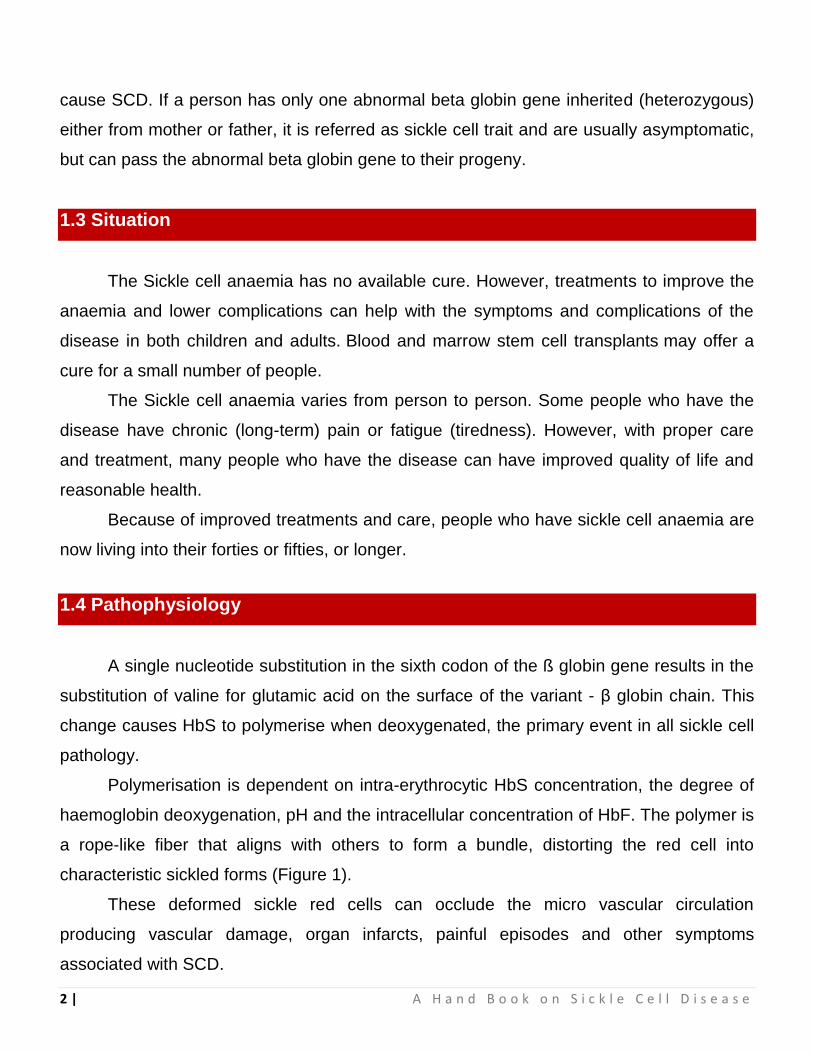
1.4 Pathophysiology
A single nucleotide substitution in the sixth codon of the ß globin gene results in the
substitution of valine for glutamic acid on the surface of the variant - β globin chain. This
change causes HbS to polymerise when deoxygenated, the primary event in all sickle cell
pathology.
Polymerisation is dependent on intra-erythrocytic HbS concentration, the degree of
haemoglobin deoxygenation, pH and the intracellular concentration of HbF. The polymer is
a rope-like fiber that aligns with others to form a bundle, distorting the red cell into
characteristic sickled forms (Figure 1).
These deformed sickle red cells can occlude the micro vascular circulation
producing vascular damage, organ infarcts, painful episodes and other symptoms
associated with SCD.

3 | A H a n d B o o k o n S i c k l e C e l l D i s e a s e
There are two essential pathological processes: haemolysis and vaso-occlusion.
Haemolysis results in anaemia and a functional deficiency of nitric oxide
which results in vascular endothelial damage and may be responsible for
complications such as pulmonary hypertension, priapism and stroke.
Vaso-occlusion causes acute and chronic ischaemia and is responsible for
acute pain and organ damage.
Sickle cell disease refers to not only
patients with sickle cell anaemia but also to
compound heterozygotes where
one β globin gene mutation includes the
sickle cell mutation and the second β globin
allele includes a gene mutation other than
the sickle cell mutation, such as mutations
associated with HbC, HbS β-thalassemia,
HbD, and HbO Arab. In sickle cell disease,
HbS is >50% of total haemoglobin.
1.5 Sickle Cell Trait
People who inherit a sickle haemoglobin gene from one parent and a normal gene
from the other parent have sickle cell trait. In sickle cell trait the HbS is <50% of total
haemoglobin. People who have sickle cell trait usually have few, if any, symptoms and
lead normal lives.
People who have sickle cell trait can pass the sickle haemoglobin gene to their
children. The following image shows an example of an inheritance pattern for sickle cell
trait.
Figure 1 : The normal Red Blood Cells and Sickle Cells

4 | A H a n d B o o k o n S i c k l e C e l l D i s e a s e
Example of an Inheritance Pattern for sickle cell trait and disease are given in figure-2
Figure 2 : Shows Different Patterns of How Sickle Haemoglobin Genes are Inherited: A person inherits two haemoglobin genes one from each parent. A normal gene will make normal haemoglobin (A). A sickle haemoglobin gene will make abnormal haemoglobin (S).
When both parents have a normal gene and an abnormal gene, each child has a 25
percent chance of inheriting two normal genes; a 50 percent chance of inheriting one
normal gene and one abnormal gene; and a 25 percent chance of inheriting two abnormal
genes (Figure 2).

5 | A H a n d B o o k o n S i c k l e C e l l D i s e a s e
Unit 2
Laboratory Diagnosis of Sickle Cell Disease
2.1 Haemoglobin Structure and Forms
Haemoglobin (Hb) is the iron-containing protein made up of four globin chains.
Depending on their structure these globin chains are known as alpha, beta, gamma, and
delta. The function of haemoglobin and its ability to transport oxygen is mainly determined
by the globin chains that are present in it. The normal haemoglobin types include
Haemoglobin A, Haemoglobin A2 and Haemoglobin F (fetal haemoglobin/HbF).
Haemoglobin A contains two alpha and two beta protein chains (α2β2) and is comprised
95%-98% of Hb found in adults. Haemoglobin A2 has two alpha and two delta protein
chains (α2δ2) and makes up to 2%-3% of Hb found in adults. Haemoglobin F has two
alpha and two gamma protein chains (α2γ2) and, is mainly produced by the fetus
during pregnancy its production usually falls shortly after birth and reaches 1%-2% of Hb
found in adults by 1-2 years. Newborn infants have two types of γ chains, Gγ (HbG2) and
Aγ (HbG1) which has glycine and alanine residue respectively at position 136.
Furthermore, the ε chain has been observed in early human embryos (Table 1). An
investigation of a haemoglobin disorder typically involves tests that determine the types
and amounts of haemoglobin present in a person's sample of blood.
Greek Designation
Greek Name
No. of Amino Acids
Observed in Chromosome
α Alpha 141 Fetal and adult life 16
β Beta 146 Adult life 11
Δ Delta 146 Adult life 11
γ Gamma 146 Fetal liver, spleen and bone marrow
11
ε Epsilon 146 Embryonic yolk
sac 11
Ζ Zeta 146 Early embryonic
Yolk sac 16
Table 1: Globin Chains in Hmoglobin

6 | A H a n d B o o k o n S i c k l e C e l l D i s e a s e
2.2 Haemoglobin S
Sickle haemoglobin (HbS) is a haemoglobin variant in which two normal α-globin
chains and two abnormal β-globin chains that contain a single amino acid substitution,
from glutamic acid to valine, on the 6th position of the beta globin chain, hence the
nomenclature of β6 gluval. The mutation in the β- globin chain results in decreased
solubility of de-oxygenated haemoglobin S that then forms rigid polymers that distort the
red cells in to the characteristic sickle shape. Classically, these red cells appear in the
form of a thin crescent with two pointed ends and lack central pallor. The polymerization of
deoxygenated haemoglobin S may cause the red cells to appear in one or more of the
following forms: envelope cells filament-shaped and crescent shaped.
2.3 Rapid Screening of Sickle Haemoglobin
To identify the
presence of sickle
haemoglobins, sickling and
solubility tests are used.
Further diagnosis is done by
using electrophoresis, HPLC
and molecular markers.
Sequence of the screening
and diagnostic tests to be
used for sickle cell disease is
depicted in figure 3.
Figure 3: Flow Chart for Sickle Cell Disease
screening

7 | A H a n d B o o k o n S i c k l e C e l l D i s e a s e
2.3.1 Solubility Test
In the solubility test is based on the relative
insolubility of haemoglobin S in the reduced state in
high phosphate buffer solution (metabisulfite is a
reducing agent). When whole blood is mixed with
the reducing agent, the haemoglobin S forms liquid
crystals and give a cloudy appearance to the
phosphate buffer solution. A transparent solution is
seen with other haemoglobins that are more
soluble in the reducing agent. A positive result is
indicated by a turbid suspension through which the
ruled lines are not visible. A negative result is
indicated by a transparent suspension through which the ruled lines are visible (Figure 4).
Whole blood anticoagulated with EDTA, heparin, or sodium citrate is acceptable.
Specimens may be stored at 4°C up to three weeks before testing. A positive control (AS)
containing 30-45% HbS and a negative control (AA) should be analyzed with each patient
specimen. Solubility test is a qualitative test and does not distinguish the difference
between haemoglobin S disease (SS) and haemoglobin S trait (AS). Preparation of
reagents and Methodology for solubility test in brief is given in Box 1.
Other abnormal haemoglobin variants (HbC) are known to cause sickling and will
give a positive solubility test. There are some physiologic sources of error that may lead to
false positive or false negative results. Erythrocytosis, hyperglobulinemia, extreme
leukocytosis, or hyperlipidemia may cause false positive results. Further an anemic
individual with Hb of < 7.0 g/dL may give a false negative result. Use of packed
erythrocytes (0.01 mL) for solubility test will correct for this error. In infants younger than 6
months and individuals with history of recent transfusion with normal erythrocytes, false
negatives may occur due to low concentration of HbS. Hence to confirm the presence of
HbS and differentiate between the two states (AS and SS), a haemoglobin electrophoresis
at an alkaline pH should be performed.
Figure 4: The Sickle Cell Solubility Test
Widely Used in Screening Projects

8 | A H a n d B o o k o n S i c k l e C e l l D i s e a s e
Box 1: Preparation of Reagents and Methodology for Solubility Test
Reagents
• Stock 2.58 M phosphate buffer: Dissolve 215 g K2HPO4 and 169 g KH2PO4 , 5g
sodium dithionate and 1g saponin in distilled water. Then make up to 1Litre with
distilled water. Adjust pH to 6.5-6.8.
• Buffer may be kept at 4°C and used as long as they are clear and uncontaminated.
Method
• Pipette 1 ml of phosphate buffer into a properly labelled tube.
• Add two drops (~20 µl whole blood) blood into the tube with a disposable Pasteur
pipette and mix well.
• Add a pinch (about 10-20 mg) of sodium dithionite or sodium metabisulphite powder
to the tubes.
• Mix and read immediately.
• Run positive and negative control bloods by following the same steps given above.
2.3.2 Sickling Test
In the Sickling test we create the conditions at which oxygen tension decline to
induce the Sickling process of HbS in RBCs. When a drop of blood is sealed between a
cover slip and a slide, the decline in oxygen tension due to oxidative processes in the
blood cells leads to sickling. In this method blood drop is added with sodium metabisulfite,
a chemical reducing agents which rapidly reduces oxyhaemoglobin to reduced
haemoglobin to accelerate sickling. In positive samples the typical sickle-shaped red blood
cells will appear (Figure 5). Occasionally the preparation may need to stand for up to 24°C.
In this case put the slides in a moist Petri dish to maintain temperature. False negative
results may be obtained if the metabilsulphite has deteriorated or if the cover slip is not
sealed properly. A positive test does not distinguish the sickle cell trait from sickle cell
disease. It is important to examine the preparation carefully and in particular near the edge
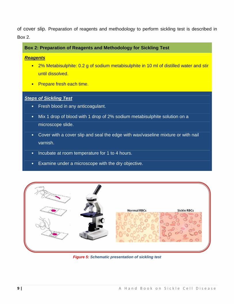
9 | A H a n d B o o k o n S i c k l e C e l l D i s e a s e
of cover slip. Preparation of reagents and methodology to perform sickling test is described in
Box 2.
Box 2: Preparation of Reagents and Methodology for Sickling Test
Reagents
• 2% Metabisulphite: 0.2 g of sodium metabisulphite in 10 ml of distilled water and stir
until dissolved.
• Prepare fresh each time.
Steps of Sickling Test
• Fresh blood in any anticoagulant.
• Mix 1 drop of blood with 1 drop of 2% sodium metabisulphite solution on a
microscope slide.
• Cover with a cover slip and seal the edge with wax/vaseline mixture or with nail
varnish.
• Incubate at room temperature for 1 to 4 hours.
• Examine under a microscope with the dry objective.
Figure 5: Schematic presentation of sickling test

10 | A H a n d B o o k o n S i c k l e C e l l D i s e a s e
2.4 Identification of Normal and Abnormal Haemoglobin Types
There are several methods of evaluating the type and relative amounts of various
normal and abnormal haemoglobin types. Haemoglobin electrophoresis is traditionally
used as the method to identify the presence of various haemoglobins. Haemoglobin
fractionation by HPLC is the most frequently used method to screen for haemoglobin
variants, including HbS. Isoelectric focusing is a highly sensitive method that is often used
at large reference laboratories. These methods evaluate the different types of
haemoglobin based on the physical and chemical properties of the different haemoglobin
molecules.
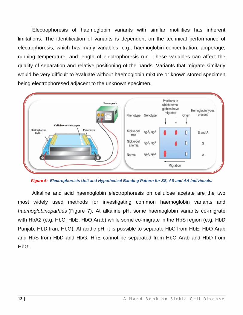
2.4.1 Electrophoresis
The electrophoresis process takes advantage of the fact that different haemoglobin
types have different electrical charges and move at different speeds in an electric field. In
an electrical field proteins that have a net negative charge will migrate from the cathode
("black," "negative") to the anode ("red", "positive") of the field. The positions of the protein
products are detected either directly by staining, or by coupled enzymatic reactions.
Substitution mutations that result in the replacement of one amino acid by another with a
different electrical charge can lead to slight changes in the overall charges of the protein.
In the case of HbS, replacement of a negatively-charged glutamate in the
standard HbA beta-globin by a neutral valine in HbS results in a protein with a slightly
reduced negative charge. Hence homozygous AA and SS individuals yield respectively
"fast" and "slow" single bands and heterozygous individual yield two bands (both alleles)
(Figure 6). Preparation of reagents and methodology to perform electrophoresis test is described
in Box 3.

11 | A H a n d B o o k o n S i c k l e C e l l D i s e a s e
Box 3: Preparation of Reagents and Methodology for Electrophoresis
Reagents and Equipment
• Electrophoresis buffer. Tris/EDTA/borate (TEB) pH 8.5. Tris-(hydroxymethyl)
aminomethane (Tris), 10.2 g, EDTA (disodium salt), 0.6 g, boric acid, 3.2 g, water to 1
litre. The buffer should be stored at 4°C and can be used up to 10 times without
deterioration.
• Fixative/stain solution, PonceauS 5g, trichloroacetic acid 7.5g, water to 1Litre.
• Destaining solution 3% (v/v) acetic acid 30ml, water to 1Litre.
• Haemolysing reagent. 0.5% (v/v) Triton X-100 in 100mg/l potassium cyanide.
• Power supply capable of delivering a constant current, 0-80mA and up to 400volts.
• A horizontal electrophoresis tank with adjustable bridge gaps and a polarity indicator.
Method
• Centrifuge samples at 1200g for 5min. Dilute 20μl of the packed red cells with 150μl of
the haemolysing reagent. Mix gently and leave for at least 5 min. If purified Hemolysate
are used, dilute 40μl of 10g/dl Hemolysate with 150μl of lysing reagent.
• With the power supply disconnected, prepare the electrophoresis tank by placing equal
amounts of TEB buffer in each of the outer buffer compartments.
• Wet two chamber wicks in the buffer, and place one along each divider/bridge support
ensuring that they make good contact with the buffer.
• Soak the cellulose acetate by lowering it slowly into a reservoir of buffer. Leave the
cellulose acetate to soak for at least 5 min before use.
• Remove the cellulose acetate strip from the buffer and blot twice between two layers of
clean blotting paper. Do not allow the cellulose acetate to dry.
• Load the applicator by depressing the tips into the sample wells twice, and apply this
first loading onto some clean blotting paper. Reload the applicator and apply the
samples to the cellulose acetate.
• Place the cellulose acetate strip across the bridges to maintain good contact.
• Electrophorese at 350 V for 25 min.
• After 25 min electrophoresis, immediately transfer the cellulose acetate to Ponceau S
and fix and stain for 5 min.
• Remove excess stain by washing for 5 min in the first acetic acid reservoir and for 10
min in each of the remaining two. Blot once, using clean blotting paper, and leave to
dry.
• Label the membranes and store in a protective plastic envelope.
12 | A H a n d B o o k o n S i c k l e C e l l D i s e a s e
Electrophoresis of haemoglobin variants with similar motilities has inherent
limitations. The identification of variants is dependent on the technical performance of
electrophoresis, which has many variables, e.g., haemoglobin concentration, amperage,
running temperature, and length of electrophoresis run. These variables can affect the
quality of separation and relative positioning of the bands. Variants that migrate similarly
would be very difficult to evaluate without haemoglobin mixture or known stored specimen
being electrophoresed adjacent to the unknown specimen.
Figure 6: Electrophoresis Unit and Hypothetical Banding Pattern for SS, AS and AA Individuals.
Alkaline and acid haemoglobin electrophoresis on cellulose acetate are the two
most widely used methods for investigating common haemoglobin variants and
haemoglobinopathies (Figure 7). At alkaline pH, some haemoglobin variants co-migrate
with HbA2 (e.g. HbC, HbE, HbO Arab) while some co-migrate in the HbS region (e.g. HbD
Punjab, HbD Iran, HbG). At acidic pH, it is possible to separate HbC from HbE, HbO Arab
and HbS from HbD and HbG. HbE cannot be separated from HbO Arab and HbD from
HbG.

13 | A H a n d B o o k o n S i c k l e C e l l D i s e a s e
Figure 7: Pattern of Haemoglobin Separation in Acid and Alkaline Electrophoresis
At acidic pH most common abnormal haemoglobins, HbS and HbC, are effectively
separated from HbA, as well as most others that migrate in similar locations by alkaline
electrophoresis. At alkaline pH mobility within the gel differs based on the overall charge of
the haemoglobin. This method allows separation of many types of haemoglobin from
normal HbA; although multiple abnormal haemoglobins may migrate in the same position.
2.4.2 Isoelectric Focusing
To overcome these limitations and increase quality of separation, it is possible to
use isoelectric focusing (IEF), an electric current is passed through a supporting medium
such as a precast agarose or polyacrylamide gel containing carrier ampholytes. These
ampholytes migrate through the medium to form a stable pH gradient ranging from pH 6.0
at the anode to pH 8.0 at the cathode. Hemolysate is applied to the gel at the cathode end,
and haemoglobin fractions migrate through the pH gradient until they are “focused” into a
sharp, distinct band at the pH equal to the isoelectric point (pI) at which the haemoglobin is
neutrally charged. It has better resolving power than many other electrophoretic
techniques as it can distinguish haemoglobin variants on the basis of their minute
differences in pI. However, HbOArab is not well separated from HbE, nor HbDPunjab from

14 | A H a n d B o o k o n S i c k l e C e l l D i s e a s e
HbG Philadelphia. Isoelectric focusing uses more specialized reagents and lengthy
procedure, hence it has largely been replaced by high performance liquid chromatography
(HPLC).
2.4.3 Capillary Electrophoresis
Most recently, automated capillary electrophoresis (CE) instruments have been
making their way in to the clinical laboratory for haemoglobin analysis. By this method,
electrophoresis is performed by adding patient sample to a thin capillary tube containing a
buffer, most often an alkaline buffer. Voltage is applied to allow separation of
haemoglobins based on their charge, similar to the traditional gel electrophoresis methods
mentioned above. Multiple samples undergo an eight-minute high-resolution separation,
concurrently.
A high-resolution haemoglobin separation is obtained, similar to IEF separation. The
ideal wavelength of 415 nm is utilized for haemoglobin detection with CE. The
electropherogram is made up of 300 consecutive readings (dots) and is divided into 15
zones. To facilitate interpretation, results are automatically positioned with regard to the
HbA and HbA2 fraction in the sample. Haemoglobins (normal and variant) are displayed
as peaks, and the zone to which a variant belongs is identified automatically by the
system. An on-board haemoglobin library is present in the form of a drop-down list and
lists all of the normal and variant haemoglobins that may be present within a particular
zone. This method has the advantage over HPLC of allowing accurate quantification of
HbA2 in the presence of Hb E. Packed red blood cell samples can be utilized for capillary
electrophoresis analysis. After removing plasma from samples, the bar-coded primary
sample tube can be loaded onto the instrument. Rest of the steps in sample processing
and separation are performed automatically by the system.

15 | A H a n d B o o k o n S i c k l e C e l l D i s e a s e
2.4.4 High Performance Liquid Chromatography
High-performance liquid
chromatography (HPLC) is an
excellent, powerful diagnostic
tool for the direct identification
of haemoglobin variants with a
high degree of precision in the
quantification of normal and
abnormal haemoglobin
fractions. Cation-exchange
HPLC has the advantage of
quantifying HbF and
HbA2 along with haemoglobin
variant screening in a single,
highly reproducible system,
making it an excellent
technology to screen for
haemoglobin variants. In
cation-exchange HPLC,
hemolysate is injected into a
chromatography column
containing a negatively
charged resin onto which the
positively charged
haemoglobins are adsorbed.
As the ionic strength of the
eluting liquid phase increases,
haemoglobin variants will
come off the column at a
Figure 8: Chromatogram Examples for Normal, AS, SS and SS/βthal
Conditions.
A. normal individual, B. SS/βthal disease, C. HbSS disease, and D. HbAS
trait. Elution times on these plots shows Hb F at 1.12-1.20 minutes, HbA at 2.33-2.49 minutes, HbA2 at 3.57-3.69 minutes and HbS at 4.40-4.44 minutes. It is obvious that in HbSS disease patient a dominant peak in the S window is present, and no detectable HbA was observed.
16 | A H a n d B o o k o n S i c k l e C e l l D i s e a s e
specific retention time, thus allowing identification of the haemoglobin variant based on the
overall charge characteristics of the protein and is continuously monitored by an optical
detector. The chromatogram is stored in and analyzed by a microcomputer. The pattern
seen by alkaline electrophoresis demonstrates some correlation with retention time by
HPLC since both methods are dependent on the charge of the haemoglobin molecule;
although the specific retention time by HPLC is dependent on the column and eluting
solution used in the instrument. In general terms, amino acid substitutions leading to more
overall negative charge will result in faster migration by alkaline electrophoresis and a
shorter retention time on the column by HPLC.
One advantage of this method is that HbC does not migrate with HbA2 as it does
on alkaline electrophoresis, thus allowing measurement of HbA2 in a patient with
heterozygous or homozygous HbC. Unfortunately, Hb E does elute with HbA2 by this
method, precluding accurate measurement of HbA2 when HbE is present. Examples of a
normal adult haemoglobin pattern by HPLC, as well as AS, SS and SS/Bthal, are shown in
Figure 8.
2.5 Newborn Screening
Early diagnosis may reduce morbidity, premature death, intellectual disability, and
other developmental disabilities because treatment can begin before the condition can
cause health problems. Newborn screening for sickle cell can be performed via the more
sensitive haemoglobin HPLC fractionation or automated capillary electrophoresis and
identifies the specific types of haemoglobin present. This test uses a few drops of blood
from pricking the baby's heel. These drops are absorbed on a screening card (Guthrie
Cards) that is taken to a laboratory for testing. Newborn dried blood spot samples are
screened for the presence of normal haemoglobins (F and A) and common haemoglobin
variants to include S, C, D, E, and Bart's. A fully automated instrument is fast and can
analyze 96 samples in few hours. The fast throughput is accomplished due to
simultaneous analyses of samples. Result interpretation is aided by automatically color-
coded curves (normal or abnormal results) and on-board haemoglobin library arranged by
zone.

17 | A H a n d B o o k o n S i c k l e C e l l D i s e a s e
2.6 Molecular Diagnostics
Molecular diagnosis combines laboratory medicine with the knowledge and
technology of molecular genetics for the detection of the various pathogenic mutations in
DNA samples in order to facilitate detection, diagnosis, sub-classification, prognosis, and
monitoring response to therapy. It has been revolutionized over the last decades,
benefiting from the discoveries in the field of molecular biology. The rate of disease gene
discovery is increasing exponentially, which facilitates the understanding diseases at
molecular level. Molecular understanding of disease is translated into diagnostic testing,
therapeutics, and eventually preventive therapies. To face the new century, the medical
practitioners not only understand molecular biology, but must also embrace the use of this
rapidly expanding body of information in their medical practice.
2.6.1 Restriction Fragment Length Polymorphism (RFLP)
The change in the sequence of the gene
can be detected by cutting DNA with the
restriction endonuclease Bsu36I, which
recognizes the sequence CC/TNAGG. The
restriction site is present in the gene for normal
haemoglobin, but is lost in the sickle cell
haemoglobin gene. A portion of DNA (268 bp),
surrounding the mutation site of the
haemoglobin gene is amplified by using the
following PCR primers. Forward: 5‟-
CAACTTCATCCACGTTCACC-3‟ and reverse
5‟- GAAGAGCCAAGGACAGGTAC-3‟. Cutting normal haemoglobin DNA with Bsu36I will
result in two DNA fragments (215 bp and 53 bp). Sickle cell haemoglobin DNA is resistant
to Bsu36I, resulting in a single DNA fragment because it lacks the Bsu36I recognition site.
Figure 9: Characterization of Sickle Cell Anaemia Using Bsu36I RFLP. Marker is 100bp
DNA Ladder, 1 is AA, 2 is SS and 3 is AS.

18 | A H a n d B o o k o n S i c k l e C e l l D i s e a s e
DNA from a heterozygous sample, with both sickle and normal haemoglobin DNA, will give
all three DNA fragments upon digestion with Bsu36I (Figure 9).
2.6.2 DNA Sequencing
Although majority of the common
haemoglobin variants can be identified
by other methods such as IEF, HPLC
and capillary electrophoresis, it is very
difficult to characterise unstable
haemoglobins and low or high oxygen
affinity haemoglobins due to their
neutral charges. This type of
uncommon haemoglobin variants
requires DNA sequencing for further identification. In general, polymerase chain reactions
(PCRs) are performed to amplify the coding regions of the β and/or α-globin genes. Then
these PCR products are directly sequenced to determine the nucleotide sequence of these
genes. The nucleotide substitution that leads to change the amino acid sequence (non-
synonymous mutation) is usually easily identified to indirectly characterise the uncommon
haemoglobin variants (Figure 10). In addition to its use in sickle cell anaemia, DNA
sequencing can also be used to identify several other nucleotide substitutions associated
with β-thalassemia.
2.7 The Future of SCD Diagnostics
The year 2010 was celebrated as the centenary year of the diagnosis of sickle cell
disease. Only just before this event UNESCO and World Health Organization recognized
the SCD as a public health priority. Although Hb electrophoresis, IEF and cation-exchange
HPLC can reliably distinguish between the different forms of Hb and provide the
information necessary for objective, definitive SCD diagnosis, currently each of these
Figure 10: DNA Sequence of HBB Gene glu6val Region.

19 | A H a n d B o o k o n S i c k l e C e l l D i s e a s e
assays must be performed in a specialized laboratory. The development of a low-cost,
portable, easy-to-use diagnostic test for SCD could make it possible for more local health
clinics to offer SCD screening, decreasing the burden on overcrowded hospitals and
centralized laboratories. Further development of novel highly-selective and efficient
approaches for isolation of nucleated fetal cells from mother‟s blood could enable prenatal
genetic screening for early detection of SCD.
20 | A H a n d B o o k o n S i c k l e C e l l D i s e a s e
Unit-3
Clinical Manifestations and Treatment of Sickle Cell Anaemia
Children with sickle cell disease should be followed by experts in the management
of this disease, most often by pediatricians. Comprehensive medical care with evidence-
based strategies delivered by experts in sickle cell disease and anticipatory guidance of
the parents about the most common complications can decrease sickle cell disease–
related mortality and morbidity. Medical care provided by a pediatrician can also decrease
frequency of emergency department visits and length of hospitalization when compared to
patients who were not seen by a pediatrician within the last year.
Infants with sickle cell anaemia have abnormal immune function and may have
functional asplenia at as early as 6 months of age. Bacterial sepsis is one of the greatest
causes for morbidity and mortality in this patient population. By 5 yr of age, most children
with sickle cell anaemia have functional asplenia.
Children with sickle cell anaemia have an additional risk factor, the deficiency of
alternative complement pathway serum opsonins against pneumococci. Regardless of
age, all patients with sickle cell anaemia are at increased risk of infection and death from
bacterial infection, particularly encapsulated organisms such as Streptococcus
pneumoniae and Haemophilus influenzae type b.
Children with sickle cell anaemia should receive prophylactic oral penicillin VK until
at least 5 yr of age (125 mg twice a day up to age 3 yr, and then 250 mg twice a day). No
established guidelines exist for penicillin prophylaxis beyond 5 yr of age, and some
clinicians continue penicillin prophylaxis, whereas others recommend discontinuation.
Continuation of penicillin prophylaxis should be considered for children beyond 5 yr of age
with previous diagnosis of pneumococcal infection, due to the increased risk of a recurrent
infection. An alternative for children who are allergic to penicillin is erythromycin ethyl
succinate 10 mg/kg twice a day. In addition to penicillin prophylaxis, routine childhood
immunizations as well as the annual administration of influenza vaccine are highly
recommended.

21 | A H a n d B o o k o n S i c k l e C e l l D i s e a s e
Human parvovirus B19 poses a unique threat for patients with sickle cell anaemia
because such infections limit the production of reticulocytes. Any child with
reticulocytopenia should be considered to have parvovirus B19 until proved otherwise.
Acute infection with parvovirus B19 is associated with red cell aplasia (aplastic crisis),
fever, pain, splenic sequestration, acute chest syndrome (ACS), glomerulonephritis, and
strokes.
3.1 General Health Management
Box 4: General Health Maintenance
1. Environmental
• Altitude: less than 1500 meters
• Avoid cold exposure
• Avoid hot exposure
4. Education
• Health education for the patient and relatives
• Information on symptoms requiring
medical advice
• Genetic counseling
• Appropriate use of analgesia at home
2. Way of Llife
• Regular hydration
• Avoidance of alcoholic beverages
• Suppression of active (or passive) tobacco use
• No cannabis or other illegal drugs
• Avoidance of strenuous exercise
• Adoption of a quiet way life
5. Psycho-Social Management
• Implementation of care pathways
• Easy access to social workers
• Open access to psychologist
• Avoidance of stress
3. Nutrition
• Folic acid supplementation 5 mg/day,
• Zinc supplementation until puberty
6. Occupational Orientation
• Avoid physically tiring jobs
• Avoid occupations with cold exposure
General health management must begin with neonatal (or prenatal) diagnosis so
that parents can be informed and the disease explained to them and the necessary
collaborative network set up, including the parents and other care givers. Disease

22 | A H a n d B o o k o n S i c k l e C e l l D i s e a s e
management must take into account the familial and genetic dimensions of the disease.
Some of the tips for general health maintenance are given in Box 4.
Females with sickle cell anaemia maintain a lower average height and weight than
those females with normal haemoglobin. This lower than average height and weight
continues until late adolescence. Puberty is usually delayed by several years. Menarche
(beginning of the menstrual period) is also delayed. It is important to reassure the
adolescent that she will eventually catch up with her peers.
Figure 11: Patients with Typical Hemolytic Face and Stunted Growth
Males with sickle cell anaemia maintain a lower average height and weight than
those males with normal haemoglobin (Figure 11). This lower than average height and
weight continues until late adolescence.Puberty is usually delayed by several years. It is
important to reassure the adolescent that he will eventually catch up with his peers.
3.2 Fever and Bacteremia
Fever in a child with sickle cell anaemia is a medical emergency, requiring prompt
medical evaluation and delivery of antibiotics due to the increased risk of bacterial infection
and concomitant high fatality rate with infection. Several clinical management strategies

23 | A H a n d B o o k o n S i c k l e C e l l D i s e a s e
have been developed for children with fever, ranging from admitting all patients with a
fever for IV antimicrobial therapy to administering a 3rd-generation cephalosporin in an
outpatient setting to patients without any of the previously established risk factors for
occult bacteremia (Box 5).
Given the observation that the
average time for a positive blood culture
with a bacterial pathogen is <20 hr in
children with sickle cell anaemia,
admission for 24 hr is probably the most
prudent strategy for children and families
without a telephone or transportation, or
with a history of inadequate follow-up.
Outpatient management should be
considered only for those with the lowest
risk for bacteremia, and treatment choice
should be considered carefully.
Children who have sickle cell
disease and who are treated with
ceftriaxone can develop severe, rapid, and
life-threatening immune hemolysis; the
established risks of outpatient
management must be reasonable against
the apparent benefits. Regardless of the clinical management strategy, all patients with
any type of sickle cell disease and fever should be evaluated and treated immediately for
occult bacteremia with either IV or IM antibiotics. Those with poor adherence, limited
financial resources, or established risk factors for bacteremia should be admitted for at
least 24 hr. The patients with positive blood cultures, pathogen-specific therapy should be
well thought-out. In the incident that Salmonella spp. or Staphylococcus
aureus bacteremia occurs, strong consideration should be given to evaluation of
osteomyelitis with a bone scan, given the increased risk of osteomyelitis in children with
Box 5: Clinical Factors Associated with
Increased Risk of Bacteremia Requiring
Admission in Febrile Children with Sickle
Cell Disease
Seriously ill appearance
Hypotension: systolic BP <70 mm Hg at 1 year
of age or <70 mm Hg + (2 × tage in years) for
older children
Poor perfusion: capillary-refill time >4 seconds
Temperature >40.0°C
A corrected white-cell count >30,000/cubic
mm or <500/cubic mm
Platelet count <100,000/cubic mm
History of pneumococcal sepsis
Severe pain
Dehydration: poor skin turgor, dry mucous
membranes, history of poor fluid intake, or
decreased output of urine
Infiltration of a segment or a larger portion of
the lung
Haemoglobin level <5.0 g/dl
24 | A H a n d B o o k o n S i c k l e C e l l D i s e a s e
sickle cell anaemia when compared to the general population. Details of Infectious risk
management in sickle cell patients were provided in Box 6.
Box 6: Infectious Risk Management
1. Penicillin V orally from 2 months to at least 5 years of age
• 125 mg twice a day up to age 3 yr, and then 250 mg twice a day
2. Prompt administration of broad spectrum or pneumococcal specific antibiotics in case of
possible bacterial infection
3. Malarial prophylaxis when appropriate
4. Immunization
• Streptococcus pneumoniae
• Haemophilus influenzae
• Meningococcus
• Influenzae
• Salmonella typhi (for at risk individuals)
5. Elimination of recurrent focal infection(dental infection, sinusitis, acute recurrent tonsillitis,
Cholecystitis, urinary infections)
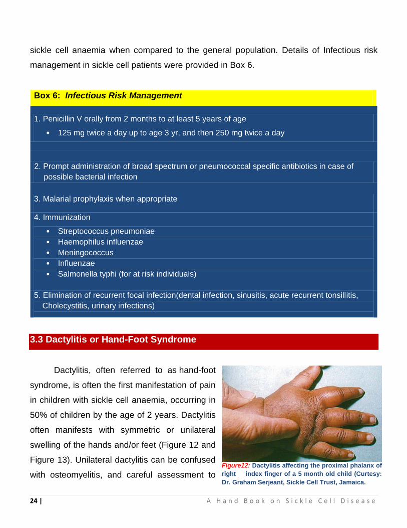
3.3 Dactylitis or Hand-Foot Syndrome
Dactylitis, often referred to as hand-foot
syndrome, is often the first manifestation of pain
in children with sickle cell anaemia, occurring in
50% of children by the age of 2 years. Dactylitis
often manifests with symmetric or unilateral
swelling of the hands and/or feet (Figure 12 and
Figure 13). Unilateral dactylitis can be confused
with osteomyelitis, and careful assessment to Figure12: Dactylitis affecting the proximal phalanx of
right index finger of a 5 month old child (Curtesy:
Dr. Graham Serjeant, Sickle Cell Trust, Jamaica.

25 | A H a n d B o o k o n S i c k l e C e l l D i s e a s e
differentiate between the two is important, because treatment differs significantly. Dactylitis
requires palliation with pain medications, such as acetaminophen with codeine, whereas
osteomyelitis requires at least 4-6 wk of IV antibiotics.
3.4 Aplastic Crisis
Aplastic crisis is a temporary
cessation of bone marrow activity
affecting predominantly the red cell
precursors (Figure 14). The
destruction of red cell precursors by
human parvovirus B19 is the main
etiology. Bone marrow aplasia,
predominantly affects red cell series
but also lowers platelets and white
cells, lasting 7-10days. The bone marrow always recovers with the development of viral
immunity but because of shortened red cell survival in SS disease may cause death. The
prevention, diagnosis and treatment strategy for aplastic crisis was given Box 7.
Figure 13 : Roentgenograms of an Infant with Sickle Cell Anaemia and acute dactylitis
Figure 14: Hands of a 12 Year Old Boy in Aplastic Crisis with Pallor
(Curtesy: Dr. Graham Serjeant, Sickle Cell Trust, Jamaica)

26 | A H a n d B o o k o n S i c k l e C e l l D i s e a s e
3.5 Splenic Sequestration
Acute splenic sequestration is a life-threatening complication occurring primarily in
infants and can occur as early as 5 wk of age which is indicated by rapid increase in the
size of spleen in a short period of time (Figure 15). Approximately 30% of children with
sickle cell anaemia have a severe splenic sequestration episode, and a significant
percentage of these episodes are fatal.
Box 8: Splenic sequestration Prevention, Diagnosis and Treatment
Anticipatory Guidance
1. Teaching parents and primary caregivers how to palpate the spleen to determine if the
spleen is enlarging.
2. The etiology of splenic sequestration is unknown.
Box 7: Aplastic Crisis Prevention, Diagnosis and Treatment
Prevention
Human parvovirus vaccine is under development but not yet available. SS siblings of affected
case have over a 50% chance of aplasia simultaneously or within 3 weeks.
Diagnosis
Marked lowering of Hb – usually to 2-4 g/dl over a few days.
Reticulocytes 0% or if present, a daily marked increase consistent with the recovery ph[[ase.
Treatment
Blood Transfusion (BT) as emergency if reticulocytes 0% and Hb > 2g/dl below steady state
level; transfusion may be performed in day care centre if uncomplicated aplasia (no features
other than pallor).
Review after 3-4 days to ensure reticulocytosis of recovery phase has occurred.
Patients may be closely monitored without transfusion if they are already in recovery phase -
daily marked increase in reticulocytes count and rising Hb.
Monitor urine for proteinuria; watch for signs of stroke.

27 | A H a n d B o o k o n S i c k l e C e l l D i s e a s e
Diagnostic Testing and Laboratory Monitoring
1. Engorgement and increase of the spleen size
2. Evidence of hypovolemia
3. Decline in haemoglobin of ≥2 g/dL from the patient‟s baseline haemoglobin
4. Reticulocytosis
5. Decrease in the platelet count may be present. These events can be accompanied by
upper respiratory tract infections, bacteremia, or viral infection.
Treatment
1. Early intervention and maintenance of hemodynamic stability using isotonic fluid or
blood transfusions.
2. If blood is required, typically 5 ml/kg of packed red blood cells (RBCs) is given.
3. Prophylactic splenectomy performed after an acute episode has resolved is the only
effective strategy for preventing future life-threatening episodes.
Repeated episodes of splenic sequestration are common, occurring in ~50% of
patients. Most recurrent episodes develop within 6 months of the previous episode.
Although blood transfusion therapy (Box 12) has been used to prevent subsequent
episodes, evidence strongly suggests this strategy does not reduce the risk of recurrent
splenic sequestration when compared to no transfusion therapy. The prevention, diagnosis
and treatment strategy for splenic sequestration was given Box 8.
Figure 15: Patient of Sickle Cell Anaemia with Chronic Massive Spleenomegaly

28 | A H a n d B o o k o n S i c k l e C e l l D i s e a s e
3.6 Pain
The chief clinical feature of sickle cell
anaemia is pain. The pain is characterized as
constant discomfort that can occur in any part
of the body but most often occurs in the
chest, abdomen, or extremities. The only
gauge for pain is the patient. Child's pain
can be assed in different ways. The Wong Baker FACES Pain Rating Scale is one among
them (Figure 16). Patients with these conditions often present with debilitating primary
effects under conditions of unclear etiology and powerful secondary effects that can alter
emotional states and facilitate misdiagnosis (Figure 17).
Box 9: Diagnostic Testing of Pain, Laboratory Monitoring and Treatment
Guidance
1. Precipitating causes of painful episodes can include physical stress, infection,
dehydration, hypoxia, local or systemic acidosis, exposure to cold, and swimming for
prolonged periods.
2. Should develop a consistent, validated pain scale, such as the Wong-Baker FACES Scale
for determining the magnitude of the pain.
3. Sleeping through the night might be an indication for decreasing pain medication by 20%
the following morning.
4. Successful treatment of painful episodes requires education of both the parents and the
patients regarding the recognition of symptoms and the optimal management strategy.
5. At home with comfort measures, such as heating blanket, relaxation techniques,
massage, and pain medication.
Figure 16: Wong Baker Faces Pain Rating
29 | A H a n d B o o k o n S i c k l e C e l l D i s e a s e
Diagnostic Testing and Laboratory Monitoring
1. Given the absence of any reliable objective laboratory or clinical parameter associated
with pain, trust between the patient and the treating physician is paramount to a
successful clinical management strategy.
Treatment
1. Patient who has ~1 painful episode per year that requires medical attention.
2. Specific therapy for pain - the use of acetaminophen or a non-steroidal agent early in the
course of pain, followed by escalation to acetaminophen with codeine or a short- or long-
acting oral opioid.
3. Hospitalization for administration of IV morphine or derivatives of morphine.
4. BT - reserve for patients with a decrease in Hb resulting in hemodynamic compromise,
respiratory distress, or a falling Hb concentration, with no expectation that a safe nadir will
be reached, such as when the child has both a falling Hb level and reticulocytes count with
a parvovirus B19 infection.
5. IV hydration does not relieve or prevent pain and is appropriate when the patient is unable
to drink as a result of the severe pain or is dehydrated.
6. Opioid dependency in children with SCD is rare and should never be used as a reason to
withhold pain medication.
7. Hydroxyurea, a myelosuppressive agent, is the only effective drug proved to reduce the
frequency of painful episodes.
The incremental increase and decrease in the use of the medication to relieve pain roughly
parallels the 8 phases associated with a chronology of pain and comfort. The average
hospital length of stay for children admitted in pain is 4.4 days. The American Pain Society
has published clinical guidelines for treating acute and chronic pain in patients with sickle
cell disease of any type. Summary of the Chronology of Pain in Children with Sickle Cell
30 | A H a n d B o o k o n S i c k l e C e l l D i s e a s e
Disease, diagnostic testing, laboratory monitoring and treatment is provided in Box 9.
Characteristics of pain in sickle cell disease and suggested measures to be used are also
documented in Table 2.
Phase Pain Characteristics Suggested Measures
(Baseline) 1 No vaso-occlusive pain; pain of
complications may be present,
such as that connected with
avascular necrosis of the hip
No comfort measures used
(Pre-pain) 2 No vaso-occlusive pain; pain of
complications may be present;
prodromal signs of impending
vaso-occlusive episode may
appear, e.g., “yellow eyes” and/or
fatigue
No comfort measures used; caregivers
may encourage child to increase fluids to
prevent pain event from occurring
(Pain start
point) 3
First signs of vaso-occlusive pain
appear, usually in mild form
Mild oral analgesic often given; fluids
increased; child usually maintains
normal activities
(Pain
acceleration) 4
Intensity of pain increases from
mild to moderate.
Some children skip this level or
move quickly from phase 3 to
phase 5
Stronger oral analgesic are given;
rubbing, heat, or other activities are often
used; child usually stays in school until
the pain becomes more severe, then
stays home and limits activities; is
usually in bed; family searches for ways
to control the pain
(Peak pain
experience) 5
Pain accelerates to high moderate
or severe levels and plateaus;
pain can remain elevated for
extended period.
Oral analgesics are given around the
clock at home; combination of comfort
measures is used; family might avoid
going to the hospital; if pain is very
distressing to the child, parent takes the
child to the emergency department

31 | A H a n d B o o k o n S i c k l e C e l l D i s e a s e
Phase Pain Characteristics Suggested Measures
Child‟s appearance, behavior, and
mood are significantly different
from normal
After child enters the hospital, families
often turn over comforting activities to
health care providers and wait to see if
the analgesics work
Family caregivers are often exhausted
from caring for the child for several days
with little or no rest
(Pain
decrease start
point) 6
Pain finally begins to decrease in
intensity from the peak pain level
Family caregivers again become active
in comforting the child but not as
intensely as during phases 4 and 5
(Steady pain
decline) 7
Pain decreases more rapidly,
becomes more tolerable for the
child
Child and family are more relaxed
Health care providers begin to wean the
child from the IV analgesic; oral opioids
given; discharge planning is started
Children may be discharged before they
are pain free
(Pain
resolution) 8
Pain intensity is at a tolerable
level, and discharge is imminent.
Child looks and acts like “normal”.
Self mood improves
May receive oral analgesics
Table 2 : Characteristics of Pain in Sickle Cell Disease
Many myths have been propagated regarding the treatment of pain in sickle cell
anaemia. The idea that painful episodes in children should be managed without opioids is
without basis and results in unnecessary suffering on the part of the patient. There is no
evidence that blood transfusion therapy during an existing painful episode, decreases the
intensity or period of the painful episode. However, patients with multiple painful episodes
requiring hospitalization within a year or with pain episodes that require hospital stay for
more than 7 days, should be evaluated for comorbidities and psychosocial stressors that
might contribute to the frequency or duration of pain.
32 | A H a n d B o o k o n S i c k l e C e l l D i s e a s e
Hydroxyurea can decrease the rate of
painful episodes by 50% and the rate of ACS
episodes and blood transfusions by ~50%.
Hydroxyurea is safe and well tolerated in
children >5 yr of age. The primary toxicities
are limited to myelosuppression that
reversed upon cessation of the drug. The
long-term toxicity associated with
hydroxyurea in children has not been
established; but all evidence to date
suggests that the benefits far outweigh the
risks.
The dose of hydroxyurea being prescribed at our institute is 10 mg/kg body weight
daily. If required, dose can be increased upto 20-30 mg/kg per day gradually, if tolerated
by patient. Monitoring children on hydroxyurea is rigorous, with initial visits every 2 weeks
to monitor for hematologic toxicity with dose escalations and then monthly after a
therapeutic dose has been identified.
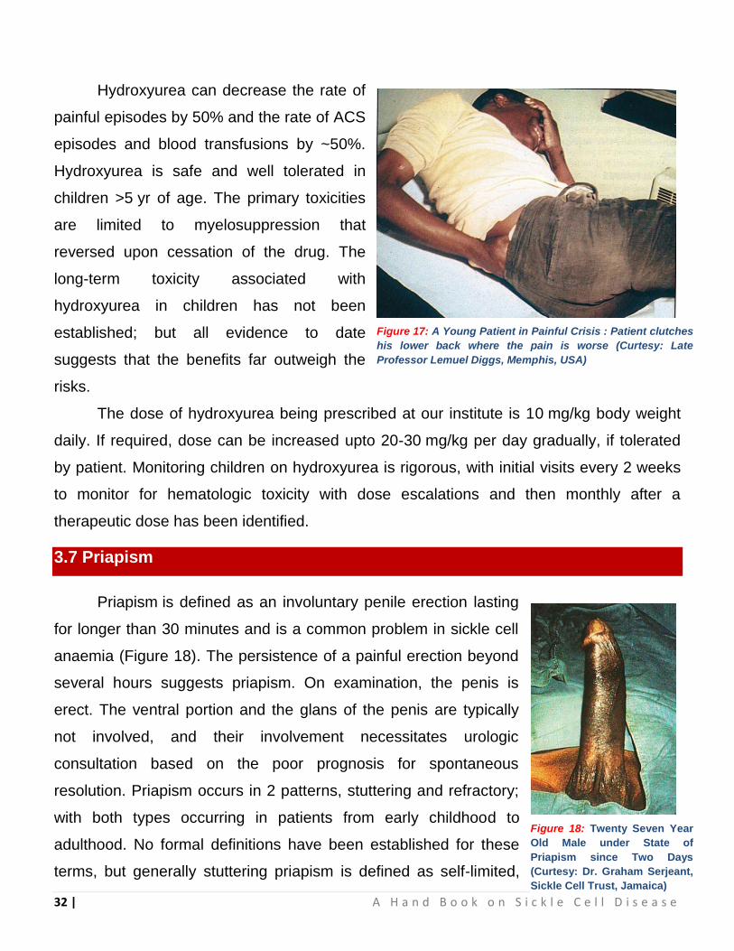
3.7 Priapism
Priapism is defined as an involuntary penile erection lasting
for longer than 30 minutes and is a common problem in sickle cell
anaemia (Figure 18). The persistence of a painful erection beyond
several hours suggests priapism. On examination, the penis is
erect. The ventral portion and the glans of the penis are typically
not involved, and their involvement necessitates urologic
consultation based on the poor prognosis for spontaneous
resolution. Priapism occurs in 2 patterns, stuttering and refractory;
with both types occurring in patients from early childhood to
adulthood. No formal definitions have been established for these
terms, but generally stuttering priapism is defined as self-limited,
Figure 17: A Young Patient in Painful Crisis : Patient clutches
his lower back where the pain is worse (Curtesy: Late
Professor Lemuel Diggs, Memphis, USA)
Figure 18: Twenty Seven Year
Old Male under State of
Priapism since Two Days
(Curtesy: Dr. Graham Serjeant,
Sickle Cell Trust, Jamaica)

33 | A H a n d B o o k o n S i c k l e C e l l D i s e a s e
intermittent bouts of priapism with several episodes over a defined period. Refractory
priapism is defined as prolonged priapism beyond several hours.
Approximately 20% of patients between 5 and 20 yr of age report having at least 1
episode of priapism. Most episodes occur between 3 AM and 9 AM. The mean age at first
episode is 12 yr, and the mean number of episodes per patient is ~16, with a mean
duration of ~2 hr. The actuarial probability of a patient‟s experiencing priapism is ~90% by
20 yr of age. Acute and preventive therapy for priapism is given in Box 10.
Box 10: Acute and Preventive Therapy for Priapism
Acute Treatment
1. Sitz bath or pain medication.
2. Priapism lasting >4 hr should be treated
by aspiration of blood from the corpora
cavernosa followed by irrigation with
dilute epinephrine to produce immediate
and sustained detumescence.
3. Urologic consultation is required to initiate
this procedure, with appropriate input
from a hematologist.
4. Either simple blood transfusion therapy
or exchange transfusion is proposed for
the acute treatment of priapism.
Preventive Therapy
1. For the prevention of recurrent priapism,
hydroxyurea appears to have promise;
2. The use of etilefrine, a sympathomimetic
amine with both α1 and β1 adrenergic
effects, appears safe and promising in the
secondary prevention of priapism.
3. The long-term effects of recurrent or
prolonged priapism episodes in
prepubertal children are not known. In
adults, infertility and impotence are
potential consequences.
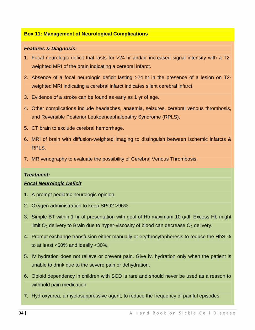
3.8 Neurologic Complications
Neurologic complications associated with sickle cell anaemia are varied and
complex. Children with other types of sickle cell disease such as HbSC or HbS β-
thalassemia plus might have overt or silent cerebral infarcts as well. Details of neurological
complications, diagnosis, treatment and prevention measures in sickle cell anaemia
patients are given in Box 11.
34 | A H a n d B o o k o n S i c k l e C e l l D i s e a s e
Box 11: Management of Neurological Complications
Features & Diagnosis:
1. Focal neurologic deficit that lasts for >24 hr and/or increased signal intensity with a T2-
weighted MRI of the brain indicating a cerebral infarct.
2. Absence of a focal neurologic deficit lasting >24 hr in the presence of a lesion on T2-
weighted MRI indicating a cerebral infarct indicates silent cerebral infarct.
3. Evidence of a stroke can be found as early as 1 yr of age.
4. Other complications include headaches, anaemia, seizures, cerebral venous thrombosis,
and Reversible Posterior Leukoencephalopathy Syndrome (RPLS).
5. CT brain to exclude cerebral hemorrhage.
6. MRI of brain with diffusion-weighted imaging to distinguish between ischemic infarcts &
RPLS.
7. MR venography to evaluate the possibility of Cerebral Venous Thrombosis.
Treatment:
Focal Neurologic Deficit
1. A prompt pediatric neurologic opinion.
2. Oxygen administration to keep SPO2 >96%.
3. Simple BT within 1 hr of presentation with goal of Hb maximum 10 g/dl. Excess Hb might
limit O2 delivery to Brain due to hyper-viscosity of blood can decrease O2 delivery.
4. Prompt exchange transfusion either manually or erythrocytapheresis to reduce the HbS %
to at least <50% and ideally <30%.
5. IV hydration does not relieve or prevent pain. Give iv. hydration only when the patient is
unable to drink due to the severe pain or dehydration.
6. Opioid dependency in children with SCD is rare and should never be used as a reason to
withhold pain medication.
7. Hydroxyurea, a myelosuppressive agent, to reduce the frequency of painful episodes.

35 | A H a n d B o o k o n S i c k l e C e l l D i s e a s e
Prevention:
Primary
Transcranial Doppler (TCD) assessment of the blood velocity in the terminal portion of the
internal carotid artery (ICA) and the proximal portion of the middle cerebral artery (MCA). Two
methods are used viz.,. non-imaging and imaging techniques.
The scan results can be divided into five categories depending on the time averaged maximal
mean (TAMM) velocity recorded, whether in the ICA or MCA or the bifurcation of the two
arteries:-
Inadequate image
Unusual low velocity
Normal velocity - 'low risk'
Borderline velocity - 'conditional'
High velocity - 'high risk'
The TAMM blood velocities used as cut-offs to define the risk limits are as follows:
Normal velocity - 'standard risk' <170 cm/s
Borderline velocity - 'conditional' 170 to 199 cm/s
High velocity - 'high risk' >200 cm/s
TCD scanning decision
Inadequate scan/
low velocities
Rescanning or alternative technique is suggested.
Normal scan <170
cm/sec
Repeat TCD in 1 year.
Conditional 170 to
199 cm/sec
Rescan between 1 & 4 months. Children <10 years
& those with higher velocities are considered to be
at higher risk & should be scanned earlier
Abnormal >200
cm/sec
Discuss risk of stroke & consider chronic
transfusion. A rescan might be considered
appropriate depending on the blood velocity &
individual clinical circumstances.
36 | A H a n d B o o k o n S i c k l e C e l l D i s e a s e
Secondary
The approach is BT therapy aimed at keeping the maximum HbS conc. <30% in first 2 years
following any new stroke and <50% thereafter. The primary toxic effect of BT is excessive
iron store.
Deferoxamine, chelating agent, is administered subcutaneously 5 of 7 nights per week for 10
hr a night , or Deferasirox can be used as a tablet taken by mouth daily.
3.9 Excessive Iron Stores
The assessment of excessive iron stores in children receiving regular blood
transfusions is difficult.
Biopsy of the liver is the gold standard for diagnosis of excessive iron stores but
it is an invasive procedure exposing children to the risk of general anesthesia,
bleeding, and pain.
The most commonly used and least-invasive method of estimating total body iron
involves serum ferritin levels; however, ferritin measurements have significant
limitations, because ferritin levels rise during acute inflammation and correlate
poorly with excessive iron in specific organs after 2 yr of regular blood
transfusion therapy.
MRI of the liver is a reasonable alternative to biopsy and more accurate than
serum ferritin in measuring iron content in heart and liver, the two most
commonly affected organs associated with increased total body iron stores. MRI
T2 and MRI R2 and R2 sequences are used to estimate iron levels in the heart
and liver.
Erythrocytapheresis is the preferred method because there is a minimum net iron
balance after the procedure. Simple transfusion therapy is the least preferable method
because this strategy results in the highest net positive iron balance after the procedure.
Despite being the preferred method, erythrocytapheresis is less commonly performed

37 | A H a n d B o o k o n S i c k l e C e l l D i s e a s e
because of the requirement for
technical expertise, access to a large
vein, and an available apheresis
machine. For patients who either will
not or cannot continue blood
transfusion therapy to prevent
subsequent strokes, hydroxyurea
therapy may be a reasonable alternative. The efficacy and toxicity of hydroxyurea as an
option for preventing secondary stroke is being addressed in a clinical trial setting.
Alternatively, human leukocyte antigen (HLA)
matched hematopoietic stem cell transplantation
from a sibling donor is a reasonable approach for
patients with strokes, although only a few children
have suitable donors. Hematopoietic stem cell
transplantation using unrelated donors is the subject
of an open clinical trial that is too premature to
comment on.
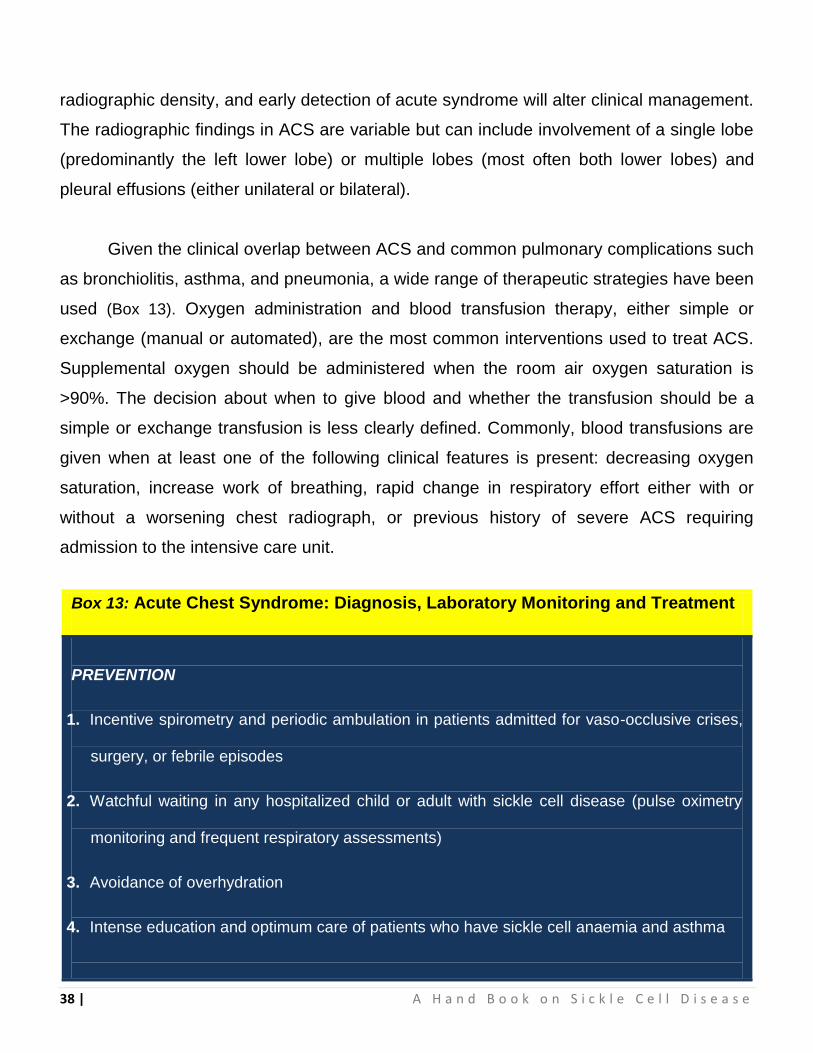
3.10 Lung Disease
The second most common reason for admission to the
hospital and a common cause of death in children with sickle
cell anaemia is acute chest syndrome (ACS). ACS refers to a
constellation of findings that include a new radiodensity on
chest radiograph, fever, respiratory distress, and pain that
occurs often in the chest, but it can also include the back
and/or abdomen only. Even in the absence of respiratory
symptoms, all patients with fever should receive a chest
radiograph (Figure 20) to identify ACS because clinical
examination alone is insufficient to identify patients with a new
Box 12: Methods of Blood Transfusion Therapy
1. Erythrocytapheresis
2. Manual exchange transfusions (phlebotomy of a
set amount of the patient‟s blood followed by
rapid administration of donated packed RBCs)
3. Simple transfusion
Figure 19: Hemolytic Facies
Figure 20: An X-ray Image
Showing Complete White Out of
Left lung in a Patient with ACS
38 | A H a n d B o o k o n S i c k l e C e l l D i s e a s e
radiographic density, and early detection of acute syndrome will alter clinical management.
The radiographic findings in ACS are variable but can include involvement of a single lobe
(predominantly the left lower lobe) or multiple lobes (most often both lower lobes) and
pleural effusions (either unilateral or bilateral).
Given the clinical overlap between ACS and common pulmonary complications such
as bronchiolitis, asthma, and pneumonia, a wide range of therapeutic strategies have been
used (Box 13). Oxygen administration and blood transfusion therapy, either simple or
exchange (manual or automated), are the most common interventions used to treat ACS.
Supplemental oxygen should be administered when the room air oxygen saturation is
>90%. The decision about when to give blood and whether the transfusion should be a
simple or exchange transfusion is less clearly defined. Commonly, blood transfusions are
given when at least one of the following clinical features is present: decreasing oxygen
saturation, increase work of breathing, rapid change in respiratory effort either with or
without a worsening chest radiograph, or previous history of severe ACS requiring
admission to the intensive care unit.
Box 13: Acute Chest Syndrome: Diagnosis, Laboratory Monitoring and Treatment
PREVENTION
1. Incentive spirometry and periodic ambulation in patients admitted for vaso-occlusive crises,
surgery, or febrile episodes
2. Watchful waiting in any hospitalized child or adult with sickle cell disease (pulse oximetry
monitoring and frequent respiratory assessments)
3. Avoidance of overhydration
4. Intense education and optimum care of patients who have sickle cell anaemia and asthma

39 | A H a n d B o o k o n S i c k l e C e l l D i s e a s e
Diagnostic Testing & Laboratory Monitoring
1. Blood culture
2. Nasopharyngeal samples for viral culture (respiratory syncytial virus, influenza)
3. Blood counts every day and appropriate chemistry
4. Continuous pulse oximetry
5. Chest radiograph
Treatment
1. Blood transfusion (simple or exchange)
2. Supplemental O2 for drop in pulse oximetry by 4% over baseline, or values <90%
3. Empirical antibiotics (cephalosporin and macrolide)
4. Continued respiratory therapy (incentive spirometry and chest physiotherapy as necessary)
5. Bronchodilators and steroids for patients with asthma
6. Optimum pain control and fluid management
3.11 Leg Ulcer
Leg ulcers are one of the complications of
sickle cell disease. They start in adolescence
and eventually appear in 75% of adults (Figure
21). Low steady state haemoglobin values are
associated with a higher incidence of ulcer
formation. A high fetal haemoglobin production
correlates with a lower incidence of leg ulcers.
The ulcers usually present over the medial
surface of the lower tibia or just posterior to the Figure 21: Non Healing Leg Ulcer in 32 Years Male with Sickle Cell Disease.
40 | A H a n d B o o k o n S i c k l e C e l l D i s e a s e
medial malleolus. Treatment is in many instances temporary and recovery may take a long
time. The current recommendations for management of leg ulcers are given in box 14. To
prevent ulceration pay close attention to improved venous circulation by using above the
knee elastic stocking.
Box 14: Management of Leg Ulcers
Acute Ulcer Chronic Ulcer
1. Surgical debridement if there is
unhealthy tissue, especially if the ulcer
is chronic and there is slow or minimal
healing.
2. Scrupulous hygiene
3. Topical antibiotics
4. Moist-wound dressing – one to four
times a day (with wet to dry gauze –
saline dressings) it helps in gentle
debridement and healing.
5. Rest
6. Elevation of leg
1. Chronic transfusion program (every 4
weeks for 6 – 12 months)
2. Consider oral zinc sulfate to promote
healing
3. Split thickness skin grafts
4. Treatment with hydroxyurea, with or
without erythropoietin, depending on
response to hydroxyurea.
3.12 Deep Jaundice
The obstruction of excretory pathway
for bilirubin is main cause, which is because
of increased bilirubin production due to
hemolysis. It may cause stone in gall bladder
and bile duct. The differential diagnosis of
deep jaundice (Figure 22) includes acute
cholestasis, viral hepatitis, and a stone
obstructing the common bile duct.
Investigation of choice must be Ultrasound of the common bile duct.
Figure 22: Patient with Deep Jaundice
41 | A H a n d B o o k o n S i c k l e C e l l D i s e a s e
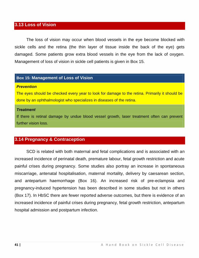
3.13 Loss of Vision
The loss of vision may occur when blood vessels in the eye become blocked with
sickle cells and the retina (the thin layer of tissue inside the back of the eye) gets
damaged. Some patients grow extra blood vessels in the eye from the lack of oxygen.
Management of loss of vision in sickle cell patients is given in Box 15.
Box 15: Management of Loss of Vision
Prevention
The eyes should be checked every year to look for damage to the retina. Primarily it should be
done by an ophthalmologist who specializes in diseases of the retina.
Treatment
If there is retinal damage by undue blood vessel growth, laser treatment often can prevent
further vision loss.
3.14 Pregnancy & Contraception
SCD is related with both maternal and fetal complications and is associated with an
increased incidence of perinatal death, premature labour, fetal growth restriction and acute
painful crises during pregnancy. Some studies also portray an increase in spontaneous
miscarriage, antenatal hospitalisation, maternal mortality, delivery by caesarean section,
and antepartum haemorrhage (Box 16). An increased risk of pre-eclampsia and
pregnancy-induced hypertension has been described in some studies but not in others
(Box 17). In HbSC there are fewer reported adverse outcomes, but there is evidence of an
increased incidence of painful crises during pregnancy, fetal growth restriction, antepartum
hospital admission and postpartum infection.

42 | A H a n d B o o k o n S i c k l e C e l l D i s e a s e
Box 16: Effect of Sickle Cell Disease on
Sexual Development
Fertility
Contraception
Frequently delayed in SS disease.
The median age of menarche
usually increased compared with
AA controls.
No good data on fertility but
similar time between initial sexual
exposure and initial pregnancy
suggests that fertility for first child
closed to normal.
Patients should be
offered the most reliable
methods; none are
contraindicated in SS
disease.
Box 17: Effect of Sickle Cell Disease on Pregnancy
On 'SS' Mother
On Child of 'SS' Mother
Greater risk of ACS & painful crisis especially in
3rd trimester and early postpartum. Also
preeclampsia and maternal death
Greater risk of fetal loss at every stage of
pregnancy. IUGR, LBW baby.
Women and men with SCD should be encouraged to have the haemoglobinopathy
status of their partner determined before they plan for pregnancy. If identified as an „at risk
couple‟ they should receive counseling and advice about reproductive options. Effective
contraception should be offered if requested.
- Penicillin prophylaxis or the equivalent should be prescribed.
- Vaccination status should be determined and updated before pregnancy.
Patients with SCD are hyposplenic and are at risk of infection, in particular
from encapsulated bacteria such as Neisseria meningitides, Streptococcus
pneumonia and Haemophilus influenzae.
Once pregnant, patients should register early for antenatal care and be monitored closely
during pregnancy. Give iron and folic acid supplementation for duration of pregnancy.
Monitor fetal growth and consider operative delivery if evidence of fetal distress, otherwise
delivery by vaginal route.

43 | A H a n d B o o k o n S i c k l e C e l l D i s e a s e
3.15 Newborn Screening
Most of the SS patients die before completing one year of their life, because
detection of SCD is not possible using routing screening and diagnostic protocols. Rational
of newborn screening is to increase the chances of detection of SCD in the first year of life.
Several of the causes of early death may be prevented and patients may be more
effectively treated. Prophylaxis and education may only be put in place if the underlying
diagnosis is known (Box 18).
Box 18: Management of Newborn Screening
Confirm Diagnosis on capillary sample at age 1 month; once confirmed, children should
be registered for regular follow-up, education, and counseling of family.
Offer screening tests for parents to affected children.
Ensure regular immunization schedules (Sickle cell patients often miss immunization
because of transient fevers, illness), including Haemophilus influenzae type b vaccine.
Teach parents how to feel for the spleen which should be done daily; or more often if
child is sick.
Advice purchase of thermometer and teach its use.
Start regular pneumococcal prophylaxis form 4 months to 4 years.
Oral penicillin-V 125 mg twice a day up to age of 3 years, and then 250 mg twice a day;
or erythromycin 125 mg bd upto age of 3 years and then 250 mg bd if allergic to
penicillin.
Pneumococcal vaccine given at age 4 years in Jamaica.
Explain about delayed physical and sexual development and slim build: advice plenty of
fresh fruit and vegetables and advice against expensive tonics to fatten child.
Arrange regular review of child every 3 months or immediately if sick. Stress importance
of attendance when well in order to establish steady state blood level and clinical
feature.
Provide walk-in facilities or immediate contact if sick.

44 | A H a n d B o o k o n S i c k l e C e l l D i s e a s e
3.16 Models of Management
Early Diagnosis and Follow-up
Newborn diagnosis essential to prevent many early deaths. Regular follow-up in
dedicated clinic with staff educated in sickle cell disease and its complications patients
must have immediate access to care and treatment if sick. Establish baseline for patients‟
clinical state and hematology by review when well. Provide programme of management
including pneumococcal prophylaxis and education on splenic palpation.
Diet
Give regular dietary advice. Regular folate supplementation is necessary in when
there is evidence of megaloblastic change or poor diet.
Haematology
Monitor hematology with reticulocytes and red cell indices every year while well until
5 years then every 2 year. Monitor renal function every 3-5 year in patients over age 30
years to detect renal failure. Always identify and treat cause for failing haemoglobin levels.
Gallstones
Common in SS disease (50% by 25 year) but rarely cause trouble no indication for
removal of asymptomatic stones. Remove if evidence of acute or chronic Cholecystitis or
blockage of the cystic or common bile duct.
Fever
Investigate febrile patients urgently and assume septicemic until proved otherwise,
acute chest, syndrome is common cause of fever,
Stroke
It is a catastrophic complication in childhood. Chronic transfusion programs may
prevent recurrence but many difficulties with such programs in the Caribbean.
Bone Pain
Find out precipitating factors for painful crisis and explore methods of avoidance.
Provide day care facilities for management of uncomplicated painful crises, remember pain

45 | A H a n d B o o k o n S i c k l e C e l l D i s e a s e
the ribs and sternum may precede acute chest syndrome. Investigate pain in hip with limp
as possible avascular necrosis of femoral head.
Leg Ulcers
Avoid trauma around the ankles if possible, Treat ulcer actively with bed rest, we
dressing, supportive elastic bandages.
Acute Chest Syndrome
It is a major cause of morbidity and mortality in sickle cell disease. Monitor cases
closely with frequent clinical and radiological appraisal and pulse oximetry. Chronic
transfusion programmes may delay onset of respiratory failure.
Pripism
Ask about priapism because it is common treatable, may have serious sequelae and
is under-reported because of embarrassment or not realized as sickle cell problem.
Renal Failure
Progressive glomerular damage associated with failing haemoglobin lever
increasingly common in patients over 30 years. Treat with top-up transfusions.
3.17 Outlook
Current Treatment modalities available for sickle cell disease are as follows:
Hydroxyurea may reduce painful crises and ACS in selected seriously affected patients but
must be monitored carefully with frequent blood tests.
Bone marrow transplantation may cure symptoms but is expensive requiring high expertise,
only applicable to approximately 15% of Patients with HLA matched siblings, and has a
10% short-term mortality longer term effects on reproduction and other aspects are
unknown.
Chronic transfusion programmers reduce recurrent strokes and may prevent some other
pathology but there are potentially serious complications.

46 | A H a n d B o o k o n S i c k l e C e l l D i s e a s e
3.18 Conclusions
In malarious areas, malaria continues to be a major cause of early death and
complications in sickle cell disease patients. Control of malaria in the Caribbean has
been associated with better survival.
Serious infections may be prevented by prophylaxis or more effectively treatment by
early presentations to clinic and appropriate antibiotics.
Education and supportive services should empower the patient to cope with their
disease. More effectively. A greate r sense of control by the patient is vital to disease
management.
.Painful Crises, an important cause of recurrent and severity in most patients after the
age of 30 years.
Cumulative end-organ damage especially of the kidneys. And the lungs become more
important with advancing age.
With good care many patients with SS disease are living productive live and surviving
to 70 years and beyond. Median survival in US and Jamaica currently estimated at 45-
55 years.

47 | A H a n d B o o k o n S i c k l e C e l l D i s e a s e
Unit-4
Counseling
For counseling to be effective, you have to first identify the patient.
The patient should know:
What Sickle Cell is?
The difference between sickle cell Trait and sickle cell disease
That the Severity of pain & frequency of crisis are unpredictable.
That this is incurable.
That Only amelioration of conditions can be done.
That this is a genetic disorder.
He/She should try:
Try to reduce pain and crisis episodes.
Try to live life normally/ try to increase life span
4.1 Spread
Approximately 12% of total population is affected in (Chhattisgarh)
Found mostly in semi-tribal areas of Chhattisgarh
High incidence of SCA/SCD in certain castes
4.2 Identification of Patient
Delayed growth, weight & height sub-normal
General complains of weakness, weak body
Highly anemic appearance, severe anaemia
Pale skin, colorless nails
Yellow skin & eyes, long time jaundice
Flat bones (forehead structure)
Continuous mild fever, long term sufferance
Short breaths/breathlessness, laborious breathing

48 | A H a n d B o o k o n S i c k l e C e l l D i s e a s e
Loss of appetite
More tired than usual
Frequent urination, dark urine
Pain in bones & ribs
Irritability & prominent mood swings
Swelling in hands & legs
Priapism
Infertility
Frequent viral & bacterial infections
Swollen spleen
4.3 Age-wise Counseling
4.3.1 Parent’s Role in Adolescents & Young Adults
Role of parents very important
Discuss nature of the disease/trait & its affect on them
Learn to identify onset of pain/crisis by self
Discuss & review ways to manage pain/crisis
Counsel them about adverse effects of tobacco, smoking, alcohol & drugs
Review school & work-place performance, watch out for drops in performance
Parents to give praise
frequently, hug often
Create a safe positive &
loving home environment
Watch what words you say
to the children
Encourage your
adolescent to try different
activities to see what
he/she enjoys (divert
attention)
Friends also play an
important role in this
COUNSELING
Counseling focuses on 3 things:
General pain management
Handling crisis
Planning for a life as
normal as possible
321
Figure 23: Component of Counseling Patients with Sickle Cell Disease
49 | A H a n d B o o k o n S i c k l e C e l l D i s e a s e
4.3.2 Adolescents & Young Adults
Patient should know who to contact,when to contact-doctor‟s name, hospital, etc.
Should know how to make appointments
Should have a personal health summary at all times
Should carry own health insurance, cards, etc.
Should understand what complications can arise, & under what conditions
Should discuss plans for independent living, vocational & economic self-reliance
Should undergo Pre-marriage screening & genetic counseling
Parents wanting patient‟s marriage & unmarried patient himself/herself should
consider the inheritance patterns
4.3.3 Young Couples/Just Married
Genetic counselor will not tell the patient what to do. Partners are free to choose
the path which is most comfortable to them.
Option for having a child should be based on inheritance pattern
Give the parents knowledge of how SCD affects the newborn baby
4.3.4 Pregnancy Phase/Pre-natal
Planning & early pre-natal care is the key
Pre-natal care should be done by an Obstetrician, who is an expert in high-risk
pregnancy
Avoid infections of all types- viral, bacterial
Healthy, high protein diet, fibrous diet is required
Pre-natal vitamins, folic acid supplements may be required
Preventing dehydration is a must
There is high possibility of premature/pre-term baby
During labour, IV fluids should be given to prevent dehydration
Oxygen (through mask) may be given during labour (depends on patient
condition)
Give the parents knowledge of how SCD affects the newborn baby

50 | A H a n d B o o k o n S i c k l e C e l l D i s e a s e
4.3.5 Neonatal/Early Infancy
High possibility of premature/pre-term baby– so prepare accordingly in advance
Newborn screening for SCA/SCD: HPLC test and other tests
Test for sepsis
High risk of infections
Vaccinations– penicillin, anti-tetanus, pneumococcal vaccination till age 5
4.3.6 Childhood and School Going Children
Should have affection, safe environment at home and in school
Two sets of books/copies advisable , one at home, one at school, so that they do
not have to carry load daily
Should be allowed to drink water frequently in school
Should be allowed to go to the bathroom as many times as required.
No strenuous exercise, long distance movement or running desirable
Should be exempt from outdoor activities when temperatures are very high or
low, instruct for alternate activity
Should be given Moderate work load in schools desirable
Should be given extra time to complete class work, tests, etc.
Children should be allowed to carry extra water bottles, medications, umbrella,
etc.
At the hint of fever/ pain/ change in complexion or pallor/ breathing difficulty/
dizziness/ differential palpitation/ swelling/ muscular weakness, they should be
immediately allowed to go home or referred to a doctor / Should know who to
contact in emergency.
4.4 Counseling for Diet Management
Take a high fibre diet, plenty of roughage
Take large amount of proteins in diet
Choose diet rich in anti-oxidants
Avoid oily and fatty foods (masala)
Take plenty of water
51 | A H a n d B o o k o n S i c k l e C e l l D i s e a s e
4.5 Do’s & Dont’s
Drink plenty of water
Modify dietary habits
Take rest and relax
Do not overwork
Avoid extreme temperature
Avoid infections
Maintain regular schedules of work and play

52 | A H a n d B o o k o n S i c k l e C e l l D i s e a s e
Bibliography
1. http://www.nhlbi.nih.gov/health/health-topics/topics/sca/
2. Rees DC, Williams TN, Gladwin MT. Sickle-cell disease. Lancet. 2010;376:2018-31.
3. Niscola P, Sorrentino F, Scaramucci L, de Fabritiis P, Cianciulli P. Pain syndromes in sickle
cell disease: an update. Pain Med. 2009;10:470-80
4. Stuart MJ, Nagel RL. Sickle-cell disease. Lancet. 2004;364:1343-60.
5. Wajcman H, Prehu C, Bardakdjian-Michau J, Prome D, Riou J, Godart C, et al. Abnormal
haemoglobins: laboratory methods. Haemoglobin. 2001;25:169-81.
6. Basset P, Beuzard Y, Garel MC, Rosa J. Isoelectric focusing of human haemoglobin: its
application to screening, to the characterization of 70 variants, and to the study of modified
fractions of normal haemoglobins. Blood. 1978;51:971-82.
7. Chen-Marotel J, Beuzard Y, Kac Trung B, Braconnier F, Rosa J, Guerrasio A, et al.
Polymorphism of human fetal haemoglobin studied by isoelectric focusing. FEBS letters.
1980;115:68-70.
8. Clarke GM, Higgins TN. Laboratory investigation of haemoglobinopathies and
thalassemias: review and update. Clinical chemistry. 2000;46:1284-90.
9. Riou J, Godart C, Hurtrel D, Mathis M, Bimet C, Bardakdjian-Michau J, et al. Cation-
exchange HPLC evaluated for presumptive identification of haemoglobin variants. Clinical
chemistry. 1997;43:34-9.
10. Kutlar F, Kutlar A, Nuguid E, Prchal J, Huisman TH. Usefulness of HPLC methodology for
the characterization of combinations of the common beta chain variants Hbs S, C, and O-
Arab, and the alpha chain variant Hb G-Philadelphia. Haemoglobin. 1993;17:55-66.
11. Tshilolo L, Kafando E, Sawadogo M, Cotton F, Vertongen F, Ferster A, et al. Neonatal
screening and clinical care programmes for sickle cell disorders in sub-Saharan Africa:
lessons from pilot studies. Public health. 2008;122:933-41.
12. Cackocic M et al: Leg ulceration in the sickle cell patient. J Am Coll Surg 187:307-309,
1998
13. Peachey R: Leg ulceration and hemolytic anaemia: an hypothesis. Br J Dermatol 98:245,
1978
14. Wolfort FC et al: Skin ulceration in sickle cell anaemia. Plast Reconstr Surg 43:71, 1969
15. Koshy M et al: Leg ulcers in patients with sickle cell disease. Blood 74:1403, 1989
53 | A H a n d B o o k o n S i c k l e C e l l D i s e a s e
16. Alleye S et al: Social effect of leg ulceration in sickle cell anaemia. South Med J 702:213,
1977
17. Royal College of Obstetricians and Gynaecologists. Management of Sickle Cell Disease in
Pregnancy.
18. A guide to sickle cell disease – prepared for the sickle cell trust (Jamaica) by Professor
Graham Serjeant.
19. Nelson Textbook of Pediatrics – 19th edition.

Who We Are
The Sickle Cell Institute Chhattisgarh (SCIC), an autonomous institute established by Government of Chhattisgarh in July 2013, is committed to increase knowledge and awareness among the people about Sickle Cell Disease (SCD), enhance methods of identification, diagnosis, and treatment to improve the quality of life of affected individuals and their families.
To fulfill this mission, the SCIC will support the development and growth of member organizations/institutions/centers in the state of Chhattisgarh and across India.
The SCIC is establishing a dedicated platform for clinical services and research initiatives that will support treatment of SCD patients. We believe that attracting experts in clinical and research fields to undertake and further the activities in the field of SCD is essential call of the time. SCIC is determined to pursue clinical and research activities in this field.
As an institution with a dedicated team clearly paying attention on the provision of improved and enhanced treatment options for patients with SCD, the Sickle Cell Institute Chhattisgarh is working for its mission and vision diligently.
Sickle Cell Institute Chhattisgarh, Raipur Genetic Lab, Department of Biochemistry, Pt. J.N.M. Medical College, Raipur (C.G.)
website-www.scic.co.in, E-mail: [email protected], Phone & Fax: 91-(771) 2885505 Toll free No: 155217