a high-throughput functional complementation assay for ... · research article a high-throughput...
TRANSCRIPT

RESEARCH ARTICLE
A High-Throughput Functional Complementation Assay for Classifi cation of BRCA1 Missense Variants Peter Bouwman 1 , Hanneke van der Gulden 1 , Ingrid van der Heijden 1 , Rinske Drost 1 , Christiaan N. Klijn 1 , Pramudita Prasetyanti 1 , Mark Pieterse 1 , Ellen Wientjens 1 , Jost Seibler 3 , Frans B.L. Hogervorst 2 , and Jos Jonkers 1
on March 10, 2020. © 2013 American Association for Cancer Research. cancerdiscovery.aacrjournals.org Downloaded from
Published OnlineFirst July 18, 2013; DOI: 10.1158/2159-8290.CD-13-0094

OCTOBER 2013�CANCER DISCOVERY | 1143
ABSTRACT Mutations in BRCA1 and BRCA2 account for the majority of hereditary breast and ovarian cancers, and therefore sequence analysis of both genes is routinely con-
ducted in patients with early-onset breast cancer. Besides mutations that clearly abolish protein func-tion or are known to increase cancer risk, a large number of sequence variants of uncertain signifi cance (VUS) have been identifi ed. Although several functional assays for BRCA1 VUSs have been described, thus far it has not been possible to conduct a high-throughput analysis in the context of the full-length protein. We have developed a relatively fast and easy cDNA-based functional assay to classify BRCA1 VUSs based on their ability to functionally complement BRCA1-defi cient mouse embryonic stem cells. Using this assay, we have analyzed 74 unclassifi ed BRCA1 missense mutants for which all predicted pathogenic variants are confi ned to the BRCA1 RING and BRCT domains.
SIGNIFICANCE: BRCA1 VUSs are frequently found in patients with hereditary breast or ovarian cancer and present a serious problem for clinical geneticists. This article describes the generation, validation, and application of a reliable high-throughput assay for the functional classifi cation of BRCA1 sequence variants of uncertain signifi cance. Cancer Discov; 3(10); 1142–55. ©2013 AACR.
Authors’ Affi liations: 1 Division of Molecular Pathology and Cancer Genomics Centre and 2 Department of Pathology, The Netherlands Cancer Institute Amsterdam, The Netherlands; and 3 TaconicArtemis GmbH, Cologne, Germany Note: Supplementary data for this article are available at Cancer Discovery Online (http://cancerdiscovery.aacrjournals.org/). Current address for C. Klijn: Genentech Inc., South San Francisco, California. Corresponding Author: Jos Jonkers, The Netherlands Cancer Institute, Plesmanlaan 121, Amsterdam, 1066 CX, The Netherlands. Phone: 31-20-5122000; Fax: 31-20-5122050; E-mail: [email protected] doi: 10.1158/2159-8290.CD-13-0094 ©2013 American Association for Cancer Research.
INTRODUCTION
Germline loss-of-function mutations in BRCA1 and BRCA2 are known to result in an approximately tenfold increased lifetime risk of developing breast or ovarian cancer. Thus far, no other genes have been identifi ed with such a strong link to hereditary breast and ovarian cancer (HBOC), and in the past decades, many women have been screened for germline muta-tions in BRCA1 or BRCA2 . This has resulted in the identifi cation of numerous pathogenic mutations as well as a large number of sequence variants for which the clinical relevance is not clear. In the most recent publication of the ENIGMA (Evidence-based Network for the Interpretation of Germline Mutant Alleles) group, an international consortium for the evaluation of BRCA1 or BRCA2 sequence variants, a total of 1,273 unique BRCA1 vari-ants is mentioned ( 1 ), but this number may increase even further because of the implementation of high-throughput sequenc-ing methods. Of the BRCA1 variants of uncertain signifi cance (VUS), 920 are nontruncating exonic mutations that may affect protein function or mRNA splicing, but for which there is not enough linkage information to indicate whether they are patho-genic. To aid genetic counseling of individuals with BRCA1/2 VUS, both genetic and functional classifi cation methods have been developed. Genetic analysis of BRCA1 or BRCA2 VUSs relies on cosegregation with disease, cooccurrence with known pathogenic mutations, and family history of cancer. These data
have been integrated into computational models to calculate the likelihood that a VUS is disease-causing ( 2 ). Additional in silico analysis of the evolutionary conservation of the amino acids affected by the mutation and the predicted impact of the mutant amino acids on protein folding are also implemented in such models. Functional assays do not rely on preexisting data, but directly test the effect of BRCA1/2 VUSs on known functions of the encoded proteins ( 3 ). Although this may seem relatively straightforward, it can be diffi cult to extrapolate data from functional assays into cancer risks for patients. For instance, in most assays only part of the BRCA1 protein is ana-lyzed. In addition, some of the more elegant assays are techni-cally demanding and not suitable to analyze large numbers of mutations. We reasoned that a good functional assay should fulfi ll three basic requirements: (i) it should investigate the biologic effects of a BRCA1 VUS in the context of the full-length protein; (ii) it should be conducted under normal physiologic conditions in a noncancerous cell type; and (iii) it should be based on a highly standardized and reproducible protocol.
These considerations led us to develop a functional assay based on physiologic expression of full-length human BRCA1cDNA in mouse embryonic stem cells that are genetically engineered to allow conditional deletion of endogenous Brca1 . Mutant BRCA1 cDNAs are generated using site-directed mutagenesis (SDM) and introduced in a defi ned genomic locus of mouse embryonic stem cells by recombinase-mediated cassette exchange (RMCE). In this way, we have analyzed 86 BRCA1 variants for their effects on cell proliferation and drug sensitivity, including 74 clinically relevant VUSs.
RESULTS
Generation of Mouse Embryonic Stem Cells Expressing BRCA1 Sequence Variants
Although BRCA1-defi cient tumor cells proliferate rap-idly in situ , loss of BRCA1 in normal cells leads to a severe proliferation defect ( 4 ). We decided to make use of this phenotype for the functional analysis of BRCA1 variants in
on March 10, 2020. © 2013 American Association for Cancer Research. cancerdiscovery.aacrjournals.org Downloaded from
Published OnlineFirst July 18, 2013; DOI: 10.1158/2159-8290.CD-13-0094

1144 | CANCER DISCOVERY�OCTOBER 2013 www.aacrjournals.org
Bouwman et al.RESEARCH ARTICLE
Brca1 -selectable conditional knockout (SCo) mouse embry-onic stem cells ( 5 ). These cells carry one Brca1 -null allele and a selectable conditional Brca1SCo allele, which contains, in addition to loxP sites around exons 5–6, a split puromycin resistance marker that is activated upon Cre-mediated dele-tion of exons 5–6. They also contain a CreERT2 allele in the Rosa26 locus, which allows for controlled activation of Cre via administration of 4-hydroxy tamoxifen (4-OHT). Thus, BRCA1-defi cient embryonic stem cells can be easily obtained via 4-OHT–induced inactivation of the Brca1SCo allele and subsequent selection for puromycin resistance ( Fig. 1 ; ref. 5 ). To allow effi cient integration of human BRCA1 variants in one specifi c genomic locus, we supplied the other Rosa26 allele with F3 and Frt recombination sites for RMCE by the
site-specifi c recombinase Flp ( Fig. 1 and Supplementary Fig. S1; ref. 6 ). Cells that have undergone successful RMCE can be selected because they express a truncated neomycin selection marker under control of the endogenous Rosa26 promoter, which further increases targeting effi ciency. Flp-mediated recombination ensures single-copy integration of BRCA1 expression constructs at the same Rosa26 locus, thus avoiding position-effect variegation and copy number–dependent differences in expression.
RMCE vectors were equipped with a human BRCA1 cDNA expression construct, which was modifi ed using SDM to introduce defi ned mutations in BRCA1 . The focus of our analysis was on Dutch and Belgian VUSs that were found in families with HBOC. We also included a number of variants
Figure 1. Schematic overview of the RMCE procedure in R26 CreERT2/RMCE ; Brca1 SCo/Δ embryonic stem cells. Before the introduction of a human BRCA1 cDNA, R26 CreERT2/RMCE ; Brca1 SCo/Δ embryonic stem cells are mouse BRCA1-profi cient and sensitive to both neomycin and puromycin. Targeting of single-copy human BRCA1 cDNA variants to the Rosa26 locus by Flp RMCE results in expression of human BRCA1 and neomycin resistance. Addition of 4-OHT leads to CreERT2-mediated deletion of mouse Brca1 exons 5 and 6, resulting in loss of mouse BRCA1 protein and concomitant expression of puromycin from the phosphoglycerate kinase (PGK) promoter. This enables selection of mouse BRCA1-defi cient, human BRCA1-expressing R26 CreERT2/hBRCA1 ; Brca1 Δ/Δ embryonic stem cells that can be used in functional complementation assays.
R26CreERT2/RMCE;Brca1SCo/Δ
R26CreERT2/hBRCA1;Brca1SCo/Δ R26CreERT2/hBRCA1;Brca1Δ/Δ
5–6 5–633 7–8
3
3 14
7–8
7–8
5–63 7–8
Introduction ofhuman BRCA1
by RMCE
143
SA
SA
Brca1SCo5–6
Brca1Δ5–13
R26CreERT2
R26RMCE
Brca1Δ5–6
Brca1Δ5–13
R26CreERT2
R26hBRCA1
Brca1SCo5–6
Brca1SCo5–6
Brca1Δ5–13
R26CreERT2
R26RMCE
Brca1Δ5–13
R26CreERT2
R26hBRCA1
143
CreERT2
CreERT2CreERT2
CreERT2
Puro
Puro
Puro PGK
PGK
PGK
Puro PGK
HygHyg FLPFLP zsgreenzsgreenSA
SA
NeoR Selection
hBRCA1–, mBrca1+, neoS, puroS
hBRCA1+, mBrca1+, neoR, puroS hBRCA1+, mBrca1–, neoR, puroR
pA hBRCA1
hBRCA1
EF1α
EF1α
Δ5′ neo
Δ5′ neohBRCA1EF1αΔ5′ neo
Cre-mediateddeletion of
mouse Brca1by 4-OHTaddition
143
PuroR
Selection
SA
SA
SA
SA
on March 10, 2020. © 2013 American Association for Cancer Research. cancerdiscovery.aacrjournals.org Downloaded from
Published OnlineFirst July 18, 2013; DOI: 10.1158/2159-8290.CD-13-0094

OCTOBER 2013�CANCER DISCOVERY | 1145
Functional Analysis of BRCA1 Sequence Variants RESEARCH ARTICLE
that were previously classifi ed using functional assays ( 7, 8 ) or a multifactorial likelihood model ( 9 ), as well as M1400V, L1407P, and M1411T, which have been reported to attenu-ate the interaction between BRCA1 and PALB2 ( 10 ). To allow validation of our functional complementation assay, we included a series of eight BRCA1 variants that are known to be deleterious or neutral according to the Breast Can-cer Information Core database (BIC; http://research.nhgri.nih.gov/bic/ ; Supplementary Table S1). These controls include the well-known pathogenic BRCA1 founder muta-tions 185delAG and 5382insC and the neutral polymor-phisms Y105C, R866C, and E1250K. The BIC designation is supported by the classifi cation according to the Interna-tional Agency for Research on Cancer ( http://iarc.fr/ ), which includes the Align-GVGD score that indicates biophysical and evolutionary alterations ( http://agvgd.iarc.fr/ ; Supple-mentary Table S1; refs. 11, 12 ). Align-GVGD scores vary between C0 (likely neutral) and C65 (likely deleterious). Sequence-verifi ed constructs that contained the intended VUS, but no other BRCA1 mutations, were transfected into R26 CreERT2/RMCE ;Brca1 SCo/ Δ embryonic stem cells to undergo RMCE ( Fig. 2 ). Neomycin-resistant clones were pooled and RMCE was confi rmed by PCR analysis. Subsequently, protein expression of the human BRCA1 variants was analyzed by Western blot analysis with a human BRCA1-specifi c antibody ( Fig. 2 and Supplementary Fig. S2). Comparison with BRCA1
protein levels in embryonic stem cells expressing human BRCA1 from a bacterial artifi cial chromosome (BAC), which is known to rescue embryonic lethality of Brca1 -null mice ( 7 , 13 ), showed that transcription of the human BRCA1 cDNA from the EF1A gene promoter results in physiologic levels of BRCA1 protein (Supplementary Fig. S1). Most BRCA1 variants were expressed at equal levels, allowing comparison of their functional activities. A number of C-terminal BRCA1 mutants showed low levels of expression (Supplementary Table S2; Supplementary Fig. S2). Real-time RT-PCR analysis showed that decreased protein expression was not caused by decreased mRNA expression (Supplementary Fig. S3), sug-gesting that the low abundance of BRCA1 protein results from posttranslational events. In fact, for most of these variants, mutation-associated protein instability has already been documented in previous publications (refs. 8 , 14 , 15 ; Supplementary Table S2).
Functional Complementation Assay of BRCA1 Sequence Variants in Mouse Brca1 -Null Embryonic Stem Cells
As a fi rst functional test, we assayed the ability of BRCA1 variants to restore the proliferation defect of switched R26 CreERT2/RMCE ;Brca1 SCo/ Δ embryonic stem cells ( Fig. 2 ). Expression of endogenous mouse Brca1 was shut off through overnight induction of Cre activity by 4-OHT, and 7 days
Figure 2. Workfl ow for the functional classifi cation of BRCA1 sequence variants in Brca1 -null embryonic stem (ES) cells. Outline of the generation of mouse Brca1 -defi cient embryonic stem cells expressing human BRCA1 variants and functional complementation assays. Indicated are the experimental steps and the time it takes one person to analyze 20 mutants.
Introduction of BRCA1 variants in R26CreERT2/RMCE;Brca1SCo/D ES cells
BRCA1Construct
+ G418
Selection ofclones with
correct RMCE
BRCA1 Expressionanalysis
SDM andsequencing
RMCE inES cells
x
+ 4-OHT + Puromycin
Functional assays
Functionalassays
Cre-Mediateddeletion of
Brca1SCo alleleProliferation and
cisplatin sensitivityassays
Selection ofBrca1ΔSCo/Δ
ES cells
Timecourse for the analysis of 20 BRCA1 variants
Weeks
3 82 765410
SDM and RMCE Functional assays
on March 10, 2020. © 2013 American Association for Cancer Research. cancerdiscovery.aacrjournals.org Downloaded from
Published OnlineFirst July 18, 2013; DOI: 10.1158/2159-8290.CD-13-0094

1146 | CANCER DISCOVERY�OCTOBER 2013 www.aacrjournals.org
Bouwman et al.RESEARCH ARTICLE
after switching, cells were plated in 96-well plates for prolife-ration assays, and were analyzed using Sulphorhodamine B staining. For each group of mutants tested, we included positive and negative controls consisting of embryonic stem cells containing, respectively, wild-type human BRCA1 cDNA and an empty RMCE vector. BRCA1 variants were evaluated on their ability to support growth compared with these con-trols (Supplementary Tables S3 and S4). Although we tested 28 mutations in the central domain encoded by exon 11 (aa 224–1366), BRCA1 variants that were unable to rescue the proliferation defect of Brca1 -null mouse embryonic stem cells to BRCA1 wild-type levels were confi ned to the conserved N- and C-terminal domains of BRCA1.
Cisplatin Sensitivity Assay for Classifi cation of BRCA1 Variants
Although the ability of BRCA1 variants to support pro-liferation appears to be indicative of VUS function, results were not always clear-cut (Supplementary Tables S3 and S4). BRCA1 is known to be important for DNA interstrand
crosslink (ICL) repair through mechanisms that are both dependent ( 5 ) and independent ( 16 ) of its function in homol-ogous recombination (HR). The role of BRCA1 in ICL repair is stressed by the occurrence of genetic reversion mutations restoring BRCA1 protein expression in platinum-resistant ovarian tumors in BRCA1 -mutation carriers ( 17, 18 ). We therefore decided to conduct a 96-well–based cisplatin sen-sitivity assay to allow a more stringent evaluation of BRCA1VUS functionality.
Half-maximal inhibitory concentrations (IC 50 ) of cisplatin were determined using a resazurin cell viability assay, and BRCA1 variants were again classifi ed in comparison to wild-type BRCA1 and an empty RMCE vector. To obtain corrected cisplatin IC 50 values, we fi tted a log-logistic curve constrained at 1 and 0. We excluded a fi t if the residual squared error (RSE) exceeded 0.1. We then applied a Bayesian predictor to classify BRCA1 VUS as pathogenic or benign. Most variants that showed less than wild-type activity in the proliferation assay also scored as functionally impaired in the cisplatin sensitivity assay and were classifi ed as deleterious ( Table 1 ,
Table 1. Functional classifi cation of BRCA1 VUS based on cisplatin response
Variant DNA change Type of mutation a Classifi cation
S4F c.11C>T VUS Not Clear
M18T c.53T>C VUS Deleterious
185delAG c.68_69delAG Deleterious control Deleterious b K45Q c.133A>C VUS Neutral
C61G c.181T>G Deleterious control Deleterious
C64G c.190T>G VUS Deleterious b
D67Y c.199G>T VUS Neutral
Y105C c.314A>G Neutral control Neutral
N132K c.396C>A VUS Neutral
P142H c.425C>A VUS Neutral
L147F c.441G>C VUS Neutral
L165P c.494T>C VUS Neutral
R170W c.508C>T VUS Neutral
S186Y c.557C>A VUS Neutral
V191I c.571G>A VUS Neutral
T231M c.692C>T VUS Neutral
D245V c.734A>T VUS Neutral
L246V c.736T>G VUS Neutral
V271L c.811G>C VUS Neutral
S308A c.922A>G; c.923G>C Artifi cial h Not Clear
L358R; C360R; E362H
c.1073T>G; c.1078T>C; c.1084G>C; c.1086G>T
Artifi cial Neutral
L668F c.2002C>T VUS Neutral
D695N c.2083G>A VUS Neutral
P798L c.2393C>T VUS Neutral
N810Y c.2428A>T VUS Neutral
T826K c.2477C>A VUS Neutral
(continued)
on March 10, 2020. © 2013 American Association for Cancer Research. cancerdiscovery.aacrjournals.org Downloaded from
Published OnlineFirst July 18, 2013; DOI: 10.1158/2159-8290.CD-13-0094

OCTOBER 2013�CANCER DISCOVERY | 1147
Functional Analysis of BRCA1 Sequence Variants RESEARCH ARTICLE
(continued)
Variant DNA change Type of mutation a Classifi cation
R841Q c.2522G>A VUS Neutral
Y856H c.2566T>C VUS Neutral
R866C c.2596C>T Neutral control Neutral
S988A c.2962T>G Artifi cial Neutral
M1008I c.3024G>A VUS Neutral
E1060A c.3179A>C VUS Neutral
S1101N c.3302G>A VUS Neutral
K1110del c.3328_3330delAAG VUS NeutralS1140G c.3418A>G VUS Neutral
E1214K c.3640G>A VUS Neutral
N1236K c.3708T>G VUS Neutral
E1250K c.3748G>A Neutral control Neutral
L1267S c.3800T>C VUS Neutral
E1282V c.3845A>T VUS Neutral
S1297del c.3891_3893delTTC VUS NeutralS1301R c.3903T>A VUS Neutral
E1346K c.4036G>A VUS Neutral
V1378I c.4132G>A VUS Neutral
M1400V c.4198A>G VUS Neutral
L1407P c.4220T>C VUS Neutral b
M1411T c.4232T>C VUS Neutral
R1443G c.4327C>G VUS Neutral
S1448G c.4342A>G VUS Neutral
S1486C c.4456A>T VUS Neutral
S1497A c.4489T>G Artifi cial Neutral
V1534M c.4600G>A VUS Neutral
R1589P c.4766G>C VUS Neutral
M1628T c.4883T>C VUS Neutral
S1651P c.4951T>C VUS Not Clear
S1651F c.4952C>T VUS Not Clear
M1652I c.4956G>A VUS Neutral
S1655F c.4964C>T VUS Deleterious b
H1686R c.5057A>G VUS Deleterious
H1686Q c.5058T>A VUS Deleterious
V1688del c.5062_5064delGTT VUS Deleterious
T1691I c.5072C>T VUS Not clear
R1699W c.5095C>T Deleterious control Deleterious b
R1699Q c.5096G>A VUS Deleterious b
G1706E c.5117G>A VUS Deleterious b
G1706A c.5117G>C VUS Neutral
A1708E c.5123C>A Deleterious control Deleterious
W1718C c.5154G>T VUS Deleterious
T1720A c.5158A>G VUS Neutral
E1735K c.5203G>A VUS Not clear
Table 1. Functional classifi cation of BRCA1 VUS based on cisplatin response (Continued)
on March 10, 2020. © 2013 American Association for Cancer Research. cancerdiscovery.aacrjournals.org Downloaded from
Published OnlineFirst July 18, 2013; DOI: 10.1158/2159-8290.CD-13-0094

1148 | CANCER DISCOVERY�OCTOBER 2013 www.aacrjournals.org
Bouwman et al.RESEARCH ARTICLE
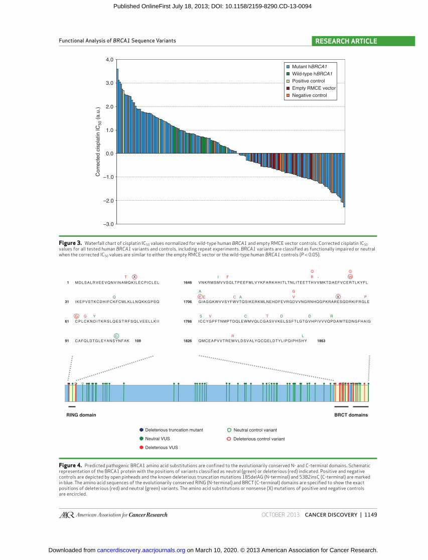
Fig. 3 ). Variants showing increased cisplatin sensitivity were tested at least twice before they were classifi ed. The positive and negative controls classifi ed as expected, although the known pathogenic truncation mutation 5382insC scored as neutral in one of three assays, stressing the need for repeat experiments. Also BRCA1 variants that were previ-ously tested in other assays conducted as expected. It should be noted that the artifi cial variants S308A and S1497A res-cued proliferation and cisplatin responses of mouse Brca1 -defi cient embryonic stem cells in BAC complementation assays, but are predicted to be deleterious based on their effects on embryonic stem cell differentiation and their response to γ-irradiation, respectively ( 7 ). The V1804D muta-tion scored as a neutral variant in our assay, which is in line with most published data ( 8, 9 ), except for the results of an embryonic stem cell–based BAC complementation assay ( 7 ). Again, all BRCA1 mutations that were classifi ed as deleteri-ous were confi ned to regions encoding the conserved N- and C-terminal domains ( Fig. 4 ), despite the observation that deletion of the central region encoded by exon 11 leads to genetic instability in mice ( 12 ). Remarkably, the three muta-tions that diminish the interaction between BRCA1 and PALB2 ( 10 ), just C-terminal of the region encoded by exon 11, had no effect on cisplatin sensitivity in this assay. Of note, there was in general good correlation between our classifi cation and the Align-GVGD score (Supplementary Table S4). Notable exceptions were the neutral control vari-ant R866C, which validated our assay but scored as likely deleterious (C65) using Align-GVGD, and G1770V, which scored as likely neutral (C0) by Align-GVGD but was classi-
fi ed as deleterious in our assay. Our assay also classifi ed some variants for which the Align-GVGD scores were less clear, emphasizing the usefulness of functional assays to comple-ment in silico analysis.
We also analyzed the possible effects of all BRCA1 variants on mRNA splicing, which may have deleterious consequences but cannot be assessed in our cDNA-based assay. The predic-tive value for exonic variants outside the consensus splice sites is questionable ( 19, 20 ), but four missense mutations were present in existing splice sites (Supplementary Table S4). Although c.5154G>T (encoding W1718C) was deleterious, three of these variants were classifi ed as neutral in our assay. One of them, c.441G>C (encoding L147F), had an increased probability to damage the splice donor site of exon 7. Another variant, c.5072C>T (encoding T1691I), might also affect splic-ing, but no defect was measured in blood samples of mutation carriers ( 19 ). The third exonic splice site mutation that was classifi ed as neutral in our cisplatin sensitivity assay, c.133A>C (K45Q), is not predicted to lead to aberrant splicing.
Results from Proliferation and Cisplatin Sensitivity Assays Correlate with HR Activity of BRCA1 Variants
BRCA1 and BRCA2 are involved in DNA repair via HR ( 12 , 21 ). Together with non-homologous end joining (NHEJ), HR forms the cellular defense against DNA double-strand breaks (DSB), a severe type of DNA damage that is lethal if unrepaired. Although HR is essentially error-free, NHEJ is error-prone, and therefore defects in HR are known to lead to genomic instability. Although it is not clear whether other
Variant DNA change Type of mutation a Classifi cation
V1736A c.5207T>C VUS Not clear b
D1739G c.5216A>G VUS Deleterious
D1739V c.5216A>T VUS Deleterious
H1746Q c.5238C>G VUS Not clear
R1753T c.5258G>C VUS Not clear b
5382insC c.5266dupC Deleterious control Not clear b
L1764P c.5291T>C VUS Deleterious b
C1767S c.5300G>C VUS Neutral
G1770V c.5309G>T VUS Deleterious b
W1782C c.5346G>T VUS Neutral
A1789T c.5365G>A VUS Deleterious
E1794D c.5382G>C VUS Neutral
V1804D c.5411T>A VUS Neutral
P1812R c.5435C>G VUS Neutral
W1837R c.5509T>C VUS Deleterious
H1862L c.5585A>T VUS Neutral a Type of mutation indicates if a variant is a VUS according to the BIC database (http://research.nhgri.nih.gov/bic/). b Functionally impaired in the DR-GFP and/or combined PARP inhibitor/cisplatin sensitivity assay.
Table 1. Functional classifi cation of BRCA1 VUS based on cisplatin response (Continued)
on March 10, 2020. © 2013 American Association for Cancer Research. cancerdiscovery.aacrjournals.org Downloaded from
Published OnlineFirst July 18, 2013; DOI: 10.1158/2159-8290.CD-13-0094
OCTOBER 2013�CANCER DISCOVERY | 1149
Functional Analysis of BRCA1 Sequence Variants RESEARCH ARTICLE
Figure 3. Waterfall chart of cisplatin IC 50 values normalized for wild-type human BRCA1 and empty RMCE vector controls. Corrected cisplatin IC 50 values for all tested human BRCA1 variants and controls, including repeat experiments. BRCA1 variants are classifi ed as functionally impaired or neutral when the corrected IC 50 values are similar to either the empty RMCE vector or the wild-type human BRCA1 controls ( P < 0.05).
4.0
–3.0
–2.0
–1.0
0.0
1.0
2.0
3.0
Cor
rect
ed c
ispl
atin
IC50
(a.
u.)
Mutant hBRCA1
Wild-type hBRCA1
Empty RMCE vector
Positive control
Negative control
Figure 4. Predicted pathogenic BRCA1 amino acid substitutions are confi ned to the evolutionarily conserved N- and C-terminal domains. Schematic representation of the BRCA1 protein with the positions of variants classifi ed as neutral (green) or deleterious (red) indicated. Positive and negative controls are depicted by open pinheads and the known deleterious truncation mutations 185delAG (N-terminal) and 5382insC (C-terminal) are marked in blue. The amino acid sequences of the evolutionarily conserved RING (N-terminal) and BRCT (C-terminal) domains are specifi ed to show the exact positions of deleterious (red) and neutral (green) variants. The amino acid substitutions or nonsense (X) mutations of positive and negative controls are encircled.
RING domain BRCT domains
Deleterious VUS
Neutral VUS
Deleterious truncation mutant
Deleterious control variant
Neutral control variant
1646
1706
1766
1826 186391 109
61
31
1 MDLSALRVEEVQNVINAMQKILECPICLEL VNKRMSMVVSGLTPEEFMLVYKFARKHHITLTNLITEETTHVVMKTDAEFVCERTLKYFL
IKEPVSTKCDHIFCKFCMLKLLNQKKGPSQ GIAGGKWVVSYFWVTQSIKERKMLNEHDFEVRGDVVNGRNHQGPKRARESQDRKIFRGLE
C P L C K N D I T K R S L Q E S T R F S Q L V E E L L K I I
CAFQLDTGLEYANSYNFAK
ICCYGPFTNMPTDQLEWMVQLCGASVVKELSSFTLGTGVHPIVVVQPDAWTEDNGFHAIG
QMCEAPVVTREWVLDSVALYQCQELDTYLIPQIPHSHY
T
Q E C A
X
Y
C
GG
F R -Q
WQ
I
EA
V
R L
TS C D D R
V PG
X
on March 10, 2020. © 2013 American Association for Cancer Research. cancerdiscovery.aacrjournals.org Downloaded from
Published OnlineFirst July 18, 2013; DOI: 10.1158/2159-8290.CD-13-0094

1150 | CANCER DISCOVERY�OCTOBER 2013 www.aacrjournals.org
Bouwman et al.RESEARCH ARTICLE
functions of BRCA1 are also important for tumor suppression ( 16 , 22 , 23–26 ), its role in HR is likely to be relevant. Therefore, we used the direct repeat (DR)-GFP assay ( 27 ) to measure the effects on HR for a subset of our BRCA1 VUSs. A number of predicted pathogenic variants and controls were shuttled into R26 CreERT2/RMCE ;Brca1 SCo/ Δ ; Pim1 DR-GFP/wt embryonic stem cells carrying the DR-GFP reporter in the Pim1 locus. Expression of endogenous mouse Brca1 was switched off, and cells were transfected with a plasmid encoding the I-SceI meganucle-ase as well as an mCherry fl uorescent marker to control for transfection effi ciency. Repair of I-SceI–induced DNA DSBs in DR-GFP via HR leads to expression of GFP, which can be monitored by fl ow cytometry. All but one of the predicted pathogenic variants tested resulted in defective HR, thereby confi rming our functional classifi cation ( Fig. 5 ). The only pre-dicted deleterious variant that did not signifi cantly differ from wild-type BRCA1 , R1699Q, seemed to support intermediate levels of HR activity.
PARP Inhibitor Sensitivity Assay for Classifi cation of BRCA1 Variants with Intermediate Activity
Deleterious effects of variants with intermediate or partial activity may escape detection in certain functional assays. Although cisplatin sensitivity assays allow robust and
Figure 5. BRCA1 sequence variants classifi ed as pathogenic do not restore HR. R26 CreERT2/hBRCA1 ; Brca1 SCo/Δ ; Pim1 DR-GFP/wt embryonic stem cells carrying the DR-GFP reporter gene in the Pim1 locus and mutant BRCA1 or controls in the Rosa26 locus were switched using 4-OHT and transfected with a vector expressing I-SceI and mCherry. Transfected cells were analyzed for GFP expression as a measure of HR activity. Expression of BRCA1 wild-type (WT) cDNA resulted in increased HR compared to the empty RMCE vector (Vector) control. Error bars indicate the SD between the results of three independent I-SceI transfections. Signifi cantly decreased HR activity compared with the wild-type control (green line) is indicated.
GF
P-P
ositi
ve c
ells
(%
)
0.0
0.5
1.0
1.5
2.0
2.5
3.0
3.5
4.0
4.5
**
ns
** *
*
ns
* **
* * * *
ns
ns
WT
Vecto
r18
5delA
G
C64G
V271L
S1651
FS16
55F
R1699
QR16
99W
G1706
AG17
06E
V1736
AR17
53T
5382
insC
L176
4PG17
70V
reproducible classifi cation of several functionally impaired BRCA1 variants, assays using other compounds may have additional value. It is known that BRCA1- and BRCA2-defi cient cells are extremely sensitive to PARP1 inhibition ( 28, 29 ), leading to a larger dynamic range between BRCA2-defi cient cells and isogenic BRCA2-profi cient controls than for cisplatin ( 30 ). We therefore tested complementation of PARP inhibitor sensitivity for a number of BRCA1 mutants and the BRCA1 wild-type control. Given the unexpected neutral effects of the M1400V, L1407P, and M1411T muta-tions in the PALB2 interaction domain, we decided to include these variants in this series, as well as the R1699Q and V1736A variants that have recently been shown to confer (intermediate) breast and ovarian cancer risk ( 31, 32 ). To allow direct comparison of results from different assays, we repeated the cisplatin sensitivity and prolifera-tion assays in parallel to the olaparib sensitivity assay. Our results indicate that BRCA1 wild-type and empty vector controls indeed show a larger difference in sensitivity for olaparib than for cisplatin ( Fig. 6 ). However, this increase in dynamic range is accompanied by an increased variation between repeat experiments for BRCA1-profi cient samples. Nevertheless, the functional defect of the R1699Q and V1736A mutations becomes more evident, and there also
on March 10, 2020. © 2013 American Association for Cancer Research. cancerdiscovery.aacrjournals.org Downloaded from
Published OnlineFirst July 18, 2013; DOI: 10.1158/2159-8290.CD-13-0094

OCTOBER 2013�CANCER DISCOVERY | 1151
Functional Analysis of BRCA1 Sequence Variants RESEARCH ARTICLE
Figure 6. PARP inhibitor sensitivity assay of BRCA1 sequence variants. R26 CreERT2/hBRCA1 ; Brca1 SCo/Δ or R26 CreERT2/RMCE ; Brca1 SCo/Δ embryonic stem cells carrying mutant BRCA1 , BRCA1 wild-type (WT), or empty RMCE vector (Vector) controls in the Rosa26 locus were switched using 4-OHT and assayed for sensitivity to cisplatin or the PARP inhibitor olaparib. The cytotoxicity assays were conducted in parallel and data were normalized to the average of the wild-type controls. Error bars indicate the SD between the results of biologic triplicates for which the cells were independently switched. Signifi cant deviation from the average IC 50 values of the wild-type control (green line) is indicated.
IC50
Rel
ativ
e to
WT
con
trol
s (%
)
0
20
40
60
80
100
120
140
160
Cisplatin
Olaparib
*
**** ****
*
***
***
****
ns
ns
ns
ns
WT
Vecto
r
M14
00V
L140
7P
M14
11T
R1699
Q
V1736
A
seems to be a less than wild-type response for the PALB2 interaction mutant L1407P. In the concurrent proliferation analysis, R1699Q and V1736A both show an intermedi-ate functional defect (Supplementary Fig. S4). Also the M1411T mutation seems to affect the response to PARP inhibition, but the difference with BRCA1 wild-type is not signifi cant. Interestingly, the L1407P and M1411T vari-ants have previously been shown to be more defective than M1400V in a gene conversion assay ( 10 ). It should also be noted that, in contrast with the large-scale classifi cation experiments, the cisplatin sensitivity assay conducted in parallel to the PARP inhibitor assay identifi ed signifi cant functional defects for L1407P and V1736A.
DISCUSSION
Over the past few years several functional assays for clas-sifi cation of BRCA1 VUSs have been developed. Several of these assays are restricted to functions of the BRCA1 protein that reside in the evolutionarily conserved RING or BRCT domains. Examples include in vitro transactivation assays for BRCT peptides ( 8 ) and measurement of ubiquitin ligase activity for protein fragments encompassing the N-terminal RING domain ( 33 ). Other assays were designed to evaluate
the functions of full-length mutant BRCA1 protein, either by monitoring general effects on proliferation or response to DNA damage ( 7 ), or by directly focusing on the role of BRCA1 in DNA repair via HR ( 34 ).
Because BRCA1 VUSs are not restricted to regions encoding the N- or C-terminal domains, and given the observation that interaction between these domains is required for recruit-ment of BRCA1 to damaged DNA ( 35 ), functional assays for the full-length protein would be ideal. In principle, such assays can be conducted in cell lines derived from BRCA1 -mutated tumors, but there are indications that the outcome of assays for BRCA1 function depends on the cellular context. As absence of BRCA1 leads to loss of cellular viability, it is thought that additional mutations are required for BRCA1-associated tumorigenesis. For example, loss of p53 alleviates the consequences of BRCA1 defi ciency both in vitro and in vivo ( 4 ) and is common in BRCA1-defi cient tumors ( 36, 37 ). Also, depletion of 53BP1 is known to suppress the defects caused by BRCA1 defi ciency ( 5 , 38 , 39 ). Therefore, aberrations in BRCA1-defi cient tumor cells may mask functional defects of BRCA1 VUSs. We reasoned that assays in normal cells that can be depleted from endogenous BRCA1 expression are most likely to reveal functional defects of BRCA1 VUSs. The usefulness of this approach has been previously shown by
on March 10, 2020. © 2013 American Association for Cancer Research. cancerdiscovery.aacrjournals.org Downloaded from
Published OnlineFirst July 18, 2013; DOI: 10.1158/2159-8290.CD-13-0094

1152 | CANCER DISCOVERY�OCTOBER 2013 www.aacrjournals.org
Bouwman et al.RESEARCH ARTICLE
BAC complementation assays in mouse embryonic stem cells ( 7 , 40 ). However, mutagenesis of large BAC clones by recombineering and functional complementation of cells with these mutant constructs is time-consuming and tech-nically demanding, and therefore cannot be conducted in a high-throughput setting. We therefore set out to develop a cDNA-based functional complementation assay in mouse embryonic stem cells that is easier to control and scale up and more suitable for routine functional classifi cation of BRCA1 sequence variants.
One advantage of BAC transgenics in complementation assays is that genes are expressed at physiologically relevant levels. This is the result of low copy-number integrations and the presence of natural regulatory elements required for proper gene expression. We decided to use RMCE to allow single-copy integration of BRCA1 cDNAs at one specifi c genomic locus. The use of RMCE effectively prevents multi-ple or partial integrations, concatemers, and position-effect variegation. As a result, all variants are expressed at equal levels. Moreover, transcription of BRCA1 cDNAs from the EF1A gene promoter results in physiologic levels of BRCA1 protein, comparable with those observed in embryonic stem cells stably transfected with a BAC containing the human BRCA1 locus. Indeed, the wild-type human BRCA1 cDNA was able to complement Brca1 -null embryonic stem cells in cellular proliferation, drug sensitivity, and HR assays. Our RMCE strategy uses BRCA1 cDNA constructs in which mutations can be swiftly introduced using SDM, enabling a higher throughput than introduction of mutations via BAC recombineering. In addition, the introduction of BRCA1 cDNAs via RMCE obviates the need to analyze multiple embry-onic stem cell clones for correct integration and expression.
As a proof of principle, we used our functional comple-mentation assay to analyze exonic BRCA1 VUSs that were identifi ed in families with HBOC in the Netherlands and Bel-gium, as well as a set of previously analyzed BRCA1 variants. A number of mutations resulted in reduced BRCA1 protein levels, most likely because of structural destabilization. In all cases, this led to diminished capacity for functional com-plementation. Also, several variants that gave rise to normal BRCA1 protein levels were unable to rescue the proliferation defect and cisplatin sensitivity of Brca1 -null embryonic stem cells. As BRCA1 loss of function mutations are associated with increased cancer risk, variants that score as functionally impaired in our embryonic stem cell assay system may be caus-ally involved in tumor formation. This notion is supported by the fact that seven of eight known pathogenic or neutral con-trol variants in our validation series were correctly classifi ed by the cisplatin sensitivity assay. The pathogenic 5382insC truncation mutation could not be classifi ed because it scored as neutral in one of three transfection series. This was prob-ably due to technical reasons, as the 5382insC mutation did not restore HR activity in Brca1 -defi cient embryonic stem cells, in contrast with R1699Q, which was recently shown to confer intermediate risk of HBOC ( 32 ).
Our assay system yielded ambivalent results for nine other variants: S4F, S308A, S1651P, S1651F, T1691I, V1736A, E1735K, H1746Q, and R1753T. T1691I and E1735K were classifi ed as functionally impaired in only one cisplatin sensitivity test, whereas values from repeat experiments could
not be taken into account because of RSE values above 0.1. S4F, S1651P, S1651F, V1736A, H1746Q, and R1753T were differently classifi ed in repeat experiments, which may refl ect technical fl aws or intermediate activity of these variants. Of note, the V1736A mutation was recently identifi ed as a pathogenic variant with hypomorphic activity in DNA repair ( 31 ). Although we did not measure signifi cant HR activity of V1736A in a DR-GFP gene conversion assay, intermedi-ate activity of this variant is supported by the results of the proliferation assays and additional cisplatin sensitivity assays. S1651F showed HR activity similar to wild-type BRCA1 , whereas R1753T was HR-defi cient. S308A is an artifi cial mutation of a BRCA1 phosphorylation site that was able to support proliferation and resistance to DNA damage in an embryonic stem cell-based BAC complementation assay. However, S308A-complemented embryonic stem cells did show increased apoptosis when cultured in embryoid bodies ( 7 ), indicating a partial defect that might explain the ambiva-lent results for this mutation in our assays.
Our results show that BRCA1 variants should ideally be assayed in triplicate to avoid misclassifi cation. This also applies to BRCA1 VUS that we classifi ed as neutral, most of which were tested only once because in our current study we focused on variants that showed functional impairment. The striking restriction of unambiguously predicted pathogenic mutants to the terminal RING and BRCT domains suggests that some plasticity is allowed for the central domain of BRCA1. However, our dataset is still limited and more experi-ments are required to gain insight into the function of this domain.
Our cDNA-based system allows for several additional func-tional assays that have been described previously for BAC transgenic embryonic stem cells ( 7 ). These include assays for defects during in vitro and in vivo embryonic stem cell differ-entiation but also treatments with other cytotoxic agents. As a proof of principle, we investigated the activity of a number of mutants in the response to the PARP inhibitor olaparib. These included M1400V, L1407, and M1411T, which were previously shown to impair PALB2 binding and have a nega-tive effect on BRCA1 function ( 10 ). Although the differences in sensitivity of BRCA1-defi cient versus BRCA1-profi cient embryonic stem cells are larger for olaparib than for cispla-tin, increased variation between repeat experiments allowed us to identify functional defects only for L1407 and not for the other two variants in the PALB2 interaction domain, M1400V and M1411T. However, the R1699Q and V1736A variants, which are known to have hypomorphic activity, clearly showed a defect in the response to olaparib. Interest-ingly, both mutations are in the BRCT domain, and it has recently been shown that mutation of this domain especially confers sensitivity to PARP inhibition ( 41 ). Together, our results show that PARP inhibitor sensitivity assays may have added value, especially for the classifi cation of BRCA1 VUSs with intermediate phenotypes.
Platinum drugs and PARP inhibitors are selectively toxic to BRCA1-defi cient cells because they target HR defi ciency. Although the role of BRCA1 in HR is thought to be essential for maintaining genomic integrity and preventing accumu-lation of (oncogenic) mutations, other activities may also contribute to its tumor suppression function. These activities
on March 10, 2020. © 2013 American Association for Cancer Research. cancerdiscovery.aacrjournals.org Downloaded from
Published OnlineFirst July 18, 2013; DOI: 10.1158/2159-8290.CD-13-0094
OCTOBER 2013�CANCER DISCOVERY | 1153
Functional Analysis of BRCA1 Sequence Variants RESEARCH ARTICLE
may include the HR-independent role for BRCA1 in ICL repair, which has been attributed to the facilitation of FANCD2 accumulation at cross-linked DNA ( 16 ). Neverthe-less, we observed a good correlation between the results of the cisplatin sensitivity assay and the results of the DR-GFP HR assay. All BRCA1 variants that failed to restore the cisplatin response in Brca1 -null embryonic stem cells were also defec-tive in catalyzing gene conversion, thereby confi rming our functional classifi cation. It will be interesting to see if this holds true for all BRCA1 variants or whether there are also pathogenic mutations that have no effect on HR.
As with any other in vitro approach, our functional com-plementation assay system might still fail to identify all pathogenic variants because it does not necessarily recapitu-late all aspects of BRCA1 function in vivo . A limitation of our cDNA-based assay is also that it cannot be used to investigate effects on mRNA splicing. Although algorithms have been designed to predict possible splice defects, the consequences of mutations outside of the consensus splice sites especially require functional validation experiments. For this purpose, BAC complementation assays ( 7 ), minigene-based splicing assays, or BRCA1 transcript analysis of patient blood samples ( 19 ) may be instrumental. However, transacting factors also affect splicing, and these may be tissue specifi c ( 42 ). A pos-sible solution would be to determine the presence of BRCA1 splice variants in tumor tissue from BRCA1 VUS carriers and use this information to generate a cDNA construct for analysis of the functional consequences. To evaluate BRCA1 VUSs, there remains a need for multifactorial models that combine results from functional assays and in silico analyses with genetic evidence and other information from mutation carriers. This also includes DNA copy number data from tumors from BRCA1 VUS carriers, as it is known that BRCA1-associated breast tumors show distinct genomic aberrations ( 43, 44 ). Our functional assay system does however provide a robust and easily implementable tool for the functional characterization of large numbers of BRCA1 VUSs within the context of the full-length protein. It is our hope that our assay system will fi nd its way to clinical genetics laboratories where it can be used to aid genetic counseling. Ideally, these tests should be coordinated on an international level and in close collaboration with the ENIGMA consortium.
METHODS
Generation of RMCE Vectors Containing Human BRCA1 Sequence Variants
Human BRCA1 cDNA from a pcDNA3-BRCA1 expression con-struct ( 45 ) was subcloned into the pRNA 251-MCS RMCE exchange vector under control of the EF1A gene promoter. BRCA1 muta-tions were introduced by site-directed mutagenesis using the Quick-Change Lightning protocol (Stratagene), and constructs were verifi ed by sequencing the entire human BRCA1 cDNA (see Supplementary Methods).
Generation of Human BRCA1 Transgenic Embryonic Stem Cells
R26 CreERT2/wt ;Brca1 SCo/ Δ embryonic stem cells were generated by gene targeting in 129/Ola E14 IB10 embryonic stem cells ( 46 ). The pres-ence of correctly targeted alleles was verifi ed using Southern blotting, Western blotting, and PCR analysis ( 5 , 47 ). The wild-type Rosa26 allele
of R26 CreERT2/wt ;Brca1 SCo/ Δ embryonic stem cells was equipped with Frt and F3 sites for Flp RMCE as described (ref. 6 ; see Supplementary Fig. S1). Introduction of human BRCA1 cDNAs via RMCE was conducted by cotransfection of R26 CreERT2/RMCE ;Brca1 SCo/ Δ embryonic stem cells with RMCE vectors and pFlpe ( 48 ) using Lipofectamine 2000 (Invitrogen). Cells that had successfully undergone RMCE were selected using 200–400 μg/mL G418. Correct RMCE was confi rmed by PCR and expression of human BRCA1 was analyzed by Western blotting using a polyclonal antibody against human BRCA1 (9010; Cell Signaling Technology). The generation of R26 CreERT2 ;Brca1 SCo/ Δ embryonic stem cells expressing human BRCA1 from the BAC clone RP11-812O5 was conducted essentially as described ( 49 ).
Cytotoxicity and Proliferation Assays Cre-mediated inactivation of the endogenous mouse Brca1 SCo allele
was achieved by overnight incubation of cells with 0.5 μmol/L 4-OHT (Sigma). One week after switching, cells were seeded in triplicate at 1,000 cells per well in 96-well plates for cisplatin or olaparib (AZD2281) sensitivity assays essentially as described ( 5 ). In addition, cells were seeded in triplicate at 500 cells per well on 96-well plates to monitor proliferation.
HR Reporter Assays For DR-GFP assays we used a modifi ed version of the p59X DR-
GFP construct (ref. 12 ; kindly provided by T. Ludwig), in which the puromycin resistance marker was inactivated by inversion of an internal SalI fragment. To allow selection of targeted integration of this construct to the Pim1 locus, we equipped the wild-type Rosa26 allele of R26 CreERT2/wt ;Brca1 SCo/ Δ embryonic stem cells with Frt and F3 sites for RMCE using a targeting vector in which zsgreen , the hygromycin resistance marker, and Flpe were replaced by a puro-mycin resistance marker. Subsequently, the DR-GFP construct was targeted to the Pim1 locus as described (26), BRCA1 variants were introduced using RMCE, and cells were subcloned to allow HR reporter assays. HR reporter assays were conducted by Lipo-fectamine 2000 transfections of an I-SceI-mCherry plasmid, which was generated by cloning CMV-mCherry (Clontech) into the cBas I-SceI expression plasmid. Three days after transfection, mCherry/GFP double-positive cells were monitored by fl ow cytometry on a fl uorescence-activated cell sorting CyAn (Beckman Coulter) using Summit software (Beckman Coulter).
Statistical Analysis We calculated the cisplatin IC 50 values from the 96-well plate-based
cisplatin sensitivity assays for VUS classifi cation by fi tting a log-logistic curve, normalized to the no-drug control, constrained between 1 and 0 using the drc package in the R programming language. We discarded fi ts that exceeded a 0.1 RSE. To allow comparison between plates, we normalized using a linear model based on the positive and negative controls, resulting in corrected IC 50 scores. We estimated normal distributions of the corrected IC 50 score for both the pooled positive and negative controls across plates. We used these estimated normals to calculate the probability of pathogenicity for each VUS.
Statistical signifi cance for the HR and cytotoxicity assays on selected groups of variants was calculated by two-tailed Student t test using Prism 6 Software. Signifi cant differences are indicated by *, P < 0.05; **, P < 0.01; ***, P < 0.001; and ****, P < 0.0001. NS stands for nonsignifi cant ( P > 0.05).
Computational Analysis Alamut software was used to obtain genomic annotations and
Align-GVGD scores (human to sea urchin) for all variants.
See Supplementary Methods for a detailed protocol for the clas-sifi cation of BRCA1 sequence variants.
on March 10, 2020. © 2013 American Association for Cancer Research. cancerdiscovery.aacrjournals.org Downloaded from
Published OnlineFirst July 18, 2013; DOI: 10.1158/2159-8290.CD-13-0094

1154 | CANCER DISCOVERY�OCTOBER 2013 www.aacrjournals.org
Bouwman et al.RESEARCH ARTICLE
Disclosure of Potential Confl icts of Interest No potential confl icts of interest were disclosed.
Authors’ Contributions Conception and design: P. Bouwman, H. van der Gulden, F.B.L. Hogervorst, J. Jonkers Development of methodology: P. Bouwman, H. van der Gulden, I. van der Heijden, R. Drost, M. Pieterse, J. Seibler, J. Jonkers Acquisition of data (provided animals, acquired and managed patients, provided facilities, etc.): P. Bouwman, H. van der Gulden, I. van der Heijden, R. Drost, P. Prasetyanti, M. Pieterse, E. Wientjens Analysis and interpretation of data (e.g., statistical analysis, biosta-tistics, computational analysis): P. Bouwman, H. van der Gulden, I. van der Heijden, C.N. Klijn, M. Pieterse, F.B.L. Hogervorst, J. Jonkers Writing, review, and/or revision of the manuscript: P. Bouwman, H. van der Gulden, I. van der Heijden, R. Drost, E. Wientjens, F.B.L. Hogervorst, J. Jonkers Administrative, technical, or material support (i.e., report-ing or organizing data, constructing databases): P. Bouwman, H. van der Gulden, I. van der Heijden, E. Wientjens, J. Seibler Study supervision: P. Bouwman, J. Jonkers
Acknowledgments The authors thank Sandra Niehaves (TaconicArtemis GmbH) for
technical assistance; D.P. Silver (Dana-Farber Cancer Institute, Bos-ton, MA) for the human BRCA1 cDNA expression construct; L. van der Weyden (Wellcome Trust Sanger Institute, Hinxton, UK) for the pFlpe expression construct; F. Stewart (BioInnovations Zentrum, Dresden, Germany) for the pFlpo expression construct; and M. Jasin (Memorial Sloan-Kettering Cancer Center, New York, NY) and T. Ludwig (Columbia University, New York, NY) for the I-SceI expres-sion plasmid and the DR-GFP reporter plasmid. The authors also thank Jo Morris (University of Birmingham, UK), Annegien Broeks, and Petra Nederlof for helpful comments on the manuscript. The Dutch/Belgium VUS workgroup is gratefully acknowledged for pro-viding data on the occurrence of BRCA1 VUS in the Netherlands and Belgium. Workgroup members are: Frans B.L. Hogervorst (Family Cancer Clinic, Netherlands Cancer Institute, Amsterdam), Johannes J.P. Gille (Department of Clinical Genetics, VU Medical Center, Amsterdam), Juul T. Wijnen and Maaike Vreeswijk (Department of Human Genetics & Department of Clinical Genetics, Leiden Univer-sity Medical Center, Leiden), Rob van der Luijt (Department of Medi-cal Genetics, University Medical Center Utrecht, Utrecht), Marinus J. Blok (Department of Clinical Genetics, University Hospital Maas-tricht, Maastricht), Ans van den Ouweland (Department of Clinical Genetics, Family Cancer Clinic, Erasmus University Medical Center, Rotterdam), Danielle Bodmer and Arjen Mensenkamp (Department of Human Genetics, Radboud University Nijmegen Medical Centre, Nijmegen), and Annemiek van der Hout (Department of Genet-ics, University Medical Center Groningen, University of Groningen, Groningen). Belgian Collaborators: Katrien Storm (Department of Medical Genetics, University and University Hospital Antwerp, Ant-werp), Kathleen Claes (Center for Medical Genetics, Ghent University Hospital, Ghent), Genevieve Michels (Center for Human Genetics, University of Leuven, Leuven), and Erik Teugels (Laboratory of Molecular Oncology, Vrije Universiteit Brussel, Brussels).
Grant Support This work was supported by grants from the Dutch Cancer Society
(NKI 2008–4116 to J. Jonkers and P. Bouwman), the Cancer Genom-ics Centre Netherlands, and the TI Center for Translational Molecu-lar Medicine (CTMM) BreastCare project.
Received March 5, 2013; revised June 20, 2013; accepted June 20, 2013; published OnlineFirst July 18, 2013.
REFERENCES 1. Spurdle AB , Healey S , Devereau A , Hogervorst FBL , Monteiro ANA ,
Nathanson KL , et al. ENIGMA–evidence-based network for the inter-pretation of germline mutant alleles: an international initiative to evaluate risk and clinical signifi cance associated with sequence varia-tion in BRCA1 and BRCA2 genes . Hum Mutat 2012 ; 33 : 2 – 7 .
2. Lindor NM , Guidugli L , Wang X , Vallée MP , Monteiro ANA , Tavtigian S , et al. A review of a multifactorial probability-based model for classifi cation of BRCA1 and BRCA2 variants of uncertain signifi -cance (VUS) . Hum Mutat 2012 ; 33 : 8 – 21 .
3. Millot GA , Carvalho MA , Caputo SM , Vreeswijk MPG , Brown MA , Webb M , et al. A guide for functional analysis of BRCA1 variants of uncertain signifi cance . Hum Mutat 2012 ; 33 : 1526 – 37 .
4. Evers B , Jonkers J . Mouse models of BRCA1 and BRCA2 defi ciency: past lessons, current understanding and future prospects . Oncogene 2006 ; 25 : 5885 – 97 .
5. Bouwman P , Aly A , Escandell JM , Pieterse M , Bartkova J , Van der Gulden H , et al. 53BP1 loss rescues BRCA1 defi ciency and is associ-ated with triple-negative and BRCA-mutated breast cancers . Nat Struct Mol Biol 2010 ; 17 : 688 – 95 .
6. Seibler J , Küter-Luks B , Kern H , Streu S , Plum L , Mauer J , et al. Single copy shRNA confi guration for ubiquitous gene knockdown in mice . Nucleic Acids Res 2005 ; 33 : e67 .
7. Chang S , Biswas K , Martin BK , Stauffer S , Sharan SK . Expression of human BRCA1 variants in mouse ES cells allows functional analysis of BRCA1 mutations . J Clin Invest 2009 ; 119 : 3160 – 71 .
8. Lee MS , Green R , Marsillac SM , Coquelle N , Williams RS , Yeung T , et al. Comprehensive analysis of missense variations in the BRCT domain of BRCA1 by structural and functional assays . Cancer Res 2010 ; 70 : 4880 – 90 .
9. Easton DF , Deffenbaugh AM , Pruss D , Frye C , Wenstrup RJ , Allen-Brady K , et al. A systematic genetic assessment of 1,433 sequence variants of unknown clinical signifi cance in the BRCA1 and BRCA2 breast cancer-predisposition genes . Am J Hum Genet 2007 ; 81 : 873 – 83 .
10. Sy SMH , Huen MSY , Chen J . PALB2 is an integral component of the BRCA complex required for homologous recombination repair . Proc Natl Acad Sci U S A 2009 ; 106 : 7155 – 60 .
11. Tavtigian SV , Deffenbaugh AM , Yin L , Judkins T , Scholl T , Samollow PB , et al. Comprehensive statistical study of 452 BRCA1 missense substitutions with classifi cation of eight recurrent substitutions as neutral . J Med Genet 2006 ; 43 : 295 – 305 .
12. Moynahan ME , Pierce AJ , Jasin M . BRCA2 is required for homology-directed repair of chromosomal breaks . Mol Cell 2001 ; 7 : 263 – 72 .
13. Chandler J , Hohenstein P , Swing DA , Tessarollo L , Sharan SK . Human BRCA1 gene rescues the embryonic lethality of Brca1 mutant mice . Genesis 2001 ; 29 : 72 – 7 .
14. De Nicolo A , Parisini E , Zhong Q , Dalla Palma M , Stoeckert KA , Domchek SM , et al. Multimodal assessment of protein functional defi ciency supports pathogenicity of BRCA1 p.V1688del . Cancer Res 2009 ; 69 : 7030 – 7 .
15. Rowling PJE , Cook R , Itzhaki LS . Toward classifi cation of BRCA1 mis-sense variants using a biophysical approach . J Biol Chem 2010 ; 285 : 20080 – 7 .
16. Bunting SF , Callén E , Kozak ML , Kim JM , Wong N , López-Contreras AJ , et al. BRCA1 functions independently of homologous recombina-tion in DNA interstrand crosslink repair . Mol Cell 2012 ; 46 : 125 – 35 .
17. Norquist B , Wurz KA , Pennil CC , Garcia R , Gross J , Sakai W , et al. Secondary somatic mutations restoring BRCA1/2 predict chemo-therapy resistance in hereditary ovarian carcinomas . J Clin Oncol 2011 ; 29 : 3008 – 15 .
18. Swisher EM , Sakai W , Karlan BY , Wurz K , Urban N , Taniguchi T . Secondary BRCA1 mutations in BRCA1-mutated ovarian carcinomas with platinum resistance . Cancer Res 2008 ; 68 : 2581 – 6 .
19. Houdayer C , Caux-Moncoutier V , Krieger S , Barrois M , Bonnet F , Bourdon V , et al. Guidelines for splicing analysis in molecular diag-nosis derived from a set of 327 combined in silico / in vitro studies on BRCA1 and BRCA2 variants . Hum Mutat 2012 ; 33 : 1228 – 38 .
on March 10, 2020. © 2013 American Association for Cancer Research. cancerdiscovery.aacrjournals.org Downloaded from
Published OnlineFirst July 18, 2013; DOI: 10.1158/2159-8290.CD-13-0094

OCTOBER 2013�CANCER DISCOVERY | 1155
Functional Analysis of BRCA1 Sequence Variants RESEARCH ARTICLE
20. Houdayer C , Dehainault C , Mattler C , Michaux D , Caux-Moncoutier V , Pagès-Berhouet S , et al. Evaluation of in silico splice tools for deci-sion-making in molecular diagnosis . Hum Mutat 2008 ; 29 : 975 – 82 .
21. Moynahan ME , Chiu JW , Koller BH , Jasin M . Brca1 controls homol-ogy-directed DNA repair . Mol Cell 1999 ; 4 : 511 – 8 .
22. Schlacher K , Wu H , Jasin M . A distinct replication fork protection pathway connects Fanconi anemia tumor suppressors to RAD51-BRCA1/2 . Cancer Cell 2012 ; 22 : 106 – 16 .
23. Joukov V , Groen AC , Prokhorova T , Gerson R , White E , Rodriguez A , et al. The BRCA1/BARD1 heterodimer modulates ran-dependent mitotic spindle assembly . Cell 2006 ; 127 : 539 – 52 .
24. Zhu Q , Pao GM , Huynh AM , Suh H , Tonnu N , Nederlof PM , et al. BRCA1 tumour suppression occurs via heterochromatin-mediated silencing . Nature 2011 ; 477 : 179 – 84 .
25. Shakya R , Reid LJ , Reczek CR , Cole F , Egli D , Lin C-S , et al. BRCA1 tumor suppression depends on BRCT phosphoprotein binding, but not its E3 ligase activity . Science 2011 ; 334 : 525 – 8 .
26. Drost R , Bouwman P , Rottenberg S , Boon U , Schut E , Klarenbeek S , et al. BRCA1 RING function is essential for tumor suppression but dispensable for therapy resistance . Cancer Cell 2011 ; 20 : 797 – 809 .
27. Pierce AJ , Johnson RD , Thompson LH , Jasin M . XRCC3 promotes homology-directed repair of DNA damage in mammalian cells . Genes Dev 1999 ; 13 : 2633 – 8 .
28. Bryant HE , Schultz N , Thomas HD , Parker KM , Flower D , Lopez E , et al. Specifi c killing of BRCA2-defi cient tumours with inhibitors of poly(ADP-ribose) polymerase . Nature 2005 ; 434 : 913 – 7 .
29. Farmer H , McCabe N , Lord CJ , Tutt ANJ , Johnson DA , Richardson TB , et al. Targeting the DNA repair defect in BRCA mutant cells as a therapeutic strategy . Nature 2005 ; 434 : 917 – 21 .
30. Evers B , Schut E , Van der Burg E , Braumuller TM , Egan DA , Holstege H , et al. A high-throughput pharmaceutical screen identifi es com-pounds with specifi c toxicity against BRCA2-defi cient tumors . Clin Cancer Res 2010 ; 16 : 99 – 108 .
31. Domchek SM , Tang J , Stopfer J , Lilli DR , Hamel N , Tischkowitz M , et al. Biallelic deleterious BRCA1 mutations in a woman with early-onset ovarian cancer . Cancer Discov 2013 ; 3 : 399 – 405 .
32. Spurdle AB , Whiley PJ , Thompson B , Feng B , Healey S , Brown MA , et al. BRCA1 R1699Q variant displaying ambiguous functional abro-gation confers intermediate breast and ovarian cancer risk . J Med Genet 2012 ; 49 : 525 – 32 .
33. Morris JR , Pangon L , Boutell C , Katagiri T , Keep NH , Solomon E . Genetic analysis of BRCA1 ubiquitin ligase activity and its relation-ship to breast cancer susceptibility . Hum Mol Genet 2006 ; 15 : 599 – 606 .
34. Ransburgh DJR , Chiba N , Ishioka C , Toland AE , Parvin JD . Identifi ca-tion of breast tumor mutations in BRCA1 that abolish its function in homologous DNA recombination . Cancer Res 2010 ; 70 : 988 – 95 .
35. Au WWY , Henderson BR . The BRCA1 RING and BRCT domains cooperate in targeting BRCA1 to ionizing radiation-induced nuclear foci . J Biol Chem 2005 ; 280 : 6993 – 7001 .
36. Manié E , Vincent-Salomon A , Lehmann-Che J , Pierron G , Turpin E , Warcoin M , et al. High frequency of TP53 mutation in BRCA1 and sporadic basal-like carcinomas but not in BRCA1 luminal breast tumors . Cancer Res 2009 ; 69 : 663 – 71 .
37. Holstege H , Joosse SA , Van Oostrom CTM , Nederlof PM , De Vries A , Jonkers J . High incidence of protein-truncating TP53 mutations in BRCA1-related breast cancer . Cancer Res 2009 ; 69 : 3625 – 33 .
38. Bunting SF , Callén E , Wong N , Chen H-T , Polato F , Gunn A , et al. 53BP1 inhibits homologous recombination in Brca1-defi cient cells by blocking resection of DNA breaks . Cell 2010 ; 141 : 243 – 54 .
39. Cao L , Xu X , Bunting SF , Liu J , Wang R-H , Cao LL , et al. A selective requirement for 53BP1 in the biological response to genomic instabil-ity induced by Brca1 defi ciency . Mol Cell 2009 ; 35 : 534 – 41 .
40. Kuznetsov SG , Liu P , Sharan SK . Mouse embryonic stem cell-based functional assay to evaluate mutations in BRCA2 . Nat Med 2008 ; 14 : 875 – 81 .
41. Li M , Yu X . Function of BRCA1 in the DNA damage response is medi-ated by ADP-ribosylation . Cancer Cell 2013 ; 23 : 693 – 704 .
42. Kornblihtt AR , Schor IE , Alló M , Dujardin G , Petrillo E , Muñoz MJ . Alternative splicing: a pivotal step between eukaryotic transcription and translation . Nat Rev Mol Cell Biol 2013 ; 14 : 153 – 65 .
43. Wessels LFA , Van Welsem T , Hart AAM , Van’t Veer LJ , Reinders MJT , Nederlof PM . Molecular classifi cation of breast carcinomas by com-parative genomic hybridization: a specifi c somatic genetic profi le for BRCA1 tumors . Cancer Res 2002 ; 62 : 7110 – 7 .
44. Van Beers EH , Van Welsem T , Wessels LFA , Li Y , Oldenburg RA , Devilee P , et al. Comparative genomic hybridization profi les in human BRCA1 and BRCA2 breast tumors highlight differential sets of genomic aberrations . Cancer Res 2005 ; 65 : 822 – 7 .
45. Chen J , Silver DP , Walpita D , Cantor SB , Gazdar AF , Tomlinson G , et al. Stable interaction between the products of the BRCA1 and BRCA2 tumor suppressor genes in mitotic and meiotic cells . Mol Cell 1998 ; 2 : 317 – 28 .
46. Robanus-Maandag E , Dekker M , Van der Valk M , Carrozza ML , Jeanny JC , Dannenberg JH , et al. p107 is a suppressor of retinoblas-toma development in pRb-defi cient mice . Genes Dev 1998 ; 12 : 1599 – 609 .
47. Liu X , Holstege H , Van der Gulden H , Treur-Mulder M , Zevenhoven J , Velds A , et al. Somatic loss of BRCA1 and p53 in mice induces mammary tumors with features of human BRCA1-mutated basal-like breast cancer . Proc Natl Acad Sci U S A 2007 ; 104 : 12111 – 6 .
48. Buchholz F , Angrand PO , Stewart AF . Improved properties of FLP recombinase evolved by cycling mutagenesis . Nat Biotechnol 1998 ; 16 : 657 – 62 .
49. Puppe J , Drost R , Liu X , Joosse SA , Evers B , Cornelissen-Steijger P , et al. BRCA1-defi cient mammary tumor cells are dependent on EZH2 expression and sensitive to Polycomb Repressive Complex 2-inhibitor 3-deazaneplanocin A . Breast Cancer Res 2009 ; 11 : R63 .
on March 10, 2020. © 2013 American Association for Cancer Research. cancerdiscovery.aacrjournals.org Downloaded from
Published OnlineFirst July 18, 2013; DOI: 10.1158/2159-8290.CD-13-0094

2013;3:1142-1155. Published OnlineFirst July 18, 2013.Cancer Discovery Peter Bouwman, Hanneke van der Gulden, Ingrid van der Heijden, et al.
Missense VariantsBRCA1Classification of A High-Throughput Functional Complementation Assay for
Updated version
10.1158/2159-8290.CD-13-0094doi:
Access the most recent version of this article at:
Material
Supplementary
http://cancerdiscovery.aacrjournals.org/content/suppl/2013/07/18/2159-8290.CD-13-0094.DC1
Access the most recent supplemental material at:
Cited articles
http://cancerdiscovery.aacrjournals.org/content/3/10/1142.full#ref-list-1
This article cites 49 articles, 20 of which you can access for free at:
Citing articles
http://cancerdiscovery.aacrjournals.org/content/3/10/1142.full#related-urls
This article has been cited by 15 HighWire-hosted articles. Access the articles at:
E-mail alerts related to this article or journal.Sign up to receive free email-alerts
Subscriptions
Reprints and
To order reprints of this article or to subscribe to the journal, contact the AACR Publications Department at
Permissions
Rightslink site. Click on "Request Permissions" which will take you to the Copyright Clearance Center's (CCC)
.http://cancerdiscovery.aacrjournals.org/content/3/10/1142To request permission to re-use all or part of this article, use this link
on March 10, 2020. © 2013 American Association for Cancer Research. cancerdiscovery.aacrjournals.org Downloaded from
Published OnlineFirst July 18, 2013; DOI: 10.1158/2159-8290.CD-13-0094