a history of pet instrumentation - wfu...
TRANSCRIPT

A history of PET instrumentation
Lars Eriksson
Siemens Molecular Imaging, University of Stockholm and Karolinska Institute

2
Research activities up to 1974
Nuclear Physics studies at the Department of Physics, University of Stockholm.Ph.D. Thesis: gamma-gamma angular correlations with a multi detector system (1973). Docent in physics in 1974
Multichannel goniometer for gamma-gamma angular corre-lations (TR Gerholm, B-G Pettersson, L. Gidefeldt, L. Eriksson, Z.H. Cho)

3
A Swedish PET project is formed
1974 first contacts with Karolinska Institute about a collaboration project of brain metabolism measured with PET- application for a Swedish PET system from Medical Research Council
L. Widen (Professor in Clinical Neurophysiology, KI)T. Greitz (Professor in Neuroradiology, KI)T.G. Gerholm (Professor in Physics, SU)
Visit to UCLA 1974-1976 at Laboratory of Nuclear Medicine [Z.H. Cho (G. Huth) ]
Working with a ring detector positron camera systemPlanned two detector rings – one ring with 64 NaI(Tl) detectors working early 1976

4
UCLA 64 ring system, planned for 2 rings- one ring finished in early 1976
(Z.H. Cho, M. Singh, J. K. Chan, L. Eriksson and others)
Z. H. Cho et al ; “Circular Ring Transverse Axial Positron Camera for 3-D .. “. IEEE Trans. Nucl. Sci. NS-23,613, 1976
One ring in operation in 1976 with 64 cylindrical NaI(Tl) detectors each 2 cm dia * 3,8 cm. The ring was 47 cm in diameter. Center to center detector distance was 2.3 cm

5
First human image from the UCLA circular ring detector system

6
-however, PET was already on its way in other places. Early PET up to ~1976
Boston (MGH) in Brownell’s group, by C. Burnham and D. Chesler and others.St Louis in M. Ter-Pogossian’s group by M. Phelps, E. Hoffman and others Brookhaven old 32 ring scanner functional in Montreal in 1975 (L. Yamamoto, C. Thompson, E. Meyer)PET development at LBL in T. Budinger’s group by S. Derenzo and othersCERN Dual HIDAC detectors (A. Jeavons, D. Townsend)NIRS, Japan (E. Tanaka and others)

7
MGH scanner PCI (below) ~1970, PCII~1975( G. Brownell, C. Burnham, D. Chesler and others)
8
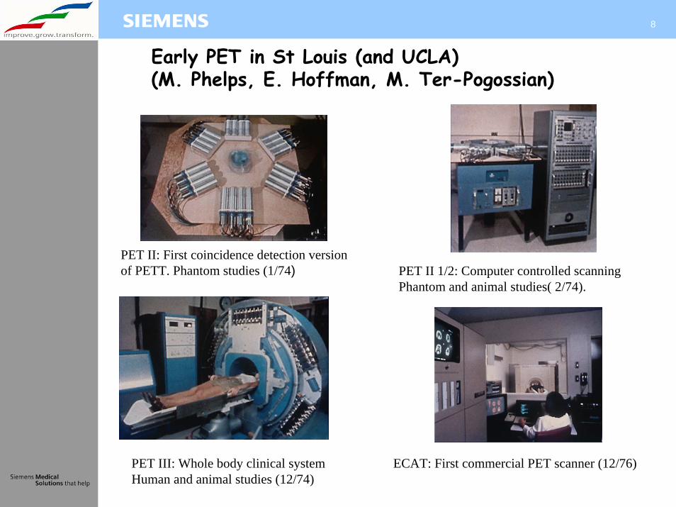
Early PET in St Louis (and UCLA)(M. Phelps, E. Hoffman, M. Ter-Pogossian)
PET II: First coincidence detection version of PETT. Phantom studies (1/74) PET II 1/2: Computer controlled scanning
Phantom and animal studies( 2/74).
PET III: Whole body clinical systemHuman and animal studies (12/74)
ECAT: First commercial PET scanner (12/76)

9
Brookhaven 32 detector ring scanner made operational for transaxial tomography and clinical work in Montreal around
1975. (L. Yamamoto, C. Thompson, E. Meyer)
Early reference to the Brookhaven scanner:S. Rankowitz, J.S. Robertson, and W.A. Higginbotham: Positron scanner for locating brain
tumors. IRE Intern. Conv. Record, Part 9:49-56 (1962)

10
PET design at CERN

11
The first four generations of PET systems

12
Fifth and sixth generation 2005-2007
5th generation: PET/CT systems where the PET system is a LSO system operated with open 3D geometry.
3D acquisition3D image reconstruction
6th generation: as above but with TOF option; (increased axial extension to 20 cm or more)

13
Some milestones during the eighties and early nineties
TOF development based on BaF2done in LETI Grenoble and Orsay, France, St Louis, Houston
Some milestones in PET instrumentationleading to the 3th, 4th and 5th generation of PET systems.
1984 BGO block detector ( M. Casey and R.Nutt)
1980 3D reconstruction algorithms for PET ( D. Townsend and others)
1988 fully 3D acquisition with ECAT (CTI, D. Townsend, T. Jones)
1990 The LSO scintillator (C. L. Melcher)High sensitivity, short decay time, high light output.
Low dead time even with open 3D geometry. Excellent time resolution implies low random rate.High light output implies fairly good energy resolution and thus low scatter fractionTOF possible
1991 PET/CT concept born (NIH funding 1995, first PET/CT images on patients 1998, D. Townsend, T. Beyer, R. Nutt)

14
The Karolinska PET project 1976 – 19881976 Start Karolinska Institutet “Brain metabolism” projectDep of Clin Neurophysiology (KI); Dep of Neuroradiology (KI)Dep of Physics (SU); Karolinska Pharmacy
Three initial objectivesConstruction of a positron camera system for brain studies.
PC95- one ring NaI(Tl) system finished in 1978 (1st generation)PC384-7B a 4 ring BGO system finished in 1981 (2nd generation Scanditronix)Work on a 3rd gen. system 1984-1987. Finally purchase from Scanditronix (PC2048-15B)
Production of 11C labeled glucose 1st attempt Swiss chard (mangold) following St Louis2nd attempt via green algae(*)- approx. 25% incorporation
Modeling of regional brain metabolism with 11C-glucoseProblems with 11CO2 egress.
Dep of Psychiatry/Psychology (KI) joins the collaboration (1982 ca).Receptor studies (e.g.D1, D2)
(*)(Scenedesmus obtusiusculus via 11CO2 and photosynthesis (1983))

15
First generation PET built for the Karolinska Institutet in 1976-1978. A 95 NaI(Tl) detector ring system (8*20*50 mm3)
(L. Widen, T. Greitz, L. Eriksson, C. Bohm and others).

16
Stepper motors For x and y motion
(wobble)
PC95 at the Karolinska Hospital (in operation 1978-1981) demonstrated by Mats Bergström
The Karolinska one ring system with 95 NaI(Tl) detectors. First system with “wobble” (1976-1978)
PC95 at the Physics Department, University of Stockholm, final phase in 1978

17
First three Karolinska systems had “wobbling” for high resolution images
A. Phantom drawing
B. Stationary image C. “wobbled” image

18
PC95 images. Ga-68 EDTA blood brain barrier studies

Picture below from 1979 of Godfrey Hounsfield, when he and Alan Cormack received the Nobel Prize, here at the Karolinska Hospital with Torgny Greitz and one of the first EMI CT scanners
Early CT Development Introduction of the EMI CT scanner (1972)
20
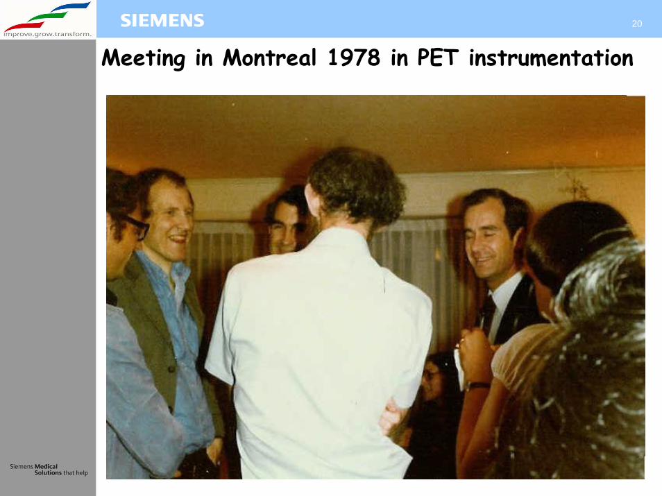
Meeting in Montreal 1978 in PET instrumentation

21
BGO suggested for PET by Z.H. Cho and M.R. Farukhi in 1977.A year later in 1978 - rear view of the 64 BGO Detector Ring of Positome II- the first PET system with BGO detectors (MNI , C. Thompson, L. Yamamoto, E. Meyer).

22
Second generation PET system at the Karolinska Hospital in 1981.This was a four detector ring BGO system (12*20*30 mm3) for brain studies built by the Karolinska Institute/Hospital and the University of Stockholm in collaboration with Scanditronix. During 1976-1990 the Karolinska PET group was headed by Professor Lennart Widén (left).

23
First image with PC384-7B – unwobbledGa-68 EDTA (glioma) ~1981

24
The Karolinska –Scanditronix system PC384-7BFixation system and a C-11 glucose study of an infarct patient

25
Signal from a BGO/GSO pair on one PMT (1983). Selection by pulse shape discrimination. Used by Scanditronix in a whole body system (Cancer Research Center in Heidelberg) and in a brain system (NIH)
26
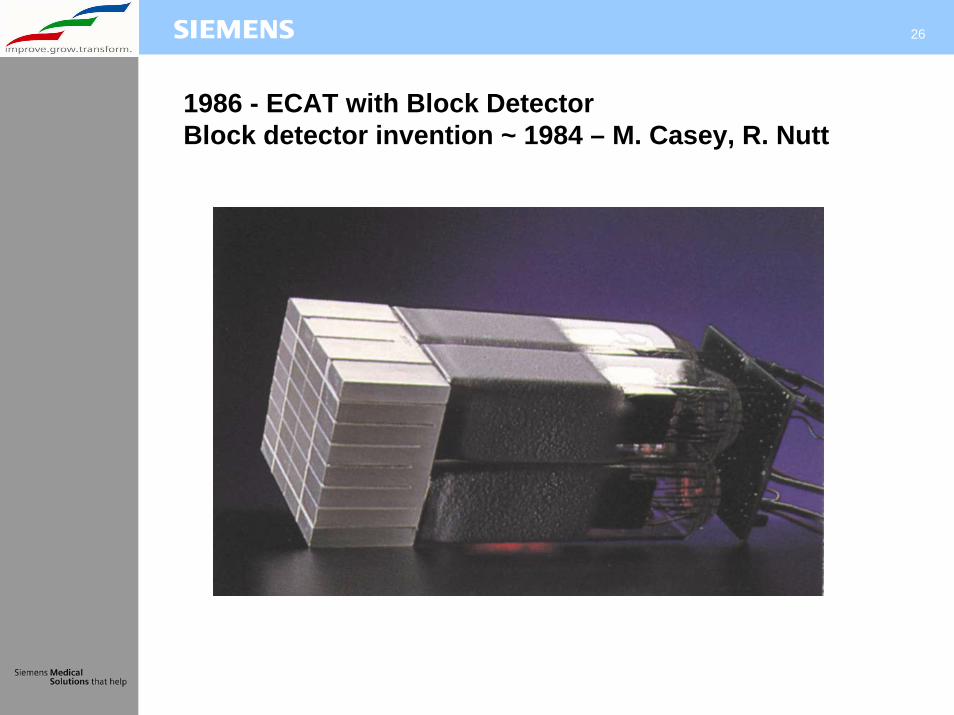
1986 - ECAT with Block DetectorBlock detector invention ~ 1984 – M. Casey, R. Nutt

27
Dual PMT and light guide for block detector attempts ( around 1985)

28
Block detector attempts in a light tight suitcase (1986)

29
Professor Christian Bohm working with detectors for future PET system (~ 1985)
30
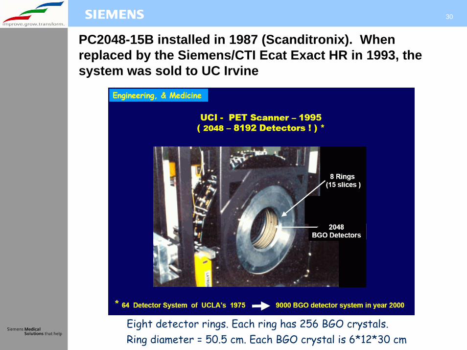
PC2048-15B installed in 1987 (Scanditronix). When replaced by the Siemens/CTI Ecat Exact HR in 1993, the system was sold to UC Irvine
Eight detector rings. Each ring has 256 BGO crystals.Ring diameter = 50.5 cm. Each BGO crystal is 6*12*30 cm

31
PC2048-15B image data (Schering SCH233900, affinity for D1 receptors (D1-antagonist)~ 1989

32
Scanditronix PET sold to General Electric in 1988-89
End of long PET collaboration between Scanditronix and the Karolinska PET group and the Department of Physics, University of Stockholm

33
Acknowledgments and special thanks to
Siemens Science and TechnologyBernard BendriemMike CaseyMaurizio ContiChristian MichelCharles Watson
Siemens Detector centerMatthias SchmandNiraj DoshiMehmet Aykac
David W. Townsend Department of MedicineUniversity of Tennessee, Knoxville
Marita Eriksson Karolinska Institute,Stockholm, Sweden
Chuck L. Melcher Scintillation materials Research CenterUniversity of Tennessee, Knoxville
Christian Bohm Department of Physics University of Stockholm, Sweden
Klaus WienhardMax Planck Institute for brain researchKöln, Germany
Ron Nutt Advanced Biomarkers Technologies, Knoxville
Professor EmeritiT.R. GerholmL. WidenT. Greitz
34

35

36
PET systems at the Karolinska Hospital during the nineties
Ecat Exact HR. (A fourth generation PET system). Purchased from CTI and installed in 1993. A block detector system with three block rings and 112 blocks per ring. Each block 23*53*30 mm3 with a 7*8 matrix cut into it. Crystal size appr. 3*6*30 mm3. Axial extension is around 16 cm. System used 2D operation with septa for transmission data and 3D operation for emission data. System still in operation used mostly for receptor studies.
Ecat Exact special (2 block ring version ~ 10 cm axial FOV). Planned to be used for cardiac studies. Later to be used for oncology screening, installed in 1994. May still be in operation as a backup system ( Dep of Nuclear Medicine, KS/KI)

37
Karolinska Ecat Exact HR installed at the NeuroClinics in 1993- still in operation

38
New PET installations at the Karolinska
Biograph 64 ( fifth generation PET system). The PET system is a Truepoint system with the TrueV option. (Siemens), a four block ring system with 48 blocks per ring. Each block a 13*13 matrix of 4*4*20 mm3 LSO pixels. Block size ~54*54mm2. Axial extension is 21.6 cm. Installed in spring 2006. The CT system is a 64 slice system. Oncology screening and evaluation of cancer therapy. ( Dep of Nuclear Medicine, KS/KI)
HRRT (fifth generation system PET system). A high resolution brain system with 8*117 blocks. Each block a 8*8 matrix of ~2*2*10mm3 LSO and LYSO pixels in a phoswich arrangement. Installed in 2007. (Astra-Zeneca-Karolinska Institutet project, L. Farde)

PET scanner
trues
neutron-deficient isotope
Clinical imaging with PET
n
np p pp
np
n
n
Δτcoincidence
O H
OH
CH2HO
HO
H
H
HHOH
18F
18FDG
sinograms
p(s,φ)
s
zφ
reconstructed image
z
x
positron range
e+e-
p n + e+ + νe
*
scatter
**
randoms
FDG distribution