a kala-modified lipid nanoparticle containing cpg-free ... · oligonucleotide fragments to the bgl...
TRANSCRIPT

Instructions for use
Title A KALA-modified lipid nanoparticle containing CpG-free plasmid DNA as a potential DNA vaccine carrier for antigenpresentation and as an immune-stimulative adjuvant
Author(s) Miura, Naoya; Shaheen, Sharif M.; Akita, Hidetaka; Nakamura, Takashi; Harashima, Hideyoshi
Citation Nucleic acids research, 43(3): 1317-1331
Issue Date 2015-02-19
Doc URL http://hdl.handle.net/2115/59281
Rights(URL) http://creativecommons.org/licenses/by/4.0/
Type article
File Information NAR43-3 1317-1331.pdf
Hokkaido University Collection of Scholarly and Academic Papers : HUSCAP
Published online 20 January 2015 Nucleic Acids Research, 2015, Vol. 43, No. 3 1317–1331doi: 10.1093/nar/gkv008
A KALA-modified lipid nanoparticle containingCpG-free plasmid DNA as a potential DNA vaccinecarrier for antigen presentation and as animmune-stimulative adjuvantNaoya Miura†, Sharif M. Shaheen†, Hidetaka Akita*,†, Takashi Nakamura andHideyoshi Harashima
Department of Molecular Design of Pharmaceutics, Faculty of Pharmaceutical Sciences, Hokkaido University, Kita12, Nishi 6, Kita-ku, Sapporo, Hokkaido 060-0812, Japan
Received September 23, 2014; Revised January 05, 2015; Accepted January 07, 2015
ABSTRACT
Technologies that delivery antigen-encoded plas-mid DNA (pDNA) to antigen presenting cell andtheir immune-activation are required for the suc-cess of DNA vaccines. Here we report on an arti-ficial nanoparticle that can achieve these; a multi-functional envelope-type nanodevice modified withKALA, a peptide that forms �-helical structure atphysiological pH (KALA-MEND). KALA modifica-tion and the removal of the CpG-motifs from thepDNA synergistically boosted transfection efficacy.In parallel, transfection with the KALA-MEND en-hances the production of multiple cytokines andchemokines and co-stimulatory molecules via theToll-like receptor 9-independent manner. Endosome-fusogenic lipid envelops and a long length of pDNAare essential for this immune stimulation. Further-more, cytoplasmic dsDNA sensors that are relatedto the STING/TBK1 pathway and inflammasome areinvolved in IFN-� and IL-1� production, respec-tively. Consequently, the robust induction of antigen-specific cytotoxic T-lymphoma activity and the re-sulting prophylactic and therapeutic anti-tumor ef-fect was observed in mice that had been immu-nized with bone marrow-derived dendritic cells exvivo transfected with antigen-encoding pDNA. Col-lectively, the KALA-MEND possesses dual functions;gene transfection system and immune-stimulativeadjuvant, those are both necessary for the success-ful DNA vaccine.
INTRODUCTION
DNA vaccines are the next-generation vaccines that maypartially substitute for attenuated vaccines or protein vac-cines. In preference to the purified or recombinantly en-gineered protein-based vaccines, a plasmid DNA (pDNA)molecule encoding an antigen gene can be readily con-structed, and can be rapidly amplified in bacteria for massproduction. Moreover, genetic engineering of the antigensuch as mutation, deletion and fusion is also easily done.Thus, DNA vaccination has great potential for applicationin infectious disease (i.e. influenza virus) and cancers, wherethe antigen frequently changes, and differed from one indi-vidual to the others.
In terms of applications of DNA vaccines, the choice ofthe target cells for gene transfection must be taken into con-sideration (1,2). Among the various types of cells, targetingantigen-presenting cells, in particular, dendritic cells (DCs),is crucial for inducing cellular immunity, which plays a par-ticularly important role in protection from tumor growthand viral infectious diseases (i.e. human immunodeficiencyvirus, herpes simplex virus) (3–5).
Many efforts, using both viral and non-viral approaches,have been made to improve transgene efficiency againstDCs (6). Since the transfection activities of viral vectorsare generally more prominent compared to non-viral vec-tors (6,7), more clinical trials have been initiated using viralvectors such as adenoviruses and modified vaccinia ankara(8). However, clinical trials using viral vector systems haveencountered serious adverse effect, including oncogenicityand excess immune responses (9,10). Another drawbackin use of viral approaches is referred to as ‘vector-specificimmunity’ (7), which is observed in patients who have al-ready generated antibody against the viruses. In this case,they cannot benefit from a vaccination because of their pre-
*To whom correspondence should be addressed. Tel: +81 11 706 3735; Fax: +81 11 706 3735; Email: [email protected] may also be addressed to Hideyoshi Harashima. Tel: +81 11 706 3919; Fax: +81 11 706 4879; Email: [email protected]†These authors contributed equally to the paper as first authors.
C© The Author(s) 2015. Published by Oxford University Press on behalf of Nucleic Acids Research.This is an Open Access article distributed under the terms of the Creative Commons Attribution License (http://creativecommons.org/licenses/by/4.0/), whichpermits unrestricted reuse, distribution, and reproduction in any medium, provided the original work is properly cited.
at (D) H
okkaido U M
ed on June 9, 2015http://nar.oxfordjournals.org/
Dow
nloaded from

1318 Nucleic Acids Research, 2015, Vol. 43, No. 3
existing immunity to viruses (11,12). Therefore, the devel-opment of non-viral methods would be highly desirable asa compliment to the viral approach, in terms of expandingthe choice of therapeutic system for use as a DNA vaccine.
Concerning non-viral vectors, we recently reported on thedevelopment of an octaarginine (R8)-modified multifunc-tional envelop-type nanodevice (R8-MEND). This particleis composed of a pDNA-condensed particle formed with apolycation, and its encapsulating liposomal envelops thatare modified with R8, a protein transduction domain thatinduce cellular uptake (13,14). While the R8-MEND exhib-ited transgene expression in dividing HeLa cells, the cor-responding activity in bone marrow-derived dendritic cells(BMDCs) or dendritic cell-derived cell line (JAWSII) wasquite poor. In an attempt to enhance the nuclear deliveryof pDNA by inducing membrane fusion between the nu-clear membrane and the envelope structure of the MEND,the surface of the lipid was modified with KALA, a peptidethat adopts an �-helical structure in a physiological environ-ment (15). To accomplish this, we developed a lipid deriva-tive of KALA (stearylated KALA; STR-KALA) that wasdesigned to allow it to be modified on the surface of thelipid envelop (16). As we expected, modifying the surfacewith STR-KALA drastically improved the transfection ef-ficiencies in JAWSII. Thus, a KALA-modified MEND witha simple structure (KALA-MEND) is also a potent candi-date for use as a carrier of pDNA targeting DCs. However,we unexpectedly found that the drastic improvement of thetransfection activity by the KALA-modification is not at-tributed to an enhancement in nuclear transport efficiency,but to the elevation of the intrinsic transcription activity ofthe preparation in JAWS II cells. (17).
In addition to the transfection of the antigen-encodinggenes, the activation of innate immunity is also requiredto elicit effective adaptive immunity during a vaccina-tion (5,18). Of note, antigen presentation without an in-nate immune response (for example, lack of co-stimulatorymolecules) induces the inactivation of adaptive immunity tothe presented antigen (anergy) (19). Very recently, microar-ray analysis revealed that transfection with the KALA-modified R8-MEND to the JAWSII cells induced a greaterperturbation in mRNA expression in host cells in com-parison with the KALA-unmodified particle. In particu-lar, a more detailed analysis indicated the transcriptionalfactors that are greatly responsible for the immune acti-vation (for example, NF-�B and STATs) are significantlyactivated (17). Collectively, these data indicated that theKALA-MEND simultaneously activates DCs upon deliv-ering pDNA to these cells.
The objective of the present study was to demonstratethe successful application of KALA-MENDs as a DNAvaccine carrier using primary cultured BMDCs. Our ini-tial effort was focused on the functional characterization ofKALA-MENDs, in an attempt to refine the encapsulatedpDNA. Accumulating evidence has revealed the therapeuticpotential of unmethylated CpG motif in DNA, a ligand forthe Toll-like receptor (TLR)9 in endosome as an immune-stimulatory adjuvant for the treatment of cancer and infec-tious diseases (20–24). However, we assumed that the useof CpG-containing pDNA might be undesirable for genetransfection in DCs when used for a DNA vaccine, since
CpG sequences that represent in the promoter and openreading frame (ORF) are potently associated with a lossof transgene expression efficiency or sustained gene expres-sion (25,26). Thus, we investigated the impact of removingCpG-motifs on gene expression in BMDCs. As describedbelow, the immunization of the BMDCs that had been pre-transfected with KALA-MEND exhibited a drastic tumor-preventing effect regardless of the presence or absence ofthe additional adjuvant, even when pDNA that was com-pletely lacking in unmethylated CpG motifs was used. Thus,we subsequently focused on the CpG motif-independentimmune stimulative function of KALA-MEND by mon-itoring the expression profile of cytokine/chemokine pro-duction using the conventional adjuvant as a compari-son. The mechanisms responsible for this immune activa-tion were also addressed. Finally, we assessed the antigen-presentation and antigen-specific cytotoxic T-lymphoma(CTL) activity, and tumor-suppressive effect conferred bythe KALA-MEND in terms of therapeutic output.
MATERIALS AND METHODS
Materials
1, 2-dioleoyl sn-glycero-3-phosphoethanolamine (DOPE)and egg phosphatidylcholine (EPC) were purchased fromAvanti Polar Lipids, Inc. (Alabaster, AL, USA). 3,3′-dioctadecyloxacarbo-cyanine perchlorate (DiO) and 1,1′-dioctadecyl-3,3,3′,3′-tetramethylindodicarbocyanine,4-chlorobenzenesulfonate salt (DiD) were purchased fromInvitrogen (Carlsbad, CA, USA). Lipofectamine R© 2000reagent was purchased from Invitrogen. Poly (dA:dT),ovalbumin (OVA), phosphatidic acid (PA) cholesteryl-hemisuccinate (CHEMS) and lipopolysaccharide (LPS)were purchased from Sigma (St. Louis, MO, USA).Stearylated octaarginine (STR-R8) and stearylatedKALA (STR-KALA) were custom-synthesized byKurabo (Osaka, Japan) as described previously (15).Synthetic oligodeoxynucleotides containing CpG motif(5′-TCCATGACGTTCCTGATGCT-3′, phosphoro-tioated) were purchased from Hokkaido System Science(Hokkaido, Japan). Mouse recombinant granulocyte-macrophage colony-stimulating factor (GM-CSF) waspurchased from R&D systems (Minneapolis, MN, USA).Phycoerythrin (PE) -labeled anti-mouse CD80 (Cat. No.:104707, clone: 16-10A1) and CD86 (Cat. No.: 105007,clone: GL-1) antibody was purchased from Biolegend (SanDiego, CA, USA). The isotype controls of each antibodywere purchased from eBioscience (San Diego, CA, USA).Mouse immunoglobulin G (IgG) 1� was purchased fromSigma (St. Louis, MO, USA). All other chemicals werecommercially available and reagent grade products.
Plasmid construction
Conventional pDNA encoding luciferase (GL3) was pre-pared by inserting a fragment encoding for GL3, ob-tained by the HindIII/XbaI digestion of the pGL3 ba-sic vector (Promega, Madison, WI, USA) into the HindIII/XbaI-digested site of pcDNA3.1 (Invitrogen, Carlsbad,CA, USA) (27). For the preparation of the CpG-free pDNA
at (D) H
okkaido U M
ed on June 9, 2015http://nar.oxfordjournals.org/
Dow
nloaded from
Nucleic Acids Research, 2015, Vol. 43, No. 3 1319
encoding luciferase, the multiple cloning site of pCpGfree-mcs (Invivogen, San Diego, CA, USA) was preliminarily re-placed with a new one that can be digested by 5′-Bgl II-PvuII-Nco I-Sca I-Xba I-Nhe I-3′ by ligating the hybridizedoligonucleotide fragments to the Bgl II/Nhe I digestionof pCpGfree-mcs (pCpGfree-NEWmcs). Thereafter, an in-sert fragment encoding the CpG-free luciferase plus oneCpG motif at just below the stop codon, Luc(+1), was ob-tained by the Nco I/Nhe I digestion of pORF-Luc::Sh-CpG (Invivogen), and ligated to the Nco I/Nhe I-digestedsite of pCpGfree-NEWmcs (pCpGfree-Luc(+1)). Finally,the last remaining CpG motif was removed by inserting aCpG motif-free nucleotide fragment into a Dra III/Nhe I-digested site of pCpGfree-Luc(+1), where we refer to it aspCpGfree-Luc(0).
For the construction of CpG-free pDNA encoding OVA,CpG-free insert encoding OVA (Supplementary Figure S1)was custom-synthesized (Invivogen), with CC just above thestart codon and AGCTAGC just below the stop codon toallow these sequences to be cleaved by NcoI (CCATGG)and Nhe I, respectively. The insert encoding CpG-free OVAwas obtained by the Nco I/Nhe I digestion and ligatedto the Nco I/Nhe I-digested site of pCpGfree-NEWmcs(pCpGfree-OVA(0)).
Preparation of MEND
Each MEND was prepared by the lipid film hydrationmethod as reported previously (30–32). In a typical run,pDNA or ODN (0.1 mg/ml) was condensed with pro-tamine (0.1 mg/ml) in 10-mM HEPES (pH 7.4), at annitrogen/phosphate (N/P) ratio of 2.2. For the compactionof pDNA with KALA peptide core (pDNA/KALA),pDNA (0.1 mg/ml) was condensed with KALA peptide (1mM) in 10-mM HEPES (pH 7.4), at an N/P ratio of 3.5. Alipid film was prepared in a glass test tube by evaporatinga chloroform and ethanol solution of the lipids, containingDOPE and PA or CHEMS at a molar ratio of 7:2 (totallipid amount: 110 nmol), a composition characterized asan endosome-fusogenic lipid (28). The prepared lipid filmwas then hydrated with a condensed pDNA solution (200�l, corresponding to 8.0-�g pDNA) for 10 min at roomtemperature (0.55 mM of total lipid concentration). In thecase of empty liposomes, 10-mM HEPES was used for lipidfilm hydration. After hydration, the tube was sonicated for1 min in a bath-type sonicator (AU-25C; Aiwa Co., Tokyo,Japan) to complete the lipid coating of the condensed DNA.Finally, STR-KALA or STR-R8 (final concentration, 10mol% of total lipid) was added to the MEND solution un-der vortexing. The diameter and �-potential of the MENDand core particle were determined using an electrophoreticlight-scattering spectrophotometer (Zetasizer; Malvern In-struments Ltd., Malvern, WR, UK).
Preparation of BMDCs of mice
Female C57BL/6 (H-2b) mice (6–8 weeks old) were ob-tained from Japan SLC, Inc. (Shizuoka, Japan). The pro-tocol for use of the mice was approved by the Pharmaceu-tical Science Animal Committee of Hokkaido University.BMDCs were prepared as described previously (29). Briefly,
bone marrow cells were cultured overnight in RPMI1640medium containing 50-�M 2-mercaptethanol, 10-mMHEPES, 1-mM sodium pyruvate, 100-U/ml penicillin-streptomycin and 10% fetal calf serum (FCS). Non-adherent cells were harvested and cultured in the samemedium supplemented with 10-ng/ml GM-CSF. On days2 and 4, non-adherent cells were removed, and the remain-ing adherent cells were cultured in fresh medium containing10-ng/ml GM-CSF. On day 6 or 7, non-adherent cells wereused in experiment as immature BMDC.
Transfection to BMDC with the MENDs for evaluating geneexpression
For transfection studies, BMDCs (4.0 × 10⁵ cells) were in-cubated with the MEND (equivalent to 0.4-�g pDNA) inserum-free medium for 30 min or 3 h. Thereafter, a mediumcontaining 10% FCS was added, and the resulting suspen-sion was incubated for an additional 21 h. GM-CSF wasadded in the medium at 10 ng/ml over through the transfec-tion studies. The cells were then washed and solubilized withthe reporter lysis buffer (Promega, Madison, WI, USA).Luciferase activity in the cell lysate was then measuredby means of a luminometer (Luminescencer-PSN; ATTO,Tokyo, Japan). The amount of protein in the cell lysate wasdetermined using a Bicinchoninic acid (BCA) protein assaykit (PIERCE, Rockford, IL, USA).
Transfection to BMDC with the MENDs for evaluating theproduction of cytokines and chemokines
For transfection studies, BMDCs (4.0 × 10⁵ cells) were in-cubated with the MEND (equivalent to 0.4-�g pDNA) orPoly (dA:dT) complexed with Lipofectamine R© 2000 reagent(0.4 �g: 1.2 �l) for 3 h in serum-free RPMI-1640 medium.In the inhibition experiment, BMDCs were incubated withmedium containing BX795 or Ac-YVAD-CMK, an in-hibitor of TBK1 or caspase-1 for 30 min before incubationwith MEND. Then, RPMI-1640 medium containing 10%FCS was then added, followed by further 3 h of incubation.Throughout the transfection studies, 10-ng/ml GM-CSFwas added in RPMI-1640 medium. Thereafter, the concen-tration of IFN-�, IL-1�, IL-6, IL-27p28, IFN-� -inducibleprotein-10 (IP-10), Monokine induced by gamma inter-feron (MIG), Macrophage inflammatory protein-1� (MIP-1�) and TNF-� in the culture supernatant was measuredby enzyme-linked immunosorbent assay (ELISA) suppliedby R&D systems (for IL-1�, IL-6, IL-27p28, IP-10, MIG,MIP-1� and TNF-�; Minneapolis, MN, USA) and PBL In-terferonSource (for IFN-�; Piscataway, NJ, USA).
Fluorescence-activated cell sorting analysis
To evaluate the the expression of CD80 and CD86 on theBMDCs, the cells (8.0 × 10⁵ cells) were incubated withthe MENDs (equivalent to 0.4-�g pDNA) or LPS (finalconcentration, 1 �g/ml) for 3 h in serum-free RPMI-1640medium. RPMI-1640 medium containing 10% FCS wasthen added, followed by a further incubation for 18 h.Throughout experiments, 10-ng/ml GM-CSF was addedin RPMI-1640 medium. Then, the cells (5.0 × 10⁵ cells)
at (D) H
okkaido U M
ed on June 9, 2015http://nar.oxfordjournals.org/
Dow
nloaded from
1320 Nucleic Acids Research, 2015, Vol. 43, No. 3
were incubated with 5 �g/ml of mouse IgG1� at 4◦C for 30min. After washing three times with fluorescence-activatedcell sorting (FACS) buffer (phosphate buffered saline (PBS)containing 0.5% bovine serum albumin and 0.1% NaN3), 5-�g/ml antibodies of PE-labeled anti-mouse CD80, CD86and each isotype control were added to the BMDCs. TheBMDCs were then incubated at 4◦C for 30 min. After wash-ing three times with FACS buffer, the BMDCs were an-alyzed by FACSCaliburTM (Becton Dickinson, FranklinLakes, NJ, USA).
Labeling of pDNA
For the visualization of pDNA, the molecule was first la-beled using a Mirus Label IT R© biotin nucleic acid label-ing kit (Mirus Corp., Madison, WI, USA). The pDNA waslabeled in the optimized buffer supplied with the kit, butthe Label IT solution was mixed at a 1/10 concentrationof the recommended protocol by diluting the solution withdistilled water. pDNA was incubated for 60 min, and thelabeled pDNA was purified by ethanol precipitation. Theconcentration of biotin-labeled pDNA was adjusted to 0.2�g/�l. Secondary, fluorescent labeling was done by Qdot R©
705 ITKTM Streptavidin Conjugate Kit (Invitrogen). A 50-�l aliquot of the biotin-labeled pDNA solution was thenincubated with a 10-�l aliquot of the Qdot R© 705 (QD705)streptavidin (final concentration, 0.02 �M) in HEPES (pH7.4) buffer at room temperature for at least 30 min. Afterquantum dot (QD) labeling, the excess QD was separatedfrom the labeled DNA by passing the solution through aSephadex G50 mini column supplied with Mirus Label IT R©
Kit, which had been pre-equilibrated with HEPES (pH 7.4)buffer.
Microscopy observation
BMDCs (4.0 × 10⁵ cells) were incubated with MEND,which encapsulated QD705-labeled pDNA prepared as de-scribed above, for 2 h in serum-free RPMI-1640 medium.RPMI-1640 medium containing 10% FCS was then added,followed by a further 4 h-incubation. Throughout ex-periments, 10-ng/ml GM-CSF was added in RPMI-1640medium. After incubation, the cells were washed twicewith RPMI-1640 medium and 20-units/ml Heparin solu-tion, and analyzed using spinning disk confocal microscopywithout fixation. Nuclei of cells were stained with Hoechst33342 for 10 min and acidic compartments were stainedwith Lysotracker R© Green DND-26 (Invitrogen) for 5 minbefore observation.
Images were acquired by a Nikon ECLIPSE TE-2000-U (Nikon, Tokyo, Japan) equipped with a spinning diskconfocal unit, CSU-X1 (Yokogawa Electric Corporation,Tokyo, Japan) and a Nikon Plan Apo 100×/1.40 oil im-mersion objective (Nikon). Control of the microscopy andacquisition of digital images were performed with the NIS-Elements software (Nikon). Images were captured with anelectron multiplier charge device camera (ImagEM; Hama-matsu Photonics, Hamamatsu, Japan). Image analysis wasperformed by Image Pro Plus (Media Cybernetics, Inc.,Rockville, MD, USA). Endosomal escape rate was calcu-lated by the following formula; ((1 − (yellow integratedpixel intensity/red integrated pixel intensity)) × 100).
Antigen presentation
BMDC cells were transfected using MENDs containingpDNA encoding the OVA plasmid as described above. Af-ter 24 h, the transfected DCs were harvested by pipetting,followed by centrifugation at 1600 rpm for 5 min. The trans-fected BMDCs (2 x 10⁵ cells) were mixed with B3Z T-cell hybridoma (1 × 10⁵ cells) in RPMI 1640 with 10%FCS in 96-well plates, and incubated for 15 h at 37◦C (30).The co-cultured cells were then washed with 200-�l PBSand then incubated with 100 �l of chlorophenol red �-D-galactopyranoside buffer (5-mM chlorophenol red �-D-galactopyranoside, 0. 5% NP-40 and 9-mM MgCl2 in PBS)for 4 h at 37◦C. After the incubation, the absorbance at 595nm of each well was measured using a micro plate reader(Benchmark Plus; Bio-Rad).
Anti-tumor effect
In the case of tumor-prevention experiments, pCpGfree-OVA(0) or non-coding pCpGfree-mcs was transfected tothe BMDCs by using KALA-MEND. After the transfec-tion for 24 h, BMDCs were harvested and were then ad-ministered (4 × 10⁵cells/40 �l) through the footpad on 14day and 7 day before the tumor inoculation. In a group,the BMDCs were additionally treated with 1 �g/ml of CpGoligonucleotide for 60 min prior to the food-pad injection.Thereafter, 8 × 10⁵ E.G7-OVA cells were subcutaneouslyinjected.
For the evaluation of the therapeutic effect against tumor,individual mice were inoculated with 8 × 10⁵ E.G7-OVAcells. In parallel, pCpGfree-OVA(0) or pCpGfree-Luc(0)was transfected to the BMDCs via the KALA-MEND orthe R8-MEND for 6 h. At 7 days and 14 days after tumorinoculation, the mice were immunized with the harvestedBMDCs by injection into the footpad (5 × 10⁵cells/40-�lPBS). Tumor volume was calculated by the following for-mula: (major axis × minor axis2) × 0.52.
In vivo CTL assay
In vivo CTL assay was performed as described previously(31). Briefly, C57BL/6 mice were subcutaneously immu-nized with each sample. After 7 days, splenocytes were pre-pared from naı̈ve C57BL/6 mice and incubated for 30 minat 37◦C with the OVA257–264 peptide in RPMI-1640 mediumcontaining 50-�M 2-mercaptoethanol, 10-mM HEPES, 1-mM sodium pyruvate, 100-units/ml penicillin-streptomycinand 10% FCS. The OVA257–264 peptide-presented spleno-cytes were then labeled by incubation for 10 min at 37◦Cwith 5-�M CFSE in PBS (CFSEhigh cells). The naı̈vesplenocytes were labeled by incubation for 10 min at 37◦Cwith 0.5-�M CFSE in PBS (CFSElow cells). CFSE-labeledcells were washed with PBS. A mixture of 5 × 10⁶ CFSEhigh
and 5 × 10⁶ CFSElow cells was intravenously injectedinto the immunized mice. After 20 h, splenocytes fromthe immunized mice were collected and single-cell suspen-sions were analyzed for the detection and quantificationof CFSE-labeled cells by FACSCaliburTM. The numbers ofCFSElow cells were essentially the same in all samples in-cluding the non-treated group. The values for lysis were
at (D) H
okkaido U M
ed on June 9, 2015http://nar.oxfordjournals.org/
Dow
nloaded from
Nucleic Acids Research, 2015, Vol. 43, No. 3 1321
the number of CFSEhigh cells corrected by the number ofCFSElow cells. In this study, non-specific lysis was not ob-served.
RESULTS
Effect of CpG motif in pDNA on the gene expression inBMDC
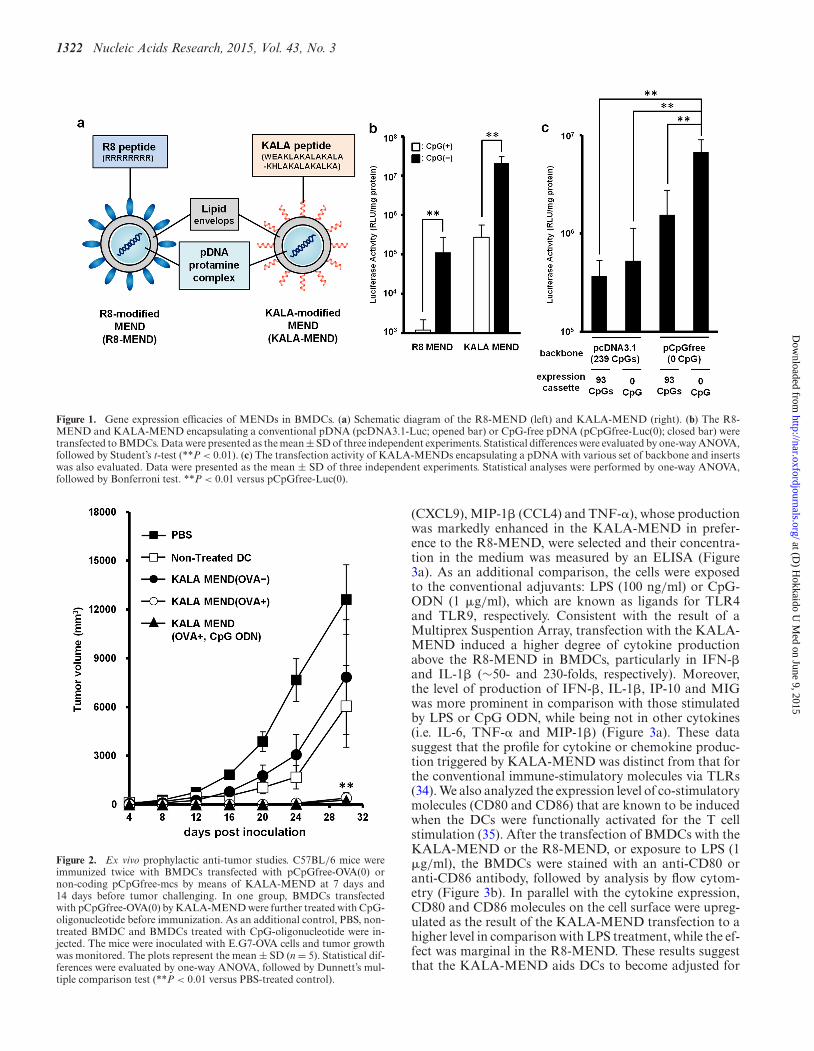
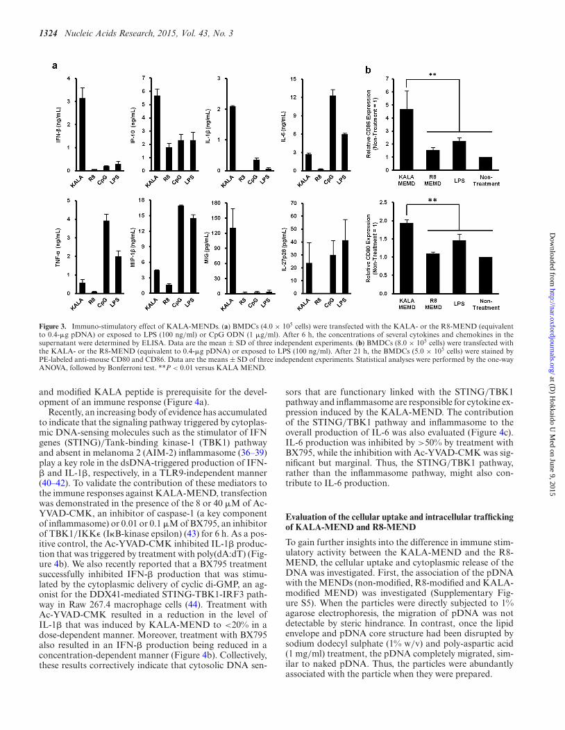
In our previous work (16), we used a mammalian expres-sion vector referred to as pcDNA3.1-Luc (332 CpGs in to-tal pDNA), in which luciferase (GL3) as a reporter gene (93CpGs in its ORF) is inserted into the pcDNA3.1 (Invitro-gen, Carlsbad, CA, USA). In initial experiments, we investi-gated the impact of the use of CpG-free pDNA (pCpGfree-Luc(0)) on gene transfection activity. In the structure ofpCpGfree-Luc(0), the CpG-free luciferase gene (Invivogen,San Diego, CA, USA) was inserted into the pCpGfree-mcs (Invivogen). The pDNA was used in preparing the R8-MEND or KALA-MEND (Figure 1a), which was thentransfected to the BMDCs generated from C57BL/6 mice.In an initial study, we prepare these particles using 1.2-dioleoyl sn-glycero-3-phosphoethanolamine (DOPE) andCHEMS as a membrane-fusogenic component of the lipidenvelop structure (32,33). The physicochemical charactersof the particles were similar regardless of the type of themodified peptide (R8 or KALA) or pDNA used (Table1). In both types of pDNA, the transfection activity washigher in the KALA-MEND than in the R8-MEND bymore than two orders of magnitude (Figure 1b). More im-portantly, the replacement of the pDNA from pcDNA3.1-Luc to the pCpGfree-Luc(0) resulted in a 50-fold enhance-ment in gene expression by KALA-MEND. The experimentin Figure 1c was designed to allow us to gain insights intothe effect of the CpG deletion in the backbone and/or ORFon the overall enhancement in gene expression. To addressthis issue, we prepared two additional types of pDNA: apcDNA3.1 vector encoding the CpG-free luciferase gene(pcDNA3.1-Luc(0); 239 CpGs) and a pCpGfree-mcs vec-tor encoding the CpG-positive luciferase gene (pCpGfree-Luc; 93 CpGs). The removal of the CpG sequences from thebackbone and/or the ORF enhanced the gene expressionin a synergistic manner (Figure 1c). It is noteworthy thata more prominent enhancement was achieved when CpGwas deleted from the backbone region. These collective datashow that KALA-MENDs loaded with pCpGfree-Luc(0)represent the best combination for gene delivery in BMDCs.
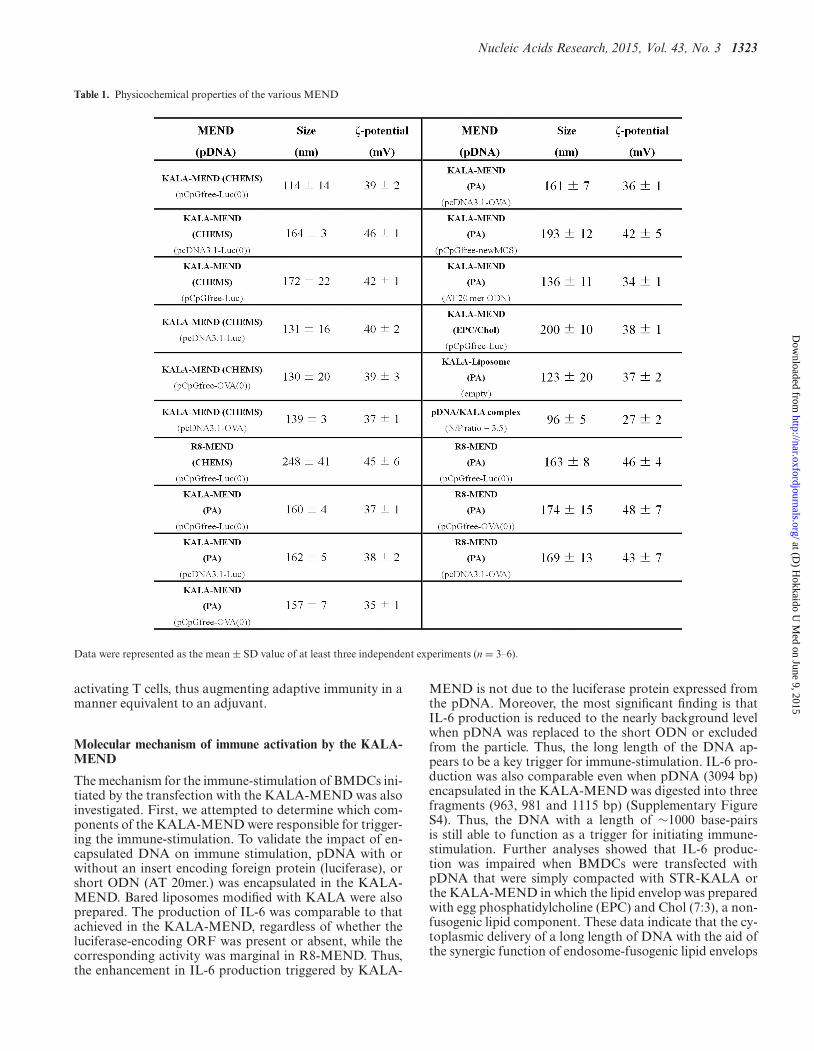
Tumor-preventing effect by the pre-immunization of BMDCstransfected with the KALA-MEND
In order to investigate the pharmacological activity as aDNA vaccine, BMDCs were ex vivo transfected with anantigen-encoding gene by KALA-MEND, and its anti-tumor effect was evaluated (Figure 2). As a model, weused E.G7-OVA tumor model, in which OVA was stablyexpressed as a model antigen. Since the deletion of theCpG motifs is one of the key factors for maximizing geneexpression (Figure 1c), we designed the CpG-free ORFsequence encoding a model antigen––OVA. The full se-quence is listed in Supplementary Figure S1. The ORF
was inserted within a pCpGfree-mcs to produce a CpG-free pDNA encoding OVA (pCpGfree-OVA(0)). IsolatedBMDCs were transfected with pCpGfree-OVA(0) by theKALA-MEND for 24 h. After the activation of the cellswith or without an adjuvant treatment (CpG-containingoligonucleotide), the immunized cells were administered tomice via the footpad at a dose of 4 × 10⁵ cells/mouse at7 days and 14 days before tumor challenging. After theinoculation of E.G7-OVA cells, tumor volume was moni-tored for up to 30 days (Figure 2). When mice were pre-immunized with BMDCs that had been transfected withpDNA free from the antigen-encoding ORF, tumor sup-pression was comparable to those pre-immunized with thenon-treated BMDCs. In contrast, when pCpGfree-OVA(0)was transfected, a drastic decrease in the rate of tumorgrowth was observed. It is also noteworthy that exten-sive tumor suppression was achieved regardless of whetheran adjuvant (CpG-containing oligonucleotide; CpG-ODN)treatment was used. These data prompted us to assumethat the KALA-MEND intrinsically functions to activateBMDCs, similar to an adjuvant.
Induction of cytokines and co-stimulatory molecules by trans-fection with the KALA-MEND
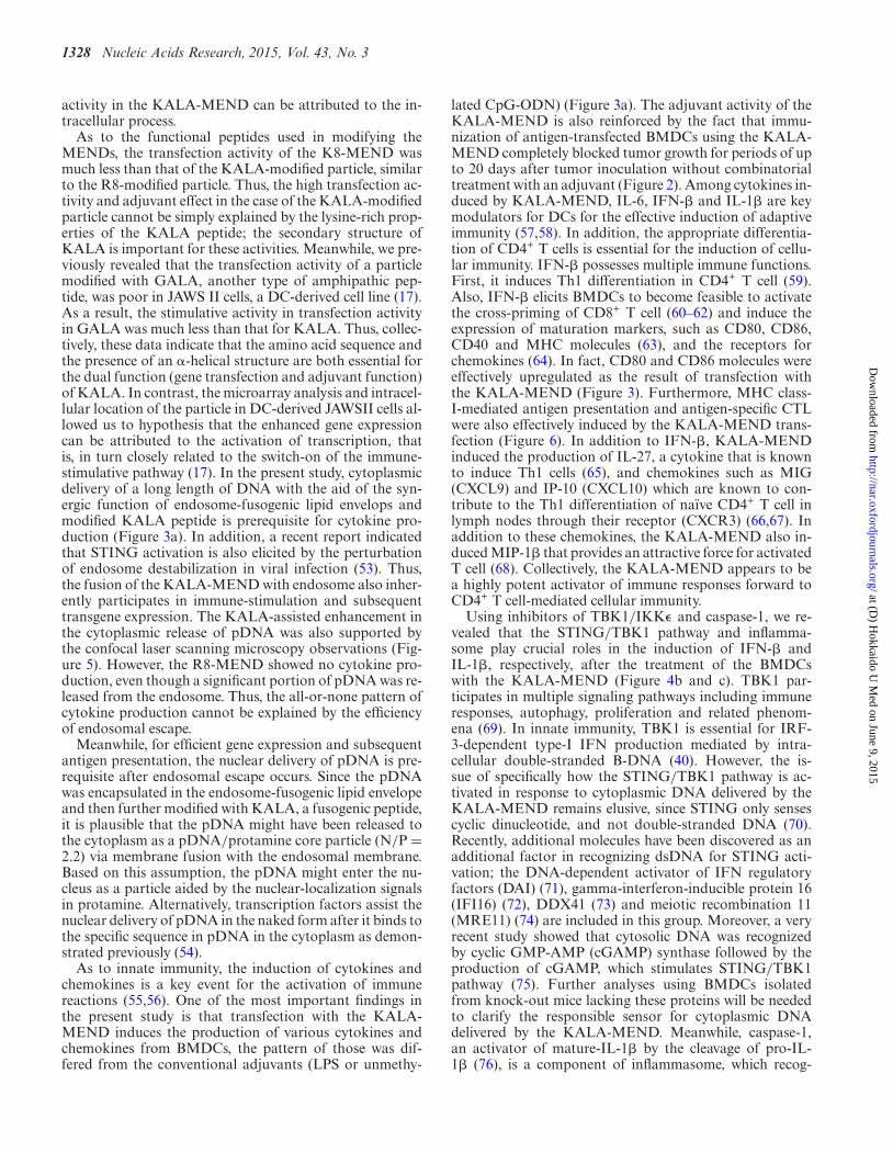
We examined the profile of cytokine production fromBMDCs after transfection with the KALA-MEND orthe R8-MEND encapsulating pCpGfree-Luc(0). In thisstudy, we used DOPE and PA as the composition in theendosome-fusogenic lipid envelope (28) since the KALA-MEND that is composed of DOPE/PA induced highercytokine production in comparison with those preparedwith DOPE/CHEMS (Supplementary Figure S2a). Thehigher transfection activity and immune-stimulative activ-ities of the KALA-MEND above the R8-MEND were re-producible when MENDs were prepared with DOPA/PA(Supplementary Figure S2b and c). In addition, the activ-ities for the KALA-MEND were drastically higher thanfor the MEND modified with octa-lysine (K8-MEND). Ofnote, the amount of protein after the transfection studyin the KALA-MEND (78 ± 1% of non-treated DCs) wasslightly lower than that in the R8-MEND and the K8-MEND (105 ± 2% and 104 ± 4% of non-treated DCs,respectively). Thus, in some cells, the over-stimulation ofthe immune-response might damage in DCs transfected bythe KALA-MEND. In other words, the lower transfec-tion activities in the R8-MEND or the K8-MEND can-not be explained from the point of view of cytotoxic-ity. Thus, the BMDCs were transfected with the KALA-MEND or the R8-MEND encapsulating pCpGfree-Luc(0).In a preliminary experiment, the production of the cy-tokines and chemokines in BMDCs in response to trans-fection was comprehensively analyzed by means of Multi-plex Suspension Array System (Milliplex MAG Kit, Milli-pore, Billerica, MA, USA, performed by Genetic Lab Co.,Ltd., Sapporo, Japan). As shown in Supplementary Fig-ure S3, the production of cytokines and chemokines wasmore prominent in the case where BMDCs were trans-fected with the KALA-MEND in comparison with theR8-MEND. Among them, eight cytokines and chemokines(IFN-�, IL-1�, IL-6, IL-27p28, IP-10 (CXCL10), MIG
at (D) H
okkaido U M
ed on June 9, 2015http://nar.oxfordjournals.org/
Dow
nloaded from
1322 Nucleic Acids Research, 2015, Vol. 43, No. 3
Figure 1. Gene expression efficacies of MENDs in BMDCs. (a) Schematic diagram of the R8-MEND (left) and KALA-MEND (right). (b) The R8-MEND and KALA-MEND encapsulating a conventional pDNA (pcDNA3.1-Luc; opened bar) or CpG-free pDNA (pCpGfree-Luc(0); closed bar) weretransfected to BMDCs. Data were presented as the mean ± SD of three independent experiments. Statistical differences were evaluated by one-way ANOVA,followed by Student’s t-test (**P < 0.01). (c) The transfection activity of KALA-MENDs encapsulating a pDNA with various set of backbone and insertswas also evaluated. Data were presented as the mean ± SD of three independent experiments. Statistical analyses were performed by one-way ANOVA,followed by Bonferroni test. **P < 0.01 versus pCpGfree-Luc(0).
Figure 2. Ex vivo prophylactic anti-tumor studies. C57BL/6 mice wereimmunized twice with BMDCs transfected with pCpGfree-OVA(0) ornon-coding pCpGfree-mcs by means of KALA-MEND at 7 days and14 days before tumor challenging. In one group, BMDCs transfectedwith pCpGfree-OVA(0) by KALA-MEND were further treated with CpG-oligonucleotide before immunization. As an additional control, PBS, non-treated BMDC and BMDCs treated with CpG-oligonucleotide were in-jected. The mice were inoculated with E.G7-OVA cells and tumor growthwas monitored. The plots represent the mean ± SD (n = 5). Statistical dif-ferences were evaluated by one-way ANOVA, followed by Dunnett’s mul-tiple comparison test (**P < 0.01 versus PBS-treated control).
(CXCL9), MIP-1� (CCL4) and TNF-�), whose productionwas markedly enhanced in the KALA-MEND in prefer-ence to the R8-MEND, were selected and their concentra-tion in the medium was measured by an ELISA (Figure3a). As an additional comparison, the cells were exposedto the conventional adjuvants: LPS (100 ng/ml) or CpG-ODN (1 �g/ml), which are known as ligands for TLR4and TLR9, respectively. Consistent with the result of aMultiprex Suspention Array, transfection with the KALA-MEND induced a higher degree of cytokine productionabove the R8-MEND in BMDCs, particularly in IFN-�and IL-1� (∼50- and 230-folds, respectively). Moreover,the level of production of IFN-�, IL-1�, IP-10 and MIGwas more prominent in comparison with those stimulatedby LPS or CpG ODN, while being not in other cytokines(i.e. IL-6, TNF-� and MIP-1�) (Figure 3a). These datasuggest that the profile for cytokine or chemokine produc-tion triggered by KALA-MEND was distinct from that forthe conventional immune-stimulatory molecules via TLRs(34). We also analyzed the expression level of co-stimulatorymolecules (CD80 and CD86) that are known to be inducedwhen the DCs were functionally activated for the T cellstimulation (35). After the transfection of BMDCs with theKALA-MEND or the R8-MEND, or exposure to LPS (1�g/ml), the BMDCs were stained with an anti-CD80 oranti-CD86 antibody, followed by analysis by flow cytom-etry (Figure 3b). In parallel with the cytokine expression,CD80 and CD86 molecules on the cell surface were upreg-ulated as the result of the KALA-MEND transfection to ahigher level in comparison with LPS treatment, while the ef-fect was marginal in the R8-MEND. These results suggestthat the KALA-MEND aids DCs to become adjusted for
at (D) H
okkaido U M
ed on June 9, 2015http://nar.oxfordjournals.org/
Dow
nloaded from
Nucleic Acids Research, 2015, Vol. 43, No. 3 1323
Table 1. Physicochemical properties of the various MEND
Data were represented as the mean ± SD value of at least three independent experiments (n = 3–6).
activating T cells, thus augmenting adaptive immunity in amanner equivalent to an adjuvant.
Molecular mechanism of immune activation by the KALA-MEND
The mechanism for the immune-stimulation of BMDCs ini-tiated by the transfection with the KALA-MEND was alsoinvestigated. First, we attempted to determine which com-ponents of the KALA-MEND were responsible for trigger-ing the immune-stimulation. To validate the impact of en-capsulated DNA on immune stimulation, pDNA with orwithout an insert encoding foreign protein (luciferase), orshort ODN (AT 20mer.) was encapsulated in the KALA-MEND. Bared liposomes modified with KALA were alsoprepared. The production of IL-6 was comparable to thatachieved in the KALA-MEND, regardless of whether theluciferase-encoding ORF was present or absent, while thecorresponding activity was marginal in R8-MEND. Thus,the enhancement in IL-6 production triggered by KALA-
MEND is not due to the luciferase protein expressed fromthe pDNA. Moreover, the most significant finding is thatIL-6 production is reduced to the nearly background levelwhen pDNA was replaced to the short ODN or excludedfrom the particle. Thus, the long length of the DNA ap-pears to be a key trigger for immune-stimulation. IL-6 pro-duction was also comparable even when pDNA (3094 bp)encapsulated in the KALA-MEND was digested into threefragments (963, 981 and 1115 bp) (Supplementary FigureS4). Thus, the DNA with a length of ∼1000 base-pairsis still able to function as a trigger for initiating immune-stimulation. Further analyses showed that IL-6 produc-tion was impaired when BMDCs were transfected withpDNA that were simply compacted with STR-KALA orthe KALA-MEND in which the lipid envelop was preparedwith egg phosphatidylcholine (EPC) and Chol (7:3), a non-fusogenic lipid component. These data indicate that the cy-toplasmic delivery of a long length of DNA with the aid ofthe synergic function of endosome-fusogenic lipid envelops
at (D) H
okkaido U M
ed on June 9, 2015http://nar.oxfordjournals.org/
Dow
nloaded from
1324 Nucleic Acids Research, 2015, Vol. 43, No. 3
Figure 3. Immuno-stimulatory effect of KALA-MENDs. (a) BMDCs (4.0 × 10⁵ cells) were transfected with the KALA- or the R8-MEND (equivalentto 0.4-�g pDNA) or exposed to LPS (100 ng/ml) or CpG ODN (1 �g/ml). After 6 h, the concentrations of several cytokines and chemokines in thesupernatant were determined by ELISA. Data are the mean ± SD of three independent experiments. (b) BMDCs (8.0 × 10⁵ cells) were transfected withthe KALA- or the R8-MEND (equivalent to 0.4-�g pDNA) or exposed to LPS (100 ng/ml). After 21 h, the BMDCs (5.0 × 10⁵ cells) were stained byPE-labeled anti-mouse CD80 and CD86. Data are the means ± SD of three independent experiments. Statistical analyses were performed by the one-wayANOVA, followed by Bonferroni test. **P < 0.01 versus KALA MEND.
and modified KALA peptide is prerequisite for the devel-opment of an immune response (Figure 4a).
Recently, an increasing body of evidence has accumulatedto indicate that the signaling pathway triggered by cytoplas-mic DNA-sensing molecules such as the stimulator of IFNgenes (STING)/Tank-binding kinase-1 (TBK1) pathwayand absent in melanoma 2 (AIM-2) inflammasome (36–39)play a key role in the dsDNA-triggered production of IFN-� and IL-1�, respectively, in a TLR9-independent manner(40–42). To validate the contribution of these mediators tothe immune responses against KALA-MEND, transfectionwas demonstrated in the presence of the 8 or 40 �M of Ac-YVAD-CMK, an inhibitor of caspase-1 (a key componentof inflammasome) or 0.01 or 0.1 �M of BX795, an inhibitorof TBK1/IKK� (I�B-kinase epsilon) (43) for 6 h. As a pos-itive control, the Ac-YVAD-CMK inhibited IL-1� produc-tion that was triggered by treatment with poly(dA:dT) (Fig-ure 4b). We also recently reported that a BX795 treatmentsuccessfully inhibited IFN-� production that was stimu-lated by the cytoplasmic delivery of cyclic di-GMP, an ag-onist for the DDX41-mediated STING-TBK1-IRF3 path-way in Raw 267.4 macrophage cells (44). Treatment withAc-YVAD-CMK resulted in a reduction in the level ofIL-1� that was induced by KALA-MEND to <20% in adose-dependent manner. Moreover, treatment with BX795also resulted in an IFN-� production being reduced in aconcentration-dependent manner (Figure 4b). Collectively,these results correctively indicate that cytosolic DNA sen-
sors that are functionary linked with the STING/TBK1pathway and inflammasome are responsible for cytokine ex-pression induced by the KALA-MEND. The contributionof the STING/TBK1 pathway and inflammasome to theoverall production of IL-6 was also evaluated (Figure 4c).IL-6 production was inhibited by >50% by treatment withBX795, while the inhibition with Ac-YVAD-CMK was sig-nificant but marginal. Thus, the STING/TBK1 pathway,rather than the inflammasome pathway, might also con-tribute to IL-6 production.
Evaluation of the cellular uptake and intracellular traffickingof KALA-MEND and R8-MEND
To gain further insights into the difference in immune stim-ulatory activity between the KALA-MEND and the R8-MEND, the cellular uptake and cytoplasmic release of theDNA was investigated. First, the association of the pDNAwith the MENDs (non-modified, R8-modified and KALA-modified MEND) was investigated (Supplementary Fig-ure S5). When the particles were directly subjected to 1%agarose electrophoresis, the migration of pDNA was notdetectable by steric hindrance. In contrast, once the lipidenvelope and pDNA core structure had been disrupted bysodium dodecyl sulphate (1% w/v) and poly-aspartic acid(1 mg/ml) treatment, the pDNA completely migrated, sim-ilar to naked pDNA. Thus, the particles were abundantlyassociated with the particle when they were prepared.
at (D) H
okkaido U M
ed on June 9, 2015http://nar.oxfordjournals.org/
Dow
nloaded from

Nucleic Acids Research, 2015, Vol. 43, No. 3 1325
Figure 4. Molecular mechanism of the cytokine expression induced by KALA-MEND. (a) BMDCs (4.0 × 10⁵ cells) were transfected with several typesof KALA or R8 peptide-modified nanoparticles. (b,c) BMDCs (4.0 × 10⁵ cells) were pre-treated with 0.01 or 0.1-�M solutions of BX795 or 8 or 40-�Msolutions of Ac-YVAD-CMK for 30 min, then transfected with KALA- or R8-MEND (equivalent to 0.4-�g pDNA) or Poly (dA:dT) conjugated withLipofectamine R© 2000 reagent (0.4 �g:1.2 �l, only Ac-YVAD-CMK treatment). After 6 h, the concentrations of several cytokines in the supernatant weremeasured by ELISA. Data are mean ± SD of three independent experiments. Statistical analyses were performed by the one-way ANOVA, followed byBonferroni test. *P < 0.05, **P < 0.01 versus no inhibition group. N.S.: not significant
The cellular uptake of the non-modified MEND, the R8-MEND and the KALA-MEND was quantified by FACS(Supplementary Figure S6). The striking observation isthat the uptake of the non-modified MEND and the R8-MEND in individual cells was heterogenous, while the R8-modification increased the fraction of cells with high up-take of particles. In contrast, the uptake of the KALA-MEND was more homogenous (with a significantly lowerstandard deviation in comparison with the non-modified orthe R8-MEND; P < 0.05). Quantitative analysis revealedthat the cellular uptake of the KALA-MEND on average(Geo Mean) was higher than that for the non-modifiedMEND or the R8-MEND.
The endosomal escape of pDNA was visualized by intra-cellular imaging using a fluorescence microscope equippedwith a confocal scanning unit (CSU-X1). Since the cyto-plasmic recognition of DNA is a plausible trigger for theimmune response described above, we assumed that theKALA-MEND was intimately involved in the delivery ofpDNA to the cytoplasm and that this process was moreefficient than that for the R8-MEND. To validate this hy-pothesis, we analyzed the co-localization of QD-labeledpDNA in acidic compartments (late endosome/lysosomes)stained with Lysotracker Green at 6 h after transfection.The signals derived from QD705 and Lysotracker Greenwere pseudo-colored in red and green, respectively. As a re-sult, the QD705-labeled pDNA transfected by the KALA-
MEND was less co-localized with acidic compartmentsthan those for the R8-MEND (Figure 5a). Endosomal es-cape efficiency was quantified in 30 independent cells interms of the percent of integrated pixel intensity of pDNAfree from the co-localization with acidic compartment tothe total integrated pixel intensity of pDNA (Figure 5b).As a result, the endosomal escape efficiency of pDNA de-livered by the KALA-MEND was significantly higher thanthat for the R8-MEND.
Finally, the association of the QD705-pDNA with DiO-labeled MENDs in the cells was evaluated (SupplementaryFigure S7). While a fraction of the pDNA free from thecolocalized with MENDs was detected, the major part ofthe pDNA was co-localized with the MENDs at the initialphase of the transfection. Thus, efficient endosomal escapewas achieved by virtue of the endosome-fusogenic lipid andKALA peptide on the surface.
Thus, a function of KALA as an inducer of cytoplasmicrelease of pDNA, concomitant with the fusogenic natureof envelop structure in MEND with endosome, is one ofthe mechanisms that explains the prominent innate immuneresponse.
MHC class-I antigen presentation and in vivo CTL assay
To evaluate antigen presentation activity, pCpGfree-OVA(0) was transfected into BMDCs using the R8-MENDor the KALA-MEND. Thereafter, the transfected BMDCs
at (D) H
okkaido U M
ed on June 9, 2015http://nar.oxfordjournals.org/
Dow
nloaded from

1326 Nucleic Acids Research, 2015, Vol. 43, No. 3
Figure 5. Quantifications of endosomal escape of KALA- or R8-MEND.BMCDs (4.0 × 10⁵ cells) were transfected with the KALA- or theR8-MEND encapsulating QD705-pDNA (equivalent to 0.4-�g pDNA,QD705-pDNA was 50% of whole pDNA; red). (a) The BMDCs were ob-served by spinning-disc confocal microscopy at 6 h after transfection. Thenuclei of cells were stained with Hoechst 33342 (blue) and acidic compart-ments were stained with Lysotracker R© Green DND-26 (green) for 10 and5 min before observation, respectively. Bars = 10 �m. (b) Quantificationof endosomal escape of KALA- or R8-MEND. Endosomal escape ratewas calculated by the following formula: ((1 − (yellow integrated pixelintensity/red integrated pixel intensity)) × 100). Each dot represents thecalculated endosomal escape rate of respective cells. Bars show mean (n =30). Statistical analysis was performed by Student’s t-test. **P < 0.01.
were co-cultured with a B3Z T-Cell hybridoma, a reportercell that produces the lacZ protein in response to therecognition of the OVA epitope displayed on MHC class-I molecules (30). As a result, significant antigen presen-tation was observed when pCpGfree-OVA(0) was trans-fected with the KALA-MEND, while this activity was be-low the detection limit in the R8-MEND (Figure 6a). Also,the use of pDNA that contains a large number of CpGmotif (pcDNA3.1-OVA) or that lacks the OVA-encodingORF (pCpGfree-Luc(0)) resulted in a diminution in antigenpresentation activity to the background level. This resultshowed that the KALA-MEND encapsulating the antigen-encoding pDNA that is free from CpG motif is a highlypotent carrier for the antigen presentation to MHC class-I molecules.
In order to confirm the induction of antigen-specificcellular immunity, we measured the CTL activities afterthe immunization of BMDC that had been transfectedby pCpGfree-OVA(0) with the KALA-MEND or the R8-MEND. As another control, BMDCs were transfected withthe KALA-MEND encapsulating the luciferase-encodingpDNA (pCpGfree-Luc(0)) as an antigen-free pDNA. Micewere immunized with the transfected BMDC via footpadadministration at a dose of 5.0 × 10⁵ cells. CTL activities
were measured at 7 days after the immunization. As a result,BMDC transfected with the KALA-MEND elicited morepotent CTL activity than that for the R8-MEND (Figure6b). Of note, the ED50 of pDNA for CTL activity was ∼0.2-�g pDNA, and the activity reached >90% when 0.8 �g ofpDNA was used. In contrast, when the pCpGfree-OVA(0)was transfected with the R8-MEND, or when pCpGfree-Luc(0) was, in turn, transfected with the KALA-MEND,the CTL activity was nearly background level even when1.6-�g pDNA was transfected. These results suggest thatthe KALA-MEND induced antigen-specific cellular immu-nity.
Tumor-treatment effect by the post-immunization of BMDCstransfected with KALA-MEND
Finally, the therapeutic effect in suppression of tumorgrowth was evaluated. In this study, mice were preliminaryinoculated with E.G7-OVA cells. Seven days after tumorchallenging (∼150 mm2 in tumor size), the BMDCs thathad been transfected with KALA-MEND or R8-MENDencapsulating pCpGfree-OVA(0) or pCpGfree-Luc(0) wasimmunized subcutaneously. At 14 days after the tumor in-oculation, the BMDCs that had been transfected with thesame types of MENDs were again immunized (Figure 7).As a result, tumor growth was significantly suppressed inthe case of BMDCs transfected with pCpGfree-OVA(0)by KALA-MEND, although the R8-MEND encapsulat-ing pCpGfree-OVA(0) or the KALA-MEND encapsulat-ing pCpGfree-Luc(0) poorly inhibits tumor growth. Conse-quently, the KALA-MEND is applicable for use in cancertherapy even when the tumor is in growing phase.
DISCUSSION
We report herein on the development of a nanotechnologywith the potential for use as a DNA vaccine, which resultsin an efficient gene expression and simultaneous stimula-tion to BMDCs to a level sufficient for antigen presentation.As a consequence, the in vivo immunization of the BMDCscaused the activation of antigen-specific CTL and a prophy-lactic and/or therapeutic effect against tumor growth. Keyfactors in this success are the use of a CpG-free pDNA anda fusogenic peptide: KALA, as a surface-modified peptide.
While some studies reported that the removal of CpGmotifs, a ligand for TLR9, resulted in a decrease in geneexpression in vitro culture cells (CHO and 293) (45) or inelectroporation-mediated intra-muscular gene transfer (46),CpG-free pDNA was generally considered to be a promis-ing gene cargo allowing the efficient and particularly per-sistent gene expression in the lung or liver-targeting genedelivery (26,47–49). Since, these desired effects in the useof CpG-free pDNA were accompanied by a decrease in cy-tokine production, avoiding immune-stimulation is consid-ered to be a key factor for efficient transgene expression,especially in the case of immune-responsive cells. Based onthis background information, we investigated the impact ofthe use of CpG-free pDNA on transfection efficiency. Asexpected, the use of pCpGfree-Luc(0) generally improvedtransgene expression by one to nearly two orders of magni-tude in comparison with pcDNA3.1-Luc in BMDCs (Fig-ure 1b). Transgene expression was synergistically enhanced
at (D) H
okkaido U M
ed on June 9, 2015http://nar.oxfordjournals.org/
Dow
nloaded from

Nucleic Acids Research, 2015, Vol. 43, No. 3 1327
Figure 6. MHC class-I restricted antigen presentation and in vivo CTL assay. (a) BMDCs were transfected with pcDNA3.1-OVA, pCpGfree-OVA(0) orpCpGfree-Luc(0) (KALA-MEND only) by means of R8-MEND or KALA-MEND. Transfected cells were co-cultured with a B3Z T-cell hybridoma for15 h at 37◦C. The co-cultured cells were then incubated with chlorophenol red �-D-galactopyranoside buffer for 4 h at 37◦C. The absorbance at 595nm was used as an index for antigen-presentation activity. The absorbance observed in non-treated BMDCs was subtracted from each group. Data aremean ± SD of three independent experiments. Non-detected (N.D.)means under the detection limit. Statistical analyses were performed by the one-wayANOVA, followed by Bonferroni test. **P < 0.01 versus pcDNA3.1-OVA. N.S. means non-significance. (b) C57BL/6 mice were immunized with BMDCstransfected with KALA-MEND (encapsulating pCpGfree-OVA(0) or pCpGfree-Luc(0)) or R8-MEND (encapsulating pCpGfree-OVA(0)). CTL activitieswere measured 1 week after immunization. Data are mean ± SD of at least three independent experiments.
Figure 7. Ex vivo therapeutic anti-tumor studies. C57BL/6 mice wereimmunized twice with BMDCs transfected with pCpGfree-OVA(0) orpCpGfree-Luc(0) by means of the KALA-MEND or the R8-MEND at7 days and 14 days after tumor challenging. As a control, PBS and non-treated BMDC were injected. Tumor growth was monitored up to 24 daysafter tumor inoculation. The plots represent the mean ± SEM (n = 5).Statistical differences were evaluated by one-way ANOVA, followed byDunnett’s multiple comparison test (**P < 0.01, *P < 0.05 versus KALA-MEND (OVA+)).
when the CpG-motifs were removed from the backbone ofthe pDNA and also from the ORF. Therefore, reducing theCpG-motifs from the pDNA is also a key factor in termsof maximizing transgene expression (Figure 1b). Neverthe-less, the most mysterious point is that the highest trans-fection activity of CpG-free pDNA cannot be explainedby the avoidance of immune-stimulation, since the multi-ple inflammatory cytokine was produced at comparable lev-els, even when CpG-free pDNA was used (Figure 3). Oneof the proposed mechanisms for the transcriptional sup-pression of the pDNA (50) as well as genomic DNA in-volves the methylation of the cytosine nucleotides in theCpG motif (51). Thus, a pDNA constructed with CpG-free human elongation factor 1� (EF1�) promoter used inthe pCpGfree-Luc(0) and/or the ORF might be more re-sistant to gene silencing compared with a set of human cy-tomegalovirus (CMV) immediate-early enhancer and pro-moter used in pcDNA3.1-Luc. This hypothesis is consistentwith the fact that EF1� promoter is known to be effectivefor cells in which the CMV promoter functions poorly, suchas embryonic stem cells (52).
Using the KALA-MEND, the highest transfection activ-ity was achieved in BMDCs (Figure 1b). While the mode ofaction of KALA from the point of view of inducing trans-gene expression remains elusive. The cellular uptake of theKALA-MEND was more homogenous and, on average,higher than that of the non-modified MEND or the R8-MEND (Supplementary Figure S6). However, the uptake ofthe KALA-MEND was higher than that for the R8-MENDby a factor of nearly 3-fold. Thus, the uptake process can-not explain the >2 orders of magnitude higher transfectionactivity. Thus, the drastic enhancement in the transfection
at (D) H
okkaido U M
ed on June 9, 2015http://nar.oxfordjournals.org/
Dow
nloaded from
1328 Nucleic Acids Research, 2015, Vol. 43, No. 3
activity in the KALA-MEND can be attributed to the in-tracellular process.
As to the functional peptides used in modifying theMENDs, the transfection activity of the K8-MEND wasmuch less than that of the KALA-modified particle, similarto the R8-modified particle. Thus, the high transfection ac-tivity and adjuvant effect in the case of the KALA-modifiedparticle cannot be simply explained by the lysine-rich prop-erties of the KALA peptide; the secondary structure ofKALA is important for these activities. Meanwhile, we pre-viously revealed that the transfection activity of a particlemodified with GALA, another type of amphipathic pep-tide, was poor in JAWS II cells, a DC-derived cell line (17).As a result, the stimulative activity in transfection activityin GALA was much less than that for KALA. Thus, collec-tively, these data indicate that the amino acid sequence andthe presence of an �-helical structure are both essential forthe dual function (gene transfection and adjuvant function)of KALA. In contrast, the microarray analysis and intracel-lular location of the particle in DC-derived JAWSII cells al-lowed us to hypothesis that the enhanced gene expressioncan be attributed to the activation of transcription, thatis, in turn closely related to the switch-on of the immune-stimulative pathway (17). In the present study, cytoplasmicdelivery of a long length of DNA with the aid of the syn-ergic function of endosome-fusogenic lipid envelops andmodified KALA peptide is prerequisite for cytokine pro-duction (Figure 3a). In addition, a recent report indicatedthat STING activation is also elicited by the perturbationof endosome destabilization in viral infection (53). Thus,the fusion of the KALA-MEND with endosome also inher-ently participates in immune-stimulation and subsequenttransgene expression. The KALA-assisted enhancement inthe cytoplasmic release of pDNA was also supported bythe confocal laser scanning microscopy observations (Fig-ure 5). However, the R8-MEND showed no cytokine pro-duction, even though a significant portion of pDNA was re-leased from the endosome. Thus, the all-or-none pattern ofcytokine production cannot be explained by the efficiencyof endosomal escape.
Meanwhile, for efficient gene expression and subsequentantigen presentation, the nuclear delivery of pDNA is pre-requisite after endosomal escape occurs. Since the pDNAwas encapsulated in the endosome-fusogenic lipid envelopeand then further modified with KALA, a fusogenic peptide,it is plausible that the pDNA might have been released tothe cytoplasm as a pDNA/protamine core particle (N/P =2.2) via membrane fusion with the endosomal membrane.Based on this assumption, the pDNA might enter the nu-cleus as a particle aided by the nuclear-localization signalsin protamine. Alternatively, transcription factors assist thenuclear delivery of pDNA in the naked form after it binds tothe specific sequence in pDNA in the cytoplasm as demon-strated previously (54).
As to innate immunity, the induction of cytokines andchemokines is a key event for the activation of immunereactions (55,56). One of the most important findings inthe present study is that transfection with the KALA-MEND induces the production of various cytokines andchemokines from BMDCs, the pattern of those was dif-fered from the conventional adjuvants (LPS or unmethy-
lated CpG-ODN) (Figure 3a). The adjuvant activity of theKALA-MEND is also reinforced by the fact that immu-nization of antigen-transfected BMDCs using the KALA-MEND completely blocked tumor growth for periods of upto 20 days after tumor inoculation without combinatorialtreatment with an adjuvant (Figure 2). Among cytokines in-duced by KALA-MEND, IL-6, IFN-� and IL-1� are keymodulators for DCs for the effective induction of adaptiveimmunity (57,58). In addition, the appropriate differentia-tion of CD4+ T cells is essential for the induction of cellu-lar immunity. IFN-� possesses multiple immune functions.First, it induces Th1 differentiation in CD4+ T cell (59).Also, IFN-� elicits BMDCs to become feasible to activatethe cross-priming of CD8+ T cell (60–62) and induce theexpression of maturation markers, such as CD80, CD86,CD40 and MHC molecules (63), and the receptors forchemokines (64). In fact, CD80 and CD86 molecules wereeffectively upregulated as the result of transfection withthe KALA-MEND (Figure 3). Furthermore, MHC class-I-mediated antigen presentation and antigen-specific CTLwere also effectively induced by the KALA-MEND trans-fection (Figure 6). In addition to IFN-�, KALA-MENDinduced the production of IL-27, a cytokine that is knownto induce Th1 cells (65), and chemokines such as MIG(CXCL9) and IP-10 (CXCL10) which are known to con-tribute to the Th1 differentiation of naı̈ve CD4+ T cell inlymph nodes through their receptor (CXCR3) (66,67). Inaddition to these chemokines, the KALA-MEND also in-duced MIP-1� that provides an attractive force for activatedT cell (68). Collectively, the KALA-MEND appears to bea highly potent activator of immune responses forward toCD4+ T cell-mediated cellular immunity.
Using inhibitors of TBK1/IKK� and caspase-1, we re-vealed that the STING/TBK1 pathway and inflamma-some play crucial roles in the induction of IFN-� andIL-1�, respectively, after the treatment of the BMDCswith the KALA-MEND (Figure 4b and c). TBK1 par-ticipates in multiple signaling pathways including immuneresponses, autophagy, proliferation and related phenom-ena (69). In innate immunity, TBK1 is essential for IRF-3-dependent type-I IFN production mediated by intra-cellular double-stranded B-DNA (40). However, the is-sue of specifically how the STING/TBK1 pathway is ac-tivated in response to cytoplasmic DNA delivered by theKALA-MEND remains elusive, since STING only sensescyclic dinucleotide, and not double-stranded DNA (70).Recently, additional molecules have been discovered as anadditional factor in recognizing dsDNA for STING acti-vation; the DNA-dependent activator of IFN regulatoryfactors (DAI) (71), gamma-interferon-inducible protein 16(IFI16) (72), DDX41 (73) and meiotic recombination 11(MRE11) (74) are included in this group. Moreover, a veryrecent study showed that cytosolic DNA was recognizedby cyclic GMP-AMP (cGAMP) synthase followed by theproduction of cGAMP, which stimulates STING/TBK1pathway (75). Further analyses using BMDCs isolatedfrom knock-out mice lacking these proteins will be neededto clarify the responsible sensor for cytoplasmic DNAdelivered by the KALA-MEND. Meanwhile, caspase-1,an activator of mature-IL-1� by the cleavage of pro-IL-1� (76), is a component of inflammasome, which recog-
at (D) H
okkaido U M
ed on June 9, 2015http://nar.oxfordjournals.org/
Dow
nloaded from
Nucleic Acids Research, 2015, Vol. 43, No. 3 1329
nizes pathogen-associated molecular patterns and danger-associated molecular patterns. Among the inflammasome,the AIM-2 inflammasome is responsible for cytosolic DNArecognition, followed by IL-1� maturation (38). Therefore,it is assumed that the AIM-2 inflammasome is a key media-tor of IL-1� production by the KALA-MEND. In conclu-sion, we succeeded in the development of a carrier for theanti-cancer DNA vaccine, which has dual function in DNAvaccines: an efficient gene carrier for the antigen-encodingpDNA and as an adjuvant triggered by the cytoplasmic de-livery of pDNA via a TLR9-independent mechanism. Fromthe standpoint of gene expression and immune-stimulation,the KALA-MEND can be considered to be an artificialvirus.
SUPPLEMENTARY DATA
Supplementary Data are available at NAR Online.
ACKNOWLEDGEMENT
The authors would like to thank Dr M.S. Feather for hishelpful advice in writing the English manuscript.
FUNDING
The Ministry of Education, Culture, Sports, Science andTechnology of Japan [LR001 to H.A.]; The MochidaMemorial Foundation for Medical and Pharmaceutical Re-search [H24-R3-13 to H.A.]; The Asahi Glass Foundation[to H.A.]. Funding for open access charge: The Asahi GlassFoundation.Conflict of interest statement. None declared.
REFERENCES1. Li,L., Saade,F. and Petrovsky,N. (2012) The future of human DNA
vaccines. J. Biotechnol., 162, 171–182.2. Shedlock,D.J. and Weiner,D.B. (2000) DNA vaccination: antigen
presentation and the induction of immunity. J. Leukoc. Biol., 68,793–806.
3. Guermonprez,P., Valladeau,J., Zitvogel,L., Thery,C. andAmigorena,S. (2002) Antigen presentation and T cell stimulation bydendritic cells. Annu. Rev. Immunol., 20, 621–667.
4. Liu,M.A. (2011) DNA vaccines: an historical perspective and view tothe future. Immunol. Rev., 239, 62–84.
5. Greenland,J.R. and Letvin,N.L. (2007) Chemical adjuvants forplasmid DNA vaccines. Vaccine, 25, 3731–3741.
6. Chen,Y.Z., Yao,X.L., Tabata,Y., Nakagawa,S. and Gao,J.Q. (2010)Gene carriers and transfection systems used in the recombination ofdendritic cells for effective cancer immunotherapy. Clin. Dev.Immunol., 2010, 565643.
7. Bins,A.D., van den Berg,J.H., Oosterhuis,K. and Haanen,J.B. (2013)Recent advances towards the clinical application of DNA vaccines.Neth. J. Med., 71, 109–117.
8. Shirakawa,T. (2009) Clinical trial design for adenoviral gene therapyproducts. Drug News Perspect., 22, 140–145.
9. Hacein-Bey-Abina,S., Von Kalle,C., Schmidt,M., McCormack,M.P.,Wulffraat,N., Leboulch,P., Lim,A., Osborne,C.S., Pawliuk,R.,Morillon,E. et al. (2003) LMO2-associated clonal T cell proliferationin two patients after gene therapy for SCID-X1. Science, 302,415–419.
10. Fox,J.L. (1999) Gene therapy safety issues come to fore. Nat.Biotechnol., 17, 1153.
11. Frahm,N., DeCamp,A.C., Friedrich,D.P., Carter,D.K., Defawe,O.D.,Kublin,J.G., Casimiro,D.R., Duerr,A., Robertson,M.N.,Buchbinder,S.P. et al. (2012) Human adenovirus-specific T cells
modulate HIV-specific T cell responses to an Ad5-vectored HIV-1vaccine. J. Clin. Invest., 122, 359–367.
12. Mingozzi,F., Maus,M.V., Hui,D.J., Sabatino,D.E., Murphy,S.L.,Rasko,J.E., Ragni,M.V., Manno,C.S., Sommer,J., Jiang,H. et al.(2007) CD8(+) T-cell responses to adeno-associated virus capsid inhumans. Nat. Med., 13, 419–422.
13. Kogure,K., Akita,H., Yamada,Y. and Harashima,H. (2008)Multifunctional envelope-type nano device (MEND) as a non-viralgene delivery system. Adv. Drug Deliv. Rev., 60, 559–571.
14. Nakamura,T., Akita,H., Yamada,Y., Hatakeyama,H. andHarashima,H. (2012) A multifunctional envelope-type nanodevice foruse in nanomedicine: concept and applications. Acc. Chem. Res., 45,1113–1121.
15. Wyman,T.B., Nicol,F., Zelphati,O., Scaria,P.V., Plank,C. andSzoka,F.C. Jr (1997) Design, synthesis, and characterization of acationic peptide that binds to nucleic acids and permeabilizesbilayers. Biochemistry, 36, 3008–3017.
16. Shaheen,S.M., Akita,H., Nakamura,T., Takayama,S., Futaki,S.,Yamashita,A., Katoono,R., Yui,N. and Harashima,H. (2011)KALA-modified multi-layered nanoparticles as gene carriers forMHC class-I mediated antigen presentation for a DNA vaccine.Biomaterials, 32, 6342–6350.
17. Akita,H., Ishii,S., Miura,N., Shaheen,S.M., Hayashi,Y.,Nakamura,T., Kaji,N., Baba,Y. and Harashima,H. (2013) A DNAmicroarray-based analysis of immune-stimulatory and transcriptionalresponses of dendritic cells to KALA-modified nanoparticles.Biomaterials, 34, 8979–8990.
18. Iwasaki,A. and Medzhitov,R. (2010) Regulation of adaptiveimmunity by the innate immune system. Science, 327, 291–295.
19. Appleman,L.J. and Boussiotis,V.A. (2003) T cell anergy andcostimulation. Immunol. Rev., 192, 161–180.
20. Kim,J.J., Nottingham,L.K., Wilson,D.M., Bagarazzi,M.L., Tsai,A.,Morrison,L.D., Javadian,A., Chalian,A.A., Agadjanyan,M.G. andWeiner,D.B. (1998) Engineering DNA vaccines via co-delivery ofco-stimulatory molecule genes. Vaccine, 16, 1828–1835.
21. Vinner,L., Nielsen,H.V., Bryder,K., Corbet,S., Nielsen,C. andFomsgaard,A. (1999) Gene gun DNA vaccination withRev-independent synthetic HIV-1 gp160 envelope gene usingmammalian codons. Vaccine, 17, 2166–2175.
22. Klinman,D.M., Yamshchikov,G. and Ishigatsubo,Y. (1997)Contribution of CpG motifs to the immunogenicity of DNAvaccines. J. Immunol., 158, 3635–3639.
23. Kojima,Y., Xin,K.Q., Ooki,T., Hamajima,K., Oikawa,T.,Shinoda,K., Ozaki,T., Hoshino,Y., Jounai,N., Nakazawa,M. et al.(2002) Adjuvant effect of multi-CpG motifs on an HIV-1 DNAvaccine. Vaccine, 20, 2857–2865.
24. Yasuda,K., Richez,C., Uccellini,M.B., Richards,R.J., Bonegio,R.G.,Akira,S., Monestier,M., Corley,R.B., Viglianti,G.A.,Marshak-Rothstein,A. et al. (2009) Requirement for DNA CpGcontent in TLR9-dependent dendritic cell activation induced byDNA-containing immune complexes. J. Immunol., 183, 3109–3117.
25. Hodges,B.L., Taylor,K.M., Joseph,M.F., Bourgeois,S.A. andScheule,R.K. (2004) Long-term transgene expression from plasmidDNA gene therapy vectors is negatively affected by CpGdinucleotides. Mol. Ther., 10, 269–278.
26. Hyde,S.C., Pringle,I.A., Abdullah,S., Lawton,A.E., Davies,L.A.,Varathalingam,A., Nunez-Alonso,G., Green,A.M., Bazzani,R.P.,Sumner-Jones,S.G. et al. (2008) CpG-free plasmids confer reducedinflammation and sustained pulmonary gene expression. Nat.Biotechnol., 26, 549–551.
27. Masuda,T., Akita,H. and Harashima,H. (2005) Evaluation of nucleartransfer and transcription of plasmid DNA condensed withprotamine by microinjection: the use of a nuclear transfer score.FEBS Lett., 579, 2143–2148.
28. Akita,H., Kudo,A., Minoura,A., Yamaguti,M., Khalil,I.A.,Moriguchi,R., Masuda,T., Danev,R., Nagayama,K., Kogure,K. et al.(2009) Multi-layered nanoparticles for penetrating the endosome andnuclear membrane via a step-wise membrane fusion process.Biomaterials, 30, 2940–2949.
29. Nakamura,T., Moriguchi,R., Kogure,K., Shastri,N. andHarashima,H. (2008) Efficient MHC class I presentation bycontrolled intracellular trafficking of antigens inoctaarginine-modified liposomes. Mol. Ther., 16, 1507–1514.
at (D) H
okkaido U M
ed on June 9, 2015http://nar.oxfordjournals.org/
Dow
nloaded from

1330 Nucleic Acids Research, 2015, Vol. 43, No. 3
30. Shastri,N. and Gonzalez,F. (1993) Endogenous generation andpresentation of the ovalbumin peptide/Kb complex to T cells. J.Immunol., 150, 2724–2736.
31. Yang,Y., Huang,C.T., Huang,X. and Pardoll,D.M. (2004) PersistentToll-like receptor signals are required for reversal of regulatory Tcell-mediated CD8 tolerance. Nat. Immunol., 5, 508–515.
32. Lee,R.J. and Huang,L. (1996) Folate-targeted, anionicliposome-entrapped polylysine-condensed DNA for tumorcell-specific gene transfer. J. Biol. Chem., 271, 8481–8487.
33. Ellens,H., Bentz,J. and Szoka,F.C. (1984) pH-induced destabilizationof phosphatidylethanolamine-containing liposomes: role of bilayercontact. Biochemistry, 23, 1532–1538.
34. Kumar,H., Kawai,T. and Akira,S. (2009) Toll-like receptors andinnate immunity. Biochem. Biophys. Res. Commun., 388, 621–625.
35. McAdam,A.J., Schweitzer,A.N. and Sharpe,A.H. (1998) The role ofB7 co-stimulation in activation and differentiation of CD4+ andCD8+ T cells. Immunol. Rev., 165, 231–247.
36. Barber,G.N. (2011) Innate immune DNA sensing pathways: STING,AIMII and the regulation of interferon production and inflammatoryresponses. Curr. Opin. Immunol., 23, 10–20.
37. Desmet,C.J. and Ishii,K.J. (2012) Nucleic acid sensing at the interfacebetween innate and adaptive immunity in vaccination. Nat. Rev.Immunol., 12, 479–491.
38. Hornung,V., Ablasser,A., Charrel-Dennis,M., Bauernfeind,F.,Horvath,G., Caffrey,D.R., Latz,E. and Fitzgerald,K.A. (2009) AIM2recognizes cytosolic dsDNA and forms a caspase-1-activatinginflammasome with ASC. Nature, 458, 514–518.
39. Ishii,K.J., Kawagoe,T., Koyama,S., Matsui,K., Kumar,H., Kawai,T.,Uematsu,S., Takeuchi,O., Takeshita,F., Coban,C. et al. (2008)TANK-binding kinase-1 delineates innate and adaptive immuneresponses to DNA vaccines. Nature, 451, 725–729.
40. Ishii,K.J., Coban,C., Kato,H., Takahashi,K., Torii,Y., Takeshita,F.,Ludwig,H., Sutter,G., Suzuki,K., Hemmi,H. et al. (2006) A Toll-likereceptor-independent antiviral response induced by double-strandedB-form DNA. Nat. Immunol., 7, 40–48.
41. Spies,B., Hochrein,H., Vabulas,M., Huster,K., Busch,D.H.,Schmitz,F., Heit,A. and Wagner,H. (2003) Vaccination with plasmidDNA activates dendritic cells via Toll-like receptor 9 (TLR9) butfunctions in TLR9-deficient mice. J. Immunol., 171, 5908–5912.
42. Wagner,H. and Bauer,S. (2006) All is not Toll: new pathways in DNArecognition. J. Exp. Med., 203, 265–268.
43. Clark,K., Plater,L., Peggie,M. and Cohen,P. (2009) Use of thepharmacological inhibitor BX795 to study the regulation andphysiological roles of TBK1 and IkappaB kinase epsilon: a distinctupstream kinase mediates Ser-172 phosphorylation and activation. J.Biol. Chem., 284, 14136–14146.
44. Miyabe,H., Hyodo,M., Nakamura,T., Sato,Y., Hayakawa,Y. andHarashima,H. (2014) A new adjuvant delivery system ‘cyclicdi-GMP/YSK05 liposome’ for cancer immunotherapy. J. Control.Release, 184, 20–27.
45. Bauer,A.P., Leikam,D., Krinner,S., Notka,F., Ludwig,C., Langst,G.and Wagner,R. (2010) The impact of intragenic CpG content on geneexpression. Nucleic Acids Res., 38, 3891–3908.
46. Kosovac,D., Wild,J., Ludwig,C., Meissner,S., Bauer,A.P. andWagner,R. (2011) Minimal doses of a sequence-optimized transgenemediate high-level and long-term EPO expression in vivo: challengingCpG-free gene design. Gene Ther., 18, 189–198.
47. Mitsui,M., Nishikawa,M., Zang,L., Ando,M., Hattori,K.,Takahashi,Y., Watanabe,Y. and Takakura,Y. (2009) Effect of thecontent of unmethylated CpG dinucleotides in plasmid DNA on thesustainability of transgene expression. J. Gene Med., 11, 435–443.
48. Lesina,E., Dames,P. and Rudolph,C. (2010) The effect of CpG motifson gene expression and clearance kinetics of aerosol administeredpolyethylenimine (PEI)-plasmid DNA complexes in the lung. J.Control. Release, 143, 243–250.
49. Ukawa,M., Akita,H., Hayashi,Y., Ishiba,R., Tange,K., Arai,M.,Kubo,K., Higuchi,Y., Shimizu,K., Konishi,S. et al.(2014) Neutralized nanoparticle composed of SS-cleavable andpH-activated lipid-like material as a long-lasting and liver-specificgene delivery system. Adv. Healthc. Mater., 3 , 1222 –1229.
50. Hong,K., Sherley,J. and Lauffenburger,D.A. (2001) Methylation ofepisomal plasmids as a barrier to transient gene expression via asynthetic delivery vector. Biomol. Eng., 18, 185–192.
51. Kobow,K. and Blumcke,I. (2012) The emerging role of DNAmethylation in epileptogenesis. Epilepsia, 53(Suppl. 9), 11–20.
52. Kim,S., Kim,G.J., Miyoshi,H., Moon,S.H., Ahn,S.E., Lee,J.H.,Lee,H.J., Cha,K.Y. and Chung,H.M. (2007) Efficiency of theelongation factor-1alpha promoter in mammalian embryonic stemcells using lentiviral gene delivery systems. Stem Cells Dev., 16,537–545.
53. Holm,C.K., Jensen,S.B., Jakobsen,M.R., Cheshenko,N.,Horan,K.A., Moeller,H.B., Gonzalez-Dosal,R., Rasmussen,S.B.,Christensen,M.H., Yarovinsky,T.O. et al. (2012) Virus-cell fusion as atrigger of innate immunity dependent on the adaptor STING. Nat.Immunol., 13, 737–743.
54. Badding,M.A., Lapek,J.D., Friedman,A.E. and Dean,D.A. (2013)Proteomic and functional analyses of protein-DNA complexes duringgene transfer. Mol. Ther., 21, 775–785.
55. Awate,S., Babiuk,L.A. and Mutwiri,G. (2013) Mechanisms of actionof adjuvants. Front. Immunol., 4, 114.
56. Spellberg,B. and Edwards,J.E. Jr (2001) Type 1/Type 2 immunity ininfectious diseases. Clin. Infect. Dis., 32, 76–102.
57. Joffre,O., Nolte,M.A., Sporri,R. and Reis e Sousa,C. (2009)Inflammatory signals in dendritic cell activation and the induction ofadaptive immunity. Immunol. Rev., 227, 234–247.
58. Eisenbarth,S.C., Colegio,O.R., O’Connor,W., Sutterwala,F.S. andFlavell,R.A. (2008) Crucial role for the Nalp3 inflammasome in theimmunostimulatory properties of aluminium adjuvants. Nature, 453,1122–1126.
59. Huber,J.P. and Farrar,J.D. (2011) Regulation of effector and memoryT-cell functions by type I interferon. Immunology, 132, 466–474.
60. Le Bon,A., Etchart,N., Rossmann,C., Ashton,M., Hou,S.,Gewert,D., Borrow,P. and Tough,D.F. (2003) Cross-priming of CD8+T cells stimulated by virus-induced type I interferon. Nat. Immunol.,4, 1009–1015.
61. Le Bon,A., Durand,V., Kamphuis,E., Thompson,C.,Bulfone-Paus,S., Rossmann,C., Kalinke,U. and Tough,D.F. (2006)Direct stimulation of T cells by type I IFN enhances the CD8+ T cellresponse during cross-priming. J. Immunol., 176, 4682–4689.
62. Beignon,A.S., Skoberne,M. and Bhardwaj,N. (2003) Type Iinterferons promote cross-priming: more functions for old cytokines.Nat. Immunol., 4, 939–941.
63. Steinman,R.M. and Hemmi,H. (2006) Dendritic cells: translatinginnate to adaptive immunity. Curr. Top. Microbiol. Immunol., 311,17–58.
64. de Veer,M.J., Holko,M., Frevel,M., Walker,E., Der,S.,Paranjape,J.M., Silverman,R.H. and Williams,B.R. (2001)Functional classification of interferon-stimulated genes identifiedusing microarrays. J. Leukoc. Biol., 69, 912–920.
65. Takeda,A., Hamano,S., Yamanaka,A., Hanada,T., Ishibashi,T.,Mak,T.W., Yoshimura,A. and Yoshida,H. (2003) Cutting edge: role ofIL-27/WSX-1 signaling for induction of T-bet through activation ofSTAT1 during initial Th1 commitment. J. Immunol., 170, 4886–4890.
66. Groom,J.R., Richmond,J., Murooka,T.T., Sorensen,E.W., Sung,J.H.,Bankert,K., von Andrian,U.H., Moon,J.J., Mempel,T.R. andLuster,A.D. (2012) CXCR3 chemokine receptor-ligand interactionsin the lymph node optimize CD4+ T helper 1 cell differentiation.Immunity, 37, 1091–1103.
67. Loetscher,M., Gerber,B., Loetscher,P., Jones,S.A., Piali,L.,Clark-Lewis,I., Baggiolini,M. and Moser,B. (1996) Chemokinereceptor specific for IP10 and mig: structure, function, and expressionin activated T-lymphocytes. J. Exp. Med., 184, 963–969.
68. Taub,D.D., Conlon,K., Lloyd,A.R., Oppenheim,J.J. and Kelvin,D.J.(1993) Preferential migration of activated CD4+ and CD8+ T cells inresponse to MIP-1 alpha and MIP-1 beta. Science, 260, 355–358.
69. Helgason,E., Phung,Q.T. and Dueber,E.C. (2013) Recent insightsinto the complexity of Tank-binding kinase 1 signaling networks: theemerging role of cellular localization in the activation and substratespecificity of TBK1. FEBS Lett., 587, 1230–1237.
70. Burdette,D.L., Monroe,K.M., Sotelo-Troha,K., Iwig,J.S., Eckert,B.,Hyodo,M., Hayakawa,Y. and Vance,R.E. (2011) STING is a directinnate immune sensor of cyclic di-GMP. Nature, 478, 515–518.
71. Takaoka,A., Wang,Z., Choi,M.K., Yanai,H., Negishi,H., Ban,T.,Lu,Y., Miyagishi,M., Kodama,T., Honda,K. et al. (2007) DAI(DLM-1/ZBP1) is a cytosolic DNA sensor and an activator of innateimmune response. Nature, 448, 501–505.
at (D) H
okkaido U M
ed on June 9, 2015http://nar.oxfordjournals.org/
Dow
nloaded from
Nucleic Acids Research, 2015, Vol. 43, No. 3 1331
72. Unterholzner,L., Keating,S.E., Baran,M., Horan,K.A., Jensen,S.B.,Sharma,S., Sirois,C.M., Jin,T., Latz,E., Xiao,T.S. et al. (2010) IFI16is an innate immune sensor for intracellular DNA. Nat. Immunol., 11,997–1004.
73. Zhang,Z., Yuan,B., Bao,M., Lu,N., Kim,T. and Liu,Y.J. (2011) Thehelicase DDX41 senses intracellular DNA mediated by the adaptorSTING in dendritic cells. Nat. Immunol., 12, 959–965.
74. Kondo,T., Kobayashi,J., Saitoh,T., Maruyama,K., Ishii,K.J.,Barber,G.N., Komatsu,K., Akira,S. and Kawai,T. (2013) DNAdamage sensor MRE11 recognizes cytosolic double-stranded DNA
and induces type I interferon by regulating STING trafficking. Proc.Natl. Acad. Sci. U.S.A., 110, 2969–2974.
75. Sun,L., Wu,J., Du,F., Chen,X. and Chen,Z.J. (2013) CyclicGMP-AMP synthase is a cytosolic DNA sensor that activates thetype I interferon pathway. Science, 339, 786–791.
76. van de Veerdonk,F.L., Netea,M.G., Dinarello,C.A. and Joosten,L.A.(2011) Inflammasome activation and IL-1beta and IL-18 processingduring infection. Trends Immunol., 32, 110–116.
at (D) H
okkaido U M
ed on June 9, 2015http://nar.oxfordjournals.org/
Dow
nloaded from