a klebsiella pneumoniae st307 outbreak clone from germany
TRANSCRIPT

RESEARCH Open Access
A Klebsiella pneumoniae ST307 outbreakclone from Germany demonstrates featuresof extensive drug resistance,hypermucoviscosity, and enhanced ironacquisitionStefan E. Heiden1†, Nils-Olaf Hübner2†, Jürgen A. Bohnert3, Claus-Dieter Heidecke4, Axel Kramer5, Veronika Balau6,Wolfgang Gierer7, Stephan Schaefer7, Tim Eckmanns8, Sören Gatermann9, Elias Eger1, Sebastian Guenther10,Karsten Becker3 and Katharina Schaufler1*
Abstract
Background: Antibiotic-resistant Klebsiella pneumoniae are a major cause of hospital- and community-acquiredinfections, including sepsis, liver abscess, and pneumonia, driven mainly by the emergence of successful high-riskclonal lineages. The K. pneumoniae sequence type (ST) 307 lineage has appeared in several different parts of theworld after first being described in Europe in 2008. From June to October 2019, we recorded an outbreak of anextensively drug-resistant ST307 lineage in four medical facilities in north-eastern Germany.
Methods: Here, we investigated these isolates and those from subsequent cases in the same facilities. Weperformed whole-genome sequencing to study phylogenetics, microevolution, and plasmid transmission, aswell as phenotypic experiments including growth curves, hypermucoviscosity, siderophore secretion, biofilmformation, desiccation resilience, serum survival, and heavy metal resistance for an in-depth characterization ofthis outbreak clone.
Results: Phylogenetics suggest a homogenous phylogram with several sub-clades containing either isolatesfrom only one patient or isolates originating from different patients, suggesting inter-patient transmission. Weidentified three large resistance plasmids, carrying either NDM-1, CTX-M-15, or OXA-48, which K. pneumoniaeST307 likely donated to other K. pneumoniae isolates of different STs and even other bacterial species (e.g.,Enterobacter cloacae) within the clinical settings. Several chromosomally and plasmid-encoded, hypervirulence-associated virulence factors (e.g., yersiniabactin, metabolite transporter, aerobactin, and heavy metal resistancegenes) were identified in addition. While growth, biofilm formation, desiccation resilience, serum survival, andheavy metal resistance were comparable to several control strains, results from siderophore secretion and(Continued on next page)
© The Author(s). 2020 Open Access This article is licensed under a Creative Commons Attribution 4.0 International License,which permits use, sharing, adaptation, distribution and reproduction in any medium or format, as long as you giveappropriate credit to the original author(s) and the source, provide a link to the Creative Commons licence, and indicate ifchanges were made. The images or other third party material in this article are included in the article's Creative Commonslicence, unless indicated otherwise in a credit line to the material. If material is not included in the article's Creative Commonslicence and your intended use is not permitted by statutory regulation or exceeds the permitted use, you will need to obtainpermission directly from the copyright holder. To view a copy of this licence, visit http://creativecommons.org/licenses/by/4.0/.The Creative Commons Public Domain Dedication waiver (http://creativecommons.org/publicdomain/zero/1.0/) applies to thedata made available in this article, unless otherwise stated in a credit line to the data.
* Correspondence: [email protected]†Stefan E. Heiden and Nils-Olaf Hübner shared the first authorship.1Institute of Pharmacy, Pharmaceutical Microbiology, University of Greifswald,Friedrich-Ludwig-Jahn-Str. 17, 17489 Greifswald, GermanyFull list of author information is available at the end of the article
Heiden et al. Genome Medicine (2020) 12:113 https://doi.org/10.1186/s13073-020-00814-6
(Continued from previous page)
hypermucoviscosity experiments revealed superiority of the ST307 clone, similar to an archetypical, hypervirulent K.pneumoniae strain (hvKP1).
Conclusions: The combination of extensive drug resistance and virulence, partly conferred through a “mosaic” plasmidcarrying both antibiotic resistance and hypervirulence-associated features, demonstrates serious public healthimplications.
Keywords: XDR Klebsiella pneumoniae, Outbreak, Hypervirulence, Plasmid transmission, “Mosaic” plasmid
BackgroundKlebsiella pneumoniae cause severe infections includingsepsis, liver abscess, and pneumonia [1, 2]. The emer-gence of multidrug-resistant (MDR) and extensivelydrug-resistant (XDR), “classic” K. pneumoniae (cKp) hasbeen mainly driven by the dissemination of high-riskclonal lineages and now constitutes a major global pub-lic health problem [3]. The majority of cKp strains causeinfection in immunocompromised patients in healthcaresettings and have demonstrated the ability to acquireantibiotic resistance elements. In addition to this cKp, asecond pathotype termed hypervirulent K. pneumoniae(hvKp) is currently circulating, particularly in Asia butwith increasing reports from other countries [2, 4]. ThehvKp’s defining features are clinical characteristics in-cluding multiple site infection and metastatic spread inthe healthy community [5] and, originally, a positive la-boratory string test indicating a hypermucoid phenotype[6]. The definition of hypervirulence is controversial,however [7]. A recent study identified potentialbiomarkers including peg-344, iroB, iucA, plasmid-bornermpA and rmpA2 genes, and high siderophoreproduction in hvKp to accurately differentiate the twopathotypes [8].While K. pneumoniae sequence type (ST) 258 has
been recognized as antibiotic-resistant, high-risk clonallineage [9], ST307 came into spotlight only morerecently [3, 10, 11]. The first K. pneumoniae ST307 iso-lates were obtained in the Netherlands in 2008 andPakistan in 2009, followed by a period of sporadic isola-tions across Europe, Asia, Africa, and the Americas [11],and originated mostly, but not uniquely, from clinicalsamples [3, 10]. A phylogenetic study suggests the emer-gence of two deep-branching ST307 lineages, one of glo-bal relevance with genomes from locations worldwideand evidence of intercontinental transfer [10]. The otherlineage included K. pneumoniae ST307 isolates fromTexas, USA [12]. Within the global lineage, some iso-lates also originated from the infections in Texas [12],which suggests that the USA may have been the originof this sequence type, especially as most of its genetic di-versity was present in that location [10]. ST307 oftencarries transferable resistance-conferring genes againstcarbapenems and newer-generation cephalosporins like
blaKPC-3, blaNDM-1, blaOXA-48, and blaCTX-M-15 [3, 10].Resistance to the novel combination ceftazidime/avibac-tam [13] and to colistin was also reported [14, 15].Besides that, the ST307 K. pneumoniae lineage com-prises a variety of additional resistance and virulence de-terminants, integrative conjugative elements, and phages[3, 10]. There are several outbreak reports on MDR K.pneumoniae ST307 in clinical settings [16–18].In this study, we analyzed carbapenemase-producing
ST307 isolates, which have been recovered from screen-ing and clinical samples within the course of an outbreak[19] that took place in four medical facilities in WesternPomerania, Germany, from June 2019 to October 2019.Additional cases were detected after the actual outbreakin the beginning of 2020. These isolates were character-ized as carrying NDM-1 and OXA-48 carbapenemase-encoding genes, mostly simultaneously, and testedcolistin-resistant. We performed whole-genome sequen-cing and phenotypic experiments to enable functionalgenomics for the in-depth understanding of this XDRoutbreak clonal lineage.
MethodsSequenced isolates and metadataBetween June 2019 and February 2020, we investigated56 enterobacterial isolates from 25 different patients in-volved in the outbreak. In addition to K. pneumoniae,we included all other Enterobacteriaceae that matchedthe carbapenem-resistant phenotype. Most isolates wereobtained from rectal (n = 23) or throat/tracheal secretion(n = 17) swabs collected as part of an extensivesurveillance program in the affected clinical institutions(Additional file 2: Table S1). Initial antibiotic susceptibil-ity testing (AST) was performed using the VITEK 2(bioMérieux, Nürtingen, Germany) system and 96-wellplate broth microdilution (Merlin, Bornheim-Hersel,Germany). Bacterial species were initially identified byMALDI-TOF MS (VITEK MS, bioMérieux, Nürtingen,Germany). For rapid detection of carbapenemase genes,a loop-mediated isothermal amplification (LAMP) assay(eazyplex SuperBug CRE, AmplexDiagnostics, Gars,Germany) was subsequently included in the UniversityMedicine laboratory diagnostic program [19]. Almost59% (33/56) of the isolates were assigned to “infection”
Heiden et al. Genome Medicine (2020) 12:113 Page 2 of 15
samples. Clinical data and outcomes of 17 initial caseshave been published elsewhere [19]. Most of these hadsevere underlying diseases. Patients were treated withceftazidime-avibactam/aztreonam with synergisticactivity. By October, six patients had died. Causal associ-ations with the outbreak clone as well as clinicaloutcomes of other cases are still under investigation,however. Epidemiologic links among the different insti-tutions were identified (Additional file 2: Table S1).Samples were incubated overnight on CHROMIDCARBA and CHROMID ESBL agar plates (bioMérieux,Nürtingen, Germany), and single colonies were pickedfor identification with VITEK MS (bioMérieux, Nürtin-gen, Germany). Antimicrobial susceptibility testing wascarried out using VITEK 2 (bioMérieux, Nürtingen,Germany), and in addition, 96-well plate broth microdi-lution was performed for determination of colistin MICs(Merlin, Bornheim-Hersel, Germany).
Whole-genome sequencingWe generated 52 K. pneumoniae, 2 E. coli, 1 C. freundii,and 1 E. cloacae whole-genome sequences on different se-quencing machines (MiGS: Illumina NextSeq 550; LGC:Illumina NextSeq 500; Eurofins: Illumina NovaSeq 6000).Two K. pneumoniae isolates (PBIO1953 [va20750],PBIO1951 [va19352]) were long-read sequenced usingONT’s Nanopore system. Additional file 2: Table S1shows the respective metadata including origin institution,sampling location, and patient pseudonym. From 13 pa-tients, more than one isolate was sequenced (11 patientswith more than one K. pneumoniae isolate; 9 patients withmore than one K. pneumoniae ST307 isolate). SomeST307 isolates were obtained from multiple sites from thesame patient (e.g., PT17). DNA was extracted using theMasterPure™ DNA Purification Kit for Blood, Version II(Lucigen, Middleton, USA). After quantification and initialquality control, DNA was shipped to MiGS (MicrobialGenome Sequencing Center, Pittsburgh, USA), LGC(LGC Genomics GmbH, Berlin, Germany), and Eurofins(Eurofins Genomics Europe Sequencing GmbH, Con-stance, Germany) and following library preparation with[20] (MiGS), the SeqWell™ Kit (LGC), and an adaptedNEBNext Ultra™ II FS DNA Library Prep Kit (Eurofins)sequenced using 2 × 150 bp paired-end reads.
Assembly and annotationRaw sequencing reads were adapter-trimmed, contaminant-filtered, and quality-trimmed using BBDuk from BBTools v.38.41 (http://sourceforge.net/projects/bbmap/). Both trimmedreads and raw reads were quality-controlled using FastQC v.0.11.8 (http://www.bioinformatics.babraham.ac.uk/projects/fastqc/). De novo genome assemblies were conducted byemploying the assembly pipeline shovill v. 1.0.4 (https://github.com/tseemann/shovill) in combination with SPAdes v.
3.13.1/3.14.0 [21]. As part of the pipeline, trimmed reads weresubsampled to assemble at a maximum coverage of 100×. Be-sides the polishing step as part of the shovill pipeline, assem-blies underwent an additional polishing step. For this, alltrimmed reads were mapped back to the contigs using bwa v.0.7.17 [22]. The obtained SAM/BAM files were sorted withSamtools v. 1.9 [23] and optical duplicates marked withGATK v. 4.1.2.0 [24]. Finally, variants were called with Pilonv. 1.23 [25]. The genomes of strains for which additionallong-read sequencing data were obtained were hybrid-assembled with Unicycler v. 0.4.8 [26]. To verify the “hybrid”nature of plasmid 1, we mapped the long-reads of PBIO1953back to the assembly using minimap2 v. 2.17 [27] and visual-ized the alignment with Tablet v. 1.19.09.03 [28] (Add-itional file 1: Fig. S1). The assembly graphs of putativeplasmid recipient isolates and the closed reference isolatePBIO1953 were inspected with Bandage v. 0.8.1 [29] and itsintegrated BLAST hit (Megablast, ≥ 99% identity, E value 1e−10) visualization (Additional file 1: Fig. S2). Genome qualityand completeness were assessed with CheckM v. 1.0.13 [30].We used Prokka v. 1.14.1 [31] to annotate draft and finishedgenomes automatically.
Genomic analysisThe in silico multi-locus sequence typing (MLST) andantibiotic resistance/virulence gene detection werecarried out using mlst v. 2.19.0 (https://github.com/tsee-mann/mlst) and ABRicate v. 0.9.9 (https://github.com/tseemann/abricate), respectively. Both tools rely on 3rd-party public databases (e.g., PubMLST [32], VFDB [33],ResFinder [34], PlasmidFinder [35], BacMet [36]). Tovisualize draft genome content, BRIG v. 0.95-dev.0004[37] and NCBI BLAST v. 2.9.0+ [38] with a threshold ofat least 99% identity were employed by aligning isolatecontigs against the respective reference. Pangenomeanalysis was performed with Roary v. 3.12.0 [39]. For in-depth typing of yersiniabactin, aerobactin, K locus, andO locus, we used Kleborate v. 1.0.0 with Kaptive [40–43]. A synteny plot comparing plasmid 1 (pPBIO1953_NDM-1) and plasmid 2 (pPBIO1953_CTX-M-15) ofPBIO1953 with K. pneumoniae virulence plasmidspK2044 [44] and pKCTC2242 [45] was created with gen-oPlotR v. 0.8.9 [46].
PhylogenyWe created a core SNP phylogeny for ST307 including(a) only the 44 ST307 isolates from this study (outbreakphylogeny) (Fig. 1) and (b) the 44 ST307 isolates as wellas 97 publicly available ST307 genomes (79 as raw reads,18 as assembly) (global phylogeny) (Additional file 2:Table S2). SNPs were called from the finished genomeof PBIO1953 using snippy v. 4.4.1 (https://github.com/tseemann/snippy). Alignments were filtered for recombi-nations using Gubbins v. 2.4.1 [47] and core SNPs
Heiden et al. Genome Medicine (2020) 12:113 Page 3 of 15

extracted using snp-sites v. 2.5.1 (83 sites for the out-break phylogeny; 1476 sites for the global phylogeny)[48]. A maximum likelihood tree was inferred withRAxML-NG v. 0.9.0 [49] using GTR+G. The best-scoring maximum likelihood tree was midpoint-rootedin iTOL v. 5.5 [50] and visualized with metadata. Anadditional phylogenetic tree of all isolates was inferredbased on whole genomes with the help of JolyTree v.1.1b.191021ac [51] and visualized with iTOL v. 5.5 [50].
Pathway analysis of variantsSingle-nucleotide polymorphisms detected among the K.pneumoniae ST307 genomes were filtered for missense,frameshift, and stop-gained variants and respective genessubjected to metabolic pathway analysis using the EcoCyc(https://ecocyc.org/) [52] database (reference: Klebsiella
pneumoniae “KpvST383_NDM_OXA-48” [NCBI Biosam-ple number: SAMN10409888] [53]).
Bacterial isolates and PCV constructionFor phenotypic experiments, we used a selection of five K.pneumoniae ST307 outbreak isolates (PBIO1953,PBIO2000, PBIO1935, PBIO1994, PBIO1993) and threenon-ST307 K. pneumoniae present during the outbreak(PBIO1951 and PBIO1961 [ST395], PBIO1979 [ST405])(Additional file 2: Table S1). They were compared againstthree K. pneumoniae with different sequence types: acommon ESBL-producing reference (ATCC700603[ST498]), one of the ubiquitously occurring K. pneumo-niae lineage ST15 (PBIO2008), and one archetypal, hyper-virulent K. pneumoniae strain (hvKP1, ST86) [6, 54]. Inaddition, we included a non-carbapenemase-producing K.pneumoniae ST307 isolate from a previous study
Fig. 1 Core SNP phylogeny of K. pneumoniae ST307 isolates. The phylogenetic tree was inferred with a maximum likelihood-based approach andis based on a core SNP alignment (83 sites). The (midpoint-rooted) tree is shown with bootstrap proportions for values ≥ 50% (1000 replicates;filled circles on branches) and additional metadata. The patient associated with isolate PBIO1964 (PT06) was initially present on a submission wardfor bacterial isolation but later on transferred to a different ward. Abbreviations: UMG, University Medicine Greifswald; WLG, clinic in Wolgast; BDH,clinic in Greifswald; KB, clinic in Karlsburg
Heiden et al. Genome Medicine (2020) 12:113 Page 4 of 15

(IMT38405 [PBIO2009]) [11] and a “plasmid-cured” vari-ant (PCV1935) stemming from PBIO1935. Based on apreviously published protocol [55], this mutant was con-structed by culturing PBIO1935 for 7 days at 42 °C andusing ceftazidime/avibactam (16 μg/mL) as selectionmarker to identify NDM-1-plasmid-cured variants. Plas-mid profile analysis (Additional file 1: Fig. S3A) performedas previously described [55], and bioinformatics analysisconfirmed partial loss of plasmid 1 (Additional file 1: Fig.S3A; dashed box) and complete loss of plasmid 3 (Add-itional file 1: Fig. S3A; dotted box) in PCV1935.
Growth curvesGrowth curves in LB medium were performed usingstandardized protocols. Experiments were performedusing three technical replicates and three biological rep-licates [56]. An E. coli K-12 strain (W3110) was used ascontrol. Growth rates were calculated as follows: μ =(ln(CFU/mL t1) − ln(CFU/mL t0))/t1 − t0.
HypermucoviscosityHypermucoviscosity experiments were performed usingthe string test. Strings of 5 mm or longer that formedafter stretching on the tip of a sterile inoculation loopwere defined positive [6]. Experiments were performedwith three technical replicates and three biologicalreplicates.
Siderophore secretionWe analyzed the study’s set of isolates for their ability tosecrete siderophores using an adapted method describedby Schwyn and Neilands [57]. Fifty microliters of over-night cultures of the isolates was grown in 5 mL freshLB medium to an OD600 of 0.6. Five microliters of thisculture was put on agar plates containing chrome azurolS-iron(III)-hexadecyltrimethylammonium bromide andincubated overnight at 37 °C. Iron uptake was deter-mined visually by color shift from blue to yellow the fol-lowing day. One ST131 E. coli strain (IMT21183) andone K-12 E. coli strain (W3110) were included as con-trols [58]. Experiments were performed with three tech-nical replicates and three biological replicates.
Biofilm formationWe used the same set of isolates as described above andperformed a biofilm formation experiment as describedin previous reports [59]. Ten microliters of overnightcultures of all isolates was transferred to 1 mL LBmedium and cultured at 37 °C until OD600 values of 0.6–0.8 were reached. One hundred microliters of cultureper well was transferred to a 96-well microtiter plate.After 3 days of static culture at 37 °C, planktonic cellswere removed from the liquid medium. The wells werewashed three times with 150 μL of double-distilled
water, and the majority of the biofilms was stained with150 μL of 0.1% crystal violet (CV) for 30 min. Then, theunbound dye was removed, and the plates were againwashed. Finally, the CV binding to the biofilm was dis-solved in 150 μL of 95% ethanol for 30 min, and biofilmformation was quantified by measuring the absorbanceat OD590 with a microplate reader (Fluostar Omega,BMG Labtech, Ortenberg, Germany). Experiments wereperformed with three technical replicates on individual96-well microtiter plates and three biological replicates.One biofilm-negative (PBIO729) and one positive con-trol (W3110) [56] were included.
Serum resistanceWe performed serum resistance experiments in humanserum (Pan-Biotech GmbH, Germany) as described pre-viously [60]. We inoculated 5 μL of overnight culture in495 μL fresh LB medium and incubated at 37 °C for 1.5h. Inoculum was centrifuged for 3 min and resuspendedin 1 mL of sterile 1× PBS. Thirty microliters was addedin triplicates to 96-well microtiter plates containing270 μL of 50% human serum. Thirty microliters of thesample was collected from each well, serially diluted,plated on LB plates, grown at 37 °C overnight, andcounted the next day (0-h count). The 96-well microtiterplates were incubated for 4 h at 37 °C. Following incuba-tion, 30 μL of the culture was plated, incubated, andagain counted (4-h count). Growth in serum was ob-tained by determining differences in the CFU after 4 h ofincubation compared to the 0-h count. Experimentswere performed with three technical replicates on indi-vidual 96-well plates and three biological replicates. AnE. coli serum-resistant control (PBIO1289, ST1159 [61])was included.
Desiccation toleranceDesiccation tolerance experiments for the study’s set ofisolates were performed as described previously withsome modifications [59]. A single colony was cultured in10mL liquid LB broth until bacterial cells reached anOD600 value of 0.6–0.8. One hundred microliters wasserially diluted, plated on LB plates, grown at 37 °Covernight, and counted the next day (C0d, 0-day count).Another 100 μL of the same culture was transferred intoa 96-well microtiter plate. Subsequently, the plate wastransferred into a sterile dryer with dehydrated silica gel.The dryer was placed in a sterile incubator (Mytron,Heilbad Heiligenstadt, Germany), which was kept at aconstant temperature of 37 °C. After 6 days of drying,the 96-well microtiter plate was removed, 100 μL/well offresh medium was added, and the plate was culturedwith 200 rpm shaking at 37 °C for 3 h. One hundred mi-croliters was collected from each well, serially diluted,plated on LB plates, grown at 37 °C overnight, and
Heiden et al. Genome Medicine (2020) 12:113 Page 5 of 15

counted the next day (C6d, 6-day count). Experimentswere performed with three technical replicates on indi-vidual 96-well microtiter plates and three biological rep-licates. An E. coli K-12 strain (W3110) was included ascontrol.
Heavy metal toleranceOvernight cultures of the study’s set of isolates were ad-justed to McFarland standard 0.5, and 50 μL of a 1:200dilution of adjusted suspensions in Mueller-Hintonbroth (Roth, Karlsruhe, Germany) was used as inoculumfor incubations for 16 to 20 h at 37°C in heavy metal-containing microtiter plates (Merlin Biocide plates,Bornheim-Hersel, Germany). The plates contained awide range of concentrations of three heavy metals: zincchloride [4–8192 μg/mL], copper sulfate [32–8192 μg/mL], and silver nitrate [0.5–64 μg/mL]. We used asealing tape to prevent dehydration of the plates. Afterincubation, the minimum inhibitory concentration wasdetermined visually and reported as the tolerance break-point. Experiments were performed with three technicaland three biological replicates. E. coli ATCC25922 wasused as control isolate.
StatisticsStatistics were performed using GraphPad Prism 8.0(https://www.graphpad.com/). After investigating Gauss-ian distributions, the non-parametric Kruskal-Wallis test[62] was applied for multiple comparisons of bacterialgroups using median values. Bonferroni adjustment wasapplied, which resulted in corrected p values of p < 0.016to assess significant changes [63]. Pairwise comparisonof growth rates (μ) between all K. pneumoniae ST307isolates and PBIO1961 was performed using the Mann-Whitney U test (p < 0.05).
ResultsGenomic analysis and phylogenyThe XDR K. pneumoniae outbreak clone was first de-tected at the University Medicine Greifswald on June 25,2019 (Additional file 2: Table S1), following bacterialscreening of a tracheal secretion sample [19]. Of 52 K.pneumoniae, 44 belonged to sequence type (ST) 307,three to ST395, three to ST11, and one each to ST405and ST147. The two E. coli were ST405 and ST362,whereas C. freundii was a ST153 and E. cloacae a ST45isolate (Additional file 2: Table S1). We focused mainlyon the phylogenetics and phenotypes of the ST307 out-break but included accessory and non-ST307 genomesto investigate transmission of resistance plasmids withinthe bacterial species and to others.When comparing all 44 ST307 against the closed reference
genome of PBIO1953, our analysis revealed 22 single-nucleotide polymorphisms (SNPs) at most (minimum, 6
[PBIO1958, PBIO1960, PBIO1932]; maximum, 22[PBIO2004]; median, 11). As expected, the phylogram showsa homogenous picture (Fig. 1; Additional file 1: Figure S4)with several sub-clades. There are sub-clades that includeonly isolates originating from the same patient (clades B [8–11 SNPs compared to PBIO1953] and C [6–8 SNPs com-pared to PBIO1953]) and sub-clades that comprise isolatesfrom different patients (clades A [14–17 SNPs compared toPBIO1953] and D [11–15 SNPs compared to PBIO1953])suggesting recent transmission between patients. Epidemio-logic data support these results, for example for sub-clade D:PT17 with isolates PBIO1965, PBIO1969, PBIO1974,PBIO1991, PBIO1995, and PT21, from whom we obtainedisolates PBIO1936 and PBIO1970, stayed on the same wardduring the same time (September 2019). On the contrary,PT06 with isolate number PBIO1964 was present on a differ-ent ward during September 2019. Note, however, that PT06was transferred later on. Interestingly, this patient underwentendoscopy examination with the same endoscope used forPT17 and PT21. It thus seems possible that K. pneumoniaeST307 was transmitted among patients either by cross-contamination through healthcare workers and surfaces orby an endoscope as has been described previously [64].When we placed the outbreak phylogeny in a global
context (Additional file 1: Fig. S4), we noticed that acluster of KPC-producing ST307 genomes originally ob-tained from the United Kingdom (UK) (Additional file 2:Table S2) was the phylogenetically closest to our ST307outbreak isolates.We then explored the distribution and character of
SNPs among the ST307 genomes (with PBIO1953 asreference) further (Additional file 1: Fig. S5). All poly-morphisms that were not in coding sequences (CDS)or not assigned as missense, frameshift, or stop-gained variants were excluded from our subsequentanalysis. We further excluded all insertion and/or de-letion mutations (Indels). Variants accumulated al-most uniquely in chromosomally encoded genes (59/66). When analyzing the data of 44 annotated genesafter exclusion of hypothetical proteins in EcoCyc, wenoticed that pathways related to membrane transport(14/44), regulation and signal transduction (9/44),amino acid and sugar metabolism (12/44), DNA-replication/conjugation (5/44), and lipopolysaccharidebiosynthesis (4/44) were often affected by mutations.Thirty-seven genomes harbored a missense SNP inthe conjugation gene traI (plasmid 3) and, simultan-eously, the topoisomerase gene gyrA, which are bothinvolved in plasmid conjugation and transfer [65, 66].Interestingly, all potential K. pneumoniae ST307 plas-mid donors belonged to this set of genomes.We often found variants in genes encoding for mem-
brane efflux. While in sub-clade C, sotB was affected, inother clusters, we observed missense mutations in phoE,
Heiden et al. Genome Medicine (2020) 12:113 Page 6 of 15
gltC, and ompC. This might be an example for pheno-copy in isolates of different sub-clades. The nitrate/ni-trite sensor gene narX, differentiating sub-clades B(PT19) and D (various patients), and sensor proteinpmrB, differentiating sub-clade C (PT23) from othersub-clades (various patients), were repeatedly and inde-pendently mutated among different patients. In addition,two other genes, narI and narJ, displayed SNPs; both areinvolved in the regulation of anaerobic respiratory geneexpression in response to nitrate and nitrite.Several genomes obtained from the same patient demon-
strated identical variants not present in other genomes, whichcould be explained by either a disruption of the infectionchain between patients or the non-advantageous character ofthe mutation for dissemination. One example is the variant inbtsT, a gene involved in pyruvate uptake and present in twoisolates from the same patient over a period of 7 days.Interestingly, the number of missense/nonsense SNPsdid not significantly increase over time during thecourse of the outbreak compared to the earliest iso-late PBIO1953 (for example PBIO1955, 8 SNPs;PBIO1956, 11 SNPs; PBIO1957, 11 SNPs; PBIO2011,11 SNPs; PBIO2012, 13 SNPs; and PBIO2018, 12SNPs; Additional file 1: Fig. S5, top).All ST307, with the exception of PBIO2003, carried
blaNDM-1, blaOXA-48, and blaCTX-M-15 resistance genes simul-taneously, which was consistent with their phenotypes. Dueto the fact that mcr-genes were not present, phenotypic re-sistance against colistin could not be explained by the expres-sion of such. We identified several missense mutations in thetwo-component systems PhoP/PhoQ (phoQ: 89T>A[Leu30Gln]) and PmrA/PmrB (pmrA: 121G>A [Ala41Thr];pmrB: 637C>A [Leu213Met], 766G>C [Gly256Arg]), typic-ally involved in colistin resistance conferred by chromosomalpoint mutations [67]. Additional mutations were present ineptA (pmrC) (80T>G [Phe27Cys]), pmrD (179C>T[Thr60Met]), arnT (1115A>G [Lys372Arg], 1211C>G[Ser404Cys]), and ugd (1061A>C [Asp354Ala]) (Add-itional file 1: Fig. S6). Three mutations (pmrB: 766G>C; eptA:80T>G; arnT: 1115A>G) were present in colistin-susceptibleisolate PBIO1979 (ST405). Among the phenotypicallycolistin-resistant isolates, four carried additional mutations inpmrA (PBIO2001: 518T>A [Ile173Asn], 533T>C[Ile178Thr]), pmrB (PBIO1953: 364G>C [Glu122Gln]), orphoP (PBIO1990, PBIO1992: 563A>C [His188Pro]). Interest-ingly, one missense mutation in pmrB (604C>A [Gln202Lys])was exclusively present in isolates from patient PT23.While the amino acid substitutions in PmrA/PmrB(Ala41Thr/Leu213Met; Gly256Arg), together with an in-sertional inactivation of mgrB, were previously reported ina colistin-resistant K. pneumoniae ST307 isolate in 2015[14], both this study’s colistin-susceptible and colistin-resistant isolates showed an uninterrupted mgrB, whosegene product is a small negative regulator of PhoQ. The
colistin-resistance phenotype is possibly explained by thecombination of several mutations in chromosomallyencoded genes.Our analysis revealed that all ST307 genomes were
positive for the chromosomally encoded “yersiniabactin(ybt) lineage 10” genetic makeup, associated with the K.pneumoniae integrative conjugative element 4 (ICEKp4).The corresponding yersiniabactin sequence type (YbST)was 20-2LV, which is most similar to YbST20 but variesin two loci (irp2: allele 61 instead of allele 208; fyuA: al-lele 39 instead of allele 2). All ST307 shared “aerobactinlineage iuc 1” signatures with aerobactin sequence type(AbST) 63-1LV and 63 (PBIO2003); thus, all ST307 iso-lates but one had a single-locus variant of AbST63 (SNPin iutA). Capsule (K) and O antigen loci of K. pneumo-niae ST307 isolates were predicted as KL102 and O2variant 2 (O2v2) with identities of ≥ 99.25% and ≥98.45%, respectively (Additional file 2: Table S1).Interestingly, the NDM-1, CTX-M-15, and OXA-48
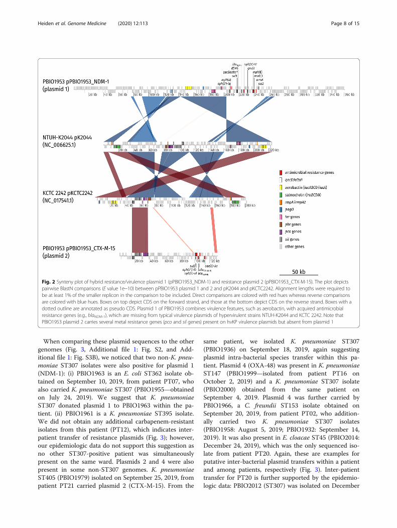
resistances were located on three different, large plas-mids, based on their respective size subsequently termedplasmids 1 (pPBIO1953_NDM-1), 2 (pPBIO1953_CTX-M-15), and 4 (pPBIO1953_OXA-48) (Additional file 1:Fig. S3 and S7). Plasmid 1 (size = 360,596 bp; incompati-bility [Inc] types: IncFIB, IncHI1B) did not only encodethe New-Delhi metallo-beta-lactamase (NDM)-1 but alsoseveral other resistance genes (dfrA5, sul1, sul2, qnrS1,aph (3′)-Ia, aph (3′)-VI, armA, mph(A), mph(E), andmsr(E)) as well as virulence (peg-344/pagO, peg-1860/pagO [metabolite transporter], iucABCD, iutA [aerobac-tin], and rmpA/rmpA2 [regulator of mucoid phenotype])and disinfectant/mineral resistance factors (qacEdelta1[disinfectant resistance], ter [tellurite resistance]) (Add-itional file 1: Fig. S7B). Plasmid 2 (size = 130,131 bp;IncFIB) carried the following antibiotic resistances:blaCTX-M-15, blaTEM-1B, sul2, aac (3)-IIa, aph (3″)-Ib,and aph (6)-Id, and, interestingly, several genes respon-sible for heavy metal resistance: ars (arsenic/antimonyresistance), sil (silver resistance), and pco (copper resist-ance). We compared plasmids 1 and 2 to two well-characterized, typical virulence plasmids of hypervirulentK. pneumoniae NTUH-K2044 [44] and KCTC 2242 [45].We found a high degree of similarity in the aforemen-tioned virulence and mineral resistance features (Fig. 2).This is of particular public health interest as the emer-gence of “mosaic” plasmids carrying AMR and virulencefactors can confer both enhanced virulence and multi-drug resistance in one single transfer. Plasmid 4 (size =63,589 bp; IncL/M) was positive for the OXA-48-encoding gene. Two additional plasmids with sizes of 72,679 bp (plasmid 3; IncFII) and 6656 bp (plasmid 5;Col440I) did not carry any resistance or virulence genes(Additional file 1: Fig. S7D and S7F).
Heiden et al. Genome Medicine (2020) 12:113 Page 7 of 15
When comparing these plasmid sequences to the othergenomes (Fig. 3, Additional file 1: Fig. S2, and Add-itional file 1: Fig. S3B), we noticed that two non-K. pneu-moniae ST307 isolates were also positive for plasmid 1(NDM-1): (i) PBIO1963 is an E. coli ST362 isolate ob-tained on September 10, 2019, from patient PT07, whoalso carried K. pneumoniae ST307 (PBIO1955—obtainedon July 24, 2019). We suggest that K. pneumoniaeST307 donated plasmid 1 to PBIO1963 within the pa-tient. (ii) PBIO1961 is a K. pneumoniae ST395 isolate.We did not obtain any additional carbapenem-resistantisolates from this patient (PT12), which indicates inter-patient transfer of resistance plasmids (Fig. 3); however,our epidemiologic data do not support this suggestion asno other ST307-positive patient was simultaneouslypresent on the same ward. Plasmids 2 and 4 were alsopresent in some non-ST307 genomes. K. pneumoniaeST405 (PBIO1979) isolated on September 25, 2019, frompatient PT21 carried plasmid 2 (CTX-M-15). From the
same patient, we isolated K. pneumoniae ST307(PBIO1936) on September 18, 2019, again suggestingplasmid intra-bacterial species transfer within this pa-tient. Plasmid 4 (OXA-48) was present in K. pneumoniaeST147 (PBIO1999—isolated from patient PT16 onOctober 2, 2019) and a K. pneumoniae ST307 isolate(PBIO2000) obtained from the same patient onSeptember 4, 2019. Plasmid 4 was further carried byPBIO1966, a C. freundii ST153 isolate obtained onSeptember 20, 2019, from patient PT02, who addition-ally carried two K. pneumoniae ST307 isolates(PBIO1958: August 5, 2019; PBIO1932: September 14,2019). It was also present in E. cloacae ST45 (PBIO2014:December 24, 2019), which was the only sequenced iso-late from patient PT20. Again, these are examples forputative inter-bacterial plasmid transfers within a patientand among patients, respectively (Fig. 3). Inter-patienttransfer for PT20 is further supported by the epidemio-logic data: PBIO2012 (ST307) was isolated on December
Fig. 2 Synteny plot of hybrid resistance/virulence plasmid 1 (pPBIO1953_NDM-1) and resistance plasmid 2 (pPBIO1953_CTX-M-15). The plot depictspairwise BlastN comparisons (E value 1e−10) between pPBIO1953 plasmid 1 and 2 and pK2044 and pKCTC2242. Alignment lengths were required tobe at least 1% of the smaller replicon in the comparison to be included. Direct comparisons are colored with red hues whereas reverse comparisonsare colored with blue hues. Boxes on top depict CDS on the forward strand, and those at the bottom depict CDS on the reverse strand. Boxes with adotted outline are annotated as pseudo CDS. Plasmid 1 of PBIO1953 combines virulence features, such as aerobactin, with acquired antimicrobialresistance genes (e.g., blaNDM-1), which are missing from typical virulence plasmids of hypervirulent strains NTUH-K2044 and KCTC 2242. Note thatPBIO1953 plasmid 2 carries several metal resistance genes (pco and sil genes) present on hvKP virulence plasmids but absent from plasmid 1
Heiden et al. Genome Medicine (2020) 12:113 Page 8 of 15
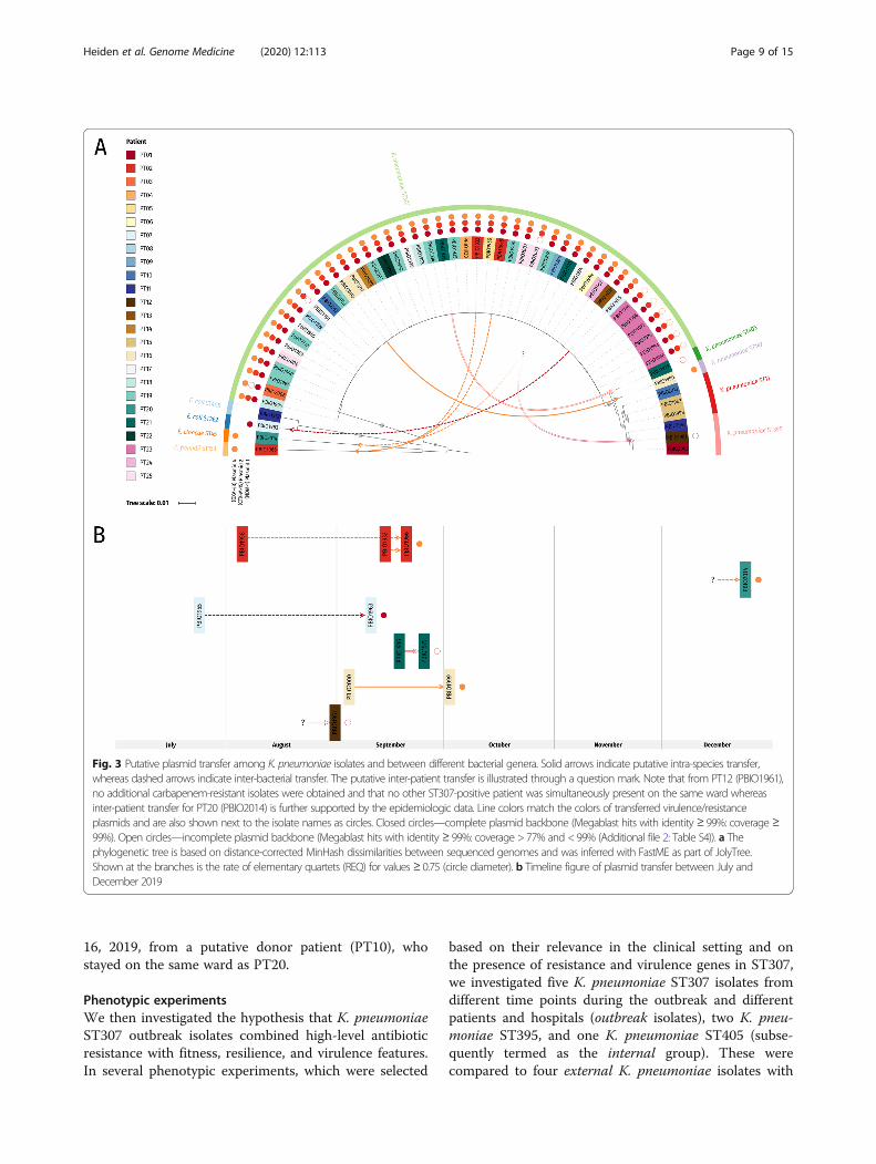
16, 2019, from a putative donor patient (PT10), whostayed on the same ward as PT20.
Phenotypic experimentsWe then investigated the hypothesis that K. pneumoniaeST307 outbreak isolates combined high-level antibioticresistance with fitness, resilience, and virulence features.In several phenotypic experiments, which were selected
based on their relevance in the clinical setting and onthe presence of resistance and virulence genes in ST307,we investigated five K. pneumoniae ST307 isolates fromdifferent time points during the outbreak and differentpatients and hospitals (outbreak isolates), two K. pneu-moniae ST395, and one K. pneumoniae ST405 (subse-quently termed as the internal group). These werecompared to four external K. pneumoniae isolates with
Fig. 3 Putative plasmid transfer among K. pneumoniae isolates and between different bacterial genera. Solid arrows indicate putative intra-species transfer,whereas dashed arrows indicate inter-bacterial transfer. The putative inter-patient transfer is illustrated through a question mark. Note that from PT12 (PBIO1961),no additional carbapenem-resistant isolates were obtained and that no other ST307-positive patient was simultaneously present on the same ward whereasinter-patient transfer for PT20 (PBIO2014) is further supported by the epidemiologic data. Line colors match the colors of transferred virulence/resistanceplasmids and are also shown next to the isolate names as circles. Closed circles—complete plasmid backbone (Megablast hits with identity ≥ 99%: coverage ≥99%). Open circles—incomplete plasmid backbone (Megablast hits with identity ≥ 99%: coverage > 77% and < 99% (Additional file 2: Table S4)). a Thephylogenetic tree is based on distance-corrected MinHash dissimilarities between sequenced genomes and was inferred with FastME as part of JolyTree.Shown at the branches is the rate of elementary quartets (REQ) for values ≥ 0.75 (circle diameter). b Timeline figure of plasmid transfer between July andDecember 2019
Heiden et al. Genome Medicine (2020) 12:113 Page 9 of 15

ST498, ST15, ST307, and ST86 including an archetypal,hypervirulent K. pneumoniae strain (hvKP1), which areunrelated to the outbreak. In addition, we included apartially plasmid-cured variant (PCV1935) and controlsfor each phenotypic experiment.We observed no significant decreased growth rates
(p > 0.1 at 1, 2, 3, 4, and 5 h) of the ST307 isolates whencompared to the internal and external groups and to theE. coli K-12 control (Fig. 4a), and also among each other.PCV1935, which carried the incomplete NDM-1 plasmid1, also showed comparable growth (μ1hST307, 1.05, vs.μ1hPCV1935, 0.67; p = 0.9999). Interestingly, K. pneumo-niae ST395 (PBIO1961) demonstrated a prolonged lagphase (μ1hST307, 1.05, vs. μ1hPBIO1961, 0.51; p = 0.038),which will be further explored in a prospective study.Hypermucoviscosity experiments revealed that all K.
pneumoniae ST307 outbreak isolates showed stronghypermucoviscosity (≥ 5 mm) (Fig. 5a), whereas in-ternal K. pneumoniae ST395 and ST405 isolates didnot (p < 0.0001). Also, the external isolates showed anegative hypermucoid phenotype, with the exceptionof K. pneumoniae ST15 and, unsurprisingly, hvKP1.Hypermucoviscosity of PCV1935 was not significantlydifferent from wildtype PBIO1935, which is interest-ing given that both plasmid-encoded rmpA andrmpA2 were lost during the curing process. On thecontrary, PBIO1961, which carried plasmid 1 (Figs. 3,4, and 5, and Additional file 1: Figure S3B and S7B),did not show hypermucoviscosity. This genome dem-onstrated a different rmpA2-truncation and K locusin comparison to the ST307 clone (Additional file 2:Table S1). Also consider that the different phenotypesmight be due to synergy-dependent processes suchthat plasmids have reduced impacts on other geneticbackgrounds than ST307. The hypermucoid pheno-type appears to be a fine-tuned process. A recentstudy [68] showed that loss of rmpC, which is anewly identified gene that contributes to capsuleregulation in hvKp, resulted in decreased capsule geneexpression while simultaneously retaining hypermu-coviscosity. Additional investigations will have to ad-dress further which regulatory mechanisms contributeto the hypermucoid phenotype in our outbreak clone.Hypermucoviscosity is associated with invasive andother aggressive types of infection, but recent litera-ture suggests that this characteristic alone is not perse responsible for a hypervirulent phenotype in Kleb-siella spp. and that both terms should not be usedsynonymously [5, 69].Similar to the hypermucoviscosity experiments, we ob-
served significant differences between the K. pneumo-niae ST307 outbreak isolates and the internal andexternal groups regarding their siderophore secretioncapacities (p < 0.0001) (Fig. 5b, c). They all exhibited a
significant higher secretion with average bleaching zonediameters of 20 mm, compared to 8.4 mm of internaland 10.8 mm of external isolates (positive control, 12mm). This is likely due to the presence of the NDM-1-plasmid-encoded aerobactin, underlined by the resultsshown for PCV1935, which demonstrated significantlyreduced siderophore secretion (p = 0.0025) (Fig. 5b, c).Note that hvKP1 also showed increased siderophore se-cretion compared to the internal group (p = 0.0126) butin tendency less than the ST307 outbreak isolates, al-though this difference was not significant (p = 0.35).PBIO1961 registered within the result range of internaland external isolates. This is likely due to a missensemutation of plasmid-encoded iutA and/or the differentcharacter of yersiniabactin (“unknown” ybt—Add-itional file 2: Table S1). Given that hypermucoviscosity,aerobactin secretion, and the metabolite transporterPEG344 [70] are suggested key virulence features of hy-pervirulent Klebsiella strains during infection, the above-average performance, in addition to extensive antibioticresistance expression, provides one step closer toexplaining why this outbreak evolved. PBIO2009 (exter-nal ST307 isolate), which does not possess typical hvKp-associated features like peg-344, iucA, and rmpA [11],showed negative hypermucoid and iron uptake pheno-types (Fig. 5a–c), possibly strengthening our assumption.We were then interested in whether the K. pneumo-
niae ST307 outbreak isolates also showed sufficient cap-acity for survival and resilience in the clinical setting andhost. Serum survival experiments revealed high survivalrates for all isolates, and together with similar strongbiofilm formation capacities (Fig. 4b, c), this suggeststhat the outbreak K. pneumoniae ST307 isolates hadgood abilities to resist clinical challenges. Comparableresults were also obtained for desiccation resilience(Fig. 4d). Despite that internal and external groupsshowed a tendency to survive 6 days of desiccation athigher rates than K. pneumoniae ST307, the differ-ence was not significant after applying Bonferronicorrection (outbreak vs. internal group: p = 0.5488;outbreak vs. external group: p = 0.0730; internal vs.external group: p > 0.9999).The genetic characterization revealed several heavy
metal efflux genes on plasmid 2. Heavy metal com-pounds, such as zinc oxide or copper sulfate, are regu-larly used as feed supplements in livestock, e.g., forprevention of gastro-intestinal disorders and growthpromotion in piglets [71], and co-selection of heavymetal and antimicrobial resistance has been increas-ingly reported [72, 73]. We thus investigated the bac-teria’s tolerance by determining minimum inhibitoryconcentrations (MICs) to copper, zinc, and silver(Additional file 2: Table S1). A MIC value of 1024 μg/mL for copper sulfate was obtained for almost all K.
Heiden et al. Genome Medicine (2020) 12:113 Page 10 of 15
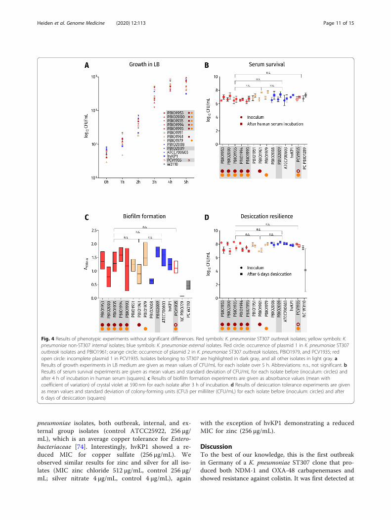
pneumoniae isolates, both outbreak, internal, and ex-ternal group isolates (control ATCC25922, 256 μg/mL), which is an average copper tolerance for Entero-bacteriaceae [74]. Interestingly, hvKP1 showed a re-duced MIC for copper sulfate (256 μg/mL). Weobserved similar results for zinc and silver for all iso-lates (MIC zinc chloride 512 μg/mL, control 256 μg/mL; silver nitrate 4 μg/mL, control 4 μg/mL), again
with the exception of hvKP1 demonstrating a reducedMIC for zinc (256 μg/mL).
DiscussionTo the best of our knowledge, this is the first outbreakin Germany of a K. pneumoniae ST307 clone that pro-duced both NDM-1 and OXA-48 carbapenemases andshowed resistance against colistin. It was first detected at
Fig. 4 Results of phenotypic experiments without significant differences. Red symbols: K. pneumoniae ST307 outbreak isolates; yellow symbols: K.pneumoniae non-ST307 internal isolates; blue symbols: K. pneumoniae external isolates. Red circle: occurrence of plasmid 1 in K. pneumoniae ST307outbreak isolates and PBIO1961; orange circle: occurrence of plasmid 2 in K. pneumoniae ST307 outbreak isolates, PBIO1979, and PCV1935; redopen circle: incomplete plasmid 1 in PCV1935. Isolates belonging to ST307 are highlighted in dark gray, and all other isolates in light gray. aResults of growth experiments in LB medium are given as mean values of CFU/mL for each isolate over 5 h. Abbreviations: n.s., not significant. bResults of serum survival experiments are given as mean values and standard deviation of CFU/mL for each isolate before (inoculum: circles) andafter 4 h of incubation in human serum (squares). c Results of biofilm formation experiments are given as absorbance values (mean withcoefficient of variation) of crystal violet at 590 nm for each isolate after 3 h of incubation. d Results of desiccation tolerance experiments are givenas mean values and standard deviation of colony-forming units (CFU) per milliliter (CFU/mL) for each isolate before (inoculum: circles) and after6 days of desiccation (squares)
Heiden et al. Genome Medicine (2020) 12:113 Page 11 of 15

Fig. 5 Results of phenotypic experiments with significant differences. Red symbols: K. pneumoniae ST307 outbreak isolates; yellow symbols: K. pneumoniae non-ST307 internal isolates; blue symbols: K. pneumoniae external isolates. Red circle: occurrence of plasmid 1 in K. pneumoniae ST307 outbreak isolates and PBIO1961;orange circle: occurrence of plasmid 2 in K. pneumoniae ST307 outbreak isolates, PBIO1979, and PCV1935; red open circle: incomplete plasmid 1 in PCV1935.Isolates belonging to ST307 are highlighted in dark gray, and all other isolates in light gray. a Results of the hypermucoviscosity test are given as mean valuesand standard deviation of the string length in millimeters (mm) for each isolate. b Results of siderophore secretion tests are given as mean values and standarddeviation of the secretion zone diameter in millimeters (mm) for each isolate. c Siderophore secretion experiment on CAS-Agar of six exemplary isolates:PBIO1953 (outbreak isolate [ST307]), PBIO1951 (internal control [ST395]), PBIO2008 (external control [ST15]), PBIO2009 (external control [ST307]), hvKP1 (externalhypervirulent control [ST86]), and PCV1935 (outbreak plasmid-cured variant [ST307]). Yellow areas around colonies indicate siderophore secretion. Abbreviationsand symbols: n.s., not significant; ****p value < 0.0001; ***p value < 0.001; **p value < 0.01
Heiden et al. Genome Medicine (2020) 12:113 Page 12 of 15
the University Medicine Greifswald in June 2019 from atracheal secretion sample [19]. As we did not detect anymcr-genes, we suggest that colistin resistance is due tochromosomal point mutations including the two-component systems PhoP/PhoQ and PmrA/PmrB, whichhave been previously described in this context [67].When placing our outbreak clone in a global frame,
a cluster of KPC-producing ST307 genomes from theUK was the phylogenetically closest. Interestingly, thiscluster was part of a study from 2017 revealing thatthese genomes harbor genetic features important forclinical and host adaptation, in particular glycogensynthesis [3]. Our outbreak isolates have seeminglydeveloped different resistance phenotypes and viru-lence strategies, and the UK cluster is thus probablynot the true, most likely recent common ancestor ofthe ST307 outbreak clone.Although we did not unequivocally verify the hypervir-
ulent character of this clone, it demonstrated hypermu-coviscosity, iron uptake, and metabolite transportercapacities—which are relevant for invasive infection andassertiveness in different host environments [41, 69,70]—comparable to an archetypal hvKp strain [54]. Thefact that these key hypervirulence features in addition todisinfectant resistance are found on mutual virulence/re-sistance plasmids in extensively drug-resistant isolates isconcerning and has tremendous public health implica-tions as these mobile genetic elements may be trans-ferred across different bacteria [75]. Our previous work,and those of others, suggests that the combination ofhigh-level drug resistance and virulence is a good com-bination for the successful spread of bacterial pathogens[3, 53, 76–79]. On the other hand, the co-carriage ofplasmid-encoded heavy metal efflux genes did not sig-nificantly impact the phenotypic tolerance in the study’sK. pneumoniae outbreak isolates, pointing towards thatthis capacity is less likely a major contributor to theclone’s success in this outbreak situation.We detected identical plasmids among ST307 and other
K. pneumoniae isolates as well as other bacterial genera,exacerbating the threat this clone poses across clinical set-tings. Note, however, that it is possible that ST307 has agreater tolerance towards possible fitness costs of the car-ried plasmids, implying that donated plasmids might havereduced impacts in other genetic backgrounds [56]. Wesuggest that the K. pneumoniae ST307 isolates are thegeneral plasmid donors; they were all isolated at an earlierdate than the putative acceptor isolates and more inde-pendent variants accumulated in some putative acceptorplasmids compared to the K. pneumoniae ST307 donorplasmids (Additional file 2: Table S3). The fact that ST395occurred three times among all isolates but only ST395PBIO1961 was positive for plasmid 1 additionallystrengthens this assumption.
Interestingly, although most hvKp show susceptibilityto antimicrobials [44], hvKp with AMR have increasinglyemerged in the last decade [80–83], which might be anongoing trend.
ConclusionsWhile the emergence of XDR, virulent, and fit pathogensis worrisome, our study helps to implement controlmeasures and calls for prospective surveillance strategiesthat take the emergence of “converging” cKp and hvKppathotypes into account.
Supplementary InformationThe online version contains supplementary material available at https://doi.org/10.1186/s13073-020-00814-6.
Additional file 1: Fig. S1. Long-read alignment for plasmid 1(pPBIO1953_NDM-1). Fig. S2. Assembly graphs of the reference isolatePBIO1953 and the putative plasmid recipient isolates. Fig. S3. Plasmido-gram of different bacterial isolates. Fig. S4. Global core SNP phylogenyof K. pneumoniae ST307 isolates. Fig. S5. Single-nucleotide polymorph-ism (SNP) distribution of K. pneumoniae ST307 isolates. Fig. S6. Missensevariants in genes putatively related to colistin resistance. Fig. S7. Com-parison of the closed genome of PBIO1953 (PT18) with the other isolates.
Additional file 2: Table S1. Overview of strains investigated in thisstudy. Table S2. Overview of publicly available Klebsiella pneumoniaeST307 genomes obtained from online sources. Table S3. Overview ofaccumulated variants in transferred plasmids. Table S4. Coverage ofisolate replicons.
AbbreviationsMDR: Multidrug-resistant; XDR: Extensively drug-resistant; cKp: Classical K.pneumoniae; hvKp: Hypervirulent K. pneumoniae; ST: Sequence type;MLST: Multi-locus sequence typing; PCV: Plasmid-cured variant; CFU: Colony-forming units; SNP: Single-nucleotide polymorphism; CDS: Coding sequence;UK: United Kingdom
AcknowledgementsWe thank Bettina Rißmann for her excellent technical assistance and Dr.Thomas A. Russo for providing the hvKP1 strain.
Authors’ contributionsK.S. and N.O.H. designed the study. S.E.H., E.E., S.S., SE.G., and J.A.B. performedthe phenotypic experiments and the computational analyses. K.S., K.B., A.K.,J.A.B., C.D.H., V.B., W.G., S.S., SÖ.G., and T.E. analyzed the data. K.S. and S.E.H.wrote the paper, and all other authors contributed to the writing. All authorsread and approved the final version of the manuscript.
FundingWe acknowledge support for the Article Processing Charge from the DFG(German Research Foundation, 393148499) and the Open Access PublicationFund of the University of Greifswald. The funding sources had no influenceon the design of the study and collection, analysis, and interpretation ofdata, and writing of the manuscript. Open Access funding enabled andorganized by Projekt DEAL.
Availability of data and materialsThe experimental and computational data that support the findings of thisresearch are available in this article and its supplementary information files.The genomic data have been deposited in the European Nucleotide Archive(ENA) at EMBL-EBI under accession number PRJEB37933 (https://www.ebi.ac.uk/ena/browser/view/PRJEB37933) [84].
Ethics approval and consent to participateEthical approval was given by the ethics committee of the University ofGreifswald, Germany (BB 133/20). Informed patient consent was waived as
Heiden et al. Genome Medicine (2020) 12:113 Page 13 of 15

samples were taken under a hospital surveillance framework for routinesampling. The research conformed to the principles of the HelsinkiDeclaration.
Consent for publicationNot applicable.
Competing interestsThe authors declare that they have no competing interests.
Author details1Institute of Pharmacy, Pharmaceutical Microbiology, University of Greifswald,Friedrich-Ludwig-Jahn-Str. 17, 17489 Greifswald, Germany. 2Central Unit forInfection Prevention and Control, University Medicine Greifswald, Greifswald,Germany. 3Friedrich Loeffler-Institute of Medical Microbiology, UniversityMedicine Greifswald, Greifswald, Germany. 4Department of General, Visceral,Thoracic and Vascular Surgery, University Medicine Greifswald, Greifswald,Germany. 5Institute for Hygiene and Environmental Medicine, UniversityMedicine Greifswald, Greifswald, Germany. 6IMD Laboratory Greifswald,Institute of Medical Diagnostics, Greifswald, Germany. 7MVZ LaboratoryLimbach Vorpommern-Rügen, Stralsund, Germany. 8Department forInfectious Disease Epidemiology, Robert Koch-Institute, Berlin, Germany.9National Reference Centre for Multidrug-Resistant Gram-Negative Bacteria,Ruhr University Bochum, Bochum, Germany. 10Institute of Pharmacy,Pharmaceutical Biology, University of Greifswald, Greifswald, Germany.
Received: 2 May 2020 Accepted: 25 November 2020
References1. Podschun R, Ullmann U. Klebsiella spp. as nosocomial pathogens:
epidemiology, taxonomy, typing methods, and pathogenicity factors. ClinMicrobiol Rev. 1998;11(4):589–603.
2. Fazili T, Sharngoe C, Endy T, Kiska D, Javaid W, Polhemus M. Klebsiella pneumoniaeliver abscess: an emerging disease. Am J Med Sci. 2016;351(3):297–304.
3. Villa L, Feudi C, Fortini D, Brisse S, Passet V, Bonura C, et al. Diversity,virulence, and antimicrobial resistance of the KPC-producing Klebsiellapneumoniae ST307 clone. Microb Genom. 2017;3(4):e000110.
4. Rossi B, Gasperini ML, Leflon-Guibout V, Gioanni A, de Lastours V, Rossi G,et al. Hypervirulent Klebsiella pneumoniae in cryptogenic liver abscesses,Paris, France. Emerg Infect Dis. 2018;24(2):221–9.
5. Russo TA, Marr CM. Hypervirulent Klebsiella pneumoniae. Clin Microbiol Rev.2019;32(3):e00001–19.
6. Shon AS, Bajwa RP, Russo TA. Hypervirulent (hypermucoviscous) Klebsiellapneumoniae: a new and dangerous breed. Virulence. 2013;4(2):107–18.
7. Liu C, Guo J. Hypervirulent Klebsiella pneumoniae (hypermucoviscous andaerobactin positive) infection over 6 years in the elderly in China:antimicrobial resistance patterns, molecular epidemiology and risk factor.Ann Clin Microbiol Antimicrob. 2019;18(1):4.
8. Russo TA, Olson R, Fang CT, Stoesser N, Miller M, MacDonald U, et al.Identification of biomarkers for differentiation of hypervirulent Klebsiellapneumoniae from classical K. pneumoniae. J Clin Microbiol. 2018;56(9):e00776–18.
9. Marsh JW, Mustapha MM, Griffith MP, Evans DR, Ezeonwuka C, Pasculle AW, et al.Evolution of outbreak-causing carbapenem-resistant Klebsiella pneumoniae ST258at a tertiary care hospital over 8 years. Mbio. 2019;10(5):e01945–19.
10. Wyres KL, Hawkey J, Hetland MAK, Fostervold A, Wick RR, Judd LM, et al.Emergence and rapid global dissemination of CTX-M-15-associated Klebsiellapneumoniae strain ST307. J Antimicrob Chemother. 2019;74(3):577–81.
11. Schaufler K, Nowak K, Dux A, Semmler T, Villa L, Kourouma L, et al. Clinicallyrelevant ESBL-producing K. pneumoniae ST307 and E. coli ST38 in an urbanWest African rat population. Front Microbiol. 2018;9:150.
12. Long SW, Olsen RJ, Eagar TN, Beres SB, Zhao P, Davis JJ, et al. Populationgenomic analysis of 1,777 extended-spectrum beta-lactamase-producingKlebsiella pneumoniae isolates, Houston, Texas: unexpected abundance ofclonal group 307. Mbio. 2017;8(3):e00489–17.
13. Giddins MJ, Macesic N, Annavajhala MK, Stump S, Khan S, McConville TH,et al. Successive emergence of ceftazidime-avibactam resistance throughdistinct genomic adaptations in blaKPC-2-harboring Klebsiella pneumoniaesequence type 307 isolates. Antimicrob Agents Chemother. 2018;62(3):e02101–17.
14. Novovic K, Trudic A, Brkic S, Vasiljevic Z, Kojic M, Medic D, et al. Molecularepidemiology of colistin-resistant, carbapenemase-producing Klebsiellapneumoniae in Serbia from 2013 to 2016. Antimicrob Agents Chemother.2017;61(5):e02550–16.
15. Saavedra SY, Diaz L, Wiesner M, Correa A, Arevalo SA, Reyes J, et al.Genomic and molecular characterization of clinical isolates ofenterobacteriaceae harboring mcr-1 in Colombia, 2002 to 2016. AntimicrobAgents Ch. 2017;61(12):e00841–17.
16. Baek EH, Kim SE, Kim S, Lee S, Cho OH, In Hong S, et al. Successfulcontrol of an extended-spectrum beta-lactamase-producing Klebsiellapneumoniae ST307 outbreak in a neonatal intensive care unit. BMCInfect Dis. 2020;20(1):166.
17. Kim JO, Song SA, Yoon EJ, Shin JH, Lee H, Jeong SH, et al. Outbreak of KPC-2-producing Enterobacteriaceae caused by clonal dissemination of Klebsiellapneumoniae ST307 carrying an IncX3-type plasmid harboring a truncatedTn4401a. Diagn Micr Infec Dis. 2017;87(4):343–8.
18. Boonstra MB, Spijkerman DCM, in’t Holt AFV, van der Laan RJ, Bode LGM, vanVianen W, et al. An outbreak of ST307 extended-spectrum beta-lactamase (ESBL)-producing Klebsiella pneumoniae in a rehabilitation center: an unusual source androute of transmission. Infect Cont Hosp Ep 2020. 41(1):31–36.
19. Haller S, Kramer R, Becker K, Bohnert JA, Eckmanns T, Hans JB, et al.Extensively drug-resistant Klebsiella pneumoniae ST307 outbreak, north-eastern Germany, June to October 2019. Euro Surveill. 2019;24(50):1900734.
20. Baym M, Kryazhimskiy S, Lieberman TD, Chung H, Desai MM, Kishony R.Inexpensive multiplexed library preparation for megabase-sized genomes.PLoS One. 2015;10(5):e0128036.
21. Bankevich A, Nurk S, Antipov D, Gurevich AA, Dvorkin M, Kulikov AS, et al.SPAdes: a new genome assembly algorithm and its applications to single-cell sequencing. J Comput Biol. 2012;19(5):455–77.
22. Li H, Durbin R. Fast and accurate short read alignment with Burrows-Wheeler transform. Bioinformatics. 2009;25(14):1754–60.
23. Li H, Handsaker B, Wysoker A, Fennell T, Ruan J, Homer N, et al. The SequenceAlignment/Map format and SAMtools. Bioinformatics. 2009;25(16):2078–9.
24. McKenna A, Hanna M, Banks E, Sivachenko A, Cibulskis K, Kernytsky A, et al.The Genome Analysis Toolkit: a MapReduce framework for analyzing next-generation DNA sequencing data. Genome Res. 2010;20(9):1297–303.
25. Walker BJ, Abeel T, Shea T, Priest M, Abouelliel A, Sakthikumar S, et al. Pilon:an integrated tool for comprehensive microbial variant detection andgenome assembly improvement. PLoS One. 2014;9(11):e112963.
26. Wick RR, Judd LM, Gorrie CL, Holt KE. Unicycler: resolving bacterial genomeassemblies from short and long sequencing reads. Plos Comput Biol. 2017;13(6):e1005595.
27. Li H. Minimap2: pairwise alignment for nucleotide sequences.Bioinformatics. 2018;34(18):3094–100.
28. Milne I, Stephen G, Bayer M, Cock PJ, Pritchard L, Cardle L, et al. UsingTablet for visual exploration of second-generation sequencing data. BriefBioinform. 2013;14(2):193–202.
29. Wick RR, Schultz MB, Zobel J, Holt KE. Bandage: interactive visualization ofde novo genome assemblies. Bioinformatics. 2015;31(20):3350–2.
30. Parks DH, Imelfort M, Skennerton CT, Hugenholtz P, Tyson GW. CheckM:assessing the quality of microbial genomes recovered from isolates, singlecells, and metagenomes. Genome Res. 2015;25(7):1043–55.
31. Seemann T. Prokka: rapid prokaryotic genome annotation. Bioinformatics.2014;30(14):2068–9.
32. Jolley KA, Bray JE, Maiden MCJ. Open-access bacterial population genomics:BIGSdb software, the PubMLST.org website and their applications. WellcomeOpen Res. 2018. 3:124.
33. Liu B, Zheng D, Jin Q, Chen L, Yang J. VFDB 2019: a comparativepathogenomic platform with an interactive web interface. Nucleic AcidsRes. 2019;47(D1):D687–D92.
34. Zankari E, Hasman H, Cosentino S, Vestergaard M, Rasmussen S, Lund O,et al. Identification of acquired antimicrobial resistance genes. J AntimicrobChemother. 2012;67(11):2640–4.
35. Carattoli A, Zankari E, Garcia-Fernandez A, Voldby Larsen M, Lund O, Villa L, et al.In silico detection and typing of plasmids using PlasmidFinder and plasmidmultilocus sequence typing. Antimicrob Agents Chemother. 2014;58(7):3895–903.
36. Pal C, Bengtsson-Palme J, Rensing C, Kristiansson E, Larsson DGJ. BacMet:antibacterial biocide and metal resistance genes database. Nucleic AcidsRes. 2014;42(D1):D737–D43.
37. Alikhan NF, Petty NK, Ben Zakour NL, Beatson SA. BLAST Ring Image Generator(BRIG): simple prokaryote genome comparisons. BMC Genomics. 2011;12:402.
Heiden et al. Genome Medicine (2020) 12:113 Page 14 of 15
38. Camacho C, Coulouris G, Avagyan V, Ma N, Papadopoulos J, Bealer K, et al.BLAST+: architecture and applications. BMC Bioinformatics. 2009;10:421.
39. Page AJ, Cummins CA, Hunt M, Wong VK, Reuter S, Holden MT, et al. Roary: rapidlarge-scale prokaryote pan genome analysis. Bioinformatics. 2015;31(22):3691–3.
40. Lam MMC, Wick RR, Wyres KL, Gorrie CL, Judd LM, Jenney AWJ, et al.Genetic diversity, mobilisation and spread of the yersiniabactin-encodingmobile element ICEKp in Klebsiella pneumoniae populations. Microb Genom.2018;4(9):e000196.
41. Lam MMC, Wyres KL, Judd LM, Wick RR, Jenney A, Brisse S, et al. Trackingkey virulence loci encoding aerobactin and salmochelin siderophoresynthesis in Klebsiella pneumoniae. Genome Med. 2018;10(1):77.
42. Wick RR, Heinz E, Holt KE, Wyres KL. Kaptive web: user-friendly capsule andlipopolysaccharide serotype prediction for Klebsiella genomes. J ClinMicrobiol. 2018;56(6):e00197–18.
43. Wyres KL, Wick RR, Gorrie C, Jenney A, Follador R, Thomson NR, et al.Identification of Klebsiella capsule synthesis loci from whole genome data.Microb Genom. 2016;2(12):e000102.
44. Wang X, Xie Y, Li G, Liu J, Li X, Tian L, et al. Whole-genome-sequencingcharacterization of bloodstream infection-causing hypervirulent Klebsiellapneumoniae of capsular serotype K2 and ST374. Virulence. 2018;9(1):510–21.
45. Shin SH, Kim S, Kim JY, Lee S, Um Y, Oh MK, et al. Complete genomesequence of the 2,3-butanediol-producing Klebsiella pneumoniae strainKCTC 2242. J Bacteriol. 2012;194(10):2736–7.
46. Guy L, Kultima JR, Andersson SG. genoPlotR: comparative gene andgenome visualization in R. Bioinformatics. 2010;26(18):2334–5.
47. Croucher NJ, Page AJ, Connor TR, Delaney AJ, Keane JA, Bentley SD, et al.Rapid phylogenetic analysis of large samples of recombinant bacterial wholegenome sequences using Gubbins. Nucleic Acids Res. 2015;43(3):e15-e.
48. Page AJ, Taylor B, Delaney AJ, Soares J, Seemann T, Keane JA, et al. SNP-sites: rapid efficient extraction of SNPs from multi-FASTA alignments. MicrobGenom. 2016;2(4):e000056.
49. Kozlov AM, Darriba D, Flouri T, Morel B, Stamatakis A. RAxML-NG: a fast,scalable and user-friendly tool for maximum likelihood phylogeneticinference. Bioinformatics. 2019;35(21):4453–5.
50. Letunic I, Bork P. Interactive Tree Of Life (iTOL) v4: recent updates and newdevelopments. Nucleic Acids Res. 2019;47(W1):W256–W9.
51. Criscuolo A. A fast alignment-free bioinformatics procedure to infer accuratedistance-based phylogenetic trees from genome assemblies. Res IdeasOutcomes. 2019;5:e36178.
52. Karp PD, Riley M, Saier M, Paulsen IT, Collado-Vides J, Paley SM, et al. TheEcoCyc database. Nucleic Acids Res. 2002;30(1):56–8.
53. Turton J, Davies F, Turton J, Perry C, Payne Z, Pike R. Hybrid resistance andvirulence plasmids in “high-risk” clones of Klebsiella pneumoniae, includingthose carrying blaNDM-5. Microorganisms. 2019;7(9):326.
54. Russo TA, Gill SR. Draft genome sequence of the hypervirulent Klebsiellapneumoniae strain hvKP1, isolated in Buffalo, New York. Genome Announc.2013;1(2):e0006513.
55. Schaufler K, Wieler LH, Semmler T, Ewers C, Guenther S. ESBL-plasmidscarrying toxin-antitoxin systems can be “cured” of wild-type Escherichia coliusing a heat technique. Gut Pathog. 2013;5(1):34.
56. Schaufler K, Semmler T, Pickard DJ, de Toro M, de la Cruz F, Wieler LH, et al.Carriage of extended-spectrum beta-lactamase-plasmids does not reducefitness but enhances virulence in some strains of pandemic E coli lineages.Front Microbiol. 2016;7:336.
57. Schwyn B, Neilands JB. Universal chemical-assay for the detection anddetermination of siderophores. Anal Biochem. 1987;160(1):47–56.
58. Schaufler K, Semmler T, Wieler LH, Trott DJ, Pitout J, Peirano G, et al.Genomic and functional analysis of emerging virulent and multidrug-resistant Escherichia coli lineage sequence type 648. Antimicrob AgentsChemother. 2019;63(6):e00243–19.
59. Du XJ, Wang XY, Dong X, Li P, Wang S. Characterization of the desiccationtolerance of Cronobacter sakazakii strains. Front Microbiol. 2018;9:2867.
60. Ranjan A, Scholz J, Semmler T, Wieler LH, Ewers C, Muller S, et al. ESBL-plasmid carriage in E coli enhances in vitro bacterial competition fitnessand serum resistance in some strains of pandemic sequence types withoutoverall fitness cost. Gut Pathog. 2018;10:24.
61. Schierack P, Heiden SE, Khan MM, Nikolaus L, Kolenda R, Stubbe M, et al.Genomic and phenotypic analysis of an ESBL-producing E. coli ST1159clonal lineage from wild birds in Mongolia. Front Microbiol. 2020;11:1699.
62. Kruskal WH, Wallis WA. Use of ranks in one-criterion variance analysis. J AmStat Assoc. 1952;47(260):583–621.
63. Sedgwick P. Multiple hypothesis testing and Bonferroni’s correction. BMJ.2014;349:g6284.
64. Marsh JW, Krauland MG, Nelson JS, Schlackman JL, Brooks AM, Pasculle AW,et al. Genomic epidemiology of an endoscope-associated outbreak ofKlebsiella pneumoniae carbapenemase (KPC)-producing K. pneumoniae. PlosOne. 2015;10(12):e0144310.
65. Haft RJF, Palacios G, Nguyen T, Mally M, Gachelet EG, Zechner EL, et al.General mutagenesis of F plasmid TraI reveals its role in conjugativeregulation. J Bacteriol. 2006;188(17):6346–53.
66. Marchese A, Debbia EA. The role of gyrA, gyrB, and dnaA functions inbacterial conjugation. Ann Microbiol. 2016;66(1):223–8.
67. Olaitan AO, Morand S, Rolain JM. Mechanisms of polymyxin resistance:acquired and intrinsic resistance in bacteria. Front Microbiol. 2014;5:643.
68. Walker KA, Miner TA, Palacios M, Trzilova D, Frederick DR, Broberg CA,et al. A Klebsiella pneumoniae regulatory mutant has reduced capsuleexpression but retains hypermucoviscosity. Mbio. 2019;10(2):e00089–19.
69. Catalan-Najera JC, Garza-Ramos U, Barrios-Camacho H. Hypervirulence andhypermucoviscosity: two different but complementary Klebsiella spp.phenotypes? Virulence. 2017;8(7):1111–23.
70. Bulger J, MacDonald U, Olson R, Beanan J, Russo TA. Metabolite transporterPEG344 is required for full virulence of hypervirulent Klebsiella pneumoniaestrain hvKP1 after pulmonary but not subcutaneous challenge. InfectImmun. 2017;85(10):e00093–17.
71. Adamse P, HJv E, Av P, Bikker P, Jd J. Trend analysis of copper andzinc in animal feed. Wageningen: Rikilt - Institute of Food Safety; 2011.
72. Baker-Austin C, Wright MS, Stepanauskas R, McArthur JV. Co-selection ofantibiotic and metal resistance. Trends Microbiol. 2006;14(4):176–82.
73. van Alen S, Kaspar U, Idelevich EA, Kock R, Becker K. Increase of zincresistance in German human derived livestock-associated MRSA between2000 and 2014. Vet Microbiol. 2018;214:7–12.
74. Ghazisaeedi F, Ciesinski L, Bednorz C, Johanns V, Pieper R, Tedin K, et al.Phenotypic zinc resistance does not correlate with antimicrobial multi-resistance in fecal E. coli isolates of piglets. Gut Pathog. 2020. 12(4).
75. Evans DR, Griffith MP, Sundermann AJ, Shutt KA, Saul MI, Mustapha MM,et al. Systematic detection of horizontal gene transfer across genera amongmultidrug-resistant bacteria in a single hospital. Elife. 2020;9:e53886.
76. Ewers C, Bethe A, Stamm I, Grobbel M, Kopp PA, Guerra B, et al. CTX-M-15-D-ST648 Escherichia coli from companion animals and horses: anotherpandemic clone combining multiresistance and extraintestinal virulence? JAntimicrob Chemother. 2014;69(5):1224–30.
77. Beceiro A, Tomas M, Bou G. Antimicrobial resistance and virulence: asuccessful or deleterious association in the bacterial world? Clin MicrobiolRev. 2013;26(2):185–230.
78. Calhau V, Ribeiro G, Mendonca N, Da Silva GJ. Prevalent combination ofvirulence and plasmidic-encoded resistance in ST 131 Escherichia coli strains.Virulence. 2013;4(8):726–9.
79. Pitout JD. Extraintestinal pathogenic Escherichia coli: a combination ofvirulence with antibiotic resistance. Front Microbiol. 2012;3:9.
80. Surgers L, Boyd A, Girard PM, Arlet G, Decre D. ESBL-producing strain ofhypervirulent Klebsiella pneumoniae K2. France Emerg Infect Dis. 2016;22(9):1687–8.
81. Zhang Y, Zeng J, Liu W, Zhao F, Hu Z, Zhao C, et al. Emergence of ahypervirulent carbapenem-resistant Klebsiella pneumoniae isolate fromclinical infections in China. J Inf Secur. 2015;71(5):553–60.
82. Shankar C, Nabarro LE, Devanga Ragupathi NK, Muthuirulandi Sethuvel DP,Daniel JL, Doss CG, et al. Draft genome sequences of three hypervirulentcarbapenem-resistant Klebsiella pneumoniae isolates from bacteremia.Genome Announc. 2016;4(6):e01081–16.
83. Gu D, Dong N, Zheng Z, Lin D, Huang M, Wang L, et al. A fatal outbreak of ST11carbapenem-resistant hypervirulent Klebsiella pneumoniae in a Chinese hospital: amolecular epidemiological study. Lancet Infect Dis. 2018;18(1):37–46.
84. Heiden SE, Hübner NO, Bohnert JA, Heidecke CD, Kramer A, Balau V, et al. AKlebsiella pneumoniae ST307 outbreak clone from Germany demonstratesfeatures of extensive drug resistance, hypermucoviscosity and enhancediron acquisition. Eur Nucleotide Arch. 2020. https://www.ebi.ac.uk/ena/browser/view/PRJEB37933.
Publisher’s NoteSpringer Nature remains neutral with regard to jurisdictional claims inpublished maps and institutional affiliations.
Heiden et al. Genome Medicine (2020) 12:113 Page 15 of 15