a l & a rom me p medicinal aromatic plants · pdf fileme medicinal aromatic plants d i c i...
TRANSCRIPT

Research Article s
Volume 5 • Issue 5 • 1000271Med Aromat Plants (Los Angel), an open access journalISSN: 2167-0412
Open AccessResearch Article
Ghazi et al., Med Aromat Plants (Los Angel) 2016, 5:5 DOI: 10.4172/2167-0412.1000271
*Corresponding author: Elizabeth P Ryan, Department of Environmentaland Radiological Health Sciences, Colorado State University, Fort Collins, CO80523, USA, Tel: +19704911536; E-mail: [email protected]
Received October 12, 2016; Accepted October 22, 2016; Published October 28, 2016
Citation: Ghazi IA, Zarei I, Mapesa JO, Wilburn JR, Leach JE, et al. (2016) Rice Bran Extracts Inhibit Invasion and Intracellular Replication of Salmonella typhimurium in Mouse and Porcine Intestinal Epithelial Cells. Med Aromat Plants (Los Angel) 5: 271. doi: 10.4172/2167-0412.1000271
Copyright: © 2016 Ghazi IA, et al. This is an open-access article distributed under the terms of the Creative Commons Attribution License, which permits unrestricted use, distribution, and reproduction in any medium, provided the original author and source are credited.
Keywords: Bacterial invasion and replication; Metabolomics; Ricebran; Salmonella; Small intestine
Abbreviations: CFU: Colony forming units; GFP: Greenfluorescent protein; IPEC-J2: Intestinal porcine epithelial; LTH: Lijiangxintuanheigu; MSIE: Mouse small intestine epithelial; RB: Rice bran; RBE: Rice bran extract; SHZ-2: Sanhuangzhan-2; UPLC-MS: Ultra-performance liquid chromatography-mass spectrometry.
IntroductionThe Center for Disease Control and Prevention estimates that 21
million Salmonella infections cause illnesses that result in more than 200,000 deaths globally each year [1,2]. Some reports suggest that Salmonella stimulates transcriptional reprogramming in host cells, produces pro-inflammatory cytokines, and promotes cytoskeletal rearrangements that contribute to diarrhea [3,4]. There are a number of barriers to treatment, including antimicrobial resistance, and limited access to and high costs of antibiotics [5]. We and others have previously demonstrated that dietary rice bran promotes resistance to Salmonella enterica serovar Typhimurium colonization in mice [6,7]. Additionally, bran from different rice varieties showed differential protection against Salmonella [8]. Recent studies also demonstrate that dietary rice bran protects against rotavirus diarrhea and promotes Th1-type immune responses to human rotavirus vaccine in gnotobiotic pigs [9,10]. While the antimicrobial activity of rice bran extracts for protection against diarrheal disease is tested on a number of pathogens, the effects of these rice bran extracts have not been previously tested for mechanisms of enteric pathogen invasion and intracellular replication.
Rice bran also delivers a distinct biochemical profile, including lipids, compared to other cereal grains that modulate immunity [11-14] and merits investigation for protective mechanisms against entericpathogens [7,9,15-17]. A number of rice bran components (e.g., lipidsand polyphenols) have been broadly investigated for protection against chronic diseases such as cancer [18-20], type II diabetes [21,22], andhyperlipidemia [23].
A majority of rice cultivars are grouped into the subspecies japonica and indica, with distinct traits for plant architecture, agronomic and physiological features [24]. Recent evidence also demonstrated that japonica and indica have distinctive seed metabolomes [25]. Given that rice bran varieties differently protect against Salmonella colonization [8], we chose to compare bran from two genetically diverse varieties, Lijiangxintuanheigu (LTH) and Sanhuangzhan-2 (SHZ-2) based on
Rice Bran Extracts Inhibit Invasion and Intracellular Replication of Salmonella typhimurium in Mouse and Porcine Intestinal Epithelial CellsIrfan A Ghazi1,7#, Iman Zarei1,9, Job O Mapesa1,8#, Jessie R Wilburn2, Jan E Leach3, Sangeeta Rao4, Corey D Broeckling5, Anna McClung6 and Elizabeth P Ryan1*1Department of Environmental and Radiological Health Sciences, Colorado State University, Fort Collins, CO 80523, USA2Department of Food Science and Nutrition, Colorado State University, Fort Collins, CO 80523, USA3Department of Bioagricultural Sciences and Pest Management, Colorado State University, Fort Collins, CO 80523, USA4Animal Population Health Institute, Department of Clinical Sciences, Colorado State University, Fort Collins, CO 80523, USA5Proteomics and Metabolomics Facility, Colorado State University, Fort Collins, CO 80523, USA6USDA-Agricultural Research Service, Dale Bumpers National Rice Research Center, Stuttgart, AR 72160, USA7Department of Plant Sciences, School of Life Sciences, University of Hyderabad, Hyderabad, Telangana, India8Department of Human Nutrition and Dietetics, Kenya Methodist University, Nairobi, Kenya9Institute of Human Nutrition and Food, College of Human Ecology, University of the Philippines Los Baños, Los Baños 4031, Laguna, Philippines#Contributed equally to this work
Abstract Dietary rice bran supplementation has been shown to inhibit Salmonella fecal shedding in animals. The aim of this
study was to determine if bran extracts from two distinct rice varieties, Lijiangxintuanheigu (LTH) and Sanhuangzhan-2 (SHZ-2), differentially inhibit Salmonella enterica serovar Typhimurium invasion and intracellular replication. Rice bran extracts were tested in vitro using mouse small intestine epithelial (MSIE) and intestinal porcine epithelial cells (IPEC-J2). Fluorescent labeled Salmonella was detected using fluorescence microscopy and culture based methods. Non-targeted metabolomics using ultra-performance liquid chromatography- mass spectrometry (UPLC-MS) was performed on LTH and SHZ-2 rice bran extracts. LTH bran extract dose-dependently reduced entry and intracellular replication of Salmonella in both MSIE and IPEC-J2 cells when compared with SHZ-2. The rice bran metabolite profiling revealed significant variations between LTH and SHZ-2. LTH had higher total numbers of metabolites (429) versus SHZ-2 (407), with increased relative abundance of lipids (i.e., galactolipids and phospholipids) and flavonoids compared with SHZ-2. SHZ-2 had higher levels of dipeptides and phenylpropanoids. Distinct metabolite differences between LTH and SHZ-2 revealed rice bran components that may be responsible for blocking Salmonella invasion and intracellular replication. Metabolomics is a powerful phenotyping tool for identifying rice bran compounds with protective effects against pathogens. Future studies may identify the rice genes responsible for these bioactive rice bran metabolites distinguishing LTH from SHZ-2, and enable genetic selection for compounds as traits that offer important health and disease fighting benefits.
Med
icina
l & Aromatic Plants
ISSN: 2167-0412
Med
icina
l & Aromatic Plants
ISSN: 2167-0412 Medicinal & ArMedicinal & Aromatic Plantsomatic Plants

Citation: Ghazi IA, Zarei I, Mapesa JO, Wilburn JR, Leach JE, et al. (2016) Rice Bran Extracts Inhibit Invasion and Intracellular Replication of Salmonella typhimurium in Mouse and Porcine Intestinal Epithelial Cells. Med Aromat Plants (Los Angel) 5: 271. doi: 10.4172/2167-0412.1000271
Page 2 of 10
Volume 5 • Issue 5 • 1000271Med Aromat Plants (Los Angel), an open access journalISSN: 2167-0412
single nucleotide polymorphisms (SNPs) previously reported from a classified set of 20 rice varieties [26,27]. We hypothesized differential inhibition of Salmonella enterica serovar Typhimurium invasion and intracellular replication in small intestinal cells by rice bran extracts. To study differences between the rice bran types, LTH and SHZ-2, for protection against Salmonella, we applied non-targeted metabolomics.
Materials and MethodsRice milling and heat stabilization
Two rice varieties, Lijiangxintuanheigu (LTH) -Oryza sativa Japonica- and Sanhuangzhan-2 (SHZ-2)- Oryza sativa Indica- were obtained from the United States Department of Agriculture- Agricultural Research Service (USDA-ARS) Dale Bumpers National Rice Research Center, Stuttgart, AR, USA. LTH is a short grain cultivar with red bran, whereas SHZ-2 is a long grain cultivar with light brown bran [26,27] (Figure S1). Seed increase of SHZ-2 was performed at Stuttgart, AR and harvested in September 2011 while that of LTH was performed in Lajas, Puerto Rico and harvested in April 2012.
The seeds were cleaned and stored at 4°C until bran preparation. Rough rice was dehulled using a Yamamoto testing husker (Model: FC2K l), and bran was removed and collected using a Yamamoto test whitening machine (Rice pal VP-31T). Testing sieve No. 20 was used to separate bran from broken rice and hulls into a clean container. Once separated, milled bran was heat stabilized at 110°C for 6 min to prevent rancidity during storage. RB was stored at -20°C until further processing for metabolite analysis.
Rice bran extract (RBE) preparation
Non-targeted metabolomics was used to analyze rice bran extracts at Metabolon Inc. (Durham, NC). Sample preparation used the automated MicroLab STAR® system from Hamilton Company. A recovery standard was added prior to extraction for quality control. Rice bran was mixed with ice-cold 80% methanol. Chemically diverse metabolites were concentrated in the supernatant after vigorous shaking (Glen Mills GenoGrinder 2000) followed by centrifugation. Rice bran extracts were applied to UPLC-MS/MS with positive and negative ion mode electrospray ionization and for intestinal cell culture-based Salmonella infections. For the cellular assays, lyopholized rice bran extract was diluted in 1 ml medium, vortexed to dissolve compounds, and filtered through a sterile 0.45 µm pore size Whatman syringe filter (Cat # 6789-1304; Whatman Inc.). Dose response experiments were completed and final working concentrations of 1 mg/ml (MSIE) and 5 mg/ml (IPEC-J2) rice bran extract were used after assessing cell mortality (data not shown and as described in Ref. [6]).
Cell culture conditions
Mouse small intestine epithelial cells (MSIE): MSIE cells were a gift from Dr. Robert Whitehead at Vanderbilt University and the Ludwig Institute for Cancer Research [28]. MSIE cells were maintained on RPMI 1640 medium (Fischer Scientific) supplemented with 10% FBS (Cat # F0500A-HI; Atlas Biologicals), 1 × antibiotic/antimycotic (Cat # 15240-062; Life Technologies), 10 µM thioglycerol (Cat # M6145; Sigma), 1 µg/ml hydrocortisone (Cat # H0135; Sigma), 2.0 mM/ml L-glutamine (Hyclone Laboratories), 1 µg/ml regular human insulin (Novo Nordik), and 10 units/ml murine IFN-γ (Peprotech). Cells were grown in 75 cm2 flasks, split at 80% confluence twice weekly, and the culture medium replaced every two days. Cells were maintained at 33°C with 5% CO2 and 95% relative humidity. Trypan blue staining was used to evaluate cell viability. Trypsinized (0.25% Trypsin-EDTA;
Life Technologies) cells were seeded in 24 well plates at a density of 5 × 10⁴ cells/ml. After 48 h incubation, the culture medium was removed and fresh medium containing rice bran extracts added for 2 h. The cell monolayer was washed twice with phosphate buffered saline (PBS, pH 7.4) before Salmonella infection.
Intestinal porcine epithelial cells- jejunum 2 (IPEC-J2): IPEC-J2 cells were a gift from Dr. Lijuan Yuan at Virginia Polytechnique Institute and State University. IPEC-J2 cells were maintained on DMEM/F12 medium (Life Technologies) supplemented with 5% FBS, 1 × antibiotic/antimycotic, 2.0 mM/ml L-Glutamine, 5 µg/ml Insulin-Transferrin-Selenium (Life Technologies), and 5 ng/ml EGF recombinant human protein solution (Life Technologies). Cells were maintained at 37°C+5% CO2, 95% relative humidity. Trypsinized cells were seeded in 24 well plates at a density of 1 × 10⁵ cells/ml, after 48 h the culture medium was removed and fresh medium containing rice bran extracts added for 2 h. The cell monolayer was washed twice with PBS before Salmonella infection.
Salmonella enterica infection: Green Fluorescent Protein (GFP) labeled Salmonella enterica serovar Typhimurium (strain 14028s) was a gift from Dr. Andres Vazquez-Torres at University of Colorado Denver. GFP Salmonella was prepared as previously described [6]. MSIE cells were infected with 100 MOI (Multiplicity of Infection) Salmonella in PBS and centrifuged at 1,200 × g for 2 min to promote bacterial interaction with cells. After 1 h of incubation the medium was removed and the cell monolayer washed twice with PBS to remove extracellular bacteria. The cells were incubated with medium containing 100 µg/ml gentamicin (Cat # G1397; Sigma) for 1 h, washed twice in PBS before a further incubation in medium containing 50 µg/ml gentamicin for 1 h (invasion) or 24 h (replication) to prevent extracellular Salmonella growth. The cells were washed twice with PBS and incubated for 30 min in medium containing 1 × DAPI/Texas red dye (Cat # ENZ-53005-C100; ENZO Life Sciences). Cells were washed twice with 1 × PBS to remove the excess staining solution before observing under a fluorescence microscope.
GFP Salmonella fluorescence (Quantification): Fluorescence was determined using Cytation 3 cell imaging multi-mode reader (BioTek, Winooski, VT, USA) with the following imaging modes at 20 × objective: Brightfield and Fluorescence (Excitation/Emission): DAPI (377/447 nm), GFP (469/525 nm), and CY5 (625/685 nm). Percent Salmonella infection was quantified by expressing the ratio of GFP Salmonella signal to cell nuclei count as a percentage [(GFP/Nuclei) × 100]. After fluorescence reading, the cells were lysed for bacterial enumeration.
Colony forming units (CFU): Salmonella growth was measured as follows: The epithelial cells were washed twice before lysed in buffer [1% TritonX-100 (Sigma) and 0.1% Sodium Dodecyl Sulphate (Sigma) in PBS] for 5 min to free the intracellular Salmonella. The released Salmonella cells were mixed and pipetted at 1 ml equivalent of cell lysate, and serially diluted 1:10 in 1 × PBS. 100 ml of each dilution factor was plated on MacConkey agar (BD Biosciences) supplemented with 50 µg/ml kanamycin (Lot # 085309; Fisher Scientific). After incubation for 16 h at 37°C, total Salmonella CFU/ml were enumerated.
Ultra-performance liquid chromatography-tandem mass spectrometry (UPLC-MS/MS)
A Waters ACQUITY UPLC and a Thermo Scientific Q-Exactive high resolution/accurate mass spectrometer interfaced with a heated electrospray ionization (HESI-II) source and Orbitrap

Citation: Ghazi IA, Zarei I, Mapesa JO, Wilburn JR, Leach JE, et al. (2016) Rice Bran Extracts Inhibit Invasion and Intracellular Replication of Salmonella typhimurium in Mouse and Porcine Intestinal Epithelial Cells. Med Aromat Plants (Los Angel) 5: 271. doi: 10.4172/2167-0412.1000271
Page 3 of 10
Volume 5 • Issue 5 • 1000271Med Aromat Plants (Los Angel), an open access journalISSN: 2167-0412
mass analyzer operated at 35,000 mass resolution was used for non-targeted metabolomics. Each sample contained eight or more injection standards at known concentrations to ensure injection and chromatographic consistency. One aliquot was analyzed using acidic positive ion optimized conditions and the other using basic negative ion optimized conditions in two independent injections using same columns (Waters UPLC BEH C18-2.1 × 100 mm, 1.7 µm). Rice bran extracts diluted in acidic conditions, were further eluted using water and methanol containing 0.1% formic acid from a C18 column. The same elution procedure was performed for basic extracts, however, ammonium bicarbonate was used instead of formic acid. The third aliquot was analyzed via negative ionization following elution from a HILIC column (Waters UPLC BEH Amide 2.1 × 150 mm, 1.7 µm) using a gradient consisting of water and acetonitrile with ammonium formate. The MS analysis alternated between MS and data-dependent MS2 scans using dynamic exclusion, and the scan range was from 80-1000 m/z.
Compound identification analysis
Metabolon’s hardware and software were used to process the LC-MS data. Compounds were identified by comparison to library entries of purified standards or recurrent unknown entities. The mass spectra of the metabolites were compared to the NIST (National Institute of Standards and Technology) library. The MS/MS scores are based on a comparison of the ions present in the experimental spectrum to the ions present in the library spectrum. More than 3000 commercially available purified standard compounds were acquired and registered into Metabolon’s Laboratory Information Management System (LIMS) for determination of analytical characteristics. A data normalization step was performed to correct variation resulting from instrument inter-day tuning differences. The relative abundance of each metabolite was further normalized by the mediumn obtained from entire dataset.
Statistical analysis
The CFU data was analyzed using GraphPad Prism 5 (GraphPad Software; San Diego California) in one-way ANOVA and the differences in sample means analyzed by Dunnett’s Multiple Comparison Test (p<0.05). CFU data were transformed to log10 scale and presented as mean ± SEM of triplicate samples representative of at least three independent experiments. A t-test/f-test was conducted on mediumn-scaled normalized relative abundance of entire data set (LTH vs. SHZ-2) to determine whether the entire metabolite means between these varieties differ.
ResultsDose and time dependent effects from LTH and SHZ-2 rice bran extracts on Salmonella intracellular replication
MSIE and IPEC-J2 were treated with 0, 0.1, 0.5, 1 and 5 mg/ml RBE from both LTH and SHZ-2. LTH RBE exhibited a dose-dependent effect that was significantly different from SHZ-2 RBE for inhibition of Salmonella intracellular replication (Figure 1A). In MSIE cells, LTH RBE significantly inhibited (p<0.05) Salmonella replication at 0.5 and 1 mg/ml compared to vehicle control (Figure 1A), whereas in IPEC-J2, the LTH RBE significantly (p<0.05) inhibited at 5 mg/ml (Figure 1B). SHZ-2 RBE showed no inhibitory effects on Salmonella replication at any of the doses examined nor on any cell line. The doses of 1 mg/ml (MSIE cells) and 5 mg/ml (IPEC-J2 cells) were used in subsequent tissue culture experiments. The LTH and SHZ-2 RBE at 1 mg/ml (MSIE) and 5 mg/ml (IPEC-J2) were next evaluated for inhibition of Salmonella replication after 2, 4 and 6 h in MSIE and IPEC-J2. LTH and SHZ-2
RBE showed no differences in Salmonella replication between the time points after 2 h incubation. Thus, 2 h was chosen for comparison in subsequent experiments.
Effect of rice bran extracts on Salmonella invasion
To determine the effect of RBE on Salmonella invasion, MSIE cells were pre-incubated with RBE for 2 h before addition of GFP-labeled Salmonella. Fluorescence microscopy of GFP-labeled Salmonella revealed that LTH inhibited Salmonella invasion when compared to SHZ-2 (Figure 2A); however, quantification by percent infection revealed no significant differences in both the control and RBE treated MSIE cells (Figure 2B). RBE from LTH showed a 2-log cycle more inhibition of Salmonella invasion when compared to SHZ-2 (Figure 2C). These findings suggest that both RBE from LTH and SHZ-2 protected MSIE cells against Salmonella invasion, but the inhibition by the LTH RBE was of greater magnitude. Given that Salmonella can invade and colonize the pig, we next tested IPEC-J2 cells. LTH RBE inhibited Salmonella replication when compared to SHZ-2 RBE (Figure 2D); however, the inhibition was not statistically significant (p>0.05) as a percent of infection (Figure 2E). Only LTH RBE demonstrated inhibition (p<0.05) of Salmonella replication in IPEC-J2 cells as determined by CFU when compared to control and SHZ-2 RBE treatment (Figure 2F).
Differential effect of LTH and SHZ-2 bran extracts on Salmonella intracellular replication
We next quantified differences between the effects of LTH and SHZ-2 RBE (1 mg/ml for MSIE and 5 mg/ml for IPEC-J2) on Salmonella replication using fluorescence microscopy and culture based methods. Assessment of GFP-labeled Salmonella (Figure 3A) showed that LTH RBE inhibited Salmonella replication in MSIE cells to a greater extent than SHZ-2; yet, the inhibition was not statistically significant (p>0.05) when quantified as a percent of infection (Figure 3B). Inhibition of Salmonella replication in MSIE cells by LTH RBE, as measured by culture methods, was greater than 1.5 log cycles (p<0.05) when compared to control and SHZ-2 RBE treatment (Figure 3C). Similarly, LTH RBE appeared to inhibit Salmonella replication in IPEC-J2 more than SHZ-2 RBE by fluorescence microscopy (Figure 3D). The inhibition was not significant (p>0.05) when quantified as a percent of infection (Figure 3E). LTH RBE demonstrated a significant (p<0.05) inhibition of Salmonella replication in IPEC-J2 cells as determined by culture counts when compared to control and SHZ-2 RBE treatment (Figure 3F). Salmonella showed higher intracellular replication capacity in IPEC-J2 cells, but with similar infection efficiency (~25%) (Figure S2, right panel).
Metabolite profiling of LTH and SHZ-2 rice bran extracts
A non-targeted metabolomics analysis based on UPLC-MS/MS resulted in the identification of 429 metabolites in LTH and 407 metabolites in SHZ-2, including amino acids and their derivatives, carbohydrates, cofactors and vitamins, hormone metabolites, lipids, nucleotides, peptides, and secondary metabolites. Differences in chemical compositions between the two varieties were in lipids, secondary metabolites and dipeptides (Table 1).
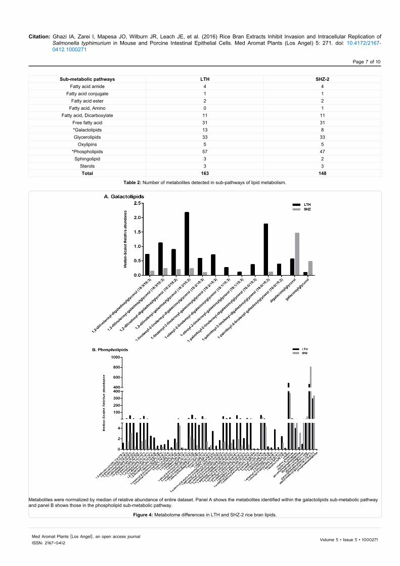
There were 163 metabolites from LTH and 148 from SHZ-2 classified in the lipid biosynthesis metabolic pathway. Among lipids, galactolipids and phospholipids showed higher variation in the number of metabolites, as well as the relative abundance between LTH and SHZ-2 (Table 2). Figure 4A and Table S1 shows the range of metabolite expression for galactolipids, such as 1,2-dilinolenoyl-

Citation: Ghazi IA, Zarei I, Mapesa JO, Wilburn JR, Leach JE, et al. (2016) Rice Bran Extracts Inhibit Invasion and Intracellular Replication of Salmonella typhimurium in Mouse and Porcine Intestinal Epithelial Cells. Med Aromat Plants (Los Angel) 5: 271. doi: 10.4172/2167-0412.1000271
Page 4 of 10
Volume 5 • Issue 5 • 1000271Med Aromat Plants (Los Angel), an open access journalISSN: 2167-0412
(A left panel) MSIE cells were treated with LTH and SHZ-2 RBE. (A right panel) IPEC-J2 cells were treated with LTH and SHZ-2 RBE. (B left panel) MSIE cells were treated with 1 mg/ml for 2, 4 and 6 h. (B right panel) IPEC-J2 cells were treated with 5 mg/ml for 2, 4 and 6 h. Significance was tested using one-way ANOVA and Dunnett’s Multiple Comparison Test. Means ± S.E.M are presented as bars (n=3) representative of three independent experiments. Bars followed by the same letter are not significantly different (p>0.05).
Figure 1: Dose and time dependent effects of Rice Bran Extracts (RBE) on Salmonella replication in intestinal epithelial cells.
digalactosylglycerol (18:3/18:3), 1-linoleoyl-2-linolenoyl-digalactosylglycerol (18:2/18:3), 1,2-dilinoleoyl-galactosylglycerol (18:2/18:2) and 1-palmitoyl-2-linoleoyl-digalactosylglycerol (16:0/18:2) with higher fold difference in LTH. Figure 4B shows that the majority of phospholipid metabolites were also present in higher abundance and number for LTH when compared to SHZ-2. LTH showed a range of 5-87-fold increased expression in 23 metabolites from phospholipids (Table S1). The biochemicals within the galactolipid and phospholipid sub-metabolic pathways are mostly long-chain polyunsaturated fatty acids. Among peptide biosynthesis metabolic pathway, eight metabolites were detected in LTH and 11 metabolites in SHZ-2. SHZ-2 was higher in both relative abundances as well as number of metabolites detected for dipeptides (Figure 5A). Leucylglycine (5-fold), alanylleucine (8-fold), and valylglycine (9-fold) were higher in SHZ-2 as compare to LTH (Table S2). Under secondary metabolites, a total of 21 metabolites were identified from LTH and 16 from SHZ-2. The subsets of secondary metabolites included benzenoid biosynthesis, flavonoid biosynthesis, phenylpropanoid biosynthesis and terpenoid biosynthesis. The variations were observed in flavonoids and phenylpropanoids (Figure 5B and 5C). LTH had higher relative abundance and total number of metabolites in flavonoids compared with SHZ-2. Three flavonoids were absent in SHZ-2 (i.e., catechin, cyanidin glucoside and dihydroquercetin). Two
other flavonoids (i.e., apigenin and chrysoeriol) were 32 and 6-fold higher, respectively, in SHZ-2, (Table S2). Although the total number of phenylpropanoids were higher in LTH, SHZ-2 had higher relative abundance in phenylpropanoids. Table S2 shows 2.5-fold difference increase in 4-hydroxycinnamate, ferulate and vanillate in SHZ-2 while p-coumaroylserotonin was almost 43 times higher in LTH.
DiscussionDietary rice bran promotes gut mucosal immune responses against
enteric pathogens [6,7,9], yet the mechanisms for how the effects differ among rice varieties and their biochemical composition is unknown. Herein we showed that LTH (japonica) significantly inhibits Salmonella invasion and intracellular replication in mouse and porcine intestinal epithelium to a greater extend when compared to SHZ-2 (indica). Although japonica and indica sub-species have the same origin, they were domesticated under different environmental conditions [24] and evidence supports variations in their seed metabalome profiles [25]. LTH and SHZ-2 were selected based on preliminary studies where they exhibited differences in inhibition of Salmonella colonization in mice [6,8,20], and because there is an available genetic population of recombinant inbred lines [29] that can facilitate subsequent identification of genes related to metabolite production [30]. Our

Citation: Ghazi IA, Zarei I, Mapesa JO, Wilburn JR, Leach JE, et al. (2016) Rice Bran Extracts Inhibit Invasion and Intracellular Replication of Salmonella typhimurium in Mouse and Porcine Intestinal Epithelial Cells. Med Aromat Plants (Los Angel) 5: 271. doi: 10.4172/2167-0412.1000271
Page 5 of 10
Volume 5 • Issue 5 • 1000271Med Aromat Plants (Los Angel), an open access journalISSN: 2167-0412
(A) MSIE cells were treated with or without 1 mg/ml of LTH or SHZ-2 for 2h before infection with or without S. typhimurium at 100 MOI for 1h and washed to remove extracellular S. typhimurium. Representative fluorescent images showing intracellular S. typhimurium (green) and nucleus (blue), images are representatives of three separate fluorescence infection experiments comprising at least three randomly selected fields of view per infection. (B) Quantification of total number of intracellular GFP S. typhimurium, determined from fluorescence images per infection, normalized per the MSIE cell count in field of view. Salmonella treatment to cells without bran extracts (white bars) was the positive control. LTH (gray bar) and SHZ-2 (black bar) extracts showed inhibitory effects on Salmonella invasion. (C) Viability of invasive S. typhimurium confirmed by CFU count on MacConkey agar, and expressed as log number of Salmonella CFU/ml. (D) IPEC-J2 cells were treated with or without 5 mg/ml of LTH or SHZ-2 for 2h before infection with or without S. typhimurium (100 MOI) for 1h. (E) Quantification of total number of intracellular GFP S. typhimurium, determined from fluorescence images per infection, normalized per the IPEC-J2 cell count in field of view. (F) Viability of invasive S. typhimurium confirmed by CFU count on MacConkey agar, and expressed as log number of Salmonella CFU/ml (panel F). Significance was tested using one-way ANOVA and Dunnett’s Multiple Comparison Test. Means ± S.E.M are presented as bars (n=3) representative of three independent experiments. Bars followed by the same letter are not significantly different (p>0.05).
Figure 2: Effect of rice bran extracts on Salmonella Invasion.
findings indicate that most of the differences in LTH and SHZ-2 composition is secondary metabolites, lipids, and the dipeptides. We showed that the red-pigmented LTH rice variety has high levels of secondary metabolites (i.e., flavonoids), and lipids (galactolipids and phospholipids), which may be associated with inhibition of Salmonella invasion and intracellular replication. These findings demonstrate the potential of RBE as a dietary alternative for prevention of Salmonella infection.
Health properties of whole grains (including pigmented rice) have been widely reported [31], and many of which are related to secondary metabolites and fatty acids contents [32,33]. These compounds have effects on bacterial clearance and disease outcomes through various mechanisms [32,33]. The findings herein support that pigmented rice offers greater protection against enteric Salmonella infection as
seen in vivo [8]. Flavonoids are classified as secondary metabolites, are abundant in many fruits and naturally occur in some cereals and legumes, and pose many beneficial health effects [34]. Flavonoids were shown to strongly inhibit arachidonic acid-derived inflammatory reactions, apoptosis, MAPK, and NF-κB pathways in macrophages [35]. An example, for a metabolite with higher abundance in LTH and among the flavonoids was catechin, which has been effective to reduce growth of Staphylococcus aureus, and Salmonella Typhimurium [36].
Studies have reported vegetable oils including rice bran oil have antimicrobial properties [15,37,38]. De Pablo et al. showed that host immunity and pathogen resistance may be influenced by the nutritional status [33]. Although, no single fatty acid is an answer for protection from food-borne pathogens (e.g., Salmonella), literature suggests that long chain polyunsaturated fatty acids could potentially

Citation: Ghazi IA, Zarei I, Mapesa JO, Wilburn JR, Leach JE, et al. (2016) Rice Bran Extracts Inhibit Invasion and Intracellular Replication of Salmonella typhimurium in Mouse and Porcine Intestinal Epithelial Cells. Med Aromat Plants (Los Angel) 5: 271. doi: 10.4172/2167-0412.1000271
Page 6 of 10
Volume 5 • Issue 5 • 1000271Med Aromat Plants (Los Angel), an open access journalISSN: 2167-0412
(A) MSIE cells were treated with or without 1 mg/ml of LTH or SHZ-2 for 2 h before infection with or without S. typhimurium (100 MOI) for 1 h and incubated for a further 24 h after removal of extracellular S. typhimurium. Representative fluorescent images showing intracellular S. typhimurium (green) and nucleus (blue), images are representatives of 3 separate fluorescence infection experiments comprising at least 3 randomly selected fields of view per infection. (B) Quantification of total number of intracellular GFP S. typhimurium, determined from fluorescence images per infection, normalized per the MSIE cell count in field of view. Salmonella treatment to cells without bran extracts (white bars) was the positive control. LTH (gray bar) and SHZ-2 (black bar) extracts showed inhibitory effects on Salmonella intracellular replication. (C) Viability of invasive S. typhimurium confirmed by CFU count on MacConkey agar, and expressed as log number of Salmonella CFU/ml. (D) IPEC-J2 cells were treated with or without 5 mg/ml of LTH or SHZ-2 for 2 h before infection with or without S. typhimurium (100 MOI) for 1 h. (E) Quantification of total number of intracellular GFP S. typhimurium, determined from fluorescence images per infection, normalized per the IPEC-J2 cell count in field of view. (F) Viability of invasive S. typhimurium confirmed by CFU count on MacConkey agar, and expressed as log number of Salmonella CFU/ml. Significance was tested using one-way ANOVA and Dunnett’s Multiple Comparison Test. Means ± S.E.M are presented as bars (n=3) representative of three independent experiments. Bars followed by the same letter are not significantly different (p >0.05).
Figure 3: Effect of rice bran extracts on S. typhimurium intracellular replication.
Metabolic pathways LTH SHZ-2Amino acids 117 115
Carbohydrates 53 51Cofactors and vitamins 28 26Hormone metabolism 4 4
*Lipids 163 148Nucleotides 35 36
*Peptide 8 11*Secondary metabolites 21 16
Total 429 407
Table 1: Number of rice bran metabolites annotated and clustered into metabolic pathways.
Citation: Ghazi IA, Zarei I, Mapesa JO, Wilburn JR, Leach JE, et al. (2016) Rice Bran Extracts Inhibit Invasion and Intracellular Replication of Salmonella typhimurium in Mouse and Porcine Intestinal Epithelial Cells. Med Aromat Plants (Los Angel) 5: 271. doi: 10.4172/2167-0412.1000271
Page 7 of 10
Volume 5 • Issue 5 • 1000271Med Aromat Plants (Los Angel), an open access journalISSN: 2167-0412
Sub-metabolic pathways LTH SHZ-2Fatty acid amide 4 4
Fatty acid conjugate 1 1Fatty acid ester 2 2
Fatty acid, Amino 0 1Fatty acid, Dicarboxylate 11 11
Free fatty acid 31 31*Galactolipids 13 8Glycerolipids 33 33
Oxylipins 5 5*Phospholipids 57 47
Sphingolipid 3 2Sterols 3 3Total 163 148
Table 2: Number of metabolites detected in sub-pathways of lipid metabolism.
Metabolites were normalized by median of relative abundance of entire dataset. Panel A shows the metabolites identified within the galactolipids sub-metabolic pathway and panel B shows those in the phospholipid sub-metabolic pathway.
Figure 4: Metabolome differences in LTH and SHZ-2 rice bran lipids.

Citation: Ghazi IA, Zarei I, Mapesa JO, Wilburn JR, Leach JE, et al. (2016) Rice Bran Extracts Inhibit Invasion and Intracellular Replication of Salmonella typhimurium in Mouse and Porcine Intestinal Epithelial Cells. Med Aromat Plants (Los Angel) 5: 271. doi: 10.4172/2167-0412.1000271
Page 8 of 10
Volume 5 • Issue 5 • 1000271Med Aromat Plants (Los Angel), an open access journalISSN: 2167-0412
Metabolites were normalized by median of relative abundance of entire dataset. Panel A indicated the metabolites identified within dipeptides sub-metabolic pathway (peptide), panel B represents flavonoids and panel C shows the metabolites identified in phenylpropanoids in LTH and SHZ-2.
Figure 5: Metabolome differences in LTH and SHZ-2 rice bran peptide and secondary metabolites.

Citation: Ghazi IA, Zarei I, Mapesa JO, Wilburn JR, Leach JE, et al. (2016) Rice Bran Extracts Inhibit Invasion and Intracellular Replication of Salmonella typhimurium in Mouse and Porcine Intestinal Epithelial Cells. Med Aromat Plants (Los Angel) 5: 271. doi: 10.4172/2167-0412.1000271
Page 9 of 10
Volume 5 • Issue 5 • 1000271Med Aromat Plants (Los Angel), an open access journalISSN: 2167-0412
alter the fate of intracellular bacterial burden based on their impact on the immune response, and therefore, fatty acids have to be properly titrated to avoid detrimental effects [39]. On the other hand, saturated fatty acids, including short-chain fatty acids, have either no effect or they have immune-enhancing and/or inflammatory effects, depending on the chain length [40-42]. For example, galactolipids such as 1,2-dilinolenoyl-digalactosylglycerol derived from Perilla frutescens was shown to inhibit superoxide generation [43,44]. We showed that SHZ-2 had higher levels of dipeptides compared to LTH. There are no reports to show that dipeptides promote enteric infections, but it is possible for dipeptides to supply essential amino acids required for bacterial growth [45]. While there are specific mechanisms for each metabolite, an emphasis is needed on emerging concepts for medicinal/aromatic plants to exhibit “phytochemical teamwork” or also referred to as bioactivity associated with a profile of plant metabolites [46].
We found that pre-treatment of intestinal epithelial cells with the LTH RBE reduced Salmonella invasion compared with the SHZ-2 RBE. This diminished invasion reduces the intracellular accumulation of Salmonella thereby suggesting that RBE may offer alternative preventive and/or treatment options. Follow up studies should examine rice bran effects on metabolic and receptor-mediumted mechanisms for protection against intracellular invasion of Salmonella.
ConclusionUnique metabolite variation between LTH and SHZ-2 revealed
rice bran components that may be responsible for blocking Salmonella invasion and intracellular replication. This study highlights a novel mechanism by which dietary rice bran can increase innate resistance against Salmonella. Multiple mechanisms for rice bran effects may involve synergies between a suite of bioactive rice bran components, the gut microbiome and the host cell. This metabolome investigation revealed a functional profile of rice bran components that merit continued research attention. Identification and characterization of metabolites from LTH rice bran may enhance our understanding of the relationship between rice genomics and bioactive bran metabolites. The set of metabolites identified that differ between rice bran varieties herein are candidates for protection against Salmonella infection, and may have important implications for reduced disease transmission.
Acknowledgments
The authors thank Dustin G. Brown for editorial comments and interpretations from metabolite analysis, Genevieve M. Foster and Andrew W. Goodyear for technical assistance, the Colorado State University postdoctoral fellowship and the Bill and Melinda Gates Foundation (OPP1015267) for supporting these studies. IAG is also grateful to University Grant Commission, New Delhi, India for the Indo-US Raman postdoctoral fellowship and University of Hyderabad.
Author Contributions
IAG, JEL and EPR conceived and designed the study. AM provided bran of the rice varieties. IAG, JOM, JRW performed the chemical and biological analyses. IZ, IAG and CB performed metabolomics analysis. IAG, JOM, IZ and EPR interpreted the results and wrote the manuscript. All authors made editorial comments, read and approved the final version of the article.
Conflict of Interests
The authors declare no conflict of interest.
References
1. CDC (2013) Typhoid Fever. Accessed on: 14 May 2013.
2. Walker CLF, Rudan I, Liu L, Harish N, Theodoratou E, et al. (2013) Global burden of childhood pneumonia and diarrhoea. Lancet 381: 1405-1416.
3. Hobbie S, Chen LM, Davis RJ, Galán JE (1997) Involvement of mitogen-activated protein kinase pathways in the nuclear responses and cytokine
production induced by Salmonella typhimurium in cultured intestinal epithelial cells. J Immunol 159: 5550-5559.
4. Bruno VM, Hannemann S, Lara-Tejero M, Flavell RA, Kleinstein SH, et al. (2009) Salmonella Typhimurium type III secretion effectors stimulate innate immune responses in cultured epithelial cells. PLoS Pathog 5: e1000538.
5. Cowan MM (1999) Plant products as antimicrobial agents. Clin Microbiol Rev 12: 564-582.
6. Kumar A, Henderson A, Forster GM, Goodyear AW, Weir TL, et al. (2012) Dietary rice bran promotes resistance to Salmonella enterica serovar Typhimurium colonization in mice. BMC Microbiol 12: 71.
7. Kim SP, Park SO, Lee SJ, Nam SH, Friedman M, et al. (2013) A polysaccharide isolated from the liquid culture of Lentinus edodes (Shiitake) mushroom mycelia containing black rice bran protects mice against a Salmonella lipopolysaccharide-induced endotoxemia. J Agric Food Chem 61: 10987-10994.
8. Goodyear A, Kumar A, Ehrhart EJ, Swanson KS, Grusak MA, et al. (2015) Dietary rice bran supplementation prevents Salmonella colonization differentially across varieties and by priming intestinal immunity. Journal of Functional Foods 18: 653-664.
9. Yang X, Wen K, Tin C, Li G, Wang H, et al. (2014) Dietary rice bran protects against rotavirus diarrhea and promotes Th1-type immune responses to human rotavirus vaccine in gnotobiotic pigs. Clin Vaccine Immunol 21: 1396-1403.
10. Yang XD, Twitchell E, Li G, Wen K, Weiss M, et al. (2015) High protective efficacy of rice bran against human rotavirus diarrhea via enhancing probiotic growth, gut barrier function, and innate immunity. Sci Rep 5: 15004.
11. Ryan EP (2011) Bioactive food components and health properties of rice bran. J Am Vet Med Assoc 238: 593-600.
12. Henderson AJ, Ollila CA, Kumar A, Borresen EC, Raina K, et al. (2012) Chemopreventive properties of dietary rice bran: current status and future prospects. Adv Nutr 3: 643-653.
13. Ghatak SB, Panchal SJ (2012) Investigation of the immunomodulatory potential of oryzanol isolated from crude rice bran oil in experimental animal models. Phytother Res 26: 1701-1708.
14. Sierra S, Lara–Villoslada F, Olivares M, Jiménez J, Boza J, et al. (2005) Increased immune response in mice consuming rice bran oil. Eur J Nutr 44: 509-516.
15. Kondo S, Teongtip R, Srichana D, Itharat A (2011) Antimicrobial activity of rice bran extracts for diarrheal disease. J Med Assoc Thai 94 Suppl 7: 117-121.
16. Friedman M (2013) Rice brans, rice bran oils, and rice hulls: composition, food and industrial uses, and bioactivities in humans, animals, and cells. J Agric Food Chem 61: 10626-10641.
17. Kim SP, Park SO, Lee SJ, Nam SH, Friedman M (2014) A Polysaccharide isolated from the liquid culture of Lentinus edodes (Shiitake) mushroom mycelia containing black rice bran protects mice against salmonellosis through upregulation of the Th1 immune reaction. J Agric Food Chem 62: 2384-2391.
18. Phutthaphadoong S, Yamada Y, Hirata A, Tomita H, Hara A, et al. (2010) Chemopreventive effect of fermented brown rice and rice bran (FBRA) on the inflammation-related colorectal carcinogenesis in ApcMin/+ mice. Oncol Rep. 23: 53-59.
19. Verschoyle RD, Greaves P, Cai H, Edwards RE, Steward WP, et al. (2007) Evaluation of the cancer chemopreventive efficacy of rice bran in genetic mouse models of breast, prostate and intestinal carcinogenesis. Br J Cancer 96: 248-254.
20. Forster GM, Raina K, Kumar A, Kumar S, Agarwal R, et al. (2013) Rice varietal differences in bioactive bran components for inhibition of colorectal cancer cell growth. Food Chemistry 141: 1545-1552.
21. Lai MH, Chen YT, Chen YY, Chang JH, Cheng HH (2012) Effects of rice bran oil on the blood lipids profiles and insulin resistance in type 2 diabetes patients. J Clin Biochem Nutr 51: 15-18.
22. Belobrajdic DP, Bird AR (2013) The potential role of phytochemicals in wholegrain cereals for the prevention of type-2 diabetes. Nutr J 12: 62.
23. Cheng HH, Ma CY, Chou TW, Chen YY, Lai MH (2010) Gamma-oryzanol ameliorates insulin resistance and hyperlipidemia in rats with streptozotocin/nicotinamide-induced type 2 diabetes. Int J Vitam Nutr Res 80: 45-53.
Citation: Ghazi IA, Zarei I, Mapesa JO, Wilburn JR, Leach JE, et al. (2016) Rice Bran Extracts Inhibit Invasion and Intracellular Replication of Salmonella typhimurium in Mouse and Porcine Intestinal Epithelial Cells. Med Aromat Plants (Los Angel) 5: 271. doi: 10.4172/2167-0412.1000271
Page 10 of 10
Volume 5 • Issue 5 • 1000271Med Aromat Plants (Los Angel), an open access journalISSN: 2167-0412
24. Khush GS (1997) Origin, dispersal, cultivation and variation of rice. Plant Mol Biol 35: 25-34.
25. Hu Shi C, Quan J, Cui S, Kleessen B, Nikoloski S, et al. (2014) Metabolic variation between japonica and indica rice cultivars as revealed by non-targeted metabolomics. Sci Rep 4: 5067.
26. McNally KL, Bruskiewich R, Mackill D, Buell RC (2006) Sequencing multiple and diverse rice varieties. Connecting whole-genome variation with phenotypes.Plant Physiol 141: 26-31.
27. McNally KL, Childs KL, Bohner R, Davidson RM, Zhao K, et al. (2009) Genomewide SNP variation reveals relationships among landraces andmodern varieties of rice. Proc Natl Acad Sci USA 106: 12273-12278.
28. Whitehead RH, VanEeden PE, Noble MD, Ataliotis P, Jat PS, et al. (2009) Establishment of conditionally immortalized epithelial cell lines from theintestinal tissue of adult normal and transgenic mice. Am J Physiol Gastrointest Liver Physiol 296: 455-460.
29. Liu B, Zhang S, Zhu X, Yang Q, Wu SZ, et al. (2004) Candidate defense genes as predictors of quantitative blast resistance in rice. Mol Plant Microbe Interact 17: 1146-1152.
30. Heuberger AL, Lewis MR, Chen MH, Brick MA, Leach JE, et al. (2010)Metabolomic and functional genomic analyses reveal varietal differences inbioactive compounds of cooked rice. PLoS One 5: e12915.
31. Huang YP, Lai HM (2016) Bioactive compounds and antioxidative activity ofcolored rice bran. Journal of Food and Drug Analysis.
32. Harrison LM, Balan KV, Babu US (2013) Dietary fatty acids and immune response to food-borne bacterial infections. Nutrients 5: 1801-1822.
33. de Pablo MA (2000) Determination of natural resistance of mice fed dietarylipids to experimental infection induced by Listeria monocytogenes. FEMSImmunol Med Microbiol 27: 127-133.
34. Gunaratne A, Wu K, Li D, Bentota A, Corke H, et al. (2013) Antioxidant activity and nutritional quality of traditional red-grained rice varieties containingproanthocyanidins. Food Chem 138: 1153-1161.
35. Chen DM, Cai X, Kwik-Uribe CL, Zeng R, Zhu XZ (2006) Inhibitory effects of
procyanidin B(2) dimer on lipid-laden macrophage formation. J Cardiovasc Pharmacol 48: 54-70.
36. Srivastava, J, et al. (2014) Antimicrobial resistance (AMR) and plant-derivedantimicrobials (PDA(m)s) as an alternative drug line to control infections.Biotech 4: 451-460.
37. Phromthong SN, Promputtha I (2012) Antimicrobial activities of vegetable oil-extracted astaxanthin from microalgae Haematococcus pluvialis. Mae FahLuang University International Conference.
38. Arpan DP, Jain Singh A (2013) Antibacterial activity of rice bran oil. RecentResearch in Science & Technology 5: 18.
39. McMurray DN, Bonilla DL, Chapkin RS (2011) n-3 Fatty acids uniquely affectanti-microbial resistance and immune cell plasma membrane organization.Chem Phys Lipids 164: 626-635.
40. Mirmonsef P, Zariffard MR, Gilbert D, Makinde H, Landay AL, et al. (2008)Short-chain fatty acids induce pro-inflammatory cytokine production alone and in combination with toll-like receptor ligands. Am J Reprod Immunol 67: 391-400.
41. Jaso-Friedmanna L, Leary JH III, Praveen K, Waldron M, Hoenig M (2008) The effects of obesity and fatty acids on the feline immune system. Vet Immunol Immunopathol 122: 146-152.
42. Hwang D (2001) Modulation of the expression of cyclooxygenase-2 by fattyacids mediated through toll-like receptor 4-derived signaling pathways. FASEB J 15: 2556-2564.
43. Takahashi M, et al. (2011) 1,2-Di-O-alpha-linolenoyl-3-O-beta-galactosyl-sn-glycerol as a superoxide generation inhibitor from Perilla frutescens var. crispa. Biosci Biotechnol Biochem 75: 2240-2242.
44. Christensen LP (2009) Galactolipids as potential health promoting compounds in vegetable foods. Recent Pat Food Nutr Agric 1: 50-58.
45. Kihara H, Snell EE (1960) Peptides and bacterial growth. IX. Release of double inhibitions with single peptides. J Biol Chem 235: 1415-1418.
46. Gomez-Casati, DF, MI Zanor, MV Busi 2013 Metabolomics in Plants and Humans: Applications in the Prevention and Diagnosis of Diseases. BioMedResearch International 792527.