a leishmania ortholog of macrophage migration inhibitory
TRANSCRIPT

A Leishmania Ortholog of Macrophage Migration InhibitoryFactor Modulates Host Macrophage Responses1
Daniela Kamir,2*¶ Swen Zierow,2*¶ Lin Leng,* Yoonsang Cho,* Yira Diaz,† Jason Griffith,*Courtney McDonald,* Melanie Merk,*¶ Robert A. Mitchell,‡ John Trent,‡ Yibang Chen,§
Yuen-Kwan Amy Kwong,§ Huabao Xiong,§ Jon Vermeire,* Michael Cappello,* Diane McMahon-Pratt,*John Walker,† Jurgen Bernhagen,¶ Elias Lolis,* and Richard Bucala3*
Parasitic organisms have evolved specialized strategies to evade immune defense mechanisms. We describe herein an ortholog ofthe cytokine, macrophage migration inhibitory factor (MIF), which is produced by the obligate intracellular parasite, Leishmaniamajor. The Leishmania MIF protein, Lm1740MIF, shows significant structural homology with human MIF as revealed by a high-resolution x-ray crystal structure (1.03 Å). Differences between the two proteins in the N-terminal tautomerization site are evident, andwe provide evidence for the selective, species-specific inhibition of MIF by small-molecule antagonists that target this site. Lm1740MIFshows significant binding interaction with the MIF receptor, CD74 (Kd � 2.9 � 10�8 M). Like its mammalian counterpart, Lm1740MIFinduces ERK1/2 MAP kinase activation in a CD74-dependent manner and inhibits the activation-induced apoptosis of macrophages.The ability of Lm1740MIF to inhibit apoptosis may facilitate the persistence of Leishmania within the macrophage and contribute toits evasion from immune destruction. The Journal of Immunology, 2008, 180: 8250–8261.
L eishmaniasis is caused by an obligate, intracellular infec-tion of macrophages with species of the eukaryotic genusLeishmania. Promastigotes invade target cells, transform
into amastigotes, and become established within the phagolyso-some (1). Although resident in macrophages, the parasite avoidstriggering antimicrobial responses; however, it may be eliminatedover time with the development of an effective adaptive immuneresponse (2).
Like all parasites, Leishmania protozoa have evolved special-ized strategies to evade immune destruction and to complete theirlife cycle (3). Several host and parasite-specific factors play a rolein the persistence of Leishmania within infected cells and in in-fluencing the clinical manifestations of the disease, which includenonhealing cutaneous ulcers and visceral involvement. Amongthese factors, the host cytokine response plays a key role by ef-fecting innate, antimicrobial responses and by promoting the dif-ferentiation of a protective T cell response (4).
The recent elucidation of the Leishmania major genome (5) hasrevealed two genes that exhibit significant sequence identity withthe mammalian cytokine, macrophage migration inhibitory factor(MIF).4 MIF is an upstream activator of innate immunity that inducessustained ERK1/2 MAPK activation and protects monocytes/macro-phages from activation-induced apoptosis (6). Several primitive eu-karyotes encode MIF-like genes that show remarkable similarity tothe mammalian counterpart, such as the human parasitic nematodesBrugia malayi (7) and Ancylostoma ceylonicum (8). The tick vectorresponsible for the transmission of anaplasmosis, Amblyomma ameri-canum, produces an MIF ortholog that is expressed in the salivarygland (9). MIF-like proteins also have been reported recently in patho-genic species of Eimeria (10), Trichinella (11), and Plasmodium (12,13), and the question has been raised as to whether these proteins playa role in the parasite-host interaction.
In this report, we describe the cloning of L. major orthologs ofMIF and the functional characterization of the ortholog,Lm1740MIF, which shows 22% sequence identity with humanMIF. Lm1740MIF was produced recombinantly and crystallo-graphically analyzed at a resolution of 1.03 Å. Lm1740MIF inter-acts functionally with the MIF receptor, CD74, and exhibits anantiapoptotic activity that may facilitate the intracellular persis-tence of Leishmania in macrophages.
Materials and MethodsMice
Mice (BALB/c, C3H/HeJ, and C3H/HeN) were purchased from CharlesRiver Laboratories. BALB/c mice deficient in the MIF receptor (CD74-KO) were provided originally by Dr. Idit Shachar (Weizmann Institute,Rehovot, Israel; Ref. 14). All mice were used at 6–8 wk of age, and
*Yale University School of Medicine, New Haven, CT 06520; †Centro Internacionalde Entrenamiento e Investigaciones Medicas, Cali, Colombia; ‡University of Louis-ville, Louisville, KY 40202; §Mt. Sinai School of Medicine, New York, NY 10029;and ¶Institute of Biochemistry, University Hospital Rheinisch-Westfaelische Tech-nische Hochschule, Aachen, Germany
Received for publication November 20, 2007. Accepted for publication April14, 2008.
The costs of publication of this article were defrayed in part by the payment of pagecharges. This article must therefore be hereby marked advertisement in accordancewith 18 U.S.C. Section 1734 solely to indicate this fact.1 This work was supported by National Institutes of Health Grants AI051306 andAI042310 (to R.B.), U19 AI65866-01 (to D.Mc.-P. and R.B.), and AI065029 (toE.L.); Deutsche Forschungsgemeinschaft Grant SFB542/A7 (to J.B.); and fellowshipsfrom the Marianne und Dr. Fritz Walter Fischer-foundation im Stifterverband derDeutschen Wissenschaft (to D.K.), the German Academic Exchange Service (to S.Z.),and the Studienstiftung des deutschen Volkes (to M.M.).2 D.K. and S.Z. contributed equally to this work.3 Address correspondence and reprint requests to Dr. Richard Bucala, Yale UniversitySchool of Medicine, TAC S525, P.O. Box 208031, 300 Cedar Street, New Haven, CT06520. E-mail address: [email protected] or Dr. Elias Lolis, Yale UniversitySchool of Medicine, SHM B345, P.O. Box 208066, 333 Cedar Street New Haven, CT06520-8066. E-mail address: [email protected]
4 Abbreviations used in this paper: MIF, macrophage migration inhibitory factor;BMM, bone marrow-derived macrophage; ISO-1, (S,R)-3-(4-hydroxyphenyl)-4,5-di-hydro-5-isoxazole acetic acid methyl ester; 4-IPP, 4-iodo-phenyl-pyrimidine;Lm1740MIF, Leishmania major MIF ortholog 1740; Lm1750MIF, L. major MIFortholog 1750; qPCR, quantitative PCR; sCD74, soluble CD74; SNP, sodium nitro-prusside; FWD, forward; BWD, backward.
Copyright © 2008 by The American Association of Immunologists, Inc. 0022-1767/08/$2.00
The Journal of Immunology
www.jimmunol.org

experiments were performed in accordance with the Yale Institutional An-imal Care and Use Committee guidelines.
Parasite and cell cultures
Bone marrow macrophages (BMM) were harvested from mouse femursand cultured for 4 days with complete medium containing RPMI 1640(Life Technologies), 20% FBS (Life Technologies), 30% L929-condi-tioned medium, and 1% penicillin/streptomycin (Life Technologies). Onday 5, cells were replated at a density of 4 � 106/ml and treated accordingto the intended experiments. Peritoneal exudate cells were obtained frommice that were injected 3–4 days previously with 2 ml of 4% sterile thio-glycolate broth (15). L. major (MHOM/IL/79/LRC-L251; Ref. 16) pro-mastigotes were cultured at 23°C in complete Schneider’s medium sup-plemented with 20% heat-inactivated FBS and gentamicin (10 �g/ml;Invitrogen).
Sequence and phylogenetic analyses
Phylogenetic analyses were conducted using sequences from the ClustalWmultiple alignment output and the Neighbor-Joining method (17) via theMEGA3.1 interface (18) with the following settings for DNA phylogeneticanalysis. Pairwise alignment parameters: gap opening penalty, 15; gap ex-tension penalty, 6.66; multiple alignment: gap opening penalty, 15; gapextension penalty, 6.66; delay divergent cutoff, 30%; DNA transitionweight, 30%; weight matrix IUB with 5000 bootstrap replicates. For pro-tein phylogenetic analysis, the following settings were chosen. Pairwisealignment parameters: gap opening penalty, 10; gap extension penalty, 0.1;multiple alignment: gap opening penalty, 10; gap extension penalty, 0.2;Gonnet protein weight matrix and delay divergent cutoff, 30%. To compareidentity, the Expert Protein Analysis System (ExPASy) proteomics serverof the Swiss Institute of Bioinformatics was used. The interface T-Coffee(19) was used for sequence alignment, and the residues are numbered inaccordance with the alignment; gaps are counted.
PCR analyses
Total RNA from L. major was isolated from procyclic, metacyclic culturesand from homogenized infected mice lymph nodes (amastigotes) using
Trizol (Invitrogen) and reverse-transcribed using the AccuScript Kit fromStratagene. To isolate metacyclics, a purification over a Percoll gradient(Invitrogen) was conducted. The RT-PCR amplifications consisted of adenaturation step (94°C, 1 min), an annealing step (50°C, 2 min), and anelongation step (72°C, 3 min). For the last cycle, the elongation step wasextended to 10 min at 72°C. Reactions were conducted for 32 cycles. L.major ADP/ATP carrier (LmjF19.0210; Ref. 20) and rRNA45 (CC144545;Ref. 21) transcripts served as internal controls. The amplification primerswere: Lm1740 forward (FWD), 5�-ATGCCGGTCATTCAAACG-3�;Lm1740 backward (BWD) 2, 5�-CTCTGGTTTGCCGAGTACA-3�;Lm1750FWD, 5�-ATGCCGTTTCTGCAGAC-3�; Lm1750BWD2, 5�-AGTCATCACGAAGTCCTC-3�; carrier FWD, 5�-ATCTCATACCCGCTGGACAC-3�; carrier BWD, 5�-TCAAGCGAGTTGCGGTAGTT-3�;rRNA45FWD, 5�-CCTACCATGCCGTGTCCTTCTA-3�; and rRNA45BWD,5�-AACGACCCCTGCAGCAATAC-3�.
Real-time quantitative PCR (qPCR) was performed with a DNA SYBRGreen kit according to the manufacturer’s instructions (Roche). Amplifi-cation for Lm1740MIF and Lm1750MIF was conducted for 3 min at 95°Cand 30 s at 95°C followed by 30 s at 60°C for 40 cycles. Amplificationfor ADP/ATP carrier and rRNA45 was conducted for 3 min at 95°C and30 s at 95°C followed by 30 s at 62°C for 40 cycles. The linearity of theassays was ensured by performing serial dilutions of the templates foreach primer set. The mean normalized expression values were calcu-lated from the obtained threshold cycle and their respective standardcurve slopes with the qgene software (22).
Molecular cloning of L. major MIF
Two forward primers, Lm1740FWD and Lm1750FWD, and two backwardprimers, Lm1740BWD, 5�-TTA GAA GTT TGT GCC ATT CC-3� andLm1750BWD, 5�-TCA AAA GTT AGT GCC GTT-3� were synthesized.The nucleotide lengths of the amplified DNA products were each 342 bp.Each PCR product was cloned into the 3�-TdR site of the linearized vectorpCR2.1 (Invitrogen), transformed into the Escherichia coli TOP10F�-com-petent cells (Invitrogen), and selected by blue-white screening. The pre-dicted sequences of the clones were confirmed by bidirectional DNA se-quencing. The recombinant plasmids were isolated using Minipreps(Promega) and purified from 2% agarose gels. The Lm1740MIF and
Table I. Data collection and refinement statistics
Native Selenomethionylated Crystal
Data collectionSpace group R3 R3Cell dimensions
a, b, c (Å) 52.32, 52.32, 96.82 52.11, 52.11, 96.83�, �, � (degrees) 90, 90, 120 90, 90, 120
Peak Inflection Remote
Wavelength (Å) 0.95 0.97912 0.97928 0.96389Resolution (Å) 50-1.03 (1.07-1.03)a 1.6 1.6 1.6Rsym or Rmerge 0.051 (0.228) 0.066 0.051 0.052I/�I 22 (6.8) 6.4 6.8 6.6Completeness (%) 95.5 (71.6) 99.7 99.6 99.7Redundancy 5.2 (3.4) 7.2 7.2 7.3
RefinementResolution (Å) 10-1.03No. of reflections 44,388Rwork/Rfree 0.139/0.157N. of atoms 1,004
Protein 880Ligand/ion 4Water 120
B factorsProtein 14.2Ligand/ion 40Water 28.7
Root mean square deviationsBond lengths (Å) 0.016Bond angles (degrees) 2.1
Ramachandran plot (%)b
Most favored 94.7Allowed 5.4
a Numbers in parentheses, highest resolution shell.b As defined in PROCHECK.
8251The Journal of Immunology

Lm1750MIF DNA sequences then were subcloned into pcDNA3.1/V5-HisTOPO (Invitrogen) for eukaryotic expression studies, and Lm1740MIFwas additionally subcloned into the prokaryotic expression vector pCRT7/CT-TOPO (Invitrogen) for recombinant protein production. A stop codonwas engineered immediately 3� to the coding region to exclude the expres-sion of the V5-His C-terminal tag.
Expression and purification of Lm1740MIF
Recombinant, native sequence Lm1740MIF protein was expressed inBL21(DE3) E. coli and induced with isopropyl-D-thiogalactoside. After4 h, the cells were harvested, washed twice in 30 mM Bis-Tris (pH 6.8), 20mM NaCl, and resuspended at 2.5% of the original volume of growthmedium. Cells were lysed in the presence of protease inhibitor mixture(Roche) using a French press. To ensure a low endotoxin (LPS) content, allbuffers used for purification of Lm1740MIF were prepared using endotox-in-free HyPure Cell Culture Grade Water (�0.005 endotoxin U/ml; Hy-Clone). Columns were treated in three cycles with 2 column volumes of 1M NaOH followed by 2 column volumes of pyrogen-free water. SolubleLm1740MIF protein was purified sequentially by anion exchange chroma-tography using Q-Sepharose Fast Flow resin (Amersham Bioscience) witha linear salt gradient from 30 mM Bis-Tris (pH 6.8), 20 mM NaCl to 30
mM Bis-Tris (pH 6.8), 400 mM NaCl, followed by a linear pH gradientfrom 30 mM Bis-Tris (pH 6.8), 20 mM NaCl to 20 mM citric buffer (pH5.5), 20 mM NaCl. The residual impurities were removed by gel filtrationover an S-75 column (HiLoad 16/60 Superdex 75; Amersham Bioscience)and eluted with 30 mM Bis-Tris (pH 6.8), 20 mM NaCl. The final purityof the MIF product was �98.0% as estimated by SDS-PAGE stained withCoomassie. The resulting Lm1740MIF preparations contained �20 pg ofLPS per �g of protein as quantified by the PyroGene Recombinant FactorC assay (Cambrex BioScience). Recombinant human and murine MIF wasprepared as described by Bernhagen et al. (23).
Crystallization and structure determination
Native and selenomethionylated Lm1740MIF were expressed and purifiedas described and concentrated to 8 mg/ml. Crystals were obtained by thehanging drop vapor diffusion method at 18°C. The reservoir solution forthe native protein included 0.1 M HEPES-Na (pH 7.5), 20% (v/v) isopro-panol, and 20% (w/v) polyethylene glycol 4000. The reservoir solution forthe selenomethionylated protein included 0.1 M trisodium citrate dihydrate(pH 5.6), 10% (v/v) isopropanol, and 20% (w/v) polyethylene glycol 4000.Crystallization drops were prepared by combining equal volumes of pro-tein solution and reservoir solution. Native crystals appeared within 3 wk,
FIGURE 1. MIF-like sequences in Leishmania. A, Physical map of the L. major chromosome 33 region encompassing nucleotides 804882–806078. Thetwo mif-related genes, Lm1740 (accession number Q4Q413) and Lm1750 (accession number Q4Q413) are shown together with consensus TATA-boxesand poly-adenylation signal sequences. B, Phylogram of parasitic MIF protein sequences including five mif-related proteins identified in the Leishmaniaspecies L. major, L. infantum, and L. braziliensis. The sequence of C. phytofermentas was used to resolve relationships among MIF-expressing parasites.The percentage of replicate trees in which the associated taxa clustered together in the bootstrap test (5000 replicates) are shown next to the branches. Theevolutionary distances were computed using the Poisson correction method and are in the units of the number of amino acid substitutions per site. The geneaccession numbers are: L. infantum, 2100 XM_001468253; L. infantum, 2090 XM_001468252; L. braziliensis, CAM40731; L. major1740, Q4Q413; L.major1750, Q4Q412� Toxoplasma gondii, DQ344450; Eimeria acervulina, DQ323516; Eimeria tenella, DQ323515; Ancyloxypha simplex, EF165010;Wichereria bancrofti, AF040629; Plasmodium yoelli yoelli, DQ494171; Plasmodium falciparum, AY561832; Plasmodium chabaudi, CAH75532; Plas-modium berghei, CAH99597; Trichuris trichiura, AJ237770; Trichinella spiralis, AY050661; Ascaris suum, AB158366; Entamoeba histolytica,XM_650516; A. ceylanicum, EF410151; B. malayi (1), AF002699; B. malayi (2), AY004865; Onchocerca volvulus mif-1. AF384027; O. volvulus mif-2,AF384028. C, Amino acid sequence alignment of selected MIF molecules constructed with the program T-Coffee (19). The consensus line indicates foreach residue: �, complete conservation; :, conservation of residue size and hydropathy; ., conservation of size or hydropathy. The color code evaluates theconsistency between a multiple alignment and every pair of aligned residues (59). Medium gray box, inconsistent bits, unlikely to be correctly aligned; lightgray, medium gray, and dark gray boxes, bits correspond to the residues more likely to be correctly aligned (with dark gray the highest reliable portion).The consensus indicates the average reliability value for every residue column. The additional gene accession numbers are: A. americanum, Q9GUA9;Gallus gallus, Q02960; Mus musculus, P34884; Homo sapiens, Q6FHV0; Macaca mulatto, Q6DN04; Sus scrofa, Q069I4.
8252 Leishmania MIF ORTHOLOGS

whereas the seleneomethionylated crystals grew overnight. For data col-lection, the crystals were cryoprotected in the reservoir solution with 20%glycerol. Diffraction data were collected at beamline X29A in theBrookhaven National Laboratory. Multiple wavelength anomalous diffrac-tion data of a selenomethionylated crystal were collected at three differentwavelengths (0.97912 Å, 0.97928 Å, and 0.96389 Å), and a native crystalwas used to collect data at a single wavelength (0.95 Å) to a resolution of1.03 Å. The data were indexed in space group R3 (a � b � 52.3 Å, c �96.8 Å) and scaled using HKL2000 (24).
The initial phase was determined with the MAD data using HKL2MAP(25). The human MIF structure (26) was used as a template to build aLm1740MIF model using XTALVIEW (27). The model was refinedagainst the native data using REFMAC (28) and CNS (29) followed bySHELX-97 (30). XTALVIEW was used to visualize the structure and tomake manual adjustments of the coordinates to improve their agreementwith the electron density map. Crystallographic data collection and refine-ment statistics are listed in Table I. The coordinates of the Lm1740MIFstructure have been deposited with the Protein Data Bank under accessioncode 3B64.
Cellular uptake studies
Fluorescein labeling was performed with N-hydroxysuccinimide ester-flu-orescein as described by manufacturer (Pierce). For uptake studies, murineRAW264.7 macrophages cultured on glass cover slides were incubatedwith MIF or Lm1740MIF (1.5 �M) for 30 min at 37°C. Cells were washedthree times with PBS and three times with 50 mM glycine, PBS, and thenfixed for 20 min at 37°C with 3.7% formaldehyde, 0.1% Triton X, PBSsolution. Actin was visualized with Texas Red-X phalloidin (Invitrogen).
Mass spectrometry
Recombinant Lm1740MIF protein was analyzed by electrospray using aC-4 capillary column and a Q-Tof Micro mass spectrometer at the Proteomicsand Mass Spectrometry Resource of the Yale Keck Laboratory (New Haven,CT). Solvent A was 5% acetonitrile, 0.1% formic acid, and 0.02% trifluoro-acetic acid. Solvent B consisted of 95% acetonitrile, 0.1% formic acid, and0.02% trifluoroacetic acid. The flow rate was 5 �l/min. The mass measurementaccuracy of this instrumentation was � 0.01–0.02%.
D-Dopachrome tautomerization assay
The model MIF substrate, D-dopachrome methyl ester (2.4 mM), was pre-pared as described previously (31). Tautomerase activity was determined atroom temperature by adding a final concentration of 0.75 mM D-dopach-rome methyl ester (� � 3700 M�1 cm�1) (32) to a 96-well plate containinghuman MIF or Lm1740MIF in 40 mM potassium phosphate buffer (pH7.4). The inhibitory effect of (S,R)-3-(4-hydroxyphenyl)-4,5-dihydro-5-isoxazole acetic acid methyl ester (ISO-1) or 4-iodophenylpyrimidine (4-IPP) was determined by preincubating LmMIF1740 or human MIF withinhibitor for 30 min before addition of the substrate. The initial velocity ofthe MIF-mediated conversion of D-dopachrome methyl ester to indolecar-boxylic acid methyl ester was measured for 30 s at � � 475 nm, and thespecific activity was expressed as converted dopachrome in millimols perminute per micromolar concentration of protein.
Migration studies
Migration assays were performed as previously described (8). Briefly, hu-man PBMCs were isolated from whole blood, washed, and resuspended in
FIGURE 1. (continued)
8253The Journal of Immunology

RPMI 1640 to 1 � 106 cells/ml. Aliquots then were placed in the upperchamber of a 24-well cell culture insert with 8-�m pore size (Falcon). Inthe lower chamber, MIF or Lm1740MIF was placed after a 30-min prein-cubation with test compounds. The MIF inhibitors included ISO-1 and4-IPP. After incubation for 3 h at 37°C, 5% CO2-transmigrated cells weremethanol fixed, stained with Giemsa, and counted under light microscopy.
MIF receptor binding studies
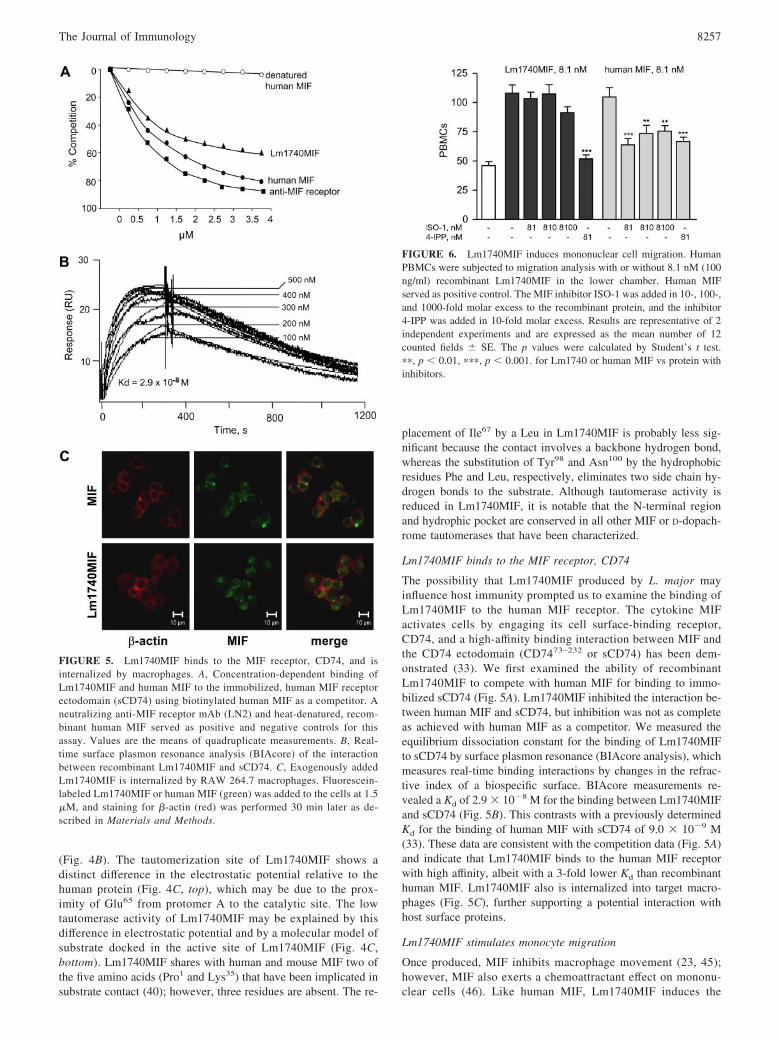
The binding of Lm1740MIF to the MIF cell surface receptor was studiedby coating individual wells of a 96-well plate with recombinant, solubleCD74 ectodomain (sCD7473–232) as described by Leng et al. (33). Plateswere washed 4 times with TTBS (pH 7.4) and blocked with superblockbuffer (Pierce) for two hrs at room temperature. Human MIF was biotin-ylated (Roche) and added at 2 �g/ml in triplicate wells with decreasingconcentrations of human MIF, heat-denatured human MIF, Lm1740MIF,or a blocking Ab directed against the MIF receptor ectodomain (cloneLN2) (33). Incubation was continued at room temperature for 2 h followedby washing with TTBS (pH 7.4). The bound, biotinylated hMIF was de-tected by adding streptavidin-conjugated alkaline phosphatase for 1 h, fol-lowed by washing and detection with p-nitrophenyl phosphate (Sigma-Aldrich). OD405 was measured using a kinetic microplate reader and valuesplotted as percent OD405 relative to wells containing biotinylated humanMIF alone. Each plot represents at least three independently performedassays, and each data point depicts a SEM � 10%.
The real-time binding interaction of Lm1740MIF with CD74 was mea-sured by surface plasmon resonance using a BIAcore 2000 optical biosen-sor (BIAcore) as previously described (33). The CM5 sensor chips and theBIA Evaluation software were from GE Healthcare. The MIF receptorectodomain (sCD7473–232) was immobilized according to manufacturer’sinstructions using the Biacore Amine Coupling Kit. Briefly, sCD7473–232
was diluted in 10 mM sodium acetate (pH 5.2) at 1 �mol. Fifty microlitersof a N-hyrdoxysuccinimide and N-ethyl-N-(dimethyaminopropyl)carbodi-imide mixture was injected at a speed of 2 �l/min for 25 min, followed byinjection of 50 �l of 1 �M purified sCD7473–232. Once the surface plasmonresonance reached 10,000 U, the injection was stopped, and the activeamine sites were blocked with 35 �l of 1 M ethanolamine (pH 8.5). Theimmobilized CM5 chip was washed overnight with 1� PBS at 20 �l/min.The derived sensor chips were washed and equilibrated in HEPES or PBS(pH 8.0; 20 �l/min), and the ligand (Lm1740MIF) was introduced at fiveserial dilutions in BIAcore buffer (1 mM DTT, 2.5 mM MgCl2, 20 mMHEPES, 1 mM EDTA, 150 mM NaCl, 0.005% P20) in 60- to 100-�linjection volumes at a flow rate of 20 �l/min. Binding was measured at25°C for 5 min, followed by 15 min of dissociation. Sensor chip regener-ation was performed for 1 min with 1 M NaCl, 50 mM NaOH. The wholeprocess was repeated three times for each dilution sample. Sensorgramresponse data were analyzed in the BIA evaluation kinetics package andthe equilibrium binding constants calculated.
Apoptosis studies
Transfection of Lm1740MIF and Lm1750MIF eukaryotic expression plas-mids into the murine RAW264.7 monocyte cell line was performed usinga Nucleofector (Amaxa) and the T-20 program described by the manufac-turer. Briefly, 2.5 � 106 cells were resuspended in 100 �l of Nucleofectorsolution together with 2 �g of plasmid DNA. After the pulse, 500 �l ofDMEM, 10% FBS was added, and the cells were seeded into 24-well platesfor apoptosis studies. Apoptosis was induced with the NO donor, sodiumnitroprusside (SNP). Transfection efficiencies were typically 30% (n � 3studies) and were measured by fluorescence microscopy at 24 h posttrans-fection with 2 �g of pmaxGFP (Amaxa). BMMs (4 � 106) were culturedin 20-mm plates, treated with murine MIF or Lm1740MIF, and incubatedovernight with and without SNP (Amersham Biosciences) (35). Apoptosiswas quantified by ELISA for cytoplasmic histone-associated DNA frag-ments (Roche). Cytoplasmic p53 content was analyzed by immunoblottingwith a pair of phospho-p53 (Ser15) and total p53 Abs (Cell Signaling Tech-nology) according to the manufacturer’s instructions. The secondary Abwas an anti-rabbit IgG Ab conjugated to HRP (Cell Signaling Technology),and detection was by chemiluminescence (GE Healthcare). The blots dis-played are representative of stimulation studies that were performed at leastthree times.
Signal transduction studies
Mouse BMMs and mouse thioglycolate-elicited peritoneal macrophages(4 � 106/plate) were rendered quiescent by incubation in 0.1% FBS beforestimulation with MIF for 2 h (36). Apoptosis was induced with SNP. Neg-ative controls were conducted by not stimulating cell death with SNP. Cellswere lysed in buffer containing 20 mM HEPES (pH 7.4), 50 mM �-glyc-erolphosphate, 2 mM EGTA, 1 mM DTT, 10 mM NaF, 1 mM NaVO4,
10% glycerol, 1% Triton X-100, and freshly added protease inhibitors(Complete, Mini, EDTA-free; Roche). For immunoblotting, cell lysateswere separated by 10% SDS-PAGE and transferred to PVDF Immobilon-Ptransfer membranes (Millipore). Immunoblotting was conducted with Absdirected against total ERK1/2 (Santa Cruz Biotechnology), and phospho-ERK-1/2 (Cell Signaling Technology) according to the manufacturer’s in-structions. The secondary Ab was an anti-rabbit IgG Ab conjugated to HRP(Cell Signaling Technology), and detection was by chemiluminescence(GE Healthcare). The blots displayed are representative of stimulationstudies that were performed at least three times.
ResultsSequence alignment and phylogenetic analyses
Two genetic loci, Lm1740 and Lm1750, were identified in the re-cently completed L. major genome (5) to have a potential openreading frame (342 bp) with 22% identity with human MIF. Bycomputational comparison with other Leishmania and Trypano-soma genes (37), both a consensus TATA-box and a polyadenyl-ation signal sequence were evident in the flanking regions of theseloci (Fig. 1A). The two putative L. major MIF orthologs(Lm1740MIF and Lm1750MIF) were predicted to encode a 112-aaprotein after excision of the initiating methionine residue. Similarto other MIF orthologs that have been described (7–9, 12, 13), theLeishmania MIF-like genes do not contain an N-terminal, secre-tory signal sequence.
Phylogenetic analysis using the Neighbor-Joining method forboth DNA and protein sequences produced similar results, ofwhich the latter is displayed in Fig. 1B. Gene duplication would bea likely origin for MIF paralogs within the same organism, as inthe case of the two L. major and Leishmania infantum MIF-relatedsequences. Lm1740MIF showed 58–99% sequence identity withMIF-like genes in other Leishmania species and 22–31% sequence
FIGURE 2. Lm1740MIF and Lm1750MIF mRNA are expressed by L.major. Real-time qPCR analysis of total RNA from L. major procyclic andmetacyclic forms cultured in vitro, and from amastigotes present within theinfected lymph nodes of mice. Experiments were conducted forLm1740MIF and Lm1750MIF transcripts relative to the internal controlmRNAs, rRNA45 (A) and ADP/ATP carrier (B). All samples were run intriplicates. The p values were calculated by Student’s t test ��, p � 0.01 forprocyclics vs metacyclics or amastigotes; �, p � 0.05 for procyclics vsmetacyclics or amastigotes.
8254 Leishmania MIF ORTHOLOGS

identity with mammalian MIFs. Except for some branch rearrange-ments, few differences were observed between the topology of theMIF DNA and protein phylograms (data not shown).
An amino acid sequence alignment of 12 selected members ofthe MIF protein family was prepared using the T-Coffee multiplesequence alignment program (Fig. 1C). Altogether, there are nineinvariant residues in MIF (Pro1, Leu21, Gly34, Lys35, Pro36, Phe52,Gly54, Gly68, and Phe116). Mammalian MIF is distinguished by thepresence of three conserved cysteines (Cys59, Cys62, and Cys83),the first two of which define a CXXC motif that mediates thiol-protein oxidoreductase activity (38). These conserved cysteines, aswell as the CXXC motif, are absent in the L. major sequences. TheMIF N-terminal proline (Pro1) by contrast appears strictly con-served, with the exception of the grossly truncated Leishmaniabraziliensis sequence. Proline-1 functions as a catalytic base inthe tautomerization of model substrates (whether a physiolog-ical substrate exists is unknown; see Ref. 38), and this residuehas been shown by x-ray crystallography studies of the mam-malian proteins to reside within a hydrophobic, substrate-bind-ing pocket (31, 39, 40).
Expression and cloning of L. major MIF
We confirmed the expression of the Lm1740MIF andLm1750MIF genes in vivo by RT-PCR analysis (data notshown) of total RNA prepared from L. major promastigotes(strain MHOM/IL/79/LRC-L251; Ref. 16). By qPCR, procyclicforms were observed to express more MIF than the metacyclicor amastigote forms, the latter of which were detected withinthe lymph nodes of infected mice (Fig. 2). The difference in theexpression levels of Lm1740MIF and Lm1750MIF was quan-tified with respect to two housekeeping genes, rRNA45 andADP-ATP carrier.
The Lm1740MIF and Lm1750MIF DNA sequences were sub-cloned into the eukaryotic expression vector, pcDNA3.1/V5-HisTOPO. Lm1740MIF DNA was additionally subcloned into theprokaryotic-expression vector pCRT7/CT-TOPO for recombinantprotein production. The coding region was engineered with a 3�stop codon to exclude the expression of the 3�-epitope tags presentin the plasmid vectors and to ensure the production of native se-quence protein. We considered native sequence to be important for
FIGURE 3. Biochemical characterization of theLm1740MIF protein. A, SDS/PAGE and Coomassiestaining analysis of recombinant Lm1740MIF proteinfractions eluted from size exclusion chromatography ona Superdex 75 10/300 GL column. STDs, protein mo-lecular mass standards. B, Electrospray ionization massspectrometry of Lm1740MIF showing a molecular mass(m/z) that lies within 0.02% accuracy of the predictedm/z (12381.22 Da). C, Tautomerization activity ofLm1740MIF and human MIF measured with the sub-strate, D-dopachrome methyl ester. A representative re-action is shown for Lm1740MIF at 1 �M and for humanMIF at 0.1 �M. The small-molecule inhibitors ISO-1 or4-IPP were preincubated with MIF at 1000- or 1-foldmolar excess, respectively. The lower tautomerase ac-tivity of Lm1740MIF made it necessary to use a higherconcentration of this protein for the inhibition studies.Data are means � SD of triplicate measurements. Thep values were calculated by Student’s t test. �, p �0.001 for human MIF vs Lm1740MIF, for 4-IPP-treated Lm1740MIF vs untreated Lm1740MIF, forISO-1- or 4-IPP-treated human MIF vs untreated hu-man MIF, and for 4-IPP-treated Lm1740MIF vs4-IPP-treated human MIF.
8255The Journal of Immunology

activity studies because covalent modification of the N-terminalproline inhibits the tautomerase activity of the protein and theC terminus mediates subunit oligomerization (26, 31). Recom-binant Lm1740MIF protein was purified by sequential fast pro-tein liquid chromatography following procedures adapted fromthe method of Bernhagen et al. (23) and yielded 15 mg of pro-tein per liter of E. coli culture. Final purification to homogene-ity was achieved by gel filtration chromatography (Fig. 3A).Electrospray ionization of purified Lm1740MIF protein gave anm/z of 12383.56, which is within 0.02% of the predicted massof the translated amino acid sequence (calculated molecularmass, 12381.22 Da; Fig. 3B).
Lm1740MIF activity studies
Mammalian MIF tautomerizes model substrates such asD-dopachrome or p-hydoxyphenylpyruvate, although the phys-iological relevance of the tautomerase activity of the proteincontinues to be debated (38). Site-directed mutagenesis andcrystallography studies of human MIF nevertheless have pro-vided significant insight into the structural determinants of thisreaction (31, 40 – 42). Pure, recombinant Lm1740MIF tau-tomerizes D-dopachrome methyl ester, but the specific activityfor the reaction is 13-fold lower than that measured for human
MIF (Lm1740MIF, 0.25 mM/min/�M; human MIF, 3.35 mM/min/�M; Fig. 3C). A low level of tautomerase activity forLm1740MIF also was observed with the substrate, p-hydoxy-phenylpyruvate (data not shown).
We next investigated whether Lm1740MIF is sensitive to hu-man MIF-catalytic site inhibitors using the tautomerase assay. Asshown in Fig. 3C, the competitive inhibitor ISO-1 (43) inhibitshuman MIF but not Lm1740MIF, suggesting a selective interac-tion of this compound with the human MIF tautomerization site.By contrast, 4-IPP, which forms a covalent modification of theMIF N-terminal proline (44) (M. Winner, J. Meier, S. Zierow,B. E. Rendon, G. Crichlow, R. Bucala, L. Leng, N. Smith, E. Lolis,J. O. Trent, and R. Mitchell, submitted for publication) inhibits thetautomerization activity of both MIF proteins with a greater inhib-itory effect on Lm1740MIF.
Three-dimensional crystal structure of Lm1740MIF
The x-ray crystal structure of Lm1740MIF was solved at 1.03 Åresolution. The structure consists of 1 protomer per asymmetricunit (Fig. 4A, top) that forms a trimer with two protomers fromadjacent asymmetrical units (bottom). The superposition of theC� backbone atoms of each monomer of human MIF andLm1740MIF results in a root mean square deviation of 1.8 Å
FIGURE 4. Conserved structurebetween Lm1740MIF and MIF. A,Schematic representation of theLm1740MIF and human MIF pro-tomers (top) and trimers (bottom)with secondary structure elementsshown in blue (� sheet), red (� helix)and cyan (random coil). B, Superim-position of the backbone ribbon dia-grams of monomeric and trimericLm1740MIF (red) and human MIF(green). In the trimeric representation,one of the protomers is displayed asbackbone trace. Also shown in the tri-mer representation are the side chainresidues implicated in substraterecognition. The substrate p-hy-droxyphenylpyruvate is displayed inyellow. C, Electrostatic surface poten-tial of the tautomerase site ofLm1740MIF and human MIF (top).The negatively charged surface poten-tial is displayed in red and the positivepotential in blue. Molecular model ofactive site residues implicated in sub-strate contact for Lm1740MIF basedon a crystal structure of MIF com-plexed with p-hydoxyphenylpyruvate(bottom; Ref. 40). Residue numberingrefers to sequence alignment of Fig.1C. The program Molscript (58) wasused to prepare A, PyMOL (59) wasused to prepare B and C (lower pan-els), and SPOCK (60) was used togenerate C (upper panels).
8256 Leishmania MIF ORTHOLOGS
(Fig. 4B). The tautomerization site of Lm1740MIF shows adistinct difference in the electrostatic potential relative to thehuman protein (Fig. 4C, top), which may be due to the prox-imity of Glu65 from protomer A to the catalytic site. The lowtautomerase activity of Lm1740MIF may be explained by thisdifference in electrostatic potential and by a molecular model ofsubstrate docked in the active site of Lm1740MIF (Fig. 4C,bottom). Lm1740MIF shares with human and mouse MIF two ofthe five amino acids (Pro1 and Lys35) that have been implicated insubstrate contact (40); however, three residues are absent. The re-
placement of Ile67 by a Leu in Lm1740MIF is probably less sig-nificant because the contact involves a backbone hydrogen bond,whereas the substitution of Tyr98 and Asn100 by the hydrophobicresidues Phe and Leu, respectively, eliminates two side chain hy-drogen bonds to the substrate. Although tautomerase activity isreduced in Lm1740MIF, it is notable that the N-terminal regionand hydrophic pocket are conserved in all other MIF or D-dopach-rome tautomerases that have been characterized.
Lm1740MIF binds to the MIF receptor, CD74
The possibility that Lm1740MIF produced by L. major mayinfluence host immunity prompted us to examine the binding ofLm1740MIF to the human MIF receptor. The cytokine MIFactivates cells by engaging its cell surface-binding receptor,CD74, and a high-affinity binding interaction between MIF andthe CD74 ectodomain (CD7473–232 or sCD74) has been dem-onstrated (33). We first examined the ability of recombinantLm1740MIF to compete with human MIF for binding to immo-bilized sCD74 (Fig. 5A). Lm1740MIF inhibited the interaction be-tween human MIF and sCD74, but inhibition was not as completeas achieved with human MIF as a competitor. We measured theequilibrium dissociation constant for the binding of Lm1740MIFto sCD74 by surface plasmon resonance (BIAcore analysis), whichmeasures real-time binding interactions by changes in the refrac-tive index of a biospecific surface. BIAcore measurements re-vealed a Kd of 2.9 � 10�8 M for the binding between Lm1740MIFand sCD74 (Fig. 5B). This contrasts with a previously determinedKd for the binding of human MIF with sCD74 of 9.0 � 10�9 M(33). These data are consistent with the competition data (Fig. 5A)and indicate that Lm1740MIF binds to the human MIF receptorwith high affinity, albeit with a 3-fold lower Kd than recombinanthuman MIF. Lm1740MIF also is internalized into target macro-phages (Fig. 5C), further supporting a potential interaction withhost surface proteins.
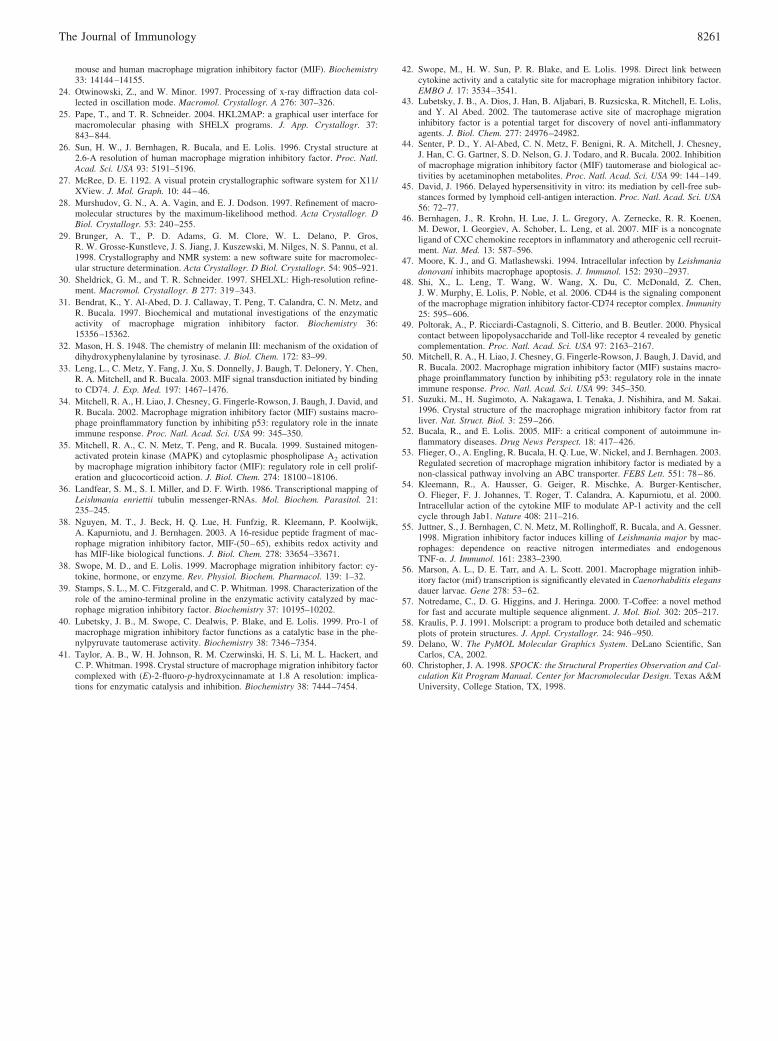
Lm1740MIF stimulates monocyte migration
Once produced, MIF inhibits macrophage movement (23, 45);however, MIF also exerts a chemoattractant effect on mononu-clear cells (46). Like human MIF, Lm1740MIF induces the
FIGURE 5. Lm1740MIF binds to the MIF receptor, CD74, and isinternalized by macrophages. A, Concentration-dependent binding ofLm1740MIF and human MIF to the immobilized, human MIF receptorectodomain (sCD74) using biotinylated human MIF as a competitor. Aneutralizing anti-MIF receptor mAb (LN2) and heat-denatured, recom-binant human MIF served as positive and negative controls for thisassay. Values are the means of quadruplicate measurements. B, Real-time surface plasmon resonance analysis (BIAcore) of the interactionbetween recombinant Lm1740MIF and sCD74. C, Exogenously addedLm1740MIF is internalized by RAW 264.7 macrophages. Fluorescein-labeled Lm1740MIF or human MIF (green) was added to the cells at 1.5�M, and staining for �-actin (red) was performed 30 min later as de-scribed in Materials and Methods.
FIGURE 6. Lm1740MIF induces mononuclear cell migration. HumanPBMCs were subjected to migration analysis with or without 8.1 nM (100ng/ml) recombinant Lm1740MIF in the lower chamber. Human MIFserved as positive control. The MIF inhibitor ISO-1 was added in 10-, 100-,and 1000-fold molar excess to the recombinant protein, and the inhibitor4-IPP was added in 10-fold molar excess. Results are representative of 2independent experiments and are expressed as the mean number of 12counted fields � SE. The p values were calculated by Student’s t test.��, p � 0.01, ���, p � 0.001. for Lm1740 or human MIF vs protein withinhibitors.
8257The Journal of Immunology

migration of peripheral blood monocytes across a Transwellmembrane (Fig. 6). The small-molecule MIF antagonist, ISO-1,binds to the human MIF tautomerization site (43) and inhibitsMIF-induced chemoattraction (8). As expected from the non-
inhibitory effect of ISO-1 on Lm1740MIF tautomerase function,ISO-1 failed to block mononuclear cell migration induced bythe latter protein, whereas the covalent inhibitor, 4-IPP, inhib-ited migration induced by MIF or Lm1740MIF.
FIGURE 7. LmMIF protects macrophages from apoptosis and activates signal transduction in a MIF receptor-dependent manner. A, MurineRAW264.7 monocytes were transfected with plasmids encoding Lm1740MIF, Lm1750MIF, murine MIF (MIF), or a control vector. The NO donor,SNP, was added 20 h later, and DNA fragmentation was assessed by ELISA. Data are the means � SD of five experiments. The p values werecalculated by Student’s t test. ��, p � 0.01, �, p � 0.05 for MIF vs vector control. ��, p � 0.01, �, p � 0.05 for Lm1740MIF vs MIF and forLm1750MIF vs MIF. ���, p � 0.001 for RAW264.7 vs vector control treated with SNP. The values for p � 0.05 are not displayed. B, Wild-type(WT) BMMs were treated with Lm1740MIF or murine MIF at the concentrations shown before the addition of the apoptosis inducer, SNP. DNAfragmentation was assessed by ELISA 36 h later. The p values were calculated by Student’s t test. ��, p � 0.01, �, p � 0.05 for MIF vs SNP control.��, p � 0.01, ��, p � 0.05 for Lm1740MIF vs MIF. ��, p � 0.01 for control vs SNP addition. The values for p � 0.05 are not displayed. Thephosphorylation of ERK1/2 and p53 was assessed by Western blotting using specific phospho-ERK1/2, total ERK1/2, phospho-p53, and total p53Abs. BMMs were treated with the NO donor SNP, as indicated. Western blotting for phosphorylation of ERK1/2 of C3H/HeJ bone marrowmacrophages served as a control against LPS contamination. C, BMMs from MIF receptor-deficient (CD74-KO) mice were prepared and studied forprotection from apoptosis, ERK1/2 phosphorylation, and phospho-p53 content as in B. The p values were calculated by Student’s t test. ���, p �0.001 for control vs SNP addition. The p values for comparisons with p � 0.05 are not displayed. Both the apoptosis studies and the Western blotsare representative of at least four independently performed experiments.
8258 Leishmania MIF ORTHOLOGS

Lm1740MIF activates ERK1/2 MAPK and inhibitsmonocyte/macrophage apoptosis
Leishmania-infected macrophages survive longer and are more viablethan uninfected macrophages (47). An important biological action ofthe cytokine MIF is to sustain monocyte/macrophage function byinhibiting activation-induced, p53-dependent apoptosis (34). Wehypothesized that Lm1740MIF may contribute to parasitism byprolonging the survival of the infected macrophage.
Transient transfection of an MIF-expressing plasmid intoRAW264.7 macrophages inhibits apoptosis by an autocrine/para-crine pathway involving the CD74 membrane receptor (34, 48).We tested the comparative ability of plasmids encoding murineMIF, Lm1740MIF, and Lm1750MIF to protect from NO-mediatedapoptosis in this model system. As shown in Fig. 7A, the twoLeishmania MIF orthologs inhibited apoptosis in a dose-dependentmanner, but at levels that were �30% lower than that of trans-fected murine MIF.
We next examined whether recombinant Lm1740MIF similarlyinhibits apoptosis in BMMs, both to confirm the biological activityof the protein and to assess its ability to influence the responses ofhost cells when present in the extracellular milieu. The effect ofLm1740MIF was less than that observed with murine MIF (Fig.7B), which is consistent both with the transfection data (Fig. 7A)and with the reduced receptor binding activity (Fig. 5, A and B).MIF signal transduction through CD74 is known to result in theactivation of the ERK1/2 MAPK pathway (35), and Lm1740MIFalso induced ERK1/2 phosphorylation in primary murine macro-phages (Fig. 7B). Because ERK1/2 activation in macrophages may
be induced by endotoxin (LPS), which is present in trace quantitiesin recombinant Lm1740MIF (�20 pg of LPS/�g of protein), weadditionally tested Lm1740MIF in macrophages from C3H/HeJmice, which have a loss of function mutation in the LPS receptor,TLR 4 (49). A dose-dependent increase in ERK1/2 phosphoryla-tion in response to Lm1740MIF was observed in these cells aswell, arguing against a role for contaminating endotoxin inLm1740MIF-induced ERK1/2 phosphorylation.
Downstream of ERK1/2 activation, the antiapoptotic action ofMIF is associated with a reduction in the intracytoplasmic contentof Ser15-phosphorylated p53, which increases in response to NOtreatment (35). Protection from apoptosis by Lm1740MIF also wasassociated with a diminution in the cellular content of Ser15-phos-phorylated p53 (Fig. 7B). Although differences in the relative abil-ity of Lm1740MIF vs murine MIF to effect changes in phosphor-ylation may be evident, quantitative comparisons are difficult tomake given the narrow linearity of signals that is apparent byWestern blotting. These results nevertheless indicate thatLm1740MIF is biologically active when added to mammalian cellsand, like mammalian MIF, stimulates ERK1/2 phosphorylationand reduces the cellular content of phospho-p53.
Finally, we examined the functional requirement for the MIFreceptor in Lm1740MIF action by studying the signaling and ap-optotic responses of macrophages obtained from mice deficient inthe MIF receptor, CD74. Murine MIF was not active in these cells,which is in agreement with prior reports (33, 48), and there was noeffect of Lm1740MIF with respect to protection from apoptosis,ERK1/2 phosphorylation, or intracellular phospho-p53 content(Fig. 7C). These data, taken together, support the conclusion thatLm1740MIF affects host cell responses, and in particular mono-cyte/macrophage survival, by engaging the MIF cell surfacereceptor.
DiscussionWe describe herein the characterization of L. major encoded or-thologs of the cytokine, MIF. The Lm1740MIF and Lm1750MIFgenes (58% identity) are expressed in significant levels in culturedpromastigotes. The two MIF-like sequences show 22% identitywhen aligned to human MIF and most likely arose by gene dupli-cation. Recombinant, native sequence Lm1740MIF protein wasproduced for structural and functional studies. An E. coli expres-sion system was used because of its high yield, the absence ofknown posttranslational modifications of MIF, and prior workshowing the utility of fast protein liquid chromatography for pre-paring pure MIF that is low in endotoxin content (23). We clonedLm1740MIF in a native sequence and not fusion protein formbecause of the known sensitivity of the N-terminal region to chem-ical modifications and evidence that the C terminus of the proteinforms contacts necessary for stable trimerization (26, 31).
The x-ray crystal structure of Lm1740MIF was solved to 1.03Å, which is the highest resolution of an MIF protein that has beenobtained to date. The overall global topology of Lm1740MIF issimilar to that of human and murine MIF (26, 51), but the catalyticsite shows distinctive features. There is a substantial difference inthe electrostatic potential within the catalytic site due to the pres-ence of Glu65, and the N-terminal tautomerase site contains threesignificant amino acid substitutions. These differences likely ex-plain the low tautomerase activity of Lm1740MIF when comparedwith human MIF, and they prompted us to investigate whethersmall-molecular MIF inhibitors also discriminate betweenLm1740MIF and human MIF. The MIF inhibitor ISO-1 did notaffect tautomerase activity or monocyte migration induced byLm1740MIF. By contrast, 4-IPP inhibited these activities in bothLm1740MIF and human MIF, with stronger inhibition observed in
FIGURE 7. (continued)
8259The Journal of Immunology

the case of Lm1740MIF. Small-molecule inhibitors of human MIFthat are designed to bind to the tautomerase site of the protein andinterfere with biological function are presently in preclinical de-velopment (52). Given the structural and functional differences inthe tautomerase sites of the human vs the Leishmania MIF pro-teins, it is possible that inhibitors may be designed to interfereselectively with Leishmania MIF.
Lm1740MIF bound with high affinity to the MIF receptorectodomain, as assessed both by an in vitro competition assayand by BIAcore analysis. Although the surface contacts be-tween MIF and the CD74 surface receptor remain unknown, theN-terminal region appears to play an important role in theseinteractions MIF (44). Nevertheless, there is sufficient structuralhomology between Lm1740MIF and human MIF to allow forhigh affinity binding to CD74. The interaction betweenLm1740MIF and CD74 led to an equal signal transductionresponse in macrophages obtained from WT and C3H/HeJmice, indicating that signaling was not due to the presence oftrace quantities of contaminating endotoxin. Lm1740MIF wasactive in three functional assays, monocyte migration, ERK1/2signaling, and protection from apoptosis, although the level ofactivity was generally lower than for mammalian MIF and ap-peared consistent with the lower Kd of Lm1740MIF for theCD74 receptor. As in the case of mouse MIF, protection fromapoptosis was strictly dependent on CD74, and the responsewas associated with a decrease in the cytoplasmic content ofSer15-phosphorylated p53.
The present results indicate that a Leishmania ortholog of thecytokine MIF has the ability to activate the human MIF receptorand influence the functional responses of monocytes/macrophages.Because Leishmania is an intracellular infection of the monocyte/macrophage, it may be hypothesized that one function of Leish-mania-encoded MIF is to sustain monocyte/macrophage survivaland contribute to the persistence of the parasite so that it maycomplete its infectious life cycle. The precise cellular pathway bywhich Lm1740MIF activates CD74 remains to be investigated; forinstance, the protein may be secreted from infected cells (53) andbind to cell surface CD74, or it may engage CD74 intracellularlyby gaining access to the endosomal compartment (54). An addi-tional question posed by these findings is whether there exist fur-ther, functional roles for Leishmania MIF with respect to the host-parasite interaction. MIF binding to CD74 has been shown recentlyto lead to the recruitment and activation of additional signalingproteins, including CD44 (48) and the chemokine receptorsCXCR2 and CXCR4 (47). Whether Lm1740MIF modulates addi-tional pathways important for intracellular parasitism or for im-mune evasion remains to be determined. It also is notable thatalthough host-derived MIF may augment the killing of L. major,its specific activity is low in comparison to cytokines such asIFN-� (55).
Although our data are consistent with a role for Leishmania MIFin modulating the host immune response, they do not exclude thepossibility of an intrinsic function for Lm1740MIF in the growthor replication of the parasite. We note that two mif-like genes havebeen described to be transcriptionally up-regulated in the dauerstage of the free-living nematode, Caenorhabditis elegans (56). Ithas been hypothesized that these genes may have a homeostaticrole during adverse conditions that cause developmental arrest(56). A physiological role for Leishmania MIF in the parasite lifecycle and a closer examination of whether these proteins functionas virulence factors may be attained by creating strains of Leish-mania lacking different MIF orthologs. Such studies also may pro-vide support for the selective, pharmacological targeting of Leish-mania MIF for therapeutic benefit.
AcknowledgmentsWe thank Ji Li for helpful scientific discussions and Cedric Notredameand Barry G. Hall for answering unhesitatingly our bioinformaticalquestions. The technical assistance of K. Goldsmith-Pestana is grate-fully acknowledged.
DisclosuresThe authors have no financial conflict of interest.
References1. Chang, K. P., and D. M. Dwyer. 1976. Multiplication of a human parasite (Leish-
mania donovani) in phagolysosomes of hamster macrophages in vitro. Science193: 678–680.
2. Olivier, M., D. J. Gregory, and G. Forget. 2005. Subversion mechanisms bywhich Leishmania parasites can escape the host immune response: a signalingpoint of view. Clin. Microbiol. Rev. 18: 293–305.
3. Bogdan, C., and M. Rollinghoff. 1998. The immune response to Leishmania:mechanisms of parasite control and evasion. Int. J. Parasitol. 28: 121–134.
4. Schartonkersten, T., and P. Scott. 1995. The role of the innate immune-responsein Th1 cell-development following Leishmania major infection. J. LeukocyteBiol. 57: 515–522.
5. Ivens, A. C., C. S. Peacock, E. A. Worthey, L. Murphy, G. Aggarwal,M. Berriman, E. Sisk, M. A. Rajandream, E. Adlem, R. Aert, et al. 2005. Thegenome of the kinetoplastid parasite, Leishmania major. Science 309: 436–442.
6. Calandra, T., and T. Roger. 2003. Macrophage migration inhibitory factor: aregulator of innate immunity. Nat. Rev. Immunol. 3: 791–800.
7. Pastrana, D. V., N. Raghavan, P. Fitzgerald, S. W. Eiseinger, C. Metz, R. Bucala,R. P. Scleimer, C. Bickel, and A. L. Scott. 1998. Filarial nematode parasitessecrete a homologue of the human cytokine macrophage migration inhibitoryfactor (MIF). Infect Immun. 66: 5955–5963.
8. Cho, Y. S., B. F. Jones, J. J. Vermeire, L. Leng, L. DiFedele, L. M. Harrison,H. B. Xiong, Y. K. A. Kwong, Y. Chen, R. Bucala, et al. 2007. Structural andfunctional characterization of a secreted hookworm macrophage migration inhib-itory factor (MIF) that interacts with the human MIF receptor CD74. J. Biol.Chem. 282: 23447–23456.
9. Jaworski, D. C., A. Jasinskas, C. N. Metz, R. Bucala, and A. G. Barbour. 2001.Identification and characterization of a homologue of the pro-inflammatory cy-tokine macrophage migration inhibitory factor in the tick, Amblyomma america-num. Insect Mol. Biol. 10: 323–331.
10. Miska, K. B., R. H. Fetterer, H. S. Lillehoj, M. C. Jenkins, P. C. Allen, andS. B. Harper. 2007. Characterisation of macrophage migration inhibitory factorfrom Eimeria species infectious to chickens. Mol. Biochem. Parasitol. 151:173–183.
11. Wu, Z., T. Boonmars, I. Nagano, T. Nakada, and Y. Takahashi. 2003. Molecularexpression and characterization of a homologue of host cytokine macrophagemigration inhibitory factor from Trichinella spp. J. Parasitol. 89: 507–515.
12. Cordery, D. V., U. Kishore, S. Kyes, M. J. Shafi, K. R. Watkins, T. N. Williams,K. Marsh, and B. C. Urban. 2007. Characterization of a Plasmodium falciparummacrophage-migration inhibitory factor homologue. J. Infect. Dis. 195: 905–912.
13. Augustijn, K. D., R. Kleemann, J. Thompson, T. Kooistra, C. E. Crawford,S. E. Reece, A. Pain, A. H. G. Siebum, C. J. Janse, and A. P. Waters. 2007.Functional characterization of the Plasmodium falciparum and P. berghei homo-logues of macrophage migration inhibitory factor. Infect. Immun. 75: 1116–1128.
14. Starlets, D., Y. Gore, I. Binsky, M. Haran, N. Harpaz, L. Shvidel,S. Becker-Herman, A. Berrebi, and I. Shachar. 2006. Cell-surface CD74 initiatesa signaling cascade leading to cell proliferation and survival. Blood 107:4807–4816.
15. Calandra, T., J. Bernhagen, R. A. Mitchell, and R. Bucala. 1994. The macrophageis an important and previously unrecognized source of macrophage migrationinhibitory factor. J. Exp. Med. 179: 1895–1902.
16. Beverley, S. M., R. B. Ismach, and D. M. Pratt. 1987. Evolution of the genusLeishmania as revealed by comparisons of nuclear-DNA restriction fragmentpatterns. Proc. Natl. Acad. Sci. USA 84: 484–488.
17. Saitou, N., and M. Nei. 1987. The neighbor-joining method: a new method forreconstructing phylogenetic trees. Mol. Biol. Evol. 4: 406–425.
18. Kumar, S., K. Tamura, and M. Nei. 2004. MEGA3: integrated software for mo-lecular evolutionary genetics analysis and sequence alignment. Brief Bioinform.5: 150–163.
19. Poirot, O., E. O’Toole, and C. Notredame. 2003. Tcoffee@igs: a web server forcomputing, evaluating, and combining multiple sequence alignments. NucleicAcids Res. 31: 3503–3506.
20. Leifso, K., G. Cohen-Freue, N. Dogra, A. Murray, and W. R. McMaster. 2007.Genomic and proteomic expression analysis of Leishmania promastigote andamastigote life stages: the Leishmania genome is constitutively expressed. Mol.Biochem. Parasitol. 152: 35–46.
21. Ouakad, M., N. Bahi-Jaber, M. Chenik, K. Dellagi, and H. Louzir. 2007. Selec-tion of endogenous reference genes for gene expression analysis in Leishmaniamajor developmental stages. Parasitol. Res. 101: 473–477.
22. Muller, P. Y., H. Janovjak, A. R. Miserez, and Z. Dobbie. 2002. Processing ofgene expression data generated by quantitative real-time RT-PCR. BioTechniques32: 1372–1379.
23. Bernhagen, J., R. A. Mitchell, T. Calandra, W. Voelter, A. Cerami, andR. Bucala. 1994. Purification, bioactivity, and secondary structure analysis of
8260 Leishmania MIF ORTHOLOGS
mouse and human macrophage migration inhibitory factor (MIF). Biochemistry33: 14144–14155.
24. Otwinowski, Z., and W. Minor. 1997. Processing of x-ray diffraction data col-lected in oscillation mode. Macromol. Crystallogr. A 276: 307–326.
25. Pape, T., and T. R. Schneider. 2004. HKL2MAP: a graphical user interface formacromolecular phasing with SHELX programs. J. App. Crystallogr. 37:843–844.
26. Sun, H. W., J. Bernhagen, R. Bucala, and E. Lolis. 1996. Crystal structure at2.6-A resolution of human macrophage migration inhibitory factor. Proc. Natl.Acad. Sci. USA 93: 5191–5196.
27. McRee, D. E. 1192. A visual protein crystallographic software system for X11/XView. J. Mol. Graph. 10: 44–46.
28. Murshudov, G. N., A. A. Vagin, and E. J. Dodson. 1997. Refinement of macro-molecular structures by the maximum-likelihood method. Acta Crystallogr. DBiol. Crystallogr. 53: 240–255.
29. Brunger, A. T., P. D. Adams, G. M. Clore, W. L. Delano, P. Gros,R. W. Grosse-Kunstleve, J. S. Jiang, J. Kuszewski, M. Nilges, N. S. Pannu, et al.1998. Crystallography and NMR system: a new software suite for macromolec-ular structure determination. Acta Crystallogr. D Biol. Crystallogr. 54: 905–921.
30. Sheldrick, G. M., and T. R. Schneider. 1997. SHELXL: High-resolution refine-ment. Macromol. Crystallogr. B 277: 319–343.
31. Bendrat, K., Y. Al-Abed, D. J. Callaway, T. Peng, T. Calandra, C. N. Metz, andR. Bucala. 1997. Biochemical and mutational investigations of the enzymaticactivity of macrophage migration inhibitory factor. Biochemistry 36:15356–15362.
32. Mason, H. S. 1948. The chemistry of melanin III: mechanism of the oxidation ofdihydroxyphenylalanine by tyrosinase. J. Biol. Chem. 172: 83–99.
33. Leng, L., C. Metz, Y. Fang, J. Xu, S. Donnelly, J. Baugh, T. Delonery, Y. Chen,R. A. Mitchell, and R. Bucala. 2003. MIF signal transduction initiated by bindingto CD74. J. Exp. Med. 197: 1467–1476.
34. Mitchell, R. A., H. Liao, J. Chesney, G. Fingerle-Rowson, J. Baugh, J. David, andR. Bucala. 2002. Macrophage migration inhibitory factor (MIF) sustains macro-phage proinflammatory function by inhibiting p53: regulatory role in the innateimmune response. Proc. Natl. Acad. Sci. USA 99: 345–350.
35. Mitchell, R. A., C. N. Metz, T. Peng, and R. Bucala. 1999. Sustained mitogen-activated protein kinase (MAPK) and cytoplasmic phospholipase A2 activationby macrophage migration inhibitory factor (MIF): regulatory role in cell prolif-eration and glucocorticoid action. J. Biol. Chem. 274: 18100–18106.
36. Landfear, S. M., S. I. Miller, and D. F. Wirth. 1986. Transcriptional mapping ofLeishmania enriettii tubulin messenger-RNAs. Mol. Biochem. Parasitol. 21:235–245.
38. Nguyen, M. T., J. Beck, H. Q. Lue, H. Funfzig, R. Kleemann, P. Koolwijk,A. Kapurniotu, and J. Bernhagen. 2003. A 16-residue peptide fragment of mac-rophage migration inhibitory factor, MIF-(50–65), exhibits redox activity andhas MIF-like biological functions. J. Biol. Chem. 278: 33654–33671.
38. Swope, M. D., and E. Lolis. 1999. Macrophage migration inhibitory factor: cy-tokine, hormone, or enzyme. Rev. Physiol. Biochem. Pharmacol. 139: 1–32.
39. Stamps, S. L., M. C. Fitzgerald, and C. P. Whitman. 1998. Characterization of therole of the amino-terminal proline in the enzymatic activity catalyzed by mac-rophage migration inhibitory factor. Biochemistry 37: 10195–10202.
40. Lubetsky, J. B., M. Swope, C. Dealwis, P. Blake, and E. Lolis. 1999. Pro-1 ofmacrophage migration inhibitory factor functions as a catalytic base in the phe-nylpyruvate tautomerase activity. Biochemistry 38: 7346–7354.
41. Taylor, A. B., W. H. Johnson, R. M. Czerwinski, H. S. Li, M. L. Hackert, andC. P. Whitman. 1998. Crystal structure of macrophage migration inhibitory factorcomplexed with (E)-2-fluoro-p-hydroxycinnamate at 1.8 A resolution: implica-tions for enzymatic catalysis and inhibition. Biochemistry 38: 7444–7454.
42. Swope, M., H. W. Sun, P. R. Blake, and E. Lolis. 1998. Direct link betweencytokine activity and a catalytic site for macrophage migration inhibitory factor.EMBO J. 17: 3534–3541.
43. Lubetsky, J. B., A. Dios, J. Han, B. Aljabari, B. Ruzsicska, R. Mitchell, E. Lolis,and Y. Al Abed. 2002. The tautomerase active site of macrophage migrationinhibitory factor is a potential target for discovery of novel anti-inflammatoryagents. J. Biol. Chem. 277: 24976–24982.
44. Senter, P. D., Y. Al-Abed, C. N. Metz, F. Benigni, R. A. Mitchell, J. Chesney,J. Han, C. G. Gartner, S. D. Nelson, G. J. Todaro, and R. Bucala. 2002. Inhibitionof macrophage migration inhibitory factor (MIF) tautomerase and biological ac-tivities by acetaminophen metabolites. Proc. Natl. Acad. Sci. USA 99: 144–149.
45. David, J. 1966. Delayed hypersensitivity in vitro: its mediation by cell-free sub-stances formed by lymphoid cell-antigen interaction. Proc. Natl. Acad. Sci. USA56: 72–77.
46. Bernhagen, J., R. Krohn, H. Lue, J. L. Gregory, A. Zernecke, R. R. Koenen,M. Dewor, I. Georgiev, A. Schober, L. Leng, et al. 2007. MIF is a noncognateligand of CXC chemokine receptors in inflammatory and atherogenic cell recruit-ment. Nat. Med. 13: 587–596.
47. Moore, K. J., and G. Matlashewski. 1994. Intracellular infection by Leishmaniadonovani inhibits macrophage apoptosis. J. Immunol. 152: 2930–2937.
48. Shi, X., L. Leng, T. Wang, W. Wang, X. Du, C. McDonald, Z. Chen,J. W. Murphy, E. Lolis, P. Noble, et al. 2006. CD44 is the signaling componentof the macrophage migration inhibitory factor-CD74 receptor complex. Immunity25: 595–606.
49. Poltorak, A., P. Ricciardi-Castagnoli, S. Citterio, and B. Beutler. 2000. Physicalcontact between lipopolysaccharide and Toll-like receptor 4 revealed by geneticcomplementation. Proc. Natl. Acad. Sci. USA 97: 2163–2167.
50. Mitchell, R. A., H. Liao, J. Chesney, G. Fingerle-Rowson, J. Baugh, J. David, andR. Bucala. 2002. Macrophage migration inhibitory factor (MIF) sustains macro-phage proinflammatory function by inhibiting p53: regulatory role in the innateimmune response. Proc. Natl. Acad. Sci. USA 99: 345–350.
51. Suzuki, M., H. Sugimoto, A. Nakagawa, I. Tenaka, J. Nishihira, and M. Sakai.1996. Crystal structure of the macrophage migration inhibitory factor from ratliver. Nat. Struct. Biol. 3: 259–266.
52. Bucala, R., and E. Lolis. 2005. MIF: a critical component of autoimmune in-flammatory diseases. Drug News Perspect. 18: 417–426.
53. Flieger, O., A. Engling, R. Bucala, H. Q. Lue, W. Nickel, and J. Bernhagen. 2003.Regulated secretion of macrophage migration inhibitory factor is mediated by anon-classical pathway involving an ABC transporter. FEBS Lett. 551: 78–86.
54. Kleemann, R., A. Hausser, G. Geiger, R. Mischke, A. Burger-Kentischer,O. Flieger, F. J. Johannes, T. Roger, T. Calandra, A. Kapurniotu, et al. 2000.Intracellular action of the cytokine MIF to modulate AP-1 activity and the cellcycle through Jab1. Nature 408: 211–216.
55. Juttner, S., J. Bernhagen, C. N. Metz, M. Rollinghoff, R. Bucala, and A. Gessner.1998. Migration inhibitory factor induces killing of Leishmania major by mac-rophages: dependence on reactive nitrogen intermediates and endogenousTNF-�. J. Immunol. 161: 2383–2390.
56. Marson, A. L., D. E. Tarr, and A. L. Scott. 2001. Macrophage migration inhib-itory factor (mif) transcription is significantly elevated in Caenorhabditis elegansdauer larvae. Gene 278: 53–62.
57. Notredame, C., D. G. Higgins, and J. Heringa. 2000. T-Coffee: a novel methodfor fast and accurate multiple sequence alignment. J. Mol. Biol. 302: 205–217.
58. Kraulis, P. J. 1991. Molscript: a program to produce both detailed and schematicplots of protein structures. J. Appl. Crystallogr. 24: 946–950.
59. Delano, W. The PyMOL Molecular Graphics System. DeLano Scientific, SanCarlos, CA, 2002.
60. Christopher, J. A. 1998. SPOCK: the Structural Properties Observation and Cal-culation Kit Program Manual. Center for Macromolecular Design. Texas A&MUniversity, College Station, TX, 1998.
8261The Journal of Immunology