a localized wnt signal orients asymmetric stem cell...
TRANSCRIPT

DOI: 10.1126/science.1231077, 1445 (2013);339 Science
et al.Shukry J. HabibVitroA Localized Wnt Signal Orients Asymmetric Stem Cell Division in
This copy is for your personal, non-commercial use only.
clicking here.colleagues, clients, or customers by , you can order high-quality copies for yourIf you wish to distribute this article to others
here.following the guidelines
can be obtained byPermission to republish or repurpose articles or portions of articles
): March 21, 2013 www.sciencemag.org (this information is current as of
The following resources related to this article are available online at
http://www.sciencemag.org/content/339/6126/1445.full.htmlversion of this article at:
including high-resolution figures, can be found in the onlineUpdated information and services,
http://www.sciencemag.org/content/suppl/2013/03/20/339.6126.1445.DC1.html can be found at: Supporting Online Material
http://www.sciencemag.org/content/339/6126/1445.full.html#relatedfound at:
can berelated to this article A list of selected additional articles on the Science Web sites
http://www.sciencemag.org/content/339/6126/1445.full.html#ref-list-1, 10 of which can be accessed free:cites 33 articlesThis article
http://www.sciencemag.org/content/339/6126/1445.full.html#related-urls1 articles hosted by HighWire Press; see:cited by This article has been
http://www.sciencemag.org/cgi/collection/developmentDevelopment
subject collections:This article appears in the following
registered trademark of AAAS. is aScience2013 by the American Association for the Advancement of Science; all rights reserved. The title
CopyrightAmerican Association for the Advancement of Science, 1200 New York Avenue NW, Washington, DC 20005. (print ISSN 0036-8075; online ISSN 1095-9203) is published weekly, except the last week in December, by theScience
on
Mar
ch 2
1, 2
013
ww
w.s
cien
cem
ag.o
rgD
ownl
oade
d fro
m

References and Notes1. P. Holland et al., Curr. Biol. 12, 1424 (2002).2. E. Kalay et al., Am. J. Hum. Genet. 90, 76 (2012).3. K. Mitchell et al., Am. J. Hum. Genet. 90, 69
(2012).4. Y. Zhang et al., Nat. Chem. Biol. 5, 217 (2009).5. H. Clevers, R. Nusse, Cell 149, 1192 (2012).6. A. P. McMahon, R. T. Moon, Cell 58, 1075 (1989).7. W. T. Montross, H. Ji, P. D. McCrea, J. Cell Sci. 113,
1759 (2000).8. R. McKendry, S. C. Hsu, R. M. Harland, R. Grosschedl,
Dev. Biol. 192, 420 (1997).
9. K. Satoh et al., Proc. Natl. Acad. Sci. U.S.A. 101, 8017(2004).
10. T. Schwarz-Romond, C. Merrifield, B. J. Nichols, M. Bienz,J. Cell Sci. 118, 5269 (2005).
11. P. Polakis, Genes Dev. 14, 1837 (2000).12. V. I. DeAlmeida et al., Cancer Res. 67, 5371 (2007).13. M. N. Kitaeva et al., Cancer Res. 57, 4478 (1997).
Acknowledgments: We thank K. O’Rourke, J. Huang, andthe next-generation sequencing and baculovirus groups fortechnical assistance. The transcriptome sequencing data forPA1 cells have been submitted to the National Center for
Biotechnology Information, NIH, Gene Expression Omnibus(www.ncbi.nlm.nih.gov/geo/) under accession no. GSE43362.
Supplementary Materialswww.sciencemag.org/cgi/content/full/science.1232253/DC1Materials and MethodsFigs. S1 to S12Table S1References (14–17)
1 November 2012; accepted 23 January 2013Published online 31 January 2013;10.1126/science.1232253
A Localized Wnt Signal OrientsAsymmetric Stem Cell Division in VitroShukry J. Habib,1,2* Bi-Chang Chen,2 Feng-Chiao Tsai,3 Konstantinos Anastassiadis,4
Tobias Meyer,3 Eric Betzig,2 Roel Nusse1*
Developmental signals such as Wnts are often presented to cells in an oriented manner. Toexamine the consequences of local Wnt signaling, we immobilized Wnt proteins on beads andintroduced them to embryonic stem cells in culture. At the single-cell level, the Wnt-bead inducedasymmetric distribution of Wnt–b-catenin signaling components, oriented the plane of mitoticdivision, and directed asymmetric inheritance of centrosomes. Before cytokinesis was completed,the Wnt-proximal daughter cell expressed high levels of nuclear b-catenin and pluripotencygenes, whereas the distal daughter cell acquired hallmarks of differentiation. We suggest thata spatially restricted Wnt signal induces an oriented cell division that generates distinct cellfates at predictable positions relative to the Wnt source.
Asymmetric cell division is a fundamentalprocess involved in many aspects of stemcell biology and cancer (1), but insight
into external cues that control asymmetric divisionsis limited (2). Although much has been learnedfrom examining asymmetric cell divisions in vivo(3–5), the complexity of tissues and the multipli-city of signals create challenges to understandinghow localized growth factors affect cell behaviorsat the single-cell level. Using in vitro studies, sin-gle cells and their divisions can be followed;however, growth factors that are added to thetissue medium present signals in a nonorientedway. To study how a given growth factor af-fects cell divisions or cell fates at the level ofthe individual cell, methods should be used topresent purified signaling molecules in an ori-ented way.
Previously we found that Wnt3a protein main-tains the self-renewal of several types of stemcells, including embryonic stem (ES) cells (6).Conversely, blocking endogenous Wnt signalsmade by ES cells leads to differentiation toward
Epiblast stem cells (EpiSCs) (6). ES cells aregrown in media supporting pluripotency thatinclude conditions that globally activate Wntsignaling (called 2i) (7, 8) and induce ES cellsto divide in mainly symmetrical patterns (9).Hence, ES cells could provide a useful exper-imental model for assessing how a local ratherthan a global Wnt signal might direct stem cellfate choices.
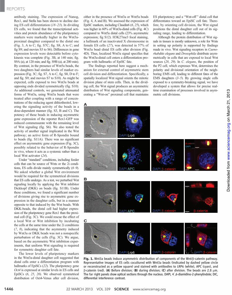
To examine the effect of a spatially localizedWnt signal on ES cells, we immobilized Wnt sig-nals to beads and observed single cells with liveimaging. Although Wnt3a maintains ES cell pluri-potency (6), the Wnt5a protein, which commonlyoperates through a non–b-catenin–dependent path-way (10), did not (figs. S1 and S8F), allowingus to use Wnt5a as a control. We chemically im-mobilized purified Wnt3a or Wnt5a proteins tobeads (figs. S2A and S3A) and confirmed theirbiological activity (figs. S2, B to D, and S3, Band C). ES cells were plated at low density inthe presence of leukemia inhibitory factor (LIF),and individual cells with a bead attached werefollowed by live cell microscopy as they divided.We examined the location of Wnt signaling com-ponents by antibody staining. In the presence ofWnt3a beads (Fig. 1A), but rarely with Wnt5abeads (fig. S4), the Wnt receptor LRP6 becameasymmetrically localized to the side of the EScell contacting the bead. Moreover, a Frizzled1-GFP (green fluorescent protein) fusion protein(fig. S5) and the adenomatous polyposis coli (APC)protein, a component of the b-catenin destruc-
tion complex, were detected in close proximityto the Wnt3a beads (Fig. 1A and fig. S6).
In ES cells contacting Wnt3a beads before di-vision, b-catenin was distributed asymmetricallyclose to the bead, overlapping with the locationof APC (Fig. 1A and fig. S6). During division,b-catenin was retained at high abundance in theprospective proximal daughter cell, both at the cellmembrane and in the nucleus (Fig. 1B). Theasymmetric distribution of these Wnt compo-nents was maintained after the cells divided: Thedaughter cell in proximity to the Wnt3a beadpreserved high amounts of LRP6 and APC, incontrast to the lower amounts in the distal cell(Fig. 1C and fig. S6).
Wnt pathway components can interact withastral microtubules and other components of themitotic spindle, including centrosomes (11, 12).We investigated the effect of Wnt beads on theasymmetric inheritance of the centrosomes byexpressing tagged Centrin1 (a component of thecentriole) and the appendage component Ninein(13, 14). Ninein marks the centrosome with theolder centriole (13–15), whereas the other cen-trosome receives new centrioles that initially lackthese structures. By the end of division, centro-somes in 78% of the cells (n = 18) that wereattached to Wnt3a beads had a high abundanceof Ninein (Fig. 2, A and B), whereas the segre-gation of Ninein was almost random in the pres-ence of the Wnt5a beads (54%; n = 15).Thus, theassociation with Wnt3a beads correlates with theasymmetric inheritance of centrosomes.
Because centrosomes orient the mitotic spin-dle, we investigated whether Wnt beads directthe orientation of cell division and partitioningof chromosomes during mitosis (16). ES cellsexpressing a histone 2B–Venus chimeric proteinto mark chromosomes (Fig. 2, C and D) wereincubated with Wnt beads and monitored duringmitosis by rapid three-dimensional imaging ofliving cells (17). In 75% of the dividing cells(n = 16), the axis of mitotic division was ori-ented in line with the Wnt3a bead (Fig. 2C andmovie S1), whereas only 12% of divisions wereoriented toward Wnt5a control beads (n = 12;Fig. 2D and movie S2).
We investigated the effect of localized Wntsignals on pluripotency gene expression, by usingvarious ES reporter cells including cells express-ing a Nanog-Venus fusion protein and GFP-basedreporters for Rex1, Sox2, and Stella (18–21).Pluripotency proteins were also followed by
1Department of Developmental Biology, Howard Hughes Med-ical Institute, Institute for Stem Cell Biology and RegenerativeMedicine, Stanford University, 265 Campus Drive, Stanford, CA94305, USA. 2Janelia Farm Research Campus, 19700 Helix Drive,Ashburn, VA 20147, USA. 3Department of Chemical and SystemsBiology, Stanford University, Stanford, CA 94305, USA. 4BIOTEC,Technische Universität Dresden Tatzberg 47-51, 01307 Dresden,Germany.
*Corresponding author. E-mail: [email protected] (R.N.);[email protected] (S.J.H.)
www.sciencemag.org SCIENCE VOL 339 22 MARCH 2013 1445
REPORTS
on
Mar
ch 2
1, 2
013
ww
w.s
cien
cem
ag.o
rgD
ownl
oade
d fro
m
antibody staining. The expression of Nanog,Rex1, and Stella has been shown to decline dur-ing ES cell differentiation (18–23). In dividingES cells, we found that the transcriptional acti-vities and protein abundance of the pluripotencymarkers were markedly higher in the Wnt3a-proximal daughter compared to the distal one(Fig. 3, A to C; fig. S7C; fig. S8, A to C; andfig S9; and movies S3 to S6). Differences in geneexpression levels were detectable before cyto-kinesis was complete (Fig. 3B, at 140 min; fig.S9A (a), at 120 min; and fig. S9B (a), at 200 min).By contrast, in the presence of Wnt5a beads, thetwo daughters had similar levels of marker ex-pression (Fig. 3C; fig. S7, A to C, fig. S8, D to F;and fig. S9; and movies S7 to S10). As might beexpected, cells exposed to two Wnt3a beads atopposing ends divided symmetrically (fig. S10).As additional controls, we generated attenuatedforms of Wnt3a, using Wnt3a beads that weretreated after coupling with a range of concen-trations of the reducing agent dithiothreitol, low-ering the signaling activity of the beads in adose-dependent manner (fig. S3, B and C). Thepotency of these beads in inducing asymmetricgene expression of the reporter Rex1-GFP wasreduced commensurate with the remaining levelof Wnt signaling (fig. S6). We also tested theactivity of another signal implicated in the Wntpathway; an active form of R-Spondin boundto beads (fig. S11A). There was no significanteffect on asymmetric gene expression (Fig. 3C),possibly related to the behavior of R-Spondinin vivo, where it acts as a systemic rather than alocal Wnt activator (24).
Under “standard” conditions, including feedercells that can be source of Wnts or the 2i condi-tions, ES cells divide mainly symmetrically (6–9).We asked whether a global Wnt environmentwould be required for the symmetrical divisionsthat ES cells undergo. As a test, we perturbed Wntsignaling locally by applying the Wnt inhibitorDickkopf (DKK) on beads (fig. S11B). Underthese conditions, we found a significant numberof divisions giving rise to asymmetric gene ex-pression in the daughter cells, but in a manneropposite to that induced by the Wnt beads. WithDKK-beads, the distal cell had higher expres-sion of the pluripotency gene Rex1 than the proxi-mal cell (Fig. 3C). We could rescue the effect ofa local Wnt or Wnt inhibition by incubatingthe cells at the same time under the 2i conditions(7, 8), indicating that the asymmetry inducedby Wnt3a or DKK beads was not a nonspecificperturbation of the cells (Fig. 3C). We argue,based on the asymmetric Wnt inhibition exper-iments, that uniform Wnt signaling is requiredfor symmetric daughter cell fate.
The lower levels of pluripotency markersin the Wnt3a-distal daughter cell suggested thatdistal cells enter a differentiation program withhallmarks of EpiSCs (22). The pluripotency geneOct4 is expressed at similar levels in ES cells andEpiSCs (6, 25, 26). We observed symmetricaldistribution of Oct4-Venus after cell division,
either in the presence of Wnt3a or Wnt5a beads(Fig. 4, A and B). We assessed the expression ofEpiSC markers, including Claudin6 (6, 25), whichwas higher in 60% of Wnt3a-distal cells (Fig. 4C)compared to Wnt5a distal cells (23% asymmetricexpression; fig S12). H3K27me3 focal staining,a hallmark of an inactivated X chromosome infemale ES cells (27), was detected in 57% ofWnt3a bead–distal ES cells after division (Fig.4D). Thus, localized Wnt3a signal specifies thatthe Wnt3a-distal cell enters a differentiation pro-gram with hallmarks of EpiSC fate.
The findings reported here suggest a mech-anism for external control of asymmetric stemcell division and differentiation. Specifically, aspatially localized Wnt signal orients the mitoticdivision plane of stem cells. Then, in the divid-ing cell, the Wnt signal produces an asymmetricdistribution of Wnt signaling components, gen-erating a “Wnt-on” proximal cell that maintains
ES pluripotency and a “Wnt-off ” distal cell thatdifferentiates toward an EpiSC cell fate. There-fore, by orienting cell division, the Wnt signalpositions the distal daughter cell out of its sig-naling range, leading to differentiation.
Although the protein distribution of Wnt sig-nals in tissues is mostly unknown, a role for Wntsin setting up polarity is supported by findingsmade in vivo. Wnt signaling receptors in Caeno-rhabditis elegans and Drosophila distribute asym-metrically in cells that are exposed to local Wntsources (28, 29). In C. elegans, the position ofthe P2 cell, which expresses Wnt, determines thepolarity and divisional orientation of the neigh-boring EMS cell, leading to different fates of theEMS daughters (3–5). By growing single cellsexposed to a symmetry-breaking signal, we havedeveloped a system that allows for precise real-time examination of processes involved in asym-metric cell divisions.
Fig. 1. Wnt3a beads induce asymmetric distribution of components of the Wnt/b-catenin pathway.Representative images of ES cells cocultured with Wnt3a beads (indicated by dashed yellow circleor reconstructed as a yellow square) and stained with antibodies to LRP6 (white), APC (cyan), andb-catenin (red). (A) Before division; (B) during division; (C) after division. The beads are 2.8 mm.The far right panels show optical sections through the nucleus. DAPI, 4´,6-diamidino-2-phenylindole; DIC,differential interference contrast.
22 MARCH 2013 VOL 339 SCIENCE www.sciencemag.org1446
REPORTS
on
Mar
ch 2
1, 2
013
ww
w.s
cien
cem
ag.o
rgD
ownl
oade
d fro
m

Fig. 2. Asymmetric inheritance of centrosomes andthe orientation of the plane of mitotic division. Time-lapse imaging of dividing single ES cells coculturedwith Wnt3a beads (indicated by a dashed yellowcircle) and (A) expressing enhanced green fluorescentprotein (EGFP)–Ninein (cyan; arrows) and DsRedex–Centrin1 (magenta). Dotted orange line indicates theboundary between two cells. (B) Immunostaining forendogenous Ninein (arrowheads). BF: bright-fieldimage. (C andD) Representative images from three-dimensional time-lapse microscopy of segregatingchromosomes in ES cells expressing H2B-Venus thatwere cocultured with Wnt3a beads (blue) or Wnt5abeads (red).
Fig. 3. Expression of the pluripotency genes Rex1 and Nanog during ES celldivision. (A) Selected frames from time-lapse imaging of a dividing Rex-1 GFPreporter ES cell in the presence of a Wnt3a bead (indicated by a dashed yellowcircle). The far right panel shows antibody staining for endogenous Rex-1 protein.(B) Selected frames from time-lapse imaging of a dividing Nanog-Venus reporterES cell in the presence of a Wnt3a bead. Signal intensities of all frames wereindividually determined, and the mean T SD intensity values plotted. Red trianglesrepresent signal intensities of the cell retaining contact with the bead after division.(C) Representative images of time-lapse microscopy of dividing Rex-1 GFP ES re-porter cells cocultured in the presence of the indicated beads in 2i or 2i-free media.Cell divisions were classified based on the relative expression of GFP and plotted.Red bar: higher GFP amounts in the bead-proximal cell; yellow bar: higher amountsof GFP in the bead-distal cell; blue bar: similar amounts of GFP in either cell.
www.sciencemag.org SCIENCE VOL 339 22 MARCH 2013 1447
REPORTS
on
Mar
ch 2
1, 2
013
ww
w.s
cien
cem
ag.o
rgD
ownl
oade
d fro
m

References and Notes1. R. A. Neumüller, J. A. Knoblich, Genes Dev. 23, 2675 (2009).2. A. D. Werts, B. Goldstein, Semin. Cell Dev. Biol. 22,
842 (2011).3. T. Walston et al., Dev. Cell 7, 831 (2004).4. B. Goldstein, H. Takeshita, K. Mizumoto, H. Sawa,
Dev. Cell 10, 391 (2006).5. K. Sugioka, K. Mizumoto, H. Sawa, Cell 146, 942 (2011).6. D. ten Berge et al., Nat. Cell Biol. 13, 1070 (2011).7. N. Sato, L. Meijer, L. Skaltsounis, P. Greengard,
A. H. Brivanlou, Nat. Med. 10, 55 (2004).8. Q.-L. Ying et al., Nature 453, 519 (2008).9. A. Surani, J. Tischler, Nature 487, 43 (2012).
10. A. J. Mikels, R. Nusse, PLoS Biol. 4, e115 (2006).11. S. Bahmanyar et al., Genes Dev. 22, 91 (2008).12. Y. M. Yamashita, D. L. Jones, M. T. Fuller, Science 301,
1547 (2003).13. M. M. Mogensen, A. Malik, M. Piel, V. Bouckson-Castaing,
M. Bornens, J. Cell Sci. 113, 3013 (2000).14. X. Wang et al., Nature 461, 947 (2009).15. M. Bornens, Science 335, 422 (2012).16. Y. M. Yamashita, M. T. Fuller, J. Cell Biol. 180, 261 (2008).17. T. A. Planchon et al., Nat. Methods 8, 417 (2011).18. B. Payer et al., Genesis 44, 75 (2006).19. Y. Toyooka, D. Shimosato, K. Murakami, K. Takahashi,
H. Niwa, Development 135, 909 (2008).20. K. Hayashi, S. M. Lopes, F. Tang, M. A. Surani,
Cell Stem Cell 3, 391 (2008).21. K. Arnold et al., Cell Stem Cell 9, 317 (2011).22. A. M. Singh, T. Hamazaki, K. E. Hankowski, N. Terada,
Stem Cells 25, 2534 (2007).23. I. Chambers et al., Nature 450, 1230 (2007).24. K. A. Kim et al., Science 309, 1256 (2005).25. P. J. Tesar et al., Nature 448, 196 (2007).26. G. Guo et al., Development 136, 1063 (2009).27. K. Plath et al., Science 300, 131 (2003).28. M. A. Hilliard, C. I. Bargmann, Dev. Cell 10, 379 (2006).29. C. Korkut et al., Cell 139, 393 (2009).
Acknowledgments: These studies were supported by theHoward Hughes Medical Institute and by grants RC1-00133-1,RB4-05825, and TR1-02149 from the California Institute forRegenerative Medicine to R.N.; by NIH grant GM063702to T.M.; and by the Center for Regenerative Therapies Dresdenand grant DFG-SPP1356 to K.A. We thank the Stanford
Neuroscience Microscopy Service, supported by NIH NS069375.Patent applications are pending for the Bessel beam planeillumination microscopy (by E.B.) and for the Wnt, DKK, andR spondin1 immobilization technology and their applications(by S.J.H. and R.N.). We also thank M. Drukker, T. Schroeder,and T. Stearns for discussions. We appreciate discussionsand comments on the manuscript by C. Logan and J. Nelson.S.J.H. was supported by a fellowship from the DeutscheForschungsgemeinschaft (DFG) and a Siebel scholarship.
Supplementary Materialswww.sciencemag.org/cgi/content/full/339/6126/1445/DC1Materials and MethodsFigs. S1 to S12Movies S1 to S10References (30–33)
3 October 2012; accepted 12 February 201310.1126/science.1231077
Type I Interferon Suppresses Type IIInterferon–Triggered HumanAnti-Mycobacterial ResponsesRosane M. B. Teles,1 Thomas G. Graeber,4 Stephan R. Krutzik,1 Dennis Montoya,1 Mirjam Schenk,1
Delphine J. Lee,5 Evangelia Komisopoulou,4 Kindra Kelly-Scumpia,1 Rene Chun,3 Shankar S. Iyer,2
Euzenir N. Sarno,6 Thomas H. Rea,7 Martin Hewison,3 John S. Adams,3 Stephen J. Popper,8
David A. Relman,8,9 Steffen Stenger,10 Barry R. Bloom,11 Genhong Cheng,2 Robert L. Modlin1,2*
Type I interferons (IFN-a and IFN-b) are important for protection against many viral infections,whereas type II interferon (IFN-g) is essential for host defense against some bacterial and parasiticpathogens. Study of IFN responses in human leprosy revealed an inverse correlation between IFN-band IFN-g gene expression programs. IFN-g and its downstream vitamin D–dependent antimicrobialgenes were preferentially expressed in self-healing tuberculoid lesions and mediated antimicrobialactivity against the pathogen Mycobacterium leprae in vitro. In contrast, IFN-b and its downstreamgenes, including interleukin-10 (IL-10), were induced in monocytes by M. leprae in vitro andpreferentially expressed in disseminated and progressive lepromatous lesions. The IFN-g–inducedmacrophage vitamin D–dependent antimicrobial peptide response was inhibited by IFN-b and byIL-10, suggesting that the differential production of IFNs contributes to protection versus pathogenesisin some human bacterial infections.
The identification of mechanisms of hostresistance versus susceptibility is central toour ability to develop new approaches to
prevent and/or treat human infectious diseases.In most instances, the human immune responserestricts the infection, preventing or limiting the
Fig. 4. Distal cells express markers of epiblast stem cell fate. (A) Selectedframes from time-lapse imaging of a dividing Oct4-Venus reporter ES cellcocultured with a Wnt3a bead (indicated by a dashed yellow circle). (B) Divisionsof Oct4-Venus ES cells cocultured with Wnt3a or Wnt5a beads were classifiedbased on the relative expression of Venus, and plotted. Red bar: higher Venusabundance in the bead-proximal cell; blue bar: similar abundance of Venus in
both cells. (C) Antibody staining for Claudin6 in an ES cell cocultured with aWnt3a bead. (D) Representative images of antibody staining for H3K27me3(arrows) in LF2 female ES cells cocultured with Wnt3a or Wnt5a beads. Dividingcells were classified based on location of the H3K27me3 stain, and plotted. Redbar: H3K27me3 stain in the bead-proximal cell; yellow bar: H3K27me3 stain inthe bead-distal cell; blue bar: no H3K27me3 stain in any cell.
22 MARCH 2013 VOL 339 SCIENCE www.sciencemag.org1448
REPORTS
on
Mar
ch 2
1, 2
013
ww
w.s
cien
cem
ag.o
rgD
ownl
oade
d fro
m