a mammalian protein kinase with potential for serine/threonine and
TRANSCRIPT

The EMBO Journal vol.10 no.2 pp.317 - 325, 1991
A mammalian protein kinase with potential forserine/threonine and tyrosine phosphorylation is relatedto cell cycle regulators
Yaacov Ben-David' *, Kenneth Letwin1'2 *Lisa Tannock1, Alan Bernstein '2 andTony Pawson'12Division of Molecular and Developmental Biology, Samuel LunenfeldResearch Institute, Mount Sinai Hospital, 600 University Avenue,Toronto, Ontario, Canada M5G IX5, and 2Department of Molecularand Medical Genetics, University of Toronto, Toronto, Canada*Equal contributions were made by the first two authors
Communicated by J.Schlessinger
In a screen of mouse erythroleukemia cDNA expressionlibraries with anti-phosphotyrosine antibodies, designedto isolate tyrosine kinase coding sequences, we identifiedseveral cDNAs encoding proteins identical or very similarto known protein-tyrosine kinases. However, twofrequently isolated cDNAs, clk and nek, encode proteinswhich are most closely related to protein kinases involvedin regulating progression through the cell cycle, andcontain motifs generally considered diagnostic of protein-serine/threonine kinases. The clk gene product containsa C-terminal cdc2-like kinase domain, most similar to theFUS3 catalytic domain. The Clk protein, expressed inbacteria, becomes efficiently phosphorylated in vitro ontyrosine as well as serine/threonine, and phosphorylatesthe exogenous substrate poly(glu, tyr) on tyrosine. Directbiochemical evidence indicates that both protein-tyrosineand protein-serine/threonine kinase activities are intrinsicto the Clk catalytic domain. These results suggest theexistence of a novel class of protein kinases, with anunusual substrate specificity, which may be involved incell cycle control.Key words: anti-phosphotyrosine antibodies/protein phos-phorylation
IntroductionProtein kinases regulate a wide array of cellular responsesto changing environmental conditions. In eukaryotes, alldescribed protein kinases are specific for phosphorylationof the hydroxyamino acids serine and threonine or tyrosine(Hunter and Cooper, 1985; Edelman et al., 1987; Hankset al., 1988), although it is likely that eukaryotic proteinkinases capable of phosphorylating histidine, arginine andlysine also exist (Wold, 1981). Phosphorylation is ofparticular significance in controlling mitogenesis andcellular differentiation. Receptors for a number of poly-peptide growth factors are transmembrane tyrosine kinases(Yarden and Ullrich, 1988), which in turn stimulateserine/threonine kinases such as protein kinase C, MAPkinase and p74raf (Hunter et al., 1984; Rossomondo et al.,1989; Morrison et al., 1989).The eukaryotic cell cycle is controlled by the p34cd(2
serine/threonine kinase, whose activity is modified throughthe cell cycle by phosphorylation and interactions withcyclins (reviewed in Lewin, 1990). p34cdc2 is enzymaticallyinactivated by phosphorylation on tyrosine, which therebyinhibits entry into mitosis (Gould and Nurse, 1989; Morlaet al., 1989). The protein kinases which phosphorylatep34cdc2 have not been biochemically characterized, althoughthe wee] gene, which negatively regulates cdc2, encodes apotential protein kinase with sequence motifs generallyconsidered diagnostic of protein-serine/threonine kinases(Russell and Nurse, 1987). Thus far no classical tyrosinekinases have been cloned from yeast.
Despite the evident interplay between serine/threonine- andtyrosine-specific protein kinases, previously identifiedeukaryotic protein kinases have shown a strict specificity foreither serine/threonine or tyrosine (see Weinmaster andPawson, 1986; Moran et al., 1988). A comparison of proteinkinase catalytic sequences has revealed two types ofconserved residue-amino acids found in all protein kinases,which probably are critical in folding or catalysis, and aminoacids conserved among tyrosine kinases but not serinekinases, and vice versa (Hanks et al., 1988). These latterare good candidates for residues involved in substratespecificity. For example the sequence HRDLXXXN isrelatively well conserved in all protein kinases (Hanks et al.,1988) and the invariant aspartate is essential for kinaseactivity (Moran et al., 1988; Tan et al., 1990). In protein-tyrosine kinases the variable (X) residues are AAR or RAA,whereas in protein serine/threonine kinases they are mostfrequently KPE, with the lysine being invariant. Similararguments can be made for the sequence immediately N-terminal of the conserved residues APE in the protein kinasesubdomain VIII (Hanks et al., 1988).cDNAs for many novel protein kinases have been cloned
using degenerate oligonucleotide probes, PCR techniques orhybridization to an existing cDNA at low stringency (Hankset al., 1988; Wilks, 1989; Hirai et al., 1987). We and othershave employed a functional screen for tyrosine kinasecDNAs in which affinity-purified anti-phosphotyrosine(P.Tyr) antibodies are used to probe a cDNA expressionlibrary (Kornbluth et al., 1988; Letwin et al., 1988; Lind-berg et al., 1988; Pasquale and Singer, 1989). A principalattraction of this approach is that it does not depend onrecognition by a tyrosine kinase-specific oligonucleotideprobe, and therefore should allow the cloning ofcDNAs thatdo not conform to the protein-tyrosine kinase consensussequence. Here we describe the isolation of cDNAs frommouse erythroleukemia cell cDNA expression libraries usinganti-P.Tyr antibodies. Two of the most frequently isolatedcDNAs encode proteins most closely related to the cdc2 andnimA cell cycle protein kinases, and contain motifs diagnosticof protein-serine/threonine kinases. However, we showbiochemically that the cdc2-like kinase catalytic domain canefficiently phosphorylate tyrosine as well as serine/threonine.
317(C Oxford University Press

Y.Ben-David et al.
Results
Isolation of protein-tyrosine kinase cDNAs from Friendmouse erythroleukemia cell expression libraries usinganti-P. Tyr antibodiesThe mRNAs from two Friend erythroleukemia cell lineswere reverse transcribed, and cDNA expression librarieswere constructed in Xgtl 1. Friend cells correspondphenotypically to immature hematopoietic cells of theerythroid lineage, and retain a capacity for erythroid differen-tiation after treatment with one of a number of chemicalinducers, such as DMSO (Friend et al., 1971). Protein-tyrosine kinases expressed in these cells might be involvedin regulating erythroid development or cellular proliferation.We therefore screened the unamplified Xgtl 1 libraries withanti-P. Tyr antibodies, a procedure which yielded a total of20 positive clones from 2 x 10⁶ phage examined. ThesecDNA clones, which based on antibody screening are likelyto encode protein-tyrosine kinases, were partially or
completely sequenced. In some instances the identity of a
specific clone was established by cross-hybridization withother isolates. The results of this analysis are summarizedin Table I.
Several clones were identified as being identical or closelyrelated to previously isolated protein-tyrosine kinases. Threeof the clones contained murine fer transcripts and one ofthese, clone F2, contained a full-length fer cDNA (Letwinet al., 1988; Hao et al., 1989). Clone C2 encoded a partiallyn message (Yamanishi et al., 1987). Clone QI containeda partial cDNA whose expected translation product is verysimilar to the recently identified rat elk PTK (Letwin et al.,1988). The relationship between the two sequences wouldsuggest that Ql is an elk-related gene rather than the murineelk equivalent (J.McGlade and V.Lhotak, unpublishedresults). The insert in clone E2 was apparently derived frommouseflg, which encodes a fibroblast growth factor-receptor(Ruta et al., 1988, 1989; Lee et al., 1989).
Identification of two novel protein kinases clk andnek, related to cdc2 and nimAThe remaining clones were categorized into three groups(Table I). The sequence of the entire 1661 nucleotide insertof the E3 cDNA was determined and found to contain a
single long open reading frame of 1519 nucleotides extend-ing from the extreme 5' end of the clone. Additional 3'sequences were derived from the overlapping RI cDNA(Figure 1). These cDNAs are probably derived from a 1.8 kbmRNA (see below), indicating that the composite sequenceis nearly full-length. The translation product initiating at the5'-most AUG codon would be 483 amino acids in lengthwith an expected molecular weight of 57 kd, although we
have not yet ruled out the possibility that the E3 cDNA lackssome 5' coding sequence. In vitro transcription and transla-tion of the E3 cDNA yielded a protein with an apparentmolecular weight of 55 kd in a rabbit reticulocyte lysate,in agreement with the size predicted from sequence analysis(data not shown). Analysis of the expected translation productof the E3 clone revealed the presence of all the characteristicprotein kinase catalytic domain sequence motifs. Surpris-ingly, the expected E3 protein contains those sequenceswhich would distinguish it as a serine/threonine-specificrather than a tyrosine-specific protein kinase. A search ofGenBank and EMBL databases confirmed E3 as a novelsequence, although it was most similar to members of the
318
Table I. Identities of clones isolated from mouse erythroleukemia cellXgtl 1 cDNA libraries by screening with anti-phosphotyrosineantibodies
Xgtll clones Identity
Fl,F2,H3 ferC2 lynQ I elk-relatedE2 flg (FGF-R)Al,A2,Bl,Hl,H4,Jl,J2,N2,N3,Pl nekCl,E3,RI clkS1 ?
Clones were identified by direct sequence analysis and, in some cases,by cross-hybridization. Since the library was not amplified, eachisolate represents a unique cDNA.
CDC28/cdc2+ family of protein kinases. Most of thekinases comprising this family have been identified in yeastand are thought to govern critical decision points in the yeastcell cycle. For this reason, we have designated the E3 cloneas clk (pronounced 'clik'), for CDC28/cdc2' -like kinase.The clk product is - 34% identical to FUS3 (Elion et al.,1990), 32% identical to YAKI (Garrett and Broach, 1989),30% identical to cdc2Hs (Lee and Nurse, 1987) and MAPkinase/ERKI (Boulton et al., 1990), 28% identical to CDC28(Lorincz and Reed, 1984), and 27% to KSS1 (Courchesneet al., 1989) over their kinase domains (Figure 2).Particularly striking is a short region of strong homologyshared by all the CDC28/cdc2+ family of kinases,corresponding to residues 451-476 of Clk, situated in theextreme carboxy terminus of the catalytic domain in a regionwhich is otherwise not well conserved among most otherprotein kinases.From the group comprising the most frequently isolated
cDNA, the entire nucleotide sequence of the N2 cDNA insertwas determined. The expected N2 translation productcontains all the sequences characteristic of protein kinasecatalytic domains (K.Letwin, Y.Ben-David and T.Pawson,manuscript in preparation). Like clk, N2 contains residueswhich are conserved among the serine/threonine-specificprotein kinases and lacks those residues generally associatedwith the tyrosine-specific protein kinases. A search of theGenBank and EMBL databases with the N2 sequencerevealed it to be a novel protein, although it showed thehighest similarity to known protein kinases. The greatestsimilarity was to the nimAl protein, a putative serine/threoninekinase involved in regulating the G2-M phase transition inthe fungus Aspergillus nidulans (Morris, 1976; Osmaniet al., 1988). Both proteins were - 30% identical overalland 43% identical across their catalytic domains. Based onits similarity to NimA, we have designated the N2 clone asnek for nimA-related kinase.The final clone, SI, does not fall into any of the previously
identified groups. Partial sequence analysis indicates that theS1 cDNA product contains a novel protein kinase catalyticdomain with several hallmarks of protein-serine/threoninekinases (L.Tannock, unpublished results). Hence all of thecDNA clones identified in the screen encode polypeptideswith sequences characteristic of protein kinase domains.
Protein kinase activity of a bacterially expressedTrpE - Cik fusion proteinThe repeated isolation of clk and nek cDNAs in a screenemploying anti-P.Tyr antibodies is paradoxical, since the

k r.2,ase aornalni
HH HHc
ATG
B
f 1 ., i _
; a 1 .- 6* I - u _ G i
*.t
I w
-. ..--.-
Fig. 1. Sequence and structure of the clk cDNA and its primary translation product. A. The 1.7 kb clk cDNA is shown with the presumed initiationand termination codons indicated. Restriction endonuclease sites are also shown (H, HindIlI; Hc, HincII; C, ClaI). The expected protein product isdepicted above the cDNA, with the catalytic domain and the positions of several highly conserved seqence motifs highlighted. B. Sequence of the clkcDNA and its primary translation product. Conserved residues diagnostic of a protein kinase catalytic domain are highlighted. The single letter aminoacid designation is used.
cDNAs encode proteins that would normally be classifiedas protein-serine/threonine kinases on the basis of theirprimary structures. There are two possible explanations forthese observations. The anti-P.Tyr antibodies mightrecognize epitopes created by the expression of clk or nekin bacteria which do not, in fact, contain P.Tyr. Alter-natively, clk and nek may possess tyrosine kinase activity.To confirm the protein kinase activity of Clk and deter-
mine its hydroxyamino acid specificity, a portion of the E3clone containing all but the extreme 5' end of the clk cDNAwas subcloned into a pATH bacterial expression vector in
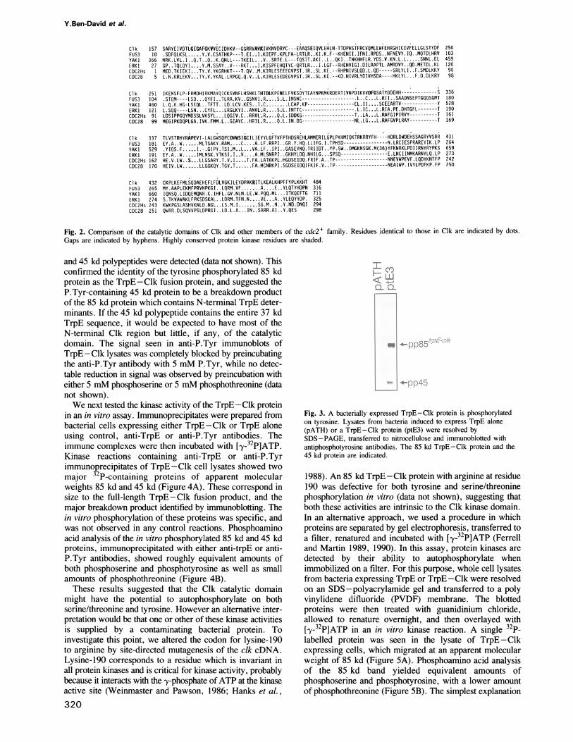
order to generate a TrpE-Clk fusion protein. This proteincontains Clk residues 79-483, including most of the N-terminal region and the entire presumptive catalytic domain.Immunoblots of lysates from cells expressing TrpE -Clkusing anti-P.Tyr antibody showed two major bands of-85 kd, corresponding to the expected size of theTrpE-Clk fusion protein, and 45 kd (Figure 3). No signalwas seen in lysates of cells expressing TrpE alone. Wheninduced bacterial lysates were immunoprecipitated with anti-P.Tyr antibodies, and the immunoprecipitates analyzed bywestern blotting with anti-TrpE antibodies, the same 85 kd
319
A
cdc-2-related mammalian protein kinase
11 I
--: l-.. .I
I N -
- ;ll ,-1 -1 - .1 -1 .-,- i
. -A 7
I. I : .
... -I -: - .. --. I - -
Y.Ben-David et al.
Clk 157 SARYEIVDTLGEGAFGKVVECIDHKV--GGRRVAVKIVKNVDRYC --- EAAQSEIQVLEHLN-TTDPHSTFRCVQMLEWFEHRGHICIVFELLGLSTYDF 250FUS3 10 .SDFQLKSL ...Y.V.CSATHKP---T,EI.E.I.KIEPF.KPLFA-LRTLR ..KI.K.F--KHENII.IFNI.RPDS ..NFNEVY.IQ ..MQTDLHRV 103YAKI 366 NRK.LVL.I .Q.T..Q..K.QNLL---TKEIL...9.V.SRTE.L---TQSIT.AKI ..L..QKI. .TNKHHFLR.YDS.V.KN.L.L ..... SNNL.EL 459ERKI 27 GP. .TQLQYI....Y.M.SSAY. .V---RKT ...I.KISPFEHQTYC-QRTLR.. .I.LGF--RHENVIGI.DILRAPTL.AMRDVY ..QD.METDL.KL 120CDC2Hs 1 MED.TKIEKI.. .TY.V.YKGRHKT ---T.QV. .M.KIRLESEEEGVPST.IR..SL.KE.--RHPNIVSLQD.L.QD ---- SRLYLI. .F.SMDLKKY 90CDC28 5 L.N.KRLEKV...TY.V.YKAL.LRPGQ.Q.V..L.KIRLESEDEGVPST.IR..SL.KE.--KD.NIVRLYDIVHSDA----HKLYL F.D.DLKRY 98
Clk 251FUS3 104YAK 1 460ERK1 121CDC2Hs 91CDC28 99
Clk 337FUS3 181YAKI 529ERK1 191CDC2Hs 162CDC28 170
Clk 432FUS3 265YAKI 660ERK1 274CDC2Hs 243CDC28 251
IKENSFLP-FRMDHIRKMAYQICKSVNFLHSNKLTHTDLKPENILFVKSDYTEAYNPKMKRDERTIVNPDIKVVDFGSATYDDEHH ------------- S 336.STQM----LSD ... QYFI. .TLRA.KV. .GSNVI.RS ..S.L.INSNC-.-L---------------- .LC... L.RII. .SAADNSEPTGQQSGMT 180L.Q.K.HG-LSIQL ..TFTT ..LD.LCV.KES ..I........ LCAP.KP-----------------EL.II....SCEEARTV-------------Y 528L.SQQ----LSN ... CYFL....LRGLKYI..ANVL.R....S.L.INTTC-------------------.L.IC...L.RIA.PE.DHTGFL-------T 190LDSIPPGQYMDSSLVKSYL ... LQGIV.C. RRVL . R.... Q.L.IDDKG -------------------T ..LA...L.RAFGIPIRVY ---------- T 161MEGIPKDQPLGA.IVK.FMM.L. GIAYC. HRIIL.R... .Q.L.IN.DG ------------------ NL.LG... L.RAFGVPLRAY --------- T 169
TLVSTRHYRAPEVI-LALGWSQPCDVWISIGCILIEYYLGFTVFPTHDSREHLAMMERILGPLPKHMIQKTRKRRYFH----HDRLDWDEHSSAGRYVSRR 431EY.A. .W. MLTSAKY.RAM...C....A.LF.RRPI ..GR.Y.HQ.LLIFG.I.TPHSD-N-.-LRCIESPRAREYIK.LP 264.YIQS.F..-...I.-.GIPY.TSI.M..L.. .VA.LF .. IPI. .GASEYNQ.TRIIDT ..YP.SW ..DMGKNSGK.M(36)YFKWRKLPDIIRNYRYPKS 659EY .A.W.. IMLNSK.YTKSI.I..V....A.MLSNRPI ..GKHYLDQ.NHILG ... SPSQ -----------------E.LNCIINMKARNYLQ.LP 273HE.V.LW. ....LLGSARY.T.V.I...T.FA.LATKKPL.HGDSEIDQ.FRIF.A. .TP --------------NNEVWPEVE.LQDYKNTFP 242HEIV.LW.....LLGGKQY.TGV.T. FA.MCNRKPI.SGDSEIDQIFKIF.V. .TP------------------ NEAIWP.IVYLPDFKP.FP 250
CKPLKEFMLSQDAEHEFLFDLVGKILEYDPAKRITLKEALKHPFFYPLKKHT 484MY.AAPLEKMFPRVNPKGI ..LQRM.VF.......A .. E. .YLQTYHDPN 316IQNSQ.LIDQEMQNR.C. IHFL.GV.NLN.LE.W.PQQ.ML. ITKQEFTG 711S.TKVAWAKLFPKSDSKAL LDRM.TFN.N....E.. .A. .YLEQYYDP. 325KWKPGSLASHVKNLD.NGL ..LS.M.I ....... SG.M. N. .Y.ND.DNQI 294QWRR.DLSQVVPSLDPRGI. LD.L.A. ..IN. .SARR.AI. .Y.QES 298
Fig. 2. Comparison of the catalytic domains of Clk and other members of the cdc2+ family. Residues identical to those in Clk are indicated by dots.Gaps are indicated by hyphens. Highly conserved protein kinase residues are shaded.
and 45 kd polypeptides were detected (data not shown). Thisconfirmed the identity of the tyrosine phosphorylated 85 kdprotein as the TrpE -Clk fusion protein, and suggested theP.Tyr-containing 45 kd protein to be a breakdown productof the 85 kd protein which contains N-terminal TrpE deter-minants. If the 45 kd polypeptide contains the entire 37 kdTrpE sequence, it would be expected to have most of theN-terminal Clk region but little, if any, of the catalyticdomain. The signal seen in anti-P.Tyr immunoblots ofTrpE -Clk lysates was completely blocked by preincubatingthe anti-P.Tyr antibody with 5 mM P.Tyr, while no detec-table reduction in signal was observed by preincubation witheither 5 mM phosphoserine or 5 mM phosphothreonine (datanot shown).We next tested the kinase activity of the TrpE-Clk protein
in an in vitro assay. Immunoprecipitates were prepared frombacterial cells expressing either TrpE-Clk or TrpE aloneusing control, anti-TrpE or anti-P.Tyr antibodies. Theimmune complexes were then incubated with [-y-32P]ATP.Kinase reactions containing anti-TrpE or anti-P.Tyrimmunoprecipitates of TrpE-Clk cell lysates showed twomajor 32P-containing proteins of apparent molecularweights 85 kd and 45 kd (Figure 4A). These correspond insize to the full-length TrpE-Clk fusion product, and themajor breakdown product identified by immunoblotting. Thein vitro phosphorylation of these proteins was specific, andwas not observed in any control reactions. Phosphoaminoacid analysis of the in vitro phosphorylated 85 kd and 45 kdproteins, immunoprecipitated with either anti-trpE or anti-P.Tyr antibodies, showed roughly equivalent amounts ofboth phosphoserine and phosphotyrosine as well as smallamounts of phosphothreonine (Figure 4B).These results suggested that the Clk catalytic domain
might have the potential to autophosphorylate on bothserine/threonine and tyrosine. However an alternative inter-pretation would be that one or other of these kinase activitiesis supplied by a contaminating bacterial protein. Toinvestigate this point, we altered the codon for lysine-190to arginine by site-directed mutagenesis of the clk cDNA.Lysine-190 corresponds to a residue which is invariant inall protein kinases and is critical for kinase activity, probablybecause it interacts with the -y-phosphate of ATP at the kinaseactive site (Weinmaster and Pawson, 1986; Hanks et al.,320
00.: LL,;s, Q-,
gq 4- 1)62
Fig. 3. A bacterially expressed TrpE-Clk protein is phosphorylatedon tyrosine. Lysates from bacteria induced to express TrpE alone(pATH) or a TrpE-Clk protein (ptE3) were resolved bySDS-PAGE, transferred to nitrocellulose and immunoblotted withantiphosphotyrosine antibodies. The 85 kd TrpE-Clk protein and the45 kd protein are indicated.
1988). An 85 kd TrpE -Clk protein with arginine at residue190 was defective for both tyrosine and serine/threoninephosphorylation in vitro (data not shown), suggesting thatboth these activities are intrinsic to the Clk kinase domain.In an alternative approach, we used a procedure in whichproteins are separated by gel electrophoresis, transferred toa filter, renatured and incubated with [-y-32P]ATP (Ferrelland Martin 1989, 1990). In this assay, protein kinases aredetected by their ability to autophosphorylate whenimmobilized on a filter. For this purpose, whole cell lysatesfrom bacteria expressing TrpE or TrpE- Clk were resolvedon an SDS-polyacrylamide gel and transferred to a polyvinylidene difluoride (PVDF) membrane. The blottedproteins were then treated with guanidinium chloride,allowed to renature overnight, and then overlayed with[_y-32P]ATP in an in vitro kinase reaction. A single 32P-labelled protein was seen in the lysate of TrpE-Clkexpressing cells, which migrated at an apparent molecularweight of 85 kd (Figure SA). Phosphoamino acid analysisof the 85 kd band yielded equivalent amounts ofphosphoserine and phosphotyrosine, with a lower amountof phosphothreonine (Figure SB). The simplest explanation

cdc-2-related mammalian protein kinase
I .o- CY)c
KJ4- LQ-.
orc c c cc
Bimmunoprecipitatianti -trpE
pS
pp85"-
r-0'':
-ppB5 -
Ps.'sp7"
4*pp45
Fig. 4. A. In vitro kinase activity of a bacterially expressed TrpE-Clk protein. Lysates from TrpE-Clk expressing cells (ptE3) or cells expressingTrpE alone (pATH) were immunoprecipitated with either control, anti-TrpE or anti-P.Tyr antibodies, using heat inactivated Saureus.Immunoprecipitates were incubated with [.y-32P]ATP and the reaction products separated by SDS-PAGE and exposed to X-ray film for 18 h. Themolecular weights (x l0-3) of protein markers are indicated. B. Phosphoamino acid analysis of the in vitro phosphorylated TrpE-Clk protein.TrpE-Clk proteins were immunoprecipitated with either anti-TrpE or anti-P.Tyr antibodies, and labeled in an in vitro kinase reaction. 3 P-labeled85 kd and 45 kd bands were isolated, hydrolyzed to free phosphoamino acids, resolved by 2-dimensional electrophoresis and exposed to X-ray filmfor 7 days using an intensifying screen. pS = phosphoserine; pT = phosphothreonine; pY = phosphotyrosine.
for these results is that the 85 kd TrpE -Clk protein is ableto phosphorylate all three hydroxyamino acids whenimmobilized on a filter. An alternative interpretation wouldbe that a co-migrating bacterial protein kinase is able totransphosphorylate specifically the TrpE-Clk protein,although this seems unlikely. The 45 kd protein identifiedby anti-P.Tyr and anti-TrpE immunoblots, and in theimmune complex kinase reaction, was not detected in thefilter kinase assay. Since this 45 kd polypeptide containsTrpE sequences it is unlikely to contain an intact kinasedomain, and would therefore not be expected toautophosphorylate. An implication of these results is that thephosphorylation of the 45 kd polypeptide in the immunecomplex kinase reaction may occur in trans, with the 45 kdprotein acting as a substrate for the active 85 kd kinase.Alternatively, the 45 kd fragment may arise by proteolysisof the 85 kd fragment after autophosphorylation.
If Clk is a protein-tyrosine kinase, it should be able tophosphorylate an exogenous substrate on tyrosine. Consis-tent with this possibility, TrpE-Clk phosphorylatedpoly(glu,tyr) in an in vitro kinase reaction (Figure 6),illustrating that Clk has protein-tyrosine kinase activitydespite its structural similarity to protein-serine/threoninekinases.
The cik gene is widely expressed and conserved inevolutionTo examine the expression of clk, we carried out a Northernblot analysis of RNAs isolated from a variety of malignantcell lines and different mouse tissues. Under stringenthybridization conditions, the clk cDNA probe identified twowidely expressed transcripts of 3.3 kb and 1.8 kb. In all thetissues that we analyzed, including spleen and thymus, the3.3 kb transcript was the predominant clk RNA species(Figure 7). In contrast, a variety of malignant lymphoid anderythroid cell lines all contained a major 1.8 kb clk RNAand a minor 3.3 kb species (data not shown). Poly(A)+RNA prepared from two mouse erythroleukemia cell lines,
A
.i
:: e
ii
;pS pT
pY
Fig. 5. TrpE-Clk phosphorylates on serine, theronine and tyrosine ina filter renaturation assay. A. Autophosphorylation of an immobilizedTrpE-Clk protein in a denaturation/renaturation kinase assay. Lysatesfrom 500 itl of TrpE-Clk expressing bacteria (ptE3) or TrpEexpressing bacteria (pATH) were resolved by SDS-PAGE andtransferred to a PVDF membrane. Blotted proteins were denatured ina guanidinium chloride solution and allowed to renature overnight at4°C. The blot was overlayed with kinase reaction buffer containing[-y-32P]ATP for 30 min, extensively washed and exposed to X-ray filmfor 2 h. B. Phosphoamino acid analysis of the autophosphorylated85 kd TrpE-Clk protein from a denaturation/renaturation kinaseassay. The PVDF membrane fragment containing theautophosphorylated protein was immersed in 6 N HCI. Phosphoaminoacids were subsequently resolved by 2-dimensional electrophoresisprior to autoradiography for 18 h.
TP3 and CB7, also contained predominantly the 1.8 kbtranscript (data not shown). Hence, there appears to bedifferential regulation of clk expression, with the 1.8 kbtranscript being favored in rapidly proliferating tumour cells.These two RNA species may be derived by differential
processing of a clk transcript, or might represent transcriptsfrom two distinct genes. Southern blot analysis of DBAmouse genomic DNA digested with the restriction enzymeBglII, using a clk cDNA probe, identified a single 17 kbband (data not shown). This suggests that clk is a single copygene, and that the 1.8 kb and 3.3 kb RNAs may be generatedby differential initiation, termination or splicing of the clktranscript. Since the 1.8 kb clk transcript is the predomi-nant species in the erythroleukemia cells from which the
321
A
205-
116-92.5-
67-
45-
ing r A
ant vl
pp45
?T.

Y.Ben-David et at.
from 26 RI mice revealed a 100% co-segregation patternbetween clk and the aldehyde oxidase (AOX) locus localizedon mouse chromosome 1 (data not shown). These resultsplace clk within 2 cM of the AOX locus, and, as no otherprotein kinase gene has been reported to map on this region,provide further evidence that it is a novel, single copy gene.
L.
-- i '1j.,J,zIL
Fig. 6. TrpE-Clk phosphorylates poly (glu, tyr) in vitro.Antiphosphotyrosine immunoprecipitates of TrpE-Clk (ptE3) or TrpEalone (pATH) were incubated in a kinase reaction buffer containing[FY_31P]ATP or [-y_32P]ATP plus poly(glu, tyr) 4:1 at a finalconcentration of 100 jig/ml. Reaction products were resolved bySDS-PAGE and exposed to X-ray film for 2 h. The molecularweights (X 10-3) of protein markers are indicated.
- -r. .. -...1.
= 1T.
p'; S
.g~~~~~~~~~~~~~~~~~~~~~~~~~~:
-I 83S
Fig. 7. Expression of clk transcripts in mouse tissues. Total cellularRNA (20 jig) from different mouse tissues was denaturated withformaldehyde, electrophoresed in an agarose gel, blotted ontonitrocellulose paper and hybridized with a clk cDNA probe. 28S RNAand 18S RNA have sizes of 4.4 kb and 2 kb respectively.
Xgtl 1 libraries were made, it seemed likely that the 1.7 kbclk cDNA insert corresponds to this smaller mRNA.Rescreening of the erythroleukemia cDNA library resultedin the repeated isolation of cDNAs of 1.6 kb or smaller withan identical restriction map to the E3 clk insert.
Since clk is related to genes found in fungi, we analyzedHindIll-digested genomic DNA from a variety by speciesof Southern blotting with a clk probe, using moderatelystringent hybridization conditions. The clk cDNA probedetected bands from the genomic DNAs of man, mouse, rat,chicken and Drosophila melanogaster (data not shown).These results suggest that the clk gene has been highlyconserved during evolution. The chromosomal location ofthe mouse clk locus was determined by following thedistribution of polymorphic alleles of the clk gene in recom-binant inbred (RI) mouse strains derived from matingsbetween C57BL/6J and DBA/25 mice. Analysis of DNA
DiscussionA mammalian protein kinase related to members ofthe cdc2 gene familyIn a screen of mouse erythroleukemia cell cDNA expres-sion libraries with anti-P.Tyr antibodies, the most frequentlyisolated classes of cDNA encode proteins related to thecdc2+ and nimA cell cycle gene products. We haveinvestigated the cdc2+ related kinase in more detail, in aneffort to understand its identification in a screen designedto identify tyrosine kinases.The cdc2+ family contains several yeast genes, including
cdc2 and its immediate Saccharomyces cerevisiae homologueCDC28;KSSJ, which promotes passage of S. cerevisiae fromGI to S phase of the cell cycle (Courchesne et al., 1989);FUS3, which is required for pheromone-induced GI arrestand for conjugation (Elion et al., 1990); and YAK], whichregulates cell growth, and is located downstream ofcAMP-dependent protein kinase on the Ras/cAMP pathway (Garrettand Broach, 1989). In Drosophila, the segment-polarity genezw3 encodes two distinct and differentially expressed proteinswhich differ in their amino-terminal regions, but share acommon C-terminal cdc2-like protein kinase domain(Siegfried et al., 1990). In mammalian cells, in addition tocdc2 itself, the mitogen activated protein (MAP) kinase(ERKl) has recently been shown to contain a cdc2-like kinasedomain most similar to KSSJ and FUS3 (Boulton et al.,1990). MAP kinase is implicated in signal transduction, sinceit is activated by a variety of mitogenic stimuli (Rossomondoet al., 1988) and is capable of re-activating dephosphorylatedS6 kinase-Il (Sturgill et al., 1988). MAP kinase itself isinactive unless phosphorylated on both tyrosine and threonine(Anderson et al., 1990). In the sense that MAP kinaserequires phosphorylation on both tyrosine and threonine forregulation of its kinase activity, it is similar to p34cdc2 ofhigher eukaryotes. Within its kinase domain, the clk geneproduct (Clk) is most closely related to FUS3. There is asequence of 26 amino acids which is highly conserved amongcdc2 family members and quite distinct from the analogoussequences of most other protein kinases. As yet, we haveno data as to the function of mammalian clk. However, theobservation that clk is related to the cdc2+ gene familyraises the possibility that it is involved in regulating signaltransduction or the cell cycle.
CIk is associated with both protein-serine/threonineand protein-tyrosine kinase activityThe most startling feature of the clk and nek cDNAs is thattheir products have all the sequence attributes of protein-serine/threonine kinases, despite the fact that they wereisolated using anti-P.Tyr antibodies. The repeated isolationof clk and nek indicates that they have some unusualproperty, since the Friend cell libraries presumably containa wealth of bonafide protein-serine/threonine kinase cDNAs.The Clk protein becomes phosphorylated on both tyrosineand serine/threonine in vitro. Since the phosphate in
322
< z::
< -
-:I
r.-. r,
f.,-) r-I
cdc-2-related mammalian protein kinase
these reactions is derived from ATP labeled in the -y posi-tion, the isolation of phosphotyrosine through some spuriousmechanism other than direct protein phosphorylation isunlikely. A critical issue is whether the phosphorylation ofTrpE-Clk on serine, threonine and tyrosine represents anintrinsic capacity of the Clk kinase domain to phosphorylateall three hydroxyamino acids, at least in an autophosphoryla-tion reaction. Strong evidence in support of this notion isprovided by the blot denaturation/renaturation kinase assay.In this experiment, the 85 kd TrpE-Clk protein becamephosphorylated on tyrosine and serine/threonine after separa-tion on a denaturing SDS -polyacrylamide gel and renatura-tion on a filter. It is difficult to explain this observation inany way other than to suggest that the TrpE-Clk proteinhas an intrinsic capacity to autophosphorylate on tyrosineas well as serine/threonine.The in vitro tyrosine and serine/threonine phosphoryla-
tion of the 45 kd TrpE-Clk fragment, which itselfapparently lacks an intact catalytic domain, suggests that Clkmight be able to transfer phosphate to tyrosine andserine/threonine residues in trans. It is of interest that almostone third (47/148) of the residues in the unique N-terminalregion of Clk are hydroxyamino acids. There is also aconcentration of basic residues in this region (32/148).TrpE-Clk was also able to phosphorylate a randompoly(glu,tyr) co-polymer, added as an exogenous substrateto an in vitro kinase reaction, confirming that it has protein-tyrosine kinase activity.
Protein kinase specificityThe Clk protein kinase domain contains the sequenceelements considered diagnostic of protein serine/threoninekinases, in agreement with the finding that Clk does indeedphosphorylate serine and threonine. However, Clk alsoappears to possess intrinsic protein-tyrosine kinase activity.There are no obvious clues in the protein sequence to explainthis observation. Within the motif HTDLKPEN, Clk has athreonine where most other kinases have arginine.Interestingly, the predicted SI cDNA product has a serineat this site (L.Tannock, unpublished results). In addition,Clk has an unusual 19 amino acid insert, relative to otherprotein kinases, in between the HTDLKPEN sequence andthe highly conserved DFG motif (Hanks et al., 1988; Moranet al., 1988). The observation that all protein kinases testedto date show a mutually exclusive preference for eitherserine/threonine or tyrosine, as well as the identification ofsequence motifs diagnostic of each subclass, has suggestedan intrinsic difference between the tyrosine- andserine/threonine-specific protein kinases. However, theevidence presented above suggests that Clk may be able tophosphorylate all three hydroxyamino acids. Other proteinkinases with the sequence motifs of protein-serine/threoninekinases may possess similar properties; a protein kinase hasrecently been identified in S. cerevisiae which isphosphorylated on tyrosine in vivo, and is associated withboth protein-serine and protein-tyrosine kinase activity invitro (Dailey et al., 1990). The nek product also has theappearance of a protein-serine/threonine kinase, and has noobvious unique sequence element in common with Clk.Experiments are in progress to determine whether the nekprotein can phosphorylate tyrosine.Two Dictyostelium discoideum protein kinases (DPYK 1
and 2) have recently been described whose catalytic domains
phosphorylate only tyrosine when expressed in bacteria (Tanand Spudich, 1990). In screening a 15 h developmentalD.discoideum expression library with anti-P.Tyr antibodieswe have also isolated a cDNA identical to DPYK2(K.Letwin, M.Aberman, G.Weeks and T.Pawson,unpublished results). The sequences of DPYK 1 and 2 canbe considered as structural mosaics of protein-serine/threonine and protein-tyrosine kinase motifs, and yetspecifically phosphorylate tyrosine in vitro. Taken with theproperties of clk, these results suggest that we have yet tounderstand fully the rules governing the substrate specificitiesof protein kinases.
Possible functions of protein-serine/threonine/tyrosinekinasesThe apparent ability of Clk to phosphorylate tyrosine maynot be maintained in vivo in mammalian cells, or may bea tolerated aberration of no functional consequence.Assuming, however, that such multi-functional kinases areof biological significance, it is of interest to consider whatrole they might play. Both clk and nek are most closelyrelated to a network of protein kinases that regulate the cellcycle. Some of these protein kinases may have developeda mechanism by which they are distinguished from theclassical protein-tyrosine and protein-serine/threoninekinases, p34CCk2 of higher eukaryotes must be phosphorylatedon both tyrosine and threonine to be held in an inactive state,and thereby allow correct timing of mitosis; conversely, thecdc2-like MAP kinase must be phosphorylated on bothtyrosine and threonine to be activated. One interpretationof these data is that at least two individual kinases of differentspecificities are required either for the inhibition of p34cdc2or the activation of MAP kinase. An alternative possibilityis that these protein kinases are themselves controlled byprotein kinases with the ability to phosphorylate serine/threonine as well as tyrosine. Such protein-serine/threonine/tyrosine kinases would therefore represent a new class ofprotein kinase, and would presumably have very specificsubstrates. We are presently analyzing the clk and nekproteins expressed in mammalian cells to determine whetherthey possess such attributes.
Materials and methodsAntibodiesTo isolate anti-phosphotyrosine antibodies, rabbit polyclonal antiserum againsta polymer of phosphotyrosine, alanine, glycine and keyhole limpet hemo-cyanin was raised and affinity purified as originally described by Kampsand Sefton (1988). Rabbit anti-TrpE antiserum was raised to the 37 kdproduct of the parental pATH expression plasmid.
Construction of cDNA libraries and isolation of cDNA clonesTwo Xgt 11 cDNA expression libraries were constructed from 5 Ugpoly(A) + RNA isolated from the erythroleukemia cell lines CB7 (Shibuyaand Mak, 1982) and DP28-9 (Ben-David et al., 1988), using a PharmaciacDNA synthesis kit. Screening of Xgtl 1 expression libraries using anti-phosphotyrosine antibody was carried out as described previously (Letwinet al., 1988).
Subcloning and nucleotide sequence determinationDNA was prepared from plaque purified phage according to standardprocedures using lambdasorb phage adsorbent and subcloned intopGEM-5Zf( +) or pGEM-7Zf( +). Clones were grouped together based on
partial DNA sequence analysis and cross-hybridization. For completesequence determination, nested deletions spanning the cDNA insert of thedesired clone were generated using the method of Henikoff (1987). Theseconstructs were rescued as ssDNA following superinfection of bacteria with
323

Y.Ben-David et al.
the coliphage M 13K07 and sequenced by the dideoxynucleotide chaintermination method using sequenase enzyme, reagents and protocols suppliedby United States Biochemicals Corporation. Sequences were obtained forboth strands.
Construction of the trpE - clk bacterial expression vectorThe plasmid pE3-2 was constructed by subcloning the 1.7 kb EcoRl cDNAinsert of XE3 into the EcoRI site of pGEM-7Zf(-). The expression vectorptE3 was constructed by subcloning the 1.4 kb HincII-HindIII fragmentof pE3-2 into the Sall (blunted with Klenow)-HindlI sites of pATH-2.This construct produced an 85 kd TrpE-Clk fusion protein composed ofthe first 330 amino acids of the TrpE protein and the C-terminal 414 aminoacids of the Clk protein.
Immunoblotting bacterial lysatesEscherichia coli DHScs harboring the expression plasmid ptE3 or a controlplasmid pATH-2 were grown and induced as described previously (Moranet al., 1988). Following a 3 h induction period, a 1 ml culture was pelleted,and the cells were lysed in 50 1l cracking buffer [ 10 mM sodium phosphate,pH 7.2; 1% (v/v) 3-mercaptoethanol, 1% (w/v) SDS, 6 M urea] andvortexed at room temperature for 30 min. The lysate was diluted with anequal volume of 2 x SDS sample buffer and S 1l aliquots were resolvedon 7.5% SDS - polyacrylamide gels and transferred to nitrocellulose.
Blotted proteins were blocked in TBST [10 mM Tris-HCI, pH 8.0;150 mM NaCI; 0.05% (v/v) Tween-20] containing 5% (w/v) bovine serumalbumin overnight. Blots were then incubated with anti-P.Tyr or anti-TrpEantibodies in TBST for 1 h, washed with TBST, incubated with a goat anti-rabbit IgG alkaline phosphatase conjugate (Sigma), washed with TBST andimmersed in alkaline phosphatase buffer containing the chromogenicsubstrates NBT and BCIP.
In vitro kinase reactionFollowing a 3 h induction period, cells from 5 ml cultures of E. coli DHSaharboring the expression vector ptE3 or the control plasmid pATH-2 werepelleted, resuspended in 1 ml TBS and lysed by sonication. Lysates wereclarified by centrifugation at 13 000 g for 30 min. Proteins were isolatedby immunoprecipitation with either a polyclonal anti-TrpE antiserum oraffinity-purified anti-P.Tyr antibodies, and heat-inactivated Staphylococcusaureus. Immunoprecipitates were washed once with KLB and twice withkinase reaction buffer (50 mM HEPES, pH 7.0; 10 mM MnCl2; 10 mMMgCl2), resuspended in 25 /d kinase reaction buffer containing 5 /ACi[y-32P]ATP (20 Ci/mmol) and incubated at room temperature for 15 min.Reactions were terminated by addition of an equal volume of 2 x SDSsample buffer and the immunoprecipitates were released by incubation at30°C for 15 min. The kinase reaction supernatant was then resolved bySDS-PAGE, transferred to PVDF membranes (Immobiolon; MilliporeCorporation) and 32P-labeled proteins visualized by autoradiography. Invitro kinase reactions using an exogenous substrate were conducted essen-tially as described above except that poly(glu, tyr) 4:1 (Sigma) was includedin the reaction buffer at a concentration of 100 /tg/ml (Braun et al., 1984).
Phosphoamino acid analysisPertinent regions of the PVDF membrane were excised and the 32P-labeledprotein hydrolyzed in 6 M HCI for 60 min at 1 10°C (Kamps and Sefton,1989). Supernatants were lyophilized, mixed with non-radioactivephosphoamino acid standards, and analyzed by 2-dimensional electrophoresis(pH 1.9 followed by pH 3.5) on thin-layer cellulose plates.
Kinase renaturation assayThe denaturation-renaturation protocol was carried out essentially asdescribed by Ferrell and Martin (1989, 1990). Whole cell lysates from 50to 500 Al induced bacteria were resolved by SDS-PAGE and transferredto PVDF membranes. Blotted proteins were immersed in denaturation solu-tion (6 M guanidinium chloride, 50 mM Tris-HCI, pH 8.3, 50 mM DTT,2 mM EDTA) for 60 min at 4°C, washed briefly with renaturation buffer(20 mM Tris-HCI, pH 7.5; 150 mM NaCI; 2 mM DTT; 2 mM EDTA;0.1% Nonidet P-40, 2% glycerol) and immersed overnight at 4°C inrenaturation buffer. Blots were blocked by immersing in renaturation buffercontaining 0.2% polyvinylpyrrolidone, 0.2% Ficoll and 20 mM sodiumpyrophosphate for 60 min at room temperature, overlaid with reaction buffer(30 mM Tris-HCI, pH 7.5; 10 mM MgCI2; 10 mM MnCl2; [-y-32P]ATP[Amersham; 3000 Ci/mmol, 50 tCi/ml, 1 ml per 10 cm2 blot area]) for30 min, and then washed extensively with TBS prior to autoradiography.
RNA extraction and Northern blottingTotal cellular RNA from different mouse tissues was isolated by the lithiumchloride precipitation procedure (Auffray and Rougeon, 1980). The isolated
RNA was extracted three times with water saturated phenol/chloroform (1: 1)and once with chloroform. The final aqueous phase was precipitated withethanol and stored in 100% ethanol at -70°C. Total RNA (20 1tg) wasfractionated using formaldehyde denaturing gel electrophoresis and thenblotted onto nitrocellulose paper. The filter was hybridized with a randomprimed clk cDNA probe corresponding to the full length E3 clone (Feinbergand Vogelstein, 1983).
AcknowledgementsWe thank R.Lindberg and T.Hunter for supplying phosphotyramine,C.A.Koch for anti-TrpE antisera, B.A.Taylor for help with the chromosomalmapping and A.Ochi for lymphoid cell lines. We are grateful to G.Cheongfor technical assistance, J.Copeland for help in the analysis of cDNA clonesand D.Queensborough for preparation of the manuscript. K.L. holds afellowship from the National Cancer Institute of Canada (NCIC). Y.B.-D.is supported by the Leukemia Research Fund. T.P. is a Terry Fox CancerResearch Scientist of the NCIC. A.B. is a McClaughlin Scientist. This workwas supported by a grant from Bristol-Myers-Squibb.
ReferencesAnderson,N.G., Maller,J.L., Tonks,N.K. and Sturgill,T.W. (1990) Nature,
343, 651-653.Auffray,C. and Rougeon,F. (1980) Eur. J. Biochem, 107, 303-314.Ben-David,Y., Prideaux,V.R., Chow,V., Benchimol,S. and Bernstein,A.
(1988) Oncogene, 3, 179-185.Boulton,T.G., Yancopoulos,G.D., Gregory,J.S., Slaughter,C.,Moomaw,C., Hsu,J. and Cobb,M.H. (1990) Science, 249, 64-67.
Braun,S., Raymond,W.E. and Racker,E. (1984) J. Biol. Chem., 259,2051 -2054.
Courchesne,W.E., Kunisawa,R. and Thorner,J. (1989) Cell, 58,1107-1119.
Dailey,D., Schieven,G.L., Lim,M.Y., Marquardt,H., Gilmore,T.,Thorner,J. and Martin,G.S. (1990) Mol. Cell. Biol., 10, 6244-6256.
Edelman,A.M., Blumenthal,D.K. and Krebs,E.G. (1987) Annu. Rev.Biochem, 56, 567-613.
Elion,E.A., Grisafi,P.L. and Fink,G.R. (1990) Cell, 60, 649-664.Feinberg,A.P. and Vogelstein,B. (1983) Anal. Biochem., 132, 6-13.Ferrell,J.E. and Martin,G.S. (1989) J. Biol. Chem. 264, 20723-20729.Ferrell,J.E. and Martin,G.S.(1990) Mol. Cell. Biol., 10, 3020-3026.Friend,C., Scher,W., Holland,J. and Sato,T. (1971) Proc. Natl. Acad. Sci.
USA, 68, 378-382.Garrett and Broach,J. (1989) Genes Dev., 3, 1335-1348.Gould,K.L. and Nurse,P. (1989) Nature, 342, 39-45.Hanks,S.K., Quinn,A.M. and Hunter,T. (1988) Science, 241, 42-52.Hao,Q., Heisterkamp,N. and Groffen,J. (1989) Mol. Cell. Biol., 9,
1587- 1593.Henikoff,S. (1987) Methods Enzymol., 155, 156-165.Hirai,H., Maru,Y., Hagiwara,K., Nishida,J. and Takaku,F. (1987) Science,
238, 1717-1719.Hunter,T. and Cooper,T. (1985) Annu. Rev. Biochem., 54, 897-930.Hunter,T., Ling,N. and Cooper,J.A. (1984) Nature, 311, 480-483.Kamps,M.P. and Sefton,B.M. (1988) Oncogene, 2, 305-315.Kamps,M.P. and Sefton,B.M. (1989) Anal. Biochem., 176, 22-27.Kornbluth,S., Paulson,K.E. and Hanafusa,H. (1988) Mol. Cell. Biol., 8,
5541 -5544.Lee,M.G. and Nurse,P. (1987) Nature, 327, 31-35.Lee,P.L., Johnson,D.E., Cousens,L.S., Fried,V.A. and Williams,L.T.
(1989) Science, 245, 57-60.Letwin,K., Yee,S.-P. and Pawson,T. (1988) Oncogene, 3, 621-627.Lewin,B. (1990) Cell, 61, 743-752.Lindberg,R.A., Thompson,D.P. and Hunter,T. (1988) Oncogene, 3,629-633.
Lorincz,A.T. and Reed,S.I. (1984) Nature, 307, 183-185.Moran,M., Koch,A., Sadowski,I. and Pawson,T. (1988) Oncogene, 3,665-672.
Morla,A., Draetta,G., Beach,D. and Wang,J.Y.J. (1989) Cell, 58,193 -203.
Morris,N.R. (1976) Genet. Res., 26, 237-254.Morrison,D.K., Kaplan,D.R., Escobedo,J.A., Rapp,U.R., Roberts,T.M.
and Williams, L.T. (1989) Cell., 58, 649-657.Osmani,S.A., Pu,R.T. and Morris,N.R. (1988) Cell, 53, 237-244.Pasquale,E.B. and Singer,S.J. (1989) Proc. Natl. Acad. Sci. USA, 86,5449-5453.
324

cdc-2-related mammalian protein kinase
Rossomondo,A.J., Payne,D.M., Weber,M.J. and Sturgill,T.W. (1989)Proc. Natl. Acad. Sci. USA, 86, 6940-6943.
Russell,P. and Nurse,P. (1987) Cell, 49, 559-567.Ruta,M., Hank,R., Ricca,G., Drohon,W., Zabelshansky,M., Lavrey,G.,
Barton,D.E., Francke,V., Schlessinger,J. and Givol,D. (1988) Oncogene,3, 9-15.
Ruta,M., Burgress,W., Givol,D., Epstein,J., Neiger,N., Kaplow,J.M.,Crumly,G., Dionne,C., Jaye,M. and Schlessinger,J. (1989) Proc. Natl.Acad. Sci. USA, 86, 8722-8726.
Shibuya,T. and Mak,T. (1982) Proc. Natl. Acad. Sci. USA, 80, 3721-3725.Siegfried,E., Perkins,LA., Capaci, T.M. and Perrimon,N. (1990) Nature,
345, 825-829.Sturgill,T.W., Ray,L.B. Erikson,E. and Maller,J.L. (1988) Nature, 334,715-718.
Tan,J.L. and Spudich,J.A. (1990) Mol. Cell Biol., 10, 3578-3583.Tan,J.C., Nocka,K., Ray,P., Tractman,P. and Besmer,P. (1990) Science,
247, 209-212.Weinmaster,G. and Pawson,T. (1986) J. Biol. Chem., 261, 328-333.Wilks,A.F. (1989) Proc. Natl. Acad. Sci. USA, 86, 1603-1607.Wold,F. (1981) Annu. Rev. Biochem., 50, 783-784.Yamanishi,Y., Fukushige,S.-I., Semab,K., Sukegawa,J., Miyajima,N.,
Natsubara,K.I., Yamamoto,T. and Toyashima,K. (1987) Mol. Cell. Biol.,7, 237-242.
Yarden,Y. and Ullrich,A. (1988) Annu. Rev. Biochem., 57, 443-478.Weinmaster,G., Zoller,M. and Pawson,T. (1986) EMBO J., 5, 69-76.
Received on October 1, 1990; revised on November 28, 1990
Note added in proofThe clk sequence data will appear in the EMBL/Genbank/DDBJ nucleotidesequence databases under the accession number X57186.
325