a mathematical model of the unfolded protein
TRANSCRIPT

Erguler et al. BMC Systems Biology 2013, 7:16http://www.biomedcentral.com/1752-0509/7/16
RESEARCH ARTICLE Open Access
A mathematical model of the unfolded proteinstress response reveals the decisionmechanism for recovery, adaptation andapoptosisKamil Erguler*, Myrtani Pieri and Constantinos Deltas*
Abstract
Background: The unfolded protein response (UPR) is a major signalling cascade acting in the quality control ofprotein folding in the endoplasmic reticulum (ER). The cascade is known to play an accessory role in a range ofgenetic and environmental disorders including neurodegenerative and cardiovascular diseases, diabetes and kidneydiseases. The three major receptors of the ER stress involved with the UPR, i.e. IRE1α, PERK and ATF6, signal through acomplex web of pathways to convey an appropriate response. The emerging behaviour ranges from adaptive tomaladaptive depending on the severity of unfolded protein accumulation in the ER; however, the decisionmechanism for the switch and its timing have so far been poorly understood.
Results: Here, we propose a mechanism by which the UPR outcome switches between survival and death. Wecompose a mathematical model integrating the three signalling branches, and perform a comprehensive bifurcationanalysis to investigate possible responses to stimuli. The analysis reveals three distinct states of behaviour, low, highand intermediate activity, associated with stress adaptation, tolerance, and the initiation of apoptosis. The decision toadapt or destruct can, therefore, be understood as a dynamic process where the balance between the stress and thefolding capacity of the ER plays a pivotal role in managing the delivery of the most appropriate response. The modeldemonstrates for the first time that the UPR is capable of generating oscillations in translation attenuation and theapoptotic signals, and this is supplemented with a Bayesian sensitivity analysis identifying a set of parameterscontrolling this behaviour.
Conclusions: This work contributes largely to the understanding of one of the most ubiquitous signalling pathwaysinvolved in protein folding quality control in the metazoan ER. The insights gained have direct consequences on themanagement of many UPR-related diseases, revealing, in addition, an extended list of candidate disease modifiers.Demonstration of stress adaptation sheds light to how preconditioning might be beneficial in manifesting the UPRoutcome to prevent untimely apoptosis, and paves the way to novel approaches for the treatment of manyUPR-related conditions.
Keywords: Endoplasmic reticulum stress, Unfolded protein response, Mathematical modelling, Translationattenuation, Chaperones, Bifurcation, Sensitivity, Oscillation
*Correspondence: [email protected]; [email protected] Medicine Research Center and Laboratory of Molecular and MedicalGenetics, Department of Biological Sciences, University of Cyprus, Kallipoleos75, 1678 Nicosia, Cyprus
© 2013 Erguler et al.; licensee BioMed Central Ltd. This is an Open Access article distributed under the terms of the CreativeCommons Attribution License (http://creativecommons.org/licenses/by/2.0), which permits unrestricted use, distribution, andreproduction in any medium, provided the original work is properly cited.

Erguler et al. BMC Systems Biology 2013, 7:16 Page 2 of 18http://www.biomedcentral.com/1752-0509/7/16
BackgroundDefects in protein folding might lead to the accumula-tion of unfolded or misfolded proteins in the endoplasmicreticulum (ER) causing stress, and the activation of theunfolded protein response (UPR) signalling cascade. TheUPR is known to play an accessory role in a range ofgenetic and environmental disorders. It is particularlyprominent in secretory cells as a bottleneck for the qual-ity control of efficient and accurate protein folding andprocessing [1].
Glucose deprivation, disruption of calcium homeosta-sis, hypoxia and aging are known to induce ER stress andthe UPR [2,3]. The UPR is also known to be involvedin a range of neurological disorders such as Alzheimer’s,Parkinson’s and prion-related diseases [4], also in manyothers including type II diabetes, atherosclerosis and heartfailure, amyotrophic lateral sclerosis (ALS), glomeru-lonephritis and acute kidney injury [5,6].
It has been demonstrated in a number of cases thatmanipulating the UPR improves the disease pheno-type [1,7]. A noteworthy example is the process calledpreconditioning in which certain ER stress inducers areadministered in order to favour an adaptive response,which prevents the destructive consequences of untimelyapoptosis [8].
In order to understand better the modulating role ofthe UPR on many glomerulopathies, and other diseaseswith which it is involved, it is necessary to acquire a betterpicture of the mechanism of the UPR and its interac-tions with cellular disease mechanisms. On mammalianER membrane there exists three well-known sensorsfor unmitigated unfolded protein accumulation: IRE1α,PERK and ATF6 [9,10]. Each of these receptors is con-nected with a unique downstream pathway processing thestress signal into an appropriate response. The emergingbehaviour ranges from adaptive, i.e. aiding protein foldingand removing unfolded proteins, to maladaptive, e.g. pro-apoptotic, depending on the degree and the duration ofunfolded protein accumulation [11].
Although each UPR pathway has been widely studied,the decision mechanism for switching between adaptiveand maladaptive responses is yet to be uncovered. Thedifferential responses of the three UPR branches againstvarious stress sources and cross-links with other signallingpathways are also under investigation.
Here, we propose a literature-based mathematicalmodel as a novel hypothesis which explains how the deci-sion could be made to generate an appropriate responseunder prolonged stress conditions of various strengths.For the first time to our knowledge, the adaptive responsemechanisms of the three signalling pathways, their cross-talk, and the associated genetic and post-translationalinteractions are being integrated into a coherent mecha-nistic model. The analysis of the resulting in silico UPR
model reveals the different behavioural states that theUPR might undergo with respect to the strength and dura-tion of the ER stress. The model demonstrates stress toler-ance, adaptation and initiation of pro-apoptotic responseprofiles, and also suggests, contrary to prior expecta-tions, that the UPR might turn gene expression on and offrepeatedly under certain conditions.
Results and discussionThe detailed mechanistic model of the UPRHere we construct a detailed ordinary differentialequations (ODE) model of the UPR based on the recentliterature [1,10,12,13]. The model comprises four mainmodules interconnected to each other. First of these iscalled the “receptor activation module”, which describesthe dynamics of all the three membrane receptors, IRE1α,PERK and ATF6, with regards to the unfolded pro-tein (UFP) accumulation. The “translation attenuationmodule”, which is associated with PERK, describes thecontrol of translation and the apoptotic signals. In addi-tion, we describe two of the “adaptive response modules”,IRE1α and ATF6 branches, which together control XBP1dynamics and BiP synthesis. We present, in Figure 1, thesimplified wiring diagram of the model outlining the com-partments and components, and the reaction channelsconnecting them. The complete list of the model compo-nents, i.e. species, parameters (Additional file 1: Table S1)and reactions (Additional file 1: Table S2) can be seen inAdditional file 1: Text 1. Throughout the text, we describethe main assumptions used in constructing the model, andin Additional file 1: Text 1, we present a summary to serveas a quick reference.
In this context, we focus on the cases of unmiti-gated ER stress, where the response mechanisms suchas chaperone-assisted protein folding and ER-associateddegradation (ERAD) are ineffective in reducing theamount or the rate of accumulation of UFP in the ER. Dis-connecting the activation of the UPR from the response itgenerates emphasises the association between inputs andoutputs, and therefore, permits an improved understand-ing of the decision mechanism. This way, we untangle thetypes and strengths of possible UPR outcomes — eitherof adaptive or maladaptive character — in response to acertain level of UFP.
The majority of the parameter values used in the modelhave not been measured experimentally. In addition, thedata available from experimental studies on mammaliansystems are not complete or sufficiently time-resolvedmaking collective parameter inference a non-trivial task.We approach this problem with the aim of obtainingbiologically plausible and testable predictions of qualita-tive behaviour. Rather than inferring a narrow range ofparameter values, we aim to analyse a wide range of theparameter space. In accordance with this objective, we

Erguler et al. BMC Systems Biology 2013, 7:16 Page 3 of 18http://www.biomedcentral.com/1752-0509/7/16
Figure 1 The wiring diagram of the UPR model. The complete UPR model comprises 27 species interconnected with 62 biochemical reactionchannels in four compartments, ER, nucleus, Golgi body, and cytoplasm. The model utilises a total of 82 parameters. For sake of simplicity, some ofthe details and the apoptotic BAX/BAK/BH3 pathway have been omitted from the figure. Please refer to Additional file 1: Text 1 for the complete setof equations and parameters.
employ arbitrary units of time and concentration, atu andacu respectively, for the species and parameters of themodel, and unless indicated otherwise we use them in themain and supplementary figures. Further studies designedto calibrate the model with experimental data for fine-tuned quantitative predictions will surely replace thesewith their canonical analogues.
In order to ease the analysis and circumvent the com-plexity, we investigate the system in four distinct modules.We perform bifurcation analyses for various parameters,investigate alternative models — testing the simplifiedversions where possible — and then, present the completepicture for which we verify the predictions with regards toexperimental observations from literature. We present themodules in this section, and the analysis of the completemodel in the following sections.
The receptor activation moduleThere are three main hypotheses for the activation ofIRE1 in yeast: BiP binds to IRE1 monomers and preventsthem from activation (no need for direct involvement ofUFP), UFP binds directly to IRE1 and facilitates the acti-vation (no need for direct involvement of BiP), or bothBiP and UFP are involved in the activation [12]. A detailed
mechanistic model developed by Pincus et al. [14] demon-strated that a mixture of both BiP and UFP regulationmight come into effect in the activation of yeast IRE1 [15].However, based on the differences in sequence betweenthe luminal parts of yeast IRE1 and mammalian IRE1α
[10], the differences in their structure [16,17], and theirdifferential abilities to prevent unfolded protein aggrega-tion [18,19], we model the activation of the mammalianIRE1α as dependent only on BiP. Compared to the mam-malian IRE1α, PERK has a closer evolutionary relation-ship to the yeast IRE1 [10]. Among the receptors, ATF6 isthe least well-known with regards to the mechanism of itsactivation. Based on the accumulated evidence, we assumethat BiP sequesters ATF6 while the unbound ATF6 istransported to the Golgi body without being oligomerisedor phosphorylated [12].
We aim to describe the receptor dynamics in a genericmodel applicable — with minor modifications — to allthe three receptors (Figure 2). We assume that the con-trol of activation is through competitive binding of BiPto the receptors and UFP, and also that the phosphory-lated/active complex is capable of reversing to its inactivemonomeric state without the need of an external phos-phatase [20-22]. Since the protein-protein interactions are

Erguler et al. BMC Systems Biology 2013, 7:16 Page 4 of 18http://www.biomedcentral.com/1752-0509/7/16
Figure 2 The generic receptor activation module for the activation dynamics of IRE1α, PERK and ATF6. The receptor is shown with thesymbol “R” with a cytoplasmic kinetic domain, and a luminal BiP-binding domain. Upon activation, the receptor oligomerises andautophosphorylates its cytoplasmic domain.
generally faster than the inflicted genetic regulatory steps,we assume these reactions take place at a faster pace — ina shorter time frame — than the rest of the system.
Our standard model for activation isddt
[ Receptor]act = vact − vdeact
= kf [ Receptor]n −kr [ Receptor]act ,
where vact is the rate of oligomerisation and activation,vdeact is the rate of deactivation and dissociation, kf andkr are the rate constants for association and dissociation,respectively, and n is the stoichiometry of the activatedcomplex. We hypothesise that UFP either directly assistsin the oligomerisation and activation of the receptor, orit stabilises the activated receptor complex. We test thedirect activation hypothesis with
vact = kf [ UFP] [ Receptor]n ,
and the stabilisation hypothesis with
vdeact = kr [ Receptor]act1 + extIRE [ UFP]
,
where extIRE represents the strength of stabilisation. Weobserve that in both cases UFP elevates the resulting acti-vation level; however, the basal activity is lower when UFPparticipates directly in the activation (the two black curvesin Figure 3). In this case, increasing cooperativity delaysthe response, but results in a steeper threshold (Additionalfile 1: Figure S1(a)). If UFP stabilises the active complex,this will be sufficient to yield a rapid and large response(Additional file 1: Figure S1(b)).
It has been experimentally observed that the activationof IRE1 and PERK follow a steep response curve, andthe process is highly cooperative [16,21,23-25]. Using ahigh association rate for the interactions of BiP, UFP andthe membrane receptors, and also increasing the coop-erativity of receptor activation, we demonstrate that it ispossible to achieve rapid activation in response to UFPwithout its direct involvement (the grey dashed curve inFigure 3). In this case, the predicted activation dynamicsincludes a transient initial lag-phase as a result of coop-erative binding, and the outcome is independent of direct
UFP binding to the receptor. The effects of various param-eter combinations can be seen in Additional file 1: FiguresS2(a and b).
As a result, the model demonstrates a steep responsecurve with a brief lag-phase complying with experimen-tal observations with the help of receptor association, kf ,and cooperativity, n, parameters without strictly requiringthe direct involvement of UFP. Since there is not enoughdata in the literature to justify, mechanistically, the directinvolvement of UFP in receptor activation, we assume, inthis context, the standard activation model without UFPinvolvement.
The IRE1α branchUpon activation, IRE1α oligomerises and autophospho-rylates rendering its cytoplasmic kinase domain active.It has been proposed that the catalytic unit of activeIRE1 is a dimer both in yeast and in mammals [13].
0 5 10 15 20
0.0
0.2
0.4
0.6
0.8
1.0
UFPT (acu)
IRE
1A (
acu)
referenceassociation & cooperativityUFP stabilizationUFP activation
n=4, kf=10
extIRE=1.6
switch=1
Figure 3 Receptor activation dynamics under differentparameterisation of the module. The figure shows the totalphosphorylated receptor (IRE1A) with respect to the total unfoldedprotein (UFPT). The grey solid curve corresponds to the referenceparameter set given in Additional file 1: Text 1.1. The grey dashedcurve describes the change from this when the rate of associationand the stoichiometry of the activated complex are increased. Thedark dotted and dashed curves refer to the effect of UFP stabilisationof the activated complex or its involvement in the direct activation,respectively.

Erguler et al. BMC Systems Biology 2013, 7:16 Page 5 of 18http://www.biomedcentral.com/1752-0509/7/16
Figure 4 The IRE1α branch of the adaptive response module. The figure shows the activated IRE1α cleaving the XBP1 mRNA (mXBP1) to yield thespliced XBP1 mRNA (mXBP1s), which translates into the XBP1 protein (XBP1s). There are two sites where the module interacts with external modules(with the ATF6 branch in the model) through regulation. These are drawn in the figure as the external regulators of BiP and XBP1 transcription.
However, higher-order oligomers have also been detectedin vitro [10,25,26]. According to this, we assume thatthe IRE1α complex is formed of 4 monomers, andeach quadromer has 2 catalytic domains. Each activedomain catalyses the unconventional splicing of the XBP1mRNA, which in turn translates into a transcription fac-tor enhancing BiP synthesis. The ATF6 branch is con-nected to the IRE1α branch through the regulation ofXBP1 and BiP mRNA (Figure 4). The module comprisesboth fast-acting protein-level interactions and lengthygenetic regulatory interactions. In order to distinguishbetween these, we set the overall kinetics of recep-tor activation faster compared to the regulatory mecha-nisms that follow. In addition, we aim to obtain 3 to 4times increase in BiP [27], and the splicing of a major-ity of the XBP1 mRNA in response to the activation ofIRE1α [23].
The module responds to UFP accumulation with asteady elevation of BiP following a short delay causedby the cooperativity of IRE1α activation (Figure 5). Formoderate UFP levels, BiP production is nearly linear withrespect to the receptor activation; however, it possessesan upper limit. That is, when the adaptive response fallsshort for managing the ER stress, and UFP accumulates toextreme levels, the pathway cannot provide additional BiPproduction; a plateau is reached.
Cleaved ATF6 is a transcription factor, which regulatesthe expression of both XBP1 and BiP [28,29], imposingexternal regulation to the IRE1α branch. We observe thatexternal regulation of BiP is primarily effective in elevatingits basal levels for weak or no stress conditions. Since moreBiP is available for concealing UFP, activation of the mod-ule is delayed, and we see an elongated initial lag-phase in
Figure 5 (dotted curve). External regulation of XBP1, onthe other hand, elevates the maximal BiP levels — higherplateau seen in Figure 5, dashed curve — allowing for themanagement of more severe stress conditions. However,there is an upper limit to BiP production, and the modelsuggests that external regulation on the IRE1α branch iseffective only to bring the folding capacity up to this limit(Additional file 1: Figure S3).
The ATF6 branchWe describe the ATF6 receptor as a monomeric trans-membrane protein whose luminal ER excision site is
0 5 10 15 20 25 30
05
1015
UFPT (acu)
BiP
T (
acu)
initialdelay
steady a
ccumulatio
n
plateau
referenceextXBPextBiP
extBiP=5
extXBP=5
Figure 5 The level of total BiP (BiPT) with increasing total UFP(UFPT) predicted by the IRE1α module. The black solid curveindicates the increase in the level of BiP when there is no externalregulation (for the reference model see Additional file 1: Text 1.2). Thedashed curve indicates the deviation from this when XBP1 mRNA isinduced externally (extXBP), and the dotted curve indicates thechange when the BiP mRNA is induced externally (extBiP) to the samedegree.

Erguler et al. BMC Systems Biology 2013, 7:16 Page 6 of 18http://www.biomedcentral.com/1752-0509/7/16
hindered by BiP [12]. Following the accumulation of UFP,we assume the rate of translocation of the unbound ATF6to the Golgi body depends linearly on receptor concen-tration. We incorporate the cleavage of its cytoplasmicdomain as an implicit process whose overall kinetics isrepresented by a single rate constant parameter (kcleavein Additional file 1: Text 1.3). The cleaved protein is atranscriptional activator of XBP1, BiP and CHOP linkingadaptive response and translation attenuation modulestogether [12]. We also model the negative regulation of thereceptor by WFS1, which is induced transcriptionally bythe activated ATF6. In turn, WFS1 enhances the degrada-tion of ATF6 on the ER membrane negatively regulatingthe UPR signalling [30,31] (Figure 6).
ATF6 has two isoforms, α and β , with different stabil-ities and activities [32]. For simplicity, we combine thetwo isoforms into a single entity, named conveniently asATF6. We assume that the parameters controlling theattributes, e.g. synthesis and degradation rates, of ATF6and its cleaved form are similar to those of IRE1α. Tuningthe remaining parameters, we match the basal ATF6 con-centration approximately to that of IRE1α. We set the rateof protein cleavage much higher than the rate of transferin order to discriminate fast enzymatic reactions with slowmembrane remodelling in consistency with the rest of themodel. The ATF6 branch with its parameters configuredaccordingly can be seen in Additional file 1: Text 1.3.
As expected from the analysis of the IRE1α mod-ule (Figure 5), we observe that ATF6 amplifies boththe basal and the maximal folding capacity (Figure 7).In order to keep the basal folding capacity to a mini-mum level, we assume that ATF6 differentially contributesto the regulation of XBP1 and BiP. As a result, the
impact in the active state becomes larger than that in theinactive state.
When the transfer of the unbound monomer is inhib-ited, we observe that the response stays unchanged; ahigh activation level is stably maintained even for noUFP. When the receptor accumulates excessively on theER membrane, BiP shifts target from IRE1α, and thisresults in the stable activation regardless of UFP. Whenonly the cleavage is inhibited, a fraction of unboundATF6 is transferred away from the ER relieving theearly activation partially. However, both the ER mem-brane and the Golgi body will eventually become sat-urated with ATF6 resulting in the UFP-independentactivation. Over-expression of ATF6 has experimentallybeen observed to enhance the UPR response regardlessof the ER stress [33,34], which conforms with the modelpredictions, i.e. ATF6 might be able to divert BiP fromIRE1α.
We observe early activation and the independence onUFP also when the WFS1 regulation is inhibited. In thiscase, ATF6 accumulates in the ER and it is functional;therefore, BiP levels rise for all UFP concentrations. Eleva-tion of BiP following the inhibition of WFS1 has also beenexperimentally observed [30,31].
As a result, the model of the ATF6 module suggests thatthe role of ATF6 in UPR activation is supplementary. Thebranch mainly regulates the basal and the maximal foldingcapacity assisting the adaptive response initiated by theIRE1α branch.
The PERK branch - the translation attenuation moduleThe translation attenuation module is built around thephosphorylation cycle of the eukaryotic initiation factor
Figure 6 The ATF6 branch of the adaptive response module. The figure shows the three compartments involved in the mechanism of ATF6activation. When BiP dissociates, the receptor is transported to the Golgi body as a monomer, and then it is cleaved by serine proteases. The cleaveddomain, ATF6p50, acts as a transcription factor regulating the XBP1 branch and the translation attenuation module. It also activates WFS1, whichcontrols the degradation of the receptor from the ER membrane.

Erguler et al. BMC Systems Biology 2013, 7:16 Page 7 of 18http://www.biomedcentral.com/1752-0509/7/16
0 5 10 15 20 25 30
05
1015
20
UFPT (acu)
BiP
T (
acu)
referenceno ATF6no transferno cleavageno inhibition
mATF6T=0
ktrans=0
kcleave=0
kdeAW=0
Figure 7 The effect of the ATF6 branch on the adaptiveresponse. The figure shows the total BiP (BiPT) levels with respect tothe total unfolded protein (UFPT). The thick black solid curvedescribes the change from the basal adaptive response, i.e. the thinsolid curve, when ATF6 branch is involved. The dashed, dotted, andthe hybrid curves, respectively, show the response when transfer orcleavage is inhibited, or when WFS1 is rendered ineffective. Thereference model with both ATF6 and XBP1 branches is given inAdditional file 1: Text 1.3.
eIF2α [35]. The membrane receptor PERK is respon-sible for phosphorylating, thus deactivating, the initia-tion factor following the accumulation of UFP. Whenthe active/unphosphorylated initiation factor levels dropbelow a certain threshold, 5’-cap dependent translationslows down substantially [36], but the translation of ATF4,CHOP and BiP selectively enhances [37-40]. AlthoughCHOP is a well-known trigger for apoptosis, it is alsoknown to activate GADD34, a phosphatase which allevi-ates the inhibition of eIF2α and reactivates translation.
We condense most of the post-translational dynam-ics into the form of an ultrasensitive-switch [41,42],where activated PERK and phosphatases–GADD34 andCReP–compete for assessing the phosphorylation statusof eIF2α. The unphosphorylated eIF2α suppresses thetranslation of ATF4 through an elaborate mechanism ofribosomal shift[43,44]. Adopting a black box approach, weinterpret the mechanism with a Hill-type kinetics, wherewe assume about 90% decrease in active eIF2α is sufficientto attenuate translation. To this basal translation atten-uation mechanism, we incorporate genetic regulation ofCHOP and GADD34, and also define the external regula-tion of CHOP by ATF6 and XBP1 [6,45] (Figure 8). Thedetails of the module with corresponding parameterisa-tion can be seen in Additional file 1: Text 1.4.1.
By analysing the bifurcation diagrams we detectthree distinct states delineated by the activity of PERK(Figure 9). While the low activity state is characterisedby low levels of CHOP and high translation rates, thehigh activity state is distinguished by high levels of CHOPand virtually suspended translation. It is evident fromFigures 9A and 9B that the rate of translation and the
expression of CHOP are complementary to each other.The states are essentially stable within wide windows ofPERK activity; however, a pair of global bifurcation points,characterised as Hopf bifurcations, exists delineating thethree activity states. These points lead to an interestingand a priori unexpected observation that an intermediateactivity state exists, where many of the system compo-nents dynamically oscillate between low and high activitystates.
We perform bifurcation analysis in order to determinethe contribution of system parameters to the propertiesof the three states. We observe that a set of parametersadjusts CHOP levels for the low activity state, elevatesthe threshold for activation, and reduces the span of theintermediate state (Additional file 1: Figure S5). The setincludes parameters responsible for directly controllingthe activation dynamics of ATF4 (kATF4, nh and eIF2aT),and also the expression of the CHOP mRNA immedi-ately downstream of ATF4 (extCHOP). In contrast, aset of parameters responsible for eIF2α phosphoryla-tion (CReP, kphos, kdephos and kmChop), results in ashift and a deformation in the intermediate state withouteffectively changing the levels of the low and high activitystates (Additional file 1: Figure S6). Finally, the parameterkmAtff, which controls the potency of CHOP activationby ATF4, affects all of the three states changing the basaland the active response levels and also the span of theintermediate state (Additional file 1: Figure S6(d)).
In order to globally determine the range of parametervalues responsible for oscillations, we employ a varia-tion of the ABC-SMC algorithm [46]. That is, we use thefollowing Heaviside step function as the distance measure,
H(x) ={
f ≥ ε 1f < ε 0 ,
where f stands for the frequency of oscillations, and ε isan arbitrary threshold. We raise ε for each generation; sothat, in the final generation, we end up with a distributionof parameter values associated with high-frequency oscil-lations. The marginal distributions given in Additionalfile 1: Figure S7, demonstrate that high cooperativity inATF4 activation (high nh) must be accompanied withlow activation threshold (low kATF4), ample eIF2α (higheIF2αT), and minimal external regulation of phosphory-lation (low CReP) and CHOP activation (low extCHOP)in order to maintain oscillations. The sensitivity matrixresulting from this distribution [47] indicates low sensi-tivity of the oscillation frequency against activated PERK,which is a strong indication of a broad intermediate activ-ity state (Additional file 1: Figure S8).
In order to further investigate the origin of oscillations,we develop a reduced time-delay model describing thedynamics of ATF4 and GADD34 (Additional file 1: Text

Erguler et al. BMC Systems Biology 2013, 7:16 Page 8 of 18http://www.biomedcentral.com/1752-0509/7/16
Figure 8 PERK branch and the translation attenuation module. The figure shows the post-translational and genetic regulatory steps involved inregulating translational rate in response to ER stress. Activated PERK phosphorylates and deactivates eIF2α, which results in increased translation ofATF4. This triggers the activation of CHOP and subsequently GADD34, which negatively regulates eIF2α phosphorylation and restores translation.Feedback provided by the ATF6 module is represented as external regulation on the expression of CHOP.
1.4.2). Similar to the extended model, we observe thethree activity states controlled by the level of active PERK(Additional file 1: Figure S9(a)). However, we observeoscillations in the intermediate state only when we intro-duce the time-delay resulting from the genetic interac-tions (Additional file 1: Figure S9(b)). The amplitude andperiod of the oscillations depend on the extent of thisdelay, i.e. the time it takes from the activation of ATF4 tothe expression of GADD34 (Additional file 1: Figure S9(b)and S9(c)).
The BAX/BAK/BH3 pathwayIn order to investigate the effect of UPR activation on thetiming of apoptosis, we connect to the UPR model themitochondrial BAX/BAK/BH3 apoptosis model of Zhanget al. [48]. For maintaining clarity and minimising thecomplexity of the ensemble, we use the condensed versiondescribed in Tyson et al. 2011 [49]. The equations and thelist of parameters as used here is given in Additional file 1:Text 1.5 (Additional file 1: Table S3).
We connect the apoptosis module with the rest of themodel by assuming that CHOP blocks the expression ofBcl-2 [35], which we describe with a Hill equation. Wealso assume that CHOP activates the transcription ofBim (BH3) [50], and replace the bifurcation parameter“Stress” with the concentration of CHOP. We preserve theparameters of the original model; however, introduce anadditional set of parameters controlling the dynamics ofBcl-2 inhibition and Bim activation. In comparison to the
bifurcation analysis presented in Tyson et al. 2011 [49],we present the behaviour of the pathway in response tovarying CHOP levels in Additional file 1: Figure S9(a), andthe time-dependent activation for a relatively high CHOPvalue in Additional file 1: Figure S9(b).
The reaction kineticsThe concentrations of both XBP1 mRNA and activatedreceptor complex can be low at some point during theactivation, splicing or deactivation. For this reason, itmight be inappropriate to use the Michaelis-Menten reac-tion kinetics as it stems from the assumptions that theenzyme concentration is fixed and the substrate concen-tration is greater than the enzyme concentration. Instead,we extend the Michaelis-Menten equation in order toaccommodate variable concentrations of both enzymeand substrate. According to this, the equation for the rateof change in the concentration of the product, [P], can bewritten as
ddt
[ P] = 12
kc
(St + Et + Km
−√
(St + Et + Km)2 − 4 St Et
),
where kc is the maximum rate of catalysis, Km is the“affinity” parameter, i.e. the amount of substrate neededfor achieving half the maximum catalytic rate, and Stand Et are the total substrate and enzyme concentrations,

Erguler et al. BMC Systems Biology 2013, 7:16 Page 9 of 18http://www.biomedcentral.com/1752-0509/7/16
0 5 10 15
02
46
810
PERKA (acu)
CH
OP
(ac
u)
lowactivity
intermediateactivity
highactivity
HB
HB
(a) CHOP
0 5 10 15
0.0
0.2
0.4
0.6
0.8
1.0
PERKA (acu)
eIF
2a (
acu)
lowactivity
intermediateactivity
highactivity
HB
HB
(b) eIF2α
Figure 9 Bifurcation diagrams showing the three activity statesproposed by the translation attenuation module. Solid darkcurves indicate stable stationary points as they evolve through thethree states in response to increasing levels of active PERK (PERKA).Dashed dark curves indicate unstable stationary points, and the solidgrey curves indicate the minimum and maximum values of CHOP (a)and eIF2α (b) oscillations. HB stands for Hopf bifurcation where alimit cycle transcends into dumped oscillations.
respectively. Details of this derivation can be followed inthe Additional file 1: Text 2.
For the downstream genetic regulatory interactions ofthe UPR, we require a generic regulatory model, whichcould accommodate many effectors acting on a singlecopy of a gene. Using the well-established models ofgenetic regulation [51], we derive the following equationdescribing the rate of change in mRNA concentration:
ddt
[ mRNA] =∑
i kci [ TF]i /Kmi
1 + ∑j [ TF]j /Kmj
,
where i denotes the ith element in the set of transcrip-tional activators, and j denotes the jth element among allregulators. In the equation, [TF] is the concentration ofa transcription regulator, kc is the maximum rate of acti-vation, and Km is the relative affinity of the TF to thegene. The model assumes competing transcription factorsenhancing/diminishing the transcription of a single or a
low-copy gene. Details of the derivation can be followedin the Additional file 1: Text 3.
This is, of course, only a first-order approximation ofthe underlying dynamics assuming adiabatic decouplingof transcription and its regulation. This assumption canbe relaxed, and the model of regulation can readily beimproved upon if desired for further studies. In this con-text, as seen in Additional file 1: Table S2, we use thisequation for the regulation of XBP1, BiP, WFS1, ATF4,CHOP and GADD34.
The three distinct activation patterns of the UPRThe complete model of the UPR includes all four ofthe functional modules as well as the apoptosis mod-ule composed into a single coherent system. In order tofacilitate the conduction of stress signals, initially in theform of receptor activation, we tune certain key param-eters to match the range of signals required to acti-vate a downstream pathway to that which is deliveredby its immediate upstream neighbour. These parametersinclude the rate of eIF2α phosphorylation and dephos-phorylation, with which we enable translation attenuationto conform the range of PERK activation. In addition, theCHOP-associated parameters of the apoptosis module,for instance, help to juxtapose the intermediate activationstates of CHOP and BAX. The complete model with theworking parameter set is listed in Additional file 1: TableS1 and Table S2.
As a result, we observe a steady accumulation of adap-tive measures, raising BiP reserves in Figure 10(a), priorto the intermediate activity state for mild stress con-ditions. When stress conditions aggravate, oscillationscommence, prevalent in CHOP and the rate of transla-tion (Figure 10b), and eventually the system encountersa bistable apoptotic switch shown in Figure 10(c). Formost of the intermediate state, i.e. for moderate stressconditions, localisation and activation of BAX are sup-pressed. However, the high activity state with elevatedapoptotic signals is reached upon breaching the threshold.Perceiving the outcome of the intermediate state is notstraightforward given that the behaviour depends heavilyon from where the state has been reached. For instance,attempts to reverse the maladaptive response by reduc-ing the rate of UFP accumulation will require a greatereffort compared to what is needed to drive the system offthe threshold. What is responsible for this difference isthe extent of the bistable region of the apoptotic module.We will address this characteristic of the model furtherfollowing the investigation of model validity.
Predictions agree with experimental observationsIn order to allow justification for model predictions withregards to previously observed experimental data, wesimulate the response against an arbitrary stress condition

Erguler et al. BMC Systems Biology 2013, 7:16 Page 10 of 18http://www.biomedcentral.com/1752-0509/7/16
10 12 14 16 18 20
7075
8085
9095
100
mUFPT (acu)
BiP
T (
acu)
mildstress
moderatestress
severestress
lowactivity
intermediateactivity
highactivity
HB
HB
(a) total BiP
10 12 14 16 18 20
12
34
5
mUFPT (acu)
CH
OP
(ac
u)
mildstress
moderatestress
severestress
lowactivity
intermediateactivity
highactivity
HB
HB
(b) CHOP
10 12 14 16 18 20
010
2030
4050
6070
mUFPT (acu)
BA
Xm
T (
acu)
mildstress
moderatestress
severestress
lowactivity
intermediateactivity
highactivity
HB
HB
(c) total BAXmFigure 10 Bifurcation diagrams showing the behaviour of the complete UPR model for various stress levels. The boundaries of the threeactivity states are also shown with horizontal arrows. The black arrows in (a) and (b) indicate the trend for milder stress conditions which areomitted from the plots. Three of the stress conditions have been marked as mild, moderate and severe for use in the analyses. These correspond tothe values 12, 15 and 18, respectively, for the rate of total unfolded protein accumulation (mUFPT). The diagrams are plotted as in Figure 9.
— moderate is appropriate in this context — and followthe initiation and the transmission of stress signals. As aresult, we observe simultaneous activation of IRE1α andPERK, as well as the cleavage of ATF6 upon UFP accu-mulation (Figure 11(a)). Following the rapid activation, weobserve a slow but steady decrease in the activated IRE1α
and PERK, which is associated with the gradual accumu-lation of BiP as a result of the initiation of the adaptiveresponse. ATF6 cleavage appears to be more resistant tobeing tuned down due to the differences in its mech-anism of activation; namely, low cooperativity and therequirement to be replenished by the newly synthesisedreceptors.
We observe a similar trend in an exemplar experimen-tal observation by DuRose et al. [23]. Their observations,as transformed likewise as our predictions for compar-ison, are given in the inset of Figure 11(a). There, wealso see the reduction in the activation of PERK as asignature of model validity. The main difference is the
trend of IRE1α activation, which is closer in relation tothat of ATF6 rather than that of PERK. This differencecould stimulate experimental studies on the differencesbetween the activation dynamics of IRE1α and PERK.However, according to the model, it could tentativelybe explained by a stronger association coefficient of theIRE1α complex.
Further to receptor activation, displacement of BiP fromthe receptors and its gradual re-association into the BiP-receptor heterodimer can be seen in Figure 11b. The levelof receptor activation is sufficient, as expected, to triggerthe splicing of the XBP1 mRNA, and the phosphorylationof eIF2α. Also in this case, the experimental observationsof DuRose et al. [23] (given in the inset of Figure 11(b)as transformed accordingly) abide well with the modelpredictions. We observe the characteristic displacementand subsequent gradual replacement of BiP on the recep-tors; however, the extent of recovery of BiP-bound recep-tors are predicted lower than the observed. DuRose

Erguler et al. BMC Systems Biology 2013, 7:16 Page 11 of 18http://www.biomedcentral.com/1752-0509/7/16
0 20 40 60 80 100
time (atu)
rela
tive
abun
danc
em
in25
%50
%75
%m
ax
0 1 2 3 4 5hours
min
50%
max
rela
tive
abun
danc
e
PERKA
IRE1A
ATF6p50/Tot.
(a) receptor activation
0 20 40 60 80 100
time (atu)
rela
tive
abun
danc
em
in25
%50
%75
%m
ax
0 1 2 3 4 5hours
min
50%
max
rela
tive
abun
danc
e BiP:IRE1a
mXBP1 Sp./Tot
(b) XBP1 splicing
time (atu)
rela
tive
abun
danc
e
0.1 1 10 75 100 125 150
min
25%
50%
75%
max
0 2 4 6 8hours
min
50%
max
rela
tive
abun
danc
e
ATF4
CHOP
GADD34
eIF2a/Tot.
(c) translation attenuation
Figure 11 Activation of the UPR following the accumulation of UFP in the ER. Synchronised activation of the three receptors, IRE1α, ATF6 andPERK, is shown in (a). Splicing of the XBP1 mRNA and the displacement of BiP from IRE1α are shown in (b). Relative timing of translation attenuationand the signal transduction through the PERK branch are plotted in (c). The time axis in (c) for early activation is given in log-scale for improvedvisual discrimination. The analogous experimental observations as adapted from Figure 8B of DuRose et al. 2006 [23] (a), Figure 7C of DuRose et al.2006 [23] (b) and Figure 3A of Marciniak et al. 2004 [53] (c) are given in the insets of the figures. The level of phosphorylated receptor is calculatedby multiplying the activated IRE1α and PERK with the stoichiometry of the activated complex. The percentage of cleaved ATF6 is calculated withrespect to the amount of ATF6 on the ER membrane and Golgi body together with cleaved ATF6. In order to facilitate the effective comparison ofpartial and total activation levels of different components, both simulations and data are re-normalised to 0-100% of their respective ranges. As aresult, the 50% mark along the y-axis represents the median of the range of values observed from 0 to 100 atu (or 150 atu for (c)) for eachcomponent. The parameters used for the simulations are given in Additional file 1: Table S2. The moderate stress condition is chosen with referenceto Figure 10. The experimental data presented are extracted from the respective publications with the aim of aiding visual comparison. The reader isreferred to these publications for the original reports of the data.
et al. addressed this issue arguing that the observed BiP-bound receptors were more than expected due to thepossibility of BiP binding to the phosphorylated/activereceptors [23].
According to the model, BiP is primarily responsi-ble for tuning down the UPR activation and resumingthe rate of translation despite unmitigated stress con-ditions. That is, accumulating BiP gradually sequestersthe receptors and, acting as a negative regulator, dimin-ishes the level of active membrane receptor. This resultsin a reduction of activated PERK, which prevents thesystem from attaining the high activity state for long.The inhibitory effect of BiP has also been reportedpreviously both in experimental [20-23] and theoretical[14,52] studies.
The model facilitates the investigation of the trans-lation attenuation kinetics in the course of developingadaptive response. As seen in Figure 11(c), activationof ATF4 begins shortly after eIF2α is phosphorylated.As expected, the immediate response to UFP accumula-tion is translation attenuation owing to its entirely post-translational kinetics. Following the activation of ATF4,CHOP and GADD34 get activated, and they act togetheron eIF2α. With the current configuration of parameters,GADD34 is not potent enough to quickly reactivate trans-lation, but it requires BiP to accumulate and weaken,indirectly though PERK and CHOP, the rate of phospho-rylation of eIF2α. Though the end of the time-course inFigure 11(c), we begin to observe the consequences ofaccumulating BiP.
Erguler et al. BMC Systems Biology 2013, 7:16 Page 12 of 18http://www.biomedcentral.com/1752-0509/7/16
A similar experimental observation was published byMarciniak et al. [53] (given in the inset of Figure 11(c) astransformed accordingly). There we see also the sequen-tial activation of ATF4, CHOP and GADD34, as well as theimmediate translational response and recovery. Althoughthe recovery of translation is too rapid compared to thepredictions, we have already shown in the comprehensiveanalysis of the PERK branch that increasing the rate ofdephosphorylation by GADD34 or decreasing the rate ofphosphorylation by CHOP (Additional file 1: Figure S6(b)and S6(c)) will be sufficient to shift the intermediate activ-ity range. This, in turn, enables an accelerated recovery oftranslation attenuation as observed.
The transient nature of eIF2α phosphorylation has pre-viously been reported [54,55], where it was primarilyattributed to the inhibitory effect of GADD34 activation.However, due to the inhibitory effect of BiP, the modelsuggests that the system might eventually traverse backto the intermediate state where oscillations in translationpersist. In Figure 11(c), we barely observe the resuming oftranslation due to the slow build up of BiP; however, in thefollowing section we investigate further the consequencesof the long-term activation of the UPR as predicted by themodel.
Preconditioning acts by developing adaptation andtoleranceIn order to demonstrate better the three phases of trans-lation attenuation we simulate the model with mild, mod-erate and severe stress conditions as marked previouslyin Figure 10. We observe, in each case, that the pri-mary response of the pathway is to turn down the rateof translation immediately (Figure 12). Since this is con-trolled by direct protein-protein interactions, and it isimmediately downstream of PERK, translational responsetakes place before the elevation of folding capacityas expected.
The system initially possesses low folding capacity;therefore, it assumes the high activity state upon activa-tion. It attenuates translation, elevates CHOP, and in themean time, activates the adaptive response, i.e. chaperonesynthesis. In this context, we concentrate on unmitigatedstress conditions where the ER stays irresponsive againstthe UPR outcome. Despite this, we observe the accumu-lation of BiP suppressing the activation in time. Suppres-sion, in turn, might result in resumption of translation,reduction of CHOP levels and aberration of apoptoticresponse. Effectiveness of the folding capacity, BiP accu-mulation in particular, determines whether the maladap-tive response can be avoided or not in cases of sustainedER stress. For instance, for mild (Figure 12(a) and mod-erate (Figure 12(b) stress conditions, we observe that theadaptive response manages to divert the outcome to lowand intermediate activity states, respectively.
Here, it is worth noting that the low activity state ischaracterised both by the level of UFP and the elevatedfolding capacity, i.e. BiP levels, in the ER. The resultingbehaviour can be interpreted as stress adaptation, wheresufficient BiP is available to suppress UPR and the mal-adaptive response. However, in the case of severe stress(Figure 12(c), BiP fails to cope with extreme UFP, andalso, to suppress UPR activation. Therefore, the adap-tive response is averted and it is replaced by a strongcommitment to apoptosis.
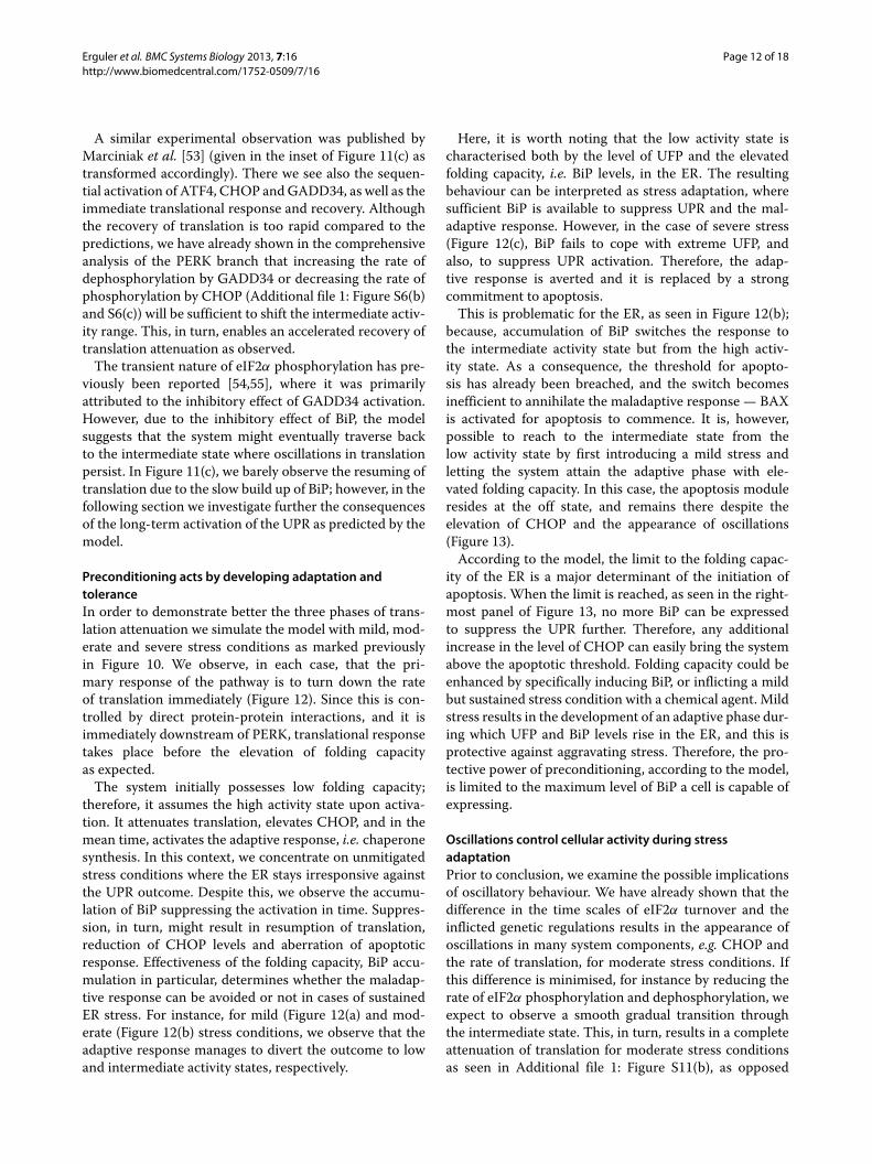
This is problematic for the ER, as seen in Figure 12(b);because, accumulation of BiP switches the response tothe intermediate activity state but from the high activ-ity state. As a consequence, the threshold for apopto-sis has already been breached, and the switch becomesinefficient to annihilate the maladaptive response — BAXis activated for apoptosis to commence. It is, however,possible to reach to the intermediate state from thelow activity state by first introducing a mild stress andletting the system attain the adaptive phase with ele-vated folding capacity. In this case, the apoptosis moduleresides at the off state, and remains there despite theelevation of CHOP and the appearance of oscillations(Figure 13).
According to the model, the limit to the folding capac-ity of the ER is a major determinant of the initiation ofapoptosis. When the limit is reached, as seen in the right-most panel of Figure 13, no more BiP can be expressedto suppress the UPR further. Therefore, any additionalincrease in the level of CHOP can easily bring the systemabove the apoptotic threshold. Folding capacity could beenhanced by specifically inducing BiP, or inflicting a mildbut sustained stress condition with a chemical agent. Mildstress results in the development of an adaptive phase dur-ing which UFP and BiP levels rise in the ER, and this isprotective against aggravating stress. Therefore, the pro-tective power of preconditioning, according to the model,is limited to the maximum level of BiP a cell is capable ofexpressing.
Oscillations control cellular activity during stressadaptationPrior to conclusion, we examine the possible implicationsof oscillatory behaviour. We have already shown that thedifference in the time scales of eIF2α turnover and theinflicted genetic regulations results in the appearance ofoscillations in many system components, e.g. CHOP andthe rate of translation, for moderate stress conditions. Ifthis difference is minimised, for instance by reducing therate of eIF2α phosphorylation and dephosphorylation, weexpect to observe a smooth gradual transition throughthe intermediate state. This, in turn, results in a completeattenuation of translation for moderate stress conditionsas seen in Additional file 1: Figure S11(b), as opposed

Erguler et al. BMC Systems Biology 2013, 7:16 Page 13 of 18http://www.biomedcentral.com/1752-0509/7/16
Total UFP Total BiP CHOP Active PERK Active BAX Active eIF2a
0 100 200 300 400 500
0.0
0.2
0.4
0.6
0.8
1.0
time (atu)
PE
RK
A &
eIF
2a (
acu)
036
7210
814
418
0B
iPT
& U
FP
T &
CH
OP
(5x
) &
BA
Xm
(ac
u)
0 100 200 300 400 500
0.0
0.2
0.4
0.6
0.8
1.0
time (atu)
PE
RK
A &
eIF
2a (
acu)
036
7210
814
418
0B
iPT
& U
FP
T &
CH
OP
(5x
) &
BA
Xm
(ac
u)(b) moderate stress
0 100 200 300 400 500
0.0
0.2
0.4
0.6
0.8
1.0
time (atu)
PE
RK
A &
eIF
2a (
acu)
036
7210
814
418
0B
iPT
& U
FP
T &
CH
OP
(5x
) &
BA
Xm
(ac
u)
(c) severe stress
(a) mild stress
Figure 12 The low, intermediate and high activity states of the UPR. The low activity state is shown in (a), where rapid but transient activationof CHOP is followed by the recovery of translational activity. In (b), the intermediate state is shown, which exhibits sustained oscillations in bothCHOP and the rate of translation. This eventually leads to the activation of BAX on mitochondrial membrane. In (c), the high activity state is shownwith elevated and sustained UPR activity, i.e. activation of PERK and expression of CHOP. The plot also shows severely reduced translation rates andthe activation of apoptotic signals. The legend is given on top of the plots. The grey shades indicate active eIF2α, which represents the relative rateof translation. The initial conditions have been extended towards the negative time axis in order to demonstrate the punctuality of the translationalresponse. The stress conditions chosen are based on Figure 10.
Erguler et al. BMC Systems Biology 2013, 7:16 Page 14 of 18http://www.biomedcentral.com/1752-0509/7/16
Total UFP Total BiP CHOP Active PERK Active BAX Active eIF2a
0 500 1000 1500
0.0
0.2
0.4
0.6
0.8
1.0
time (atu)
PE
RK
A &
eIF
2a (
acu)
036
7210
814
418
0
BiP
T &
UF
PT
& C
HO
P (
5x)
& B
AX
m (
acu)no stress adaptation tolerance cell death
Figure 13 The UPR response against stepwise escalation of the ER stress levels. The mild, moderate and severe stress conditions as inFigure 12 are administered sequentially for a duration of 500 time units each. The elevation of active PERK, BiP and CHOP, the status of translation,and the activation of BAX on mitochondrial membrane are shown with respect to accumulating UFP. The grey shade indicates active eIF2α, whichrepresents the relative rate of translation. The initial conditions have been extended towards the negative time axis in order to demonstrate thepunctuality of the translational response. The plot is drawn as in Figure 12, and the legend is given on top of the plot.
to minor changes seen for mild and severe conditions(Additional file 1: Figure S11(a) and S11(c) in Additionalfile 1: Text 1.6).
On the other hand, when the stress conditions progres-sively worsen, we observe that the low and high activitystates change nominally; however, translation becomespermanently attenuated upon entry to the intermediateactivity state (Additional file 1: Figure S11(d)). We havedeemed such a transition in Figure 13 as the developmentof stress toleration, where translation resumes at least forsome periods. It is, however, more appropriate to con-sider the non-oscillatory intermediate state as senescence,because of the lack of translation seen together with noapoptotic activity. As a result, the model suggests thatthe existence of oscillations provides a means for transla-tion, and hence the routine cellular activity, to be partiallyrestored.
DiscussionThe UPR is composed of a complicated mesh of biochem-ical and genetic regulatory interactions. These range fromunconventional mRNA splicing, global translational dis-ruption and the activation of hundreds of genes with a sin-gle aim to deliver the right response at the right time [56].The decision and timing of an appropriate response areimplemented within the intricate wiring of this signallingcascade, which we aimed to decipher by constructingits detailed mechanistic model. The model incorporatedthe three main signalling pathways, i.e. IRE1α, PERK andATF6, the interconnections between these pathways, andthe downstream genetic regulatory interactions. To thebest of our knowledge, this model is the first in its extentand in the detail it incorporates.
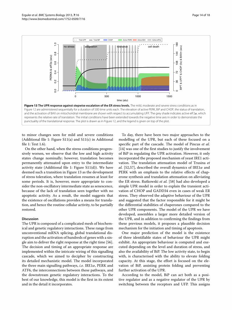
To day, there have been two major approaches to themodelling of the UPR, but each of these focused on aspecific part of the cascade. The model of Pincus et al.[14] was one of the first studies to justify the involvementof BiP in regulating the UPR activation. However, it onlyincorporated the proposed mechanism of yeast IRE1 acti-vation. The translation attenuation model of Trusina etal. [52,57], described the overall dynamics of IRE1α andPERK with an emphasis to the relative effects of chap-erone synthesis and translation attenuation on alleviatingthe ER stress. Rutkowski et al. [58] had also developed asimple UPR model in order to explain the transient acti-vation of CHOP and GADD34 even in cases of weak ERstress. They observed the adaptive behaviour of the UPRand suggested that the factor responsible for it might bethe differential stabilities of chaperones compared to theother UPR components. The model of the UPR we havedeveloped, assembles a larger more detailed version ofthe UPR, and in addition to confirming the findings fromthese previous models, it proposes a plausible decisionmechanism for the initiation and timing of apoptosis.
One major prediction of the model is the existenceof three identifiable states of behaviour the UPR mightexhibit. An appropriate behaviour is computed and exe-cuted depending on the level and duration of stress, andalso the availability of BiP. The low activity state, to beginwith, is characterised with the ability to elevate foldingcapacity. At this stage, the effort is focused on the ele-vation of BiP, assisting protein folding and preventingfurther activation of the UPR.
According to the model, BiP can act both as a posi-tive regulator and as a negative regulator of the UPR byswitching between the receptors and UFP. This assigns

Erguler et al. BMC Systems Biology 2013, 7:16 Page 15 of 18http://www.biomedcentral.com/1752-0509/7/16
the chaperone a pivotal role during the low activity statewhere it helps to coordinate the development of stressadaptation. BiP has previously been associated experi-mentally with adaptation [21,58-60], which we predict tooccur when sufficient chaperone accumulates to suppressUPR signalling and prevent the elevation of CHOP, thesignal for apoptosis.
Adaptation is compromised when the limit of chaper-one synthesis is reached. For severe stress conditions, thisresults in the elevation of apoptotic signals and the irre-versible activation of the BAX/BAK/BH3 pathway. At thisstage, the rate of translation is sustained at a minimumlevel, which might be unfavourable for apoptotic activitydue to the inability to synthesise certain proteins [61,62].We speculate that direct binding of BAX (and BAK) toIRE1α [63] on the ER membrane may be essential to acti-vate an alternative pathway, for instance the JNK pathwayand the unspecific mRNA decay mechanism [24]. Thisin turn may promote apoptosis especially when it is aug-mented with the disruption of the Ca+2 balance — causedby the activated BAX.
The model predicts an intermediate activity state dur-ing which CHOP is activated but has yet to reach its upperlimit. During this state, we observed oscillations in manysystem components, including the rate of translation, forthe first time to our knowledge. Oscillations occur as aresult of differences in the kinetics of eIF2α phosphory-lation/dephosphorylation and genetic regulation, and thisplays a crucial role in resuming translation at least for briefperiods of time. We speculate that translation at this stagemight be beneficial in the continuation of vital cellularfunctions, or, especially if the apoptosis is initiated, in thesynthesis of apoptotic genes.
The current configuration of the model parameters per-mits the alignment of the intermediate activity region ofCHOP with the bistable range of BAX. As CHOP levelsraise the system moves across the bistable regime exceed-ing the activation threshold just before CHOP reachesits upper limit. Therefore, the maladaptive behaviour atthe intermediate state depends heavily on from where itis reached. For instance, applying enough stress to bringthe system to the intermediate activity state from anunstressed ER will cause the elevation of apoptotic sig-nals. This is mainly because of the shortage of time forBiP to accumulate to suppress UPR signalling, leading tothe appearance of first the high and then the intermediateactivity state. Here, the importance of existence of an earlystage of adaptation becomes obvious. Developing adap-tation, or preconditioning in clinical terms [5,8,59,64,65],enables the elevation of folding capacity, and BiP, restingthe system at the low activity state. When the intermediatestate is reached from there, BAX remains low at the inac-tive branch of the bistable regime providing protectionfrom apoptosis.
Regardless of where it is reached from the interme-diate state exhibits oscillations in system components.However, they can be exhausted if the time differencebetween the phosphorylation of eIF2α and the activationof GADD34 is reduced. By doing so, we noticed that themajor contribution of oscillatory behaviour to the out-come of the UPR is the resuming of translational activity.In the no-oscillation case, during the intermediate state,there is absolutely no translational activity upon UPR acti-vation. Moreover, if the system resides on the inactivebranch of the apoptotic switch, in addition to translationattenuation, the activation of BAX will be permanentlysuppressed. It is only natural to expect this state of senes-cence to end shortly due to the gradual degradation ofcritical cellular functions.
We hypothesise that senescence and apoptosis mightbe preferred or avoided depending on the cell type. Forinstance, some of the vital cell types that cannot bereplaced when damaged, e.g. nerve cells or podocytes ofkidney, might be adapted to exhibit oscillations so thattranslation is resumed in part as a survival response. Onthe other hand, it might be beneficial for a lymphocyteto self-destruct promptly in case of any malevolent con-sequences of cellular damage. Testing the validity of thishypothesis, however, extends beyond the intended scopeof this research.
The precise mechanisms of receptor dynamics, geneticregulation and crosstalk with other stress signalling path-ways are currently unknown. This contributes greatly tothe inevitable incompleteness of modelling approachesalike. However, with this research, we presented a math-ematical model, which is, being faithful to the exist-ing literature, highly predictive despite the absence ofa perfect quantitative match between the predictionsand the experimental observations. The modular step-by-step approach of constructing the model has beena major factor in easing the analysis and supplyingthis predictive power. The choice of the parameter val-ues originated from the bifurcation analyses with refer-ence to the experimental observations from literature.As experimental observations accumulate, the inaccu-racies and disagreements between the predictions andthe observations will form a strong basis for improv-ing and extending this model. Consequently, such studieswill necessitate the accommodation of data variability interms of intrinsic stochastic fluctuations of the system.In order to address this issue, we are currently workingtowards relaxing the deterministic assumption and study-ing the three types of UPR output under the influence ofintrinsic noise.
Nevertheless, a particular configuration of parametersmight be valid for a certain cell type under certainextra- or intracellular conditions at a specific develop-mental stage. We argue that it is possible to tune the

Erguler et al. BMC Systems Biology 2013, 7:16 Page 16 of 18http://www.biomedcentral.com/1752-0509/7/16
model of the UPR to represent the signalling cascadeduring most of such specific conditions. Consequently,the model should yield a response similar to what hasbeen investigated in this work. An important step towardsthe validation of the model predictions, is to design atitration experiment where the ER is subjected to dif-ferent stress conditions and the formation of the threedistinct types of behaviour is observed: the low activitystate with adaptive behaviour, the intermediate activitystate with oscillations and bistability in apoptotic sig-nals, and the high activity state with strong commitmentto apoptosis.
An interesting experimental challenge as a natural con-sequence of this research would be to look for modifiergenes in the UPR for related diseases. It might be possiblethat, for instance, any mutation or malfunctioning result-ing in the manipulation of the intermediate activity stateresults in adopting the high activity state prematurely. Inthis case, translation may be attenuated and apoptoticsignals elevated even though the ER stress is mild or mod-erate. In light of this, one of the major undertakings of ourgroup is currently the investigation of the contribution ofthe UPR to the vast phenotypic heterogeneity of AlportSyndrome and Thin Basement Membrane Nephropathy[1,66].
ConclusionHere we develop, for the first time, a combined mecha-nistic model of the three signalling pathways of the UPRcascade. The model incorporates highly detailed enzy-matic and genetic regulatory interactions based on therecent literature. The analysis of the model reveals that thebalance between the ER stress and the folding capacity ofthe ER plays a pivotal role in managing the transformationfrom an adaptive to a maladaptive response. Accordingto this, there exists three distinct states of behaviour theUPR may adopt: low, intermediate and high activity states.We demonstrate, for the first time, that under the rightcircumstances, the intermediate state may exhibit oscil-lations in translation attenuation and apoptotic signals.Demonstration of stress adaptation provides a mechanis-tic explanation as to how preconditioning might preventthe initiation of apoptosis. The model can be configuredto represent the UPR of a specific cell type under cer-tain experimental conditions. The experimental validationof the model predictions is currently one of the majorundertakings of our group.
MethodsThe complete list of differential equations, derivations ofreaction kinetics, and the choice of parameter values areexplained in detail in the Additional file 1. The SBMLv2.4version of the model is submitted to the BioModelsDatabase [67] with the identifier BIOMD0000000446.
The bifurcation analysis of the model is performedwith XPPAUT5.41. The wiring diagrams are created inCellDesignerTM [68].
Additional file
Additional file 1: Supplementary Text. The Supplementary Textincludes detailed technical information about the mathematical model, itsassumptions and supplementary analyses to the main manuscriptconcerning the effects of a broader range of the model parameters.
AbbreviationsUFP: unfolded protein; UFPT: total UFP; BiP/GRP78: immunoglobulin bindingprotein / glucose regulated protein; IRE1α: inositol requiring protein 1α; PERK:protein kinase RNA-like ER kinase; ATF6: activating transcription factor 6; ATF4:activating transcription factor 4; GADD34: growth arrest and DNA damage-34;CHOP/GADD153: CCAAT/enhancer-binding protein homologous protein;XBP1: X-box binding protein 1; eIF2α: eukaryotic initiation factor 2α; CReP:constitutive repressor of eIF2α phosphorylation; WFS1: Wolfram syndrome 1.
Competing interestsThe authors declare that they have no competing interests.
Authors’ contributionsKE, MP and CD conceived the project. KE designed the study, developed themodel and performed the analysis. KE and CD contributed to the final versionof the manuscript. All authors read and approved the final manuscript.
AcknowledgementsWe acknowledge Prof David Ron, Dr Stefan Marciniak and Prof Maho Niwa forsupporting this work through sharing their data. We acknowledge Dr AlexeiKorennykh for his helpful insights on the dynamics of receptor activation.Many thanks to Charalambos Stefanou for useful discussions about the UPRpathway. The authors are indebted to Dr Tina Toni for her invaluablecomments and suggestions on the manuscript. This work was funded by theEuropean Regional Development Fund and the Republic of Cyprus throughthe Research Promotion Foundation (Strategic Infrastructure Project NEWINFRASTRUCTURE/STRATEGIC/0308/24), and through the University of CyprusArticles 3/311 and 3/346 to CD. The funders had no role in study design,model development and analysis, decision to publish or preparation of themanuscript.
Received: 22 October 2012 Accepted: 28 January 2013Published: 21 February 2013
References1. Inagi R: Endoplasmic reticulum stress as a progression factor for
kidney injury. Curr Opin Pharmacol 2010, 10(2):156–165. [http://www.sciencedirect.com/science/article/pii/S1471489209002070]
2. Kaufman RJ: Orchestrating the unfolded protein response in healthand disease. J Clin Invest 2002, 110(10):1389–98.
3. Naidoo N: ER and aging-Protein folding and the ER stress response.Ageing Res Rev 2009, 8(3):150–9.
4. Hetz C, Glimcher LH: XBP-1 and the UPRosome: Mastering SecretoryCell Function. Curr Immunol Rev 2008, 4:1–10 .
5. Cybulsky AV: Endoplasmic reticulum stress in proteinuric kidneydisease. Kidney Int 2010, 77(3):187–193.
6. Chakrabarti A, Chen AW, Varner JD: A review of the mammalianunfolded protein response. Biotechnol Bioeng 2011, 108(12):2777–2793.
7. Morris JA, Dorner AJ, Edwards CA, Hendershot LM, Kaufman RJ:Immunoglobulin Binding Protein (BiP) function is required toprotect cells from endoplasmic reticulum stress but is not requiredfor the secretion of selective proteins. J Biol Chem 1997,272(7):4327–4334.
8. Inagi R, Kumagai T, Nishi H, Kawakami T, Miyata T, Fujita T, Nangaku M:Preconditioning with endoplasmic reticulum stress amelioratesmesangioproliferative glomerulonephritis. J Am Soc Nephrology 2008,19(5):915–922.

Erguler et al. BMC Systems Biology 2013, 7:16 Page 17 of 18http://www.biomedcentral.com/1752-0509/7/16
9. Kohno K: Stress-sensing mechanisms in the unfolded proteinresponse similarities and differences between yeast and mammals.J Biochem 2010, 147:27–33.
10. Kimata Y, Kohno K: Endoplasmic reticulum stress-sensing mechanismsin yeast and mammalian cells. Curr Opin Cell Biol 2011, 23(2):135–142.
11. Tabas I, Ron D: Integrating the mechanisms of apoptosis induced byendoplasmic reticulum stress. Nat Cell Biol 2011, 13(3):184–90.
12. Ron D, Walter P: Signal integration in the endoplasmic reticulumunfolded protein response. Nat Rev Mol Cell Biol 2007, 8(7):519–529.[http://www.nature.com/nrm/journal/v8/n7/full/nrm2199.html]
13. Hetz C, Glimcher LH: Fine-tuning of the unfolded protein responseAssembling the IRE1alpha interactome. Mol Cell 2009, 35(5):551–61.
14. Pincus D, Chevalier MW, Aragon T, Anken EV, Vidal SE, El-Samad H, WalterP: BiP binding to the ER-Stress sensor Ire1 tunes the homeostaticbehavior of the unfolded protein response. Plos Biol 2010,8(7):e1000415. [http://www.plosbiology.org/article/fetchObjectAttachment.action?uri=info:doi/10.1371/journal.pbio.1000415&representation=PDF]
15. Onn A, Ron D: Modeling the endoplasmic reticulum unfolded proteinresponse. Nat Struct Mol Biol 2010, 17(8):924–925.
16. Li H, Korennykh AV, Behrman SL, Walter P: Mammalian endoplasmicreticulum stress sensor IRE1 signals by dynamic clustering. Proc NatlAcad Sci USA 2010, 107(37):16113–16118.
17. Ali MMU, Bagratuni T, Davenport EL, Nowak PR, Silva-Santisteban MC,Hardcastle A, Mcandrews C, Rowlands MG, Morgan GJ, Aherne W, CollinsI, Davies FE, Pearl LH: Structure of the Ire1 autophosphorylationcomplex and implications for the unfolded protein response. EMBO J2011, 30(5):894–905.
18. Oikawa D, Kimata Y, Kohno K, Iwawaki T: Activation of mammalian IRE1[alpha] upon ER stress depends on dissociation of BiP rather than ondirect interaction with unfolded proteins. Exp Cell Res 2009,315(15):2496–2504.
19. Gardner BM, Walter P: Unfolded proteins are Ire1-activating ligandsthat directly induce the unfolded protein response. Science 2011,333(6051):1891–4.
20. Sicheri F, Silverman R: Putting the brakes on the unfolded proteinresponse. J Cell Biol 2011, 193:17.
21. Rubio C, Pincus D, Korennykh A, Schuck S, El-Samad H, Walter P:Homeostatic adaptation to endoplasmic reticulum stress dependson Ire1 kinase activity. J Cell Biol 2011, 193:171.
22. Chawla A, Chakrabarti S, Ghosh G, Niwa M: Attenuation of yeast UPR isessential for survival and is mediated by IRE1 kinase. J Cell Biol 2011,193:41.
23. DuRose J, Tam A, Niwa M: Intrinsic capacities of molecular sensors ofthe unfolded protein response to sense alternate forms ofendoplasmic reticulum stress. Mol Biol Cell 2006, 17(7):3095.
24. Han D, Lerner A, Walle LV, Upton J, Xu W, Hagen A: IRE1 [alpha] KinaseActivation Modes Control Alternate Endoribonuclease Outputs toDetermine Divergent Cell Fates. Cell 2009, 138(3):562–575. [http://www.sciencedirect.com/science/article/pii/S0092867409008927]
25. Korennykh AV, Egea PF, Korostelev AA, Finer-Moore J, Zhang C, ShokatKM, Stroud RM, Walter P: The unfolded protein response signalsthrough high-order assembly of Ire1. Nature 2009, 457(7230):[http://www.nature.com/nature/journal/v457/n7230/full/nature07661.html]
26. Korennykh AV: Personal Communication; 2012 .27. Bull VH, Thiede B: Proteome analysis of tunicamycin-induced ER
stress. ELECTROPHORESIS 2012, 33(12):1814–1823.28. Yoshida H, Matsui, T, Yamamoto, A, Okada, T, Mori, K: XBP1 mRNA is
induced by ATF6 and spliced by IRE1 in response to ER stress toproduce a highly active transcription factor. Cell 2001,107(7):881–91.
29. Yamamoto K, Yoshida H, Kokame K, Kaufman RJ, Mori K: Differentialcontributions of ATF6 and XBP1 to the activation of endoplasmicreticulum stress-responsive cis-acting elements ERSE, UPRE andERSE-II. J Biochem 2004, 136(3):343–50.
30. Yamada T, Ishihara H, Tamura A, Takahashi R, Yamaguchi S, Takei D, TokitaA, Satake C, Tashiro F, Katagiri H, Aburatani H, ichi Miyazaki, J, Oka Y: WFS1-deficiency increases endoplasmic reticulum stress, impairs cell cycleprogression and triggers the apoptotic pathway specifically inpancreatic beta-cells. Human Mol Genet 2006, 15(10):1600–1609.
31. Fonseca SG, Ishigaki S, Oslowski CM, Lu S, Lipson KL, Ghosh R, Hayashi E,Ishihara H, Oka Y, Permutt MA, Urano F: Wolfram syndrome 1 genenegatively regulates ER stress signaling in rodent and human cells. JClin Invest 2010, 120(3):744–755.
32. Thuerauf DJ, Marcinko M, Belmont PJ, Glembotski CC: Effects of theisoform-specific characteristics of ATF6 alpha and ATF6 beta onendoplasmic reticulum stress response gene expression and cellviability. J Biol Chem 2007, 282(31):22865–22878.
33. Yoshida H, Haze K, Yanagi H, Yura T, Mori K: Identification of thecis-acting endoplasmic reticulum stress response elementresponsible for transcriptional induction of mammalianglucose-regulated proteins Involvement of basic leucine zippertranscription factors. J Biol Chem 1998, 273(50):33741–33749.
34. Haze K, Yoshida H, Yanagi H, Yura T, Mori K: Mammalian transcriptionfactor ATF6 is synthesized as a transmembrane protein andactivated by proteolysis in response to endoplasmic reticulumstress. Mol Biol Cell 1999, 10(11):3787–3799.
35. Szegezdi E, Logue SE, Gorman AM, Samali A: Mediators of endoplasmicreticulum stress-induced apoptosis. EMBO Rep 2006, 7(9):880–885.
36. Wek RC: eIF-2 kinases: regulators of general and gene-specifictranslation initiation. Trends Biochem Sci 1994, 19(11):491–496.
37. Harding HP, Novoa I, Zhang Y, Zeng H, Wek R, Schapira M, Ron D:Regulated translation initiation controls stress-induced geneexpression in mammalian cells. Mol Cell 2000, 6(5):1099–1108.
38. Gulow K, Bienert D, Haas IG: BiP is feed-back regulated by control ofprotein translation efficiency. J Cell Sci 2002, 115(Pt 11):2443–2452.
39. van Huizen R, Martindale JL, Gorospe M, Holbrook NJ: P58IPK, a novelendoplasmic reticulum stress-inducible protein and potentialnegative regulator of eIF2alpha signaling. J Biol Chem 2003,278(18):15558–15564.
40. Lopez-Lastra M, Rivas A, Barrıa MI: Protein synthesis in eukaryotes: thegrowing biological relevance of cap-independent translationinitiation. Biol Res 2005, 38(2-3):121–146.
41. Goldbeter A, Koshland D: An amplified sensitivity arising fromcovalent modification in biological systems. Proc Nat Acad Sci. . . 1981.[http://www.jstor.org/discover/10.2307/11361?uid=3739192&uid=2134&uid=2&uid=70&uid=4&sid=21102148328741]
42. Tyson J, Chen K, Novak B: Sniffers, buzzers, toggles and blinkersdynamics of regulatory and signaling pathways in the cell. Curr OpinCell Biol 2003. [http://linkinghub.elsevier.com/retrieve/pii/S0955067403000176]
43. Lu PD, Harding HP, Ron D: Translation reinitiation at alternative openreading frames regulates gene expression in an integrated stressresponse. J Cell Biol 2004, 167:27–33.
44. Vattem KM, Wek RC: Reinitiation involving upstream ORFs regulatesATF4 mRNA translation in mammalian cells. Proc Natl Acad Sci USA2004, 101(31):11269–11274.
45. Lee AH, Iwakoshi NN, Glimcher LH: XBP-1 regulates a subset ofendoplasmic reticulum resident chaperone genes in the unfoldedprotein response. Mol Cell Biol 2003, 23(21):7448–7459.
46. Toni T, Welch D, Strelkowa N, Ipsen A, Stumpf MP: ApproximateBayesian computation scheme for parameter inference and modelselection in dynamical systems. J R Soc Interface 2008, 6(31):187–202.
47. Erguler K, Stumpf MPH: Practical limits for reverse engineering ofdynamical systems: a statistical analysis of sensitivity andparameter inferability in systems biology models. Mol BioSyst 2011,7(5):1593–1602.
48. Zhang T, Brazhnik P, Tyson JJ: Computational analysis of dynamicalresponses to the intrinsic pathway of programmed cell death.Biophys J 2009, 97(2):415–434.
49. Tyson JJ, Baumann WT, Chen C, Verdugo A, Tavassoly I, Wang Y, WeinerLM, Clarke R: Dynamic modelling of oestrogen signalling and cell fatein breast cancer cells. Nat Rev Cancer 2011, 11(7):523–532. [http://www.nature.com/nrc/journal/v11/n7/full/nrc3081.html]
50. Puthalakath H, O’Reilly LA, Gunn P, Lee L, Kelly PN, Huntington ND,Hughes PD, Michalak EM, McKimm-Breschkin J, Motoyama N, Gotoh T,Akira S, Bouillet P, Strasser A: ER stress triggers apoptosis by activatingBH3-only protein Bim. Cell 2007, 129(7):1337–1349.
51. Kaern M, Elston TC, Blake WJ, Collins JJ: Stochasticity in gene expression:from theories to phenotypes. Nat Rev Genet 2005, 6(6):451–464.

Erguler et al. BMC Systems Biology 2013, 7:16 Page 18 of 18http://www.biomedcentral.com/1752-0509/7/16
52. Trusina A, Papa FR, Tang C: Rationalizing translation attenuation inthe network architecture of the unfolded protein response. Proc NatlAcad Sci USA 2008, 105(51):20280–20285. [http://www.pnas.org/content/105/51/20280.long]
53. Marciniak SJ, Yun CY, Oyadomari S, Novoa I, Zhang Y, Jungreis R, Nagata K,Harding HP, Ron D: CHOP induces death by promoting proteinsynthesis and oxidation in the stressed endoplasmic reticulum.Genes Dev 2004, 18(24):3066–3077.
54. Brush MH, Weiser DC, Shenolikar S: Growth arrest and DNAdamage-inducible protein GADD34 targets protein phosphatase 1alpha to the endoplasmic reticulum and promotesdephosphorylation of the alpha subunit of eukaryotic translationinitiation factor 2. Mol Cell Biol 2003, 23(4):1292–1303.
55. Armstrong JL, Flockhart R, Veal GJ, Lovat PE, Redfern CPF: Regulation ofendoplasmic reticulum stress-induced cell death by ATF4 inneuroectodermal tumor cells. J Biol Chem 2010, 285(9):6091–6100.
56. McCracken AA, Brodsky JL: A molecular portrait of the response tounfolded proteins. Genome Biol 2000, 1(2):REVIEWS1013.
57. Trusina A, Tang C: The unfolded protein response and translationattenuation: a modelling approach. Diabetes Obes Metab 2010, 12Suppl 2:27–31. [http://onlinelibrary.wiley.com/doi/10.1111/j.1463-1326.2010.01274.x/abstract;jsessionid=BC778943D52056AB3E3931A9BEB9E251.d02t03]
58. Rutkowski DT, Arnold SM, Miller CN, Wu J, Li J, Gunnison KM, Mori K, AkhaAAS, Raden D, Kaufman RJ: Adaptation to ER stress is mediated bydifferential stabilities of pro-survival and pro-apoptotic mRNAs andproteins. Plos Biol 2006, 4(11):e374. [http://www.plosbiology.org/article/info%3Adoi%2F10.1371%2Fjournal.pbio.0040374]
59. Rutkowski DT, Kaufman RJ: That which does not kill me makes mestronger: adapting to chronic ER stress. Trends Biochem Sci 2007,32(10):469–76.
60. Tsang KY, Chan D, Bateman JF, Cheah KSE: In vivo cellular adaptation toER stress: survival strategies with double-edged consequences. J CellSci 2010, 123(Pt 13):2145–2154.
61. Scheuner D, Patel R, Wang F, Lee K, Kumar K, Wu J, Nilsson A, Karin M,Kaufman RJ: Double-stranded RNA-dependent protein kinasephosphorylation of the -subunit of eukaryotic translation initiationfactor 2 mediates Apoptosis. J Biol Chem 2006, 281(30):21458–21468.
62. Back SH, Scheuner D, Han J, Song B, Ribick M, Wang J, Gildersleeve RD,Pennathur S, Kaufman RJ: Translation attenuation through eIF2α;phosphorylation prevents oxidative stress and maintains thedifferentiated state in β Cells. Cell Metabolism 2009, 10:13–26.[http://dx.doi.org/10.1016/j.cmet.2009.06.002]
63. Hetz C, Bernasconi P, Fisher J, Lee AH, Bassik MC, Antonsson B, Brandt GS,Iwakoshi NN, Schinzel A, Glimcher LH, Korsmeyer SJ: Proapoptotic BAXand BAK modulate the unfolded protein response by a directinteraction with IRE1alpha. Science 2006, 312(5773):572–576.
64. Peyrou M, Cribb AE: Effect of endoplasmic reticulum stresspreconditioning on cytotoxicity of clinically relevant nephrotoxinsin renal cell lines. Toxicol In Vitro 2007, 21(5):878–886.
65. Li J, Wang JJ, Zhang SX: Preconditioning with endoplasmic reticulumstress mitigates retinal endothelial inflammation via activation ofX-box binding protein 1. J Biol Chem 2011, 286(6):4912–4921.
66. Gubler MC: Inherited diseases of the glomerular basementmembrane. Nat Clin Pract Nephrol 2008, 4:24–37.
67. Li C, Donizelli M, Rodriguez N, Dharuri H, Endler L, Chelliah V, Li L, He E,Henry A, Stefan MI, Snoep JL, Hucka M, Novere NL, Laibe C: BioModelsDatabase: An enhanced, curated and annotated resource forpublished quantitative kinetic models. BMC Syst Biol 2010, 4:92.
68. Funahashi A, Morohashi M, Kitano H: CellDesigner: a process diagrameditor for gene-regulatory and biochemical networks. BIOSILICO 2003,1(5):159–162. [https://openwetware.org/images/c/c0/Funahashi.pdf]
doi:10.1186/1752-0509-7-16Cite this article as: Erguler et al.: A mathematical model of the unfoldedprotein stress response reveals the decision mechanism for recovery,adaptation and apoptosis. BMC Systems Biology 2013 7:16.
Submit your next manuscript to BioMed Centraland take full advantage of:
• Convenient online submission
• Thorough peer review
• No space constraints or color figure charges
• Immediate publication on acceptance
• Inclusion in PubMed, CAS, Scopus and Google Scholar
• Research which is freely available for redistribution
Submit your manuscript at www.biomedcentral.com/submit