a member of the ferlin calcium sensor family is essential ... · a member of the ferlin calcium...
TRANSCRIPT

A Member of the Ferlin Calcium Sensor Family Is Essential forToxoplasma gondii Rhoptry Secretion
Bradley I. Coleman,a* Sudeshna Saha,a* Seiko Sato,b Klemens Engelberg,a David J. P. Ferguson,c* Isabelle Coppens,d
Melissa B. Lodoen,b Marc-Jan Gubbelsa
aDepartment of Biology, Boston College, Chestnut Hill, Massachusetts, USAbDepartment of Molecular Biology & Biochemistry and the Institute for Immunology, University of California,Irvine, California, USA
cNuffield Department of Clinical Laboratory Science, University of Oxford John Radcliffe Hospital, Oxford,United Kingdom
dDepartment of Molecular Microbiology and Immunology, Johns Hopkins University Bloomberg School ofPublic Health, Baltimore, Maryland, USA
ABSTRACT Invasion of host cells by apicomplexan parasites such as Toxoplasmagondii is critical for their infectivity and pathogenesis. In Toxoplasma, secretion of es-sential egress, motility, and invasion-related proteins from microneme organelles isregulated by oscillations of intracellular Ca2�. Later stages of invasion are consideredCa2� independent, including the secretion of proteins required for host cell entryand remodeling from the parasite’s rhoptries. We identified a family of three Toxo-plasma proteins with homology to the ferlin family of double C2 domain-containingCa2� sensors. In humans and model organisms, such Ca2� sensors orchestrate Ca2�-dependent exocytic membrane fusion with the plasma membrane. Here we focus onone ferlin that is conserved across the Apicomplexa, T. gondii FER2 (TgFER2). Unex-pectedly, conditionally TgFER2-depleted parasites secreted their micronemes nor-mally and were completely motile. However, these parasites were unable to invadehost cells and were therefore not viable. Knockdown of TgFER2 prevented rhoptrysecretion, and these parasites failed to form the moving junction at the parasite-hostinterface necessary for host cell invasion. Collectively, these data demonstrate the re-quirement of TgFER2 for rhoptry secretion in Toxoplasma tachyzoites and suggest apossible Ca2� dependence of rhoptry secretion. These findings provide the firstmechanistic insights into this critical yet poorly understood aspect of apicomplexanhost cell invasion.
IMPORTANCE Apicomplexan protozoan parasites, such as those causing malaria andtoxoplasmosis, must invade the cells of their hosts in order to establish a pathogenicinfection. Timely release of proteins from a series of apical organelles is required forinvasion. Neither the vesicular fusion events that underlie secretion nor the observedreliance of the various processes on changes in intracellular calcium concentrationsis completely understood. We identified a group of three proteins with strong ho-mology to the calcium-sensing ferlin family, which are known to be involved in pro-tein secretion in other organisms. Surprisingly, decreasing the amounts of one ofthese proteins (TgFER2) did not have any effect on the typically calcium-dependentsteps in invasion. Instead, TgFER2 was essential for the release of proteins from or-ganelles called rhoptries. These data provide a tantalizing first look at the mecha-nisms controlling the very poorly understood process of rhoptry secretion, which isessential for the parasite’s infection cycle.
KEYWORDS Toxoplasma gondii, calcium, ferlin, micronemes, protein secretion,rhoptries
Received 13 July 2018 Accepted 20 August2018 Published 2 October 2018
Citation Coleman BI, Saha S, Sato S,Engelberg K, Ferguson DJP, Coppens I,Lodoen MB, Gubbels M-J. 2018. A member ofthe ferlin calcium sensor family is essentialfor Toxoplasma gondii rhoptry secretion.mBio 9:e01510-18. https://doi.org/10.1128/mBio.01510-18.
Editor Anita A. Koshy, University of Arizona
Copyright © 2018 Coleman et al. This is anopen-access article distributed under the termsof the Creative Commons Attribution 4.0International license.
Address correspondence to Marc-Jan Gubbels,[email protected].
* Present address: Bradley I. Coleman, HarvardMedical School, Boston, Massachusetts, USA;Sudeshna Saha, University of California, SanDiego, School of Medicine, San Diego,California, USA; David J. P. Ferguson,Department of Biological and MedicalSciences, Oxford Brookes University, Oxford,United Kingdom.
B.I.C. and S.S. contributed equally to this article.
RESEARCH ARTICLEHost-Microbe Biology
crossm
September/October 2018 Volume 9 Issue 5 e01510-18 ® mbio.asm.org 1
on August 24, 2019 by guest
http://mbio.asm
.org/D
ownloaded from

The apicomplexan parasite Toxoplasma gondii infects one in every three humans.Clinical symptoms of toxoplasmosis derive from the tissue destruction and inflam-
mation caused by repeated rounds of host cell invasion, intracellular replication, andlytic egress of the tachyzoite life stage. Egress is mediated by intracellular Ca2� ([Ca2�]i)fluctuations that trigger release of proteins from the microneme organelles (1). Follow-ing egress, parasites move via gliding motility to a new host cell, triggered by additionalparasite [Ca2�]i oscillations that facilitate further release of micronemes (2). Subsequenthost cell invasion also relies on micronemal proteins (3). The micronemes are localizedat the apical end of the parasite and are released from the apical tip (4). Following initialrecognition of a host cell, the parasite engages in a tighter interaction with the targetcell, mediated by proteins secreted from the rhoptries. The club-shaped rhoptries areanchored at the parasite’s apical end, from where they secrete their contents (5, 6).Proteins in the apical rhoptry neck (RONs) are secreted into the host cell before therhoptry bulb proteins (ROPs) are released (7). RONs function in tightening the parasite-host attachment by forming a moving junction (MJ), whereas ROPs modulate a varietyof host cell pathways to accommodate intracellular replication (6). Rhoptry secretionmust be preceded by microneme secretion and requires recognition of a host cell.Although the molecular details of the underlying signal transduction pathways and themechanism of rhoptry exocytosis remain obscure (8), rhoptry release is generallyassumed to be Ca2� independent. The last step in establishing infection of a new cellis secretion of host cell-remodeling proteins from the parasite’s dense granules, whichagain is believed to be Ca2� independent.
The Ca2� signal during egress and invasion is transduced by several molecularmechanisms, including calmodulin (2), calcineurin (9), Ca2�-dependent protein kinases(CDPK1 [10] and CDPK3 [11–13]), and at the site of microneme exocytic membranefusion by the T. gondii DOC2.1 protein (14), referred to as TgDOC2 here. Unlike otherwell-studied Ca2�-triggered exocytosis models, the only identifiable domain in TgDOC2is the namesake double C2 domain (“DOC2”), making its organization unconventional.In model organisms, Ca2�-mediated vesicle fusion with the plasma membrane isorganized by at least three DOC2 domain proteins, of which at least one contains atransmembrane domain (15–18). C2 domains bind to other proteins or phospholipids.Ca2� can make these C2 domain interactions conditional through its association withpositionally conserved Asp residues in a select number of C2 domains (15, 19). Theferlins are unique among the DOC2 domain proteins as they contain five to seven C2domains rather than two and are relatively large (�200 kDa). The ferlins comprise anancient eukaryotic protein family present in most unicellular organisms (except amoe-bas and fungi), including the Apicomplexa and all multicellular organisms (excepthigher plants) (20). Although ferlins are relatively understudied due to their absencefrom neurons and yeast, they typically function in membrane fusion, vesicle trafficking,and membrane repair. Dysfunction of human ferlins can cause deafness and musculardystrophy (21).
To better understand the machinery underlying Toxoplasma Ca2�-mediated exocy-tosis, we evaluated the DOC2 domain family in the Apicomplexa. Next to the uncon-ventional TgDOC2 (14), we identified three DOC2 proteins of the ferlin family, two ofwhich are widely conserved across Apicomplexa. We determined that TgFER2, a con-served representative, is essential for host cell invasion and required for rhoptrysecretion. These findings provide critical insight into the poorly understood mecha-nisms of rhoptry secretion while raising the possibility that, contrary to commonassumptions, rhoptry secretion might be a Ca2�-dependent process.
RESULTSThe T. gondii genome encodes three ferlin proteins. Next to TgDOC2, a series
of BLAST searches of the Toxoplasma genome identified four additional proteinscontaining two or more C2 domains, of which three also contained a transmembranedomain. Two proteins had clear homology to the ferlin family of Ca2�-sensitivemembrane fusion proteins (Fig. 1A). We named these proteins TgFER1
Coleman et al. ®
September/October 2018 Volume 9 Issue 5 e01510-18 mbio.asm.org 2
on August 24, 2019 by guest
http://mbio.asm
.org/D
ownloaded from

(TGME49_309420) and TgFER2 (TGME49_260470). The other DOC2 proteins,TGME49_295472 and TGME49_295468, are adjacent in the genome but the genescoding for them are annotated as a single gene in the ontological region in Neosporaand Eimeria spp. This merged protein also possesses the global architecture of a ferlinand was named TgFER3. However, it diverges from the family by its extensive degen-eration of C2 domains and its bigger size of 297 kDa (Fig. 1A).
Using human otoferlin as a reference, the C2 domains in T. gondii ferlins 1 to 3 followthe typical paired C2 pattern (Fig. 1A). The absence of the C2A domain in TgFER1 andTgFER2 is not unusual as this domain is missing in the majority of studied ferlins (20).All ferlins studied to date contain the FerI domain of as yet unknown function. Thisdomain is present in TgFER1, slightly degenerate in TgFER2, and undetectable inTgFER3. We queried the conservation of ferlins in representative apicomplexan organ-isms and their closest free-living relatives, the Chromerids (22). Clear orthologs ofTgFER1 and TgFER2 were universally present, but TgFER3 orthologs were restricted tothe Coccidia (Neospora, Sarcocystis, and Eimeria) and, somewhat surprisingly, to thechromerid Vitrella brassicaformis (Fig. 1B). This suggests that TgFER3 was present in thelast common ancestor of Chromerids and Apicomplexa but was lost from all apicompl-exan lineages except the Coccidia. Overall, the Toxoplasma genome harbors four DOC2family proteins: our previously reported TgDOC2 and FER1 to -3.
TgFER2 is essential for completing the lytic cycle. Given the documented roles ofDOC2 and ferlins in Ca2�-mediated secretion, we hypothesized that apicomplexanferlins are involved in microneme secretion. To test this hypothesis, we probed thefunction of TgFER2 by replacing its promoter with a tetracycline-regulatable promoter(23) and simultaneously inserted a single N-terminal Myc epitope to provide localiza-tion data (Fig. 2A and B). Western blots of total parasite lysates probed with anti-Mycantibodies marked a single protein consistent with the 160-kDa predicted molecularweight of TgFER2 (Fig. 2C). Regulation of TgFER2 was demonstrated by exposing FER2conditional knockdown (Fer2-cKD) parasites to anhydrous tetracycline (ATc) for 48 h toblock TgFER2 transcription. Myc-TgFER2 was undetectable by Western blotting (Fig. 2C)or immunofluorescence assay (IFA) (Fig. 2D), confirming efficient protein knockdown.TgFER2-depleted parasites did not form plaques after 7 or 14 days (Fig. 2E). Noobservable changes in the morphology or growth rate of intracellularly replicatingparasites were observed (see Fig. S1A in the supplemental material). TgFER2 therefore
FIG 1 (A) Toxoplasma encodes three ferlin proteins, and human otoferlin is shown for comparison. Ferlins are defined by 5 to 7 C2 domains(labeled A to F) and a C-terminal transmembrane (TM) domain, as well as typically a “FerI” domain of unknown function. TgFER3 contains anN-terminal signal peptide, which, in combination with the C-terminal TM domain, could signal glycosylphosphatidylinositol (GPI) anchor additionat the C terminus. Yellow shading in TgFER3 represents coiled-coil domains. Degenerate C2 domains are defined as having a P value below thecutoff in Pfam database searches. (B) Phylogenetic analysis of apicomplexan, chromerid, and human ferlins. Abbreviations are grouped as follows.Human ferlins: L1 to L6, FR1L1 to -6 (FR1L1, dysferlin [O75923.1]; FR1L2, otoferlin [Q9HC10.3]; FR1L3, myoferlin [Q9NZM1.1]; FR1L4, A9Z1Z3.1;FR1L5, A0AVI2.2; FR1L6, Q2WGJ9.2). Ot, green algae Ostreococcus tauri (Q01FJ7). Chromerids: Vb, Vitrella brassicaformis (VbFER1, Vbre_12074 plusVbra_12075; VbFER2, Vbra_9198); and Cv, Chromera velia (CvFER1, Cvel_17519.2; CvFER2, Cvel_9223). Apicomplexa: Tg, Toxoplasma gondii (TgFER1,TGME49_309420; TgFER2, TGME49_260470; TgFER3, TGME49_295472 plus TGME49_295468); Nc, Neospora caninum (NcFER1, NCLIV_053770;NcFER2, NCLIV_026570; NcFER3, NCLIV_002280); Em, Eimeria maxima (EmFER1, EMWEY_00002120; EmFER2, EMWEY_00009280; EmFER3, EM-WEY_00017650); Pf, Plasmodium falciparum (PfFER1, PF3D7_0806300; PfFER2, PF3D7_1455600); Pb, Plasmodium berghei (PbFER1,PBANKA_122440; PbFER2, PBANKA_131930); Cp, Cryptosporidium parvum (CpFER1, cgd8_2910; CpFER2, cgd2_2320); and Gn, Gregarina niphan-drodes (GnFER1, GNI_063830; GnFER2, GNI_073830). Alignment and the unrooted Jukes-Cantor phylogenetic tree were generated in Geneious(v.6.1.6) (62) from a MUSCLE alignment using neighbor joining. Note that the FER1 and FER2 nodes for T. gondii and N. caninum are barelydiscernible at this scale.
T. gondii Ferlin 2 Is Required for Rhoptry Secretion ®
September/October 2018 Volume 9 Issue 5 e01510-18 mbio.asm.org 3
on August 24, 2019 by guest
http://mbio.asm
.org/D
ownloaded from

does not function in cell division or replication but is essential for completion ofToxoplasma’s lytic cycle.
TgFER2 does not localize to the micronemes. Myc-TgFER2 localization by IFArevealed a dispersed pattern not reminiscent of any Toxoplasma feature (Fig. 2D). Sincemembrane trafficking proteins are notoriously hard to accurately localize (e.g., synap-totagmin IV is observed on different membranes in the secretory pathway, dependenton cell type, condition, and fixation method [24–26]), we tested fixatives like parafor-maldehyde, methanol, and acetone. Although the control SAG1 pattern varied perfixative, the Myc pattern remained unchanged (see Fig. S2A in the supplementalmaterial). Because it is conceivable that TgFER2 localization changes upon egressand/or in response to a change in [Ca2�]i, we also probed extracellular parasites �
treatment with the Ca2� ionophore A23187. Under any extracellular condition, TgFER2appears as defined cytoplasmic puncta suggestive of either inclusion bodies or amembranous structure of unknown identity (see Fig. S2B). This staining pattern differedfrom the more disperse intracellular signal, which is likely a result of the differentphysiological states (e.g., [Ca2�]i) of the parasites under these conditions.
Since the transmembrane domain is predictive of membrane association, weresolved the localization by immunoelectron microscopy (IEM). In intracellularparasites, a comparatively small number of gold particles were distributed through-out the cytoplasm (Fig. 3A). Gold particles were notably enriched at the cytoplasmicside of the inner membrane complex (IMC) (Fig. 3A and B) and within the internalstructures of the conoid at the apical end (Fig. 3A and C to E). In extracellularparasites, we observed a different pattern with gold particles patched on the cytoplas-
FIG 2 Generation and validation of a TgFER2 conditional knockdown (cKD) parasite. (A) Schematic representation of singlehomologous promoter replacement with the anhydrous tetracycline (ATc)-regulatable promoter TetO7sag4. Note that aMyc epitope tag is simultaneously added on the N terminus. Sites of diagnostic primer pairs used in panel B are indicated.(B) Diagnostic PCR of the parent line (TaTiΔKu80) and the FER2-cKD promoter replacement line using the primer pairsdepicted in panel A. (C) Western blot demonstrating the conditional expression of the Myc-tagged TgFER2 allele. TgFER2is downregulated to undetectable levels after 48 h of ATc treatment. Anti-IMC1 is used as a loading control. (D)Immunofluorescence demonstrating the loss of Myc-TgFER2 expression upon ATc treatment for 20 h. Parasites were fixedwith 100% methanol. DAPI (4=,6-diamidino-2-phenylindole) labels DNA, and anti-SAG1 marks the plasma membrane. (E)Plaque assays of the parent (TaTiΔKu80) and FER2-cKD lines � ATc treatment for the times indicated. No plaques areobserved upon loss of TgFER2 expression.
Coleman et al. ®
September/October 2018 Volume 9 Issue 5 e01510-18 mbio.asm.org 4
on August 24, 2019 by guest
http://mbio.asm
.org/D
ownloaded from

mic side of the rhoptries (Fig. 3F to H). The lack of consensus across multiple IFA andIEM experiments and the technical limitations of both methods restrain us fromdrawing any definitive conclusion about FER2 localization in the tachyzoites. Nonethe-less, TgFER2 labeling was never observed on the micronemes, which was inconsistentwith our initially hypothesized role for FER2 in microneme secretion.
TgFER2 is not required for microneme secretion and conoid extrusion. Tounravel the lethality of TgFER2 depletion, we first assayed parasite egress. After 48 or96 h of ATc treatment, FER2-cKD parasites egressed normally when treated with Ca2�
ionophore A23187 (Fig. S1B). This suggests that the micronemes are secreted normally.The morphology and distribution of micronemes in TgFER2-depleted parasites are alsonormal by IFA and transmission electron microscopy (TEM) (see Fig. S3 in the supple-mental material). Since TgFER2 was detected in the conoid, we also examined conoidextrusion as another Ca2�-regulated process (27). Figure S1C in the supplementalmaterial shows that conoid extrusion is normal in the TgFER2-depleted mutant.
Next we directly tested microneme protein secretion through Mic2 release (28). Bothuntriggered, low-level constitutive secretion and Ca2� ionophore-induced micronemesecretion occurred normally in the absence of TgFER2 (Fig. 4A and B). It is now apparentthat micronemes are not uniform and that distinct populations within the parasitecontain different proteins (29). We therefore reasoned that TgFER2 might act differen-tially on these populations and that this might explain our observations. Mic2 issecreted from a Rab5a/c-dependent population of micronemes. Another component of
FIG 3 Subcellular localization of TgFER2. Shown is immunoelectron microscopy of intracellular (A to D) and extracellular (F to H) Toxoplasma tachyzoitesexpressing an N-terminal Myc epitope-tagged TgFER2 from the endogenous locus under the TetO7sag4 promoter. In intracellular parasites, Myc antibodiesdirect gold particle clusters to the cytoplasmic side of the IMC, and there is a strong enrichment inside the conoid, but no strong association with micronemesand minor association with the rhoptries. In extracellular parasites, gold particles are predominantly observed in clusters on the cytoplasmic side of the rhoptrymembranes next to localization inside the conoid. (Note the experimental variation: Myc antibody binding was not as efficient in extracellular parasites.) “R”marks the rhoptries, asterisks mark micronemes, and arrowheads mark gold beads in the conoid. Panel E is a magnification of the region marked in panel D.Scale bars are 250 nm.
T. gondii Ferlin 2 Is Required for Rhoptry Secretion ®
September/October 2018 Volume 9 Issue 5 e01510-18 mbio.asm.org 5
on August 24, 2019 by guest
http://mbio.asm
.org/D
ownloaded from

this population, Mic10, was also secreted normally in TgFER2-depleted parasites(Fig. 4A). We examined secretion from the Rab5a/c-independent population Mic pro-teins 3, 5, 8, and 11 containing population by assessing Mic8 secretion by Westernblotting. This proceeded normally in the absence of TgFER2. Labeling of Mic3, -5, and-8 by IFA on nonpermeabilized parasites (30, 31) also confirmed secretion of theseproteins to the surface of both FER2-replete and -depleted parasites (Fig. 4C; seeFig. S4A and B in the supplemental material). Thus, secretion of all micronemes isTgFER2 independent.
Surface antigen SAG1 and Mic8 were deposited in trails behind parasites � ATc,implying that TgFER2-depleted parasites are still motile (Fig. S4A and B). This wasconfirmed by scoring the total number of motile parasites and the type of motilitydisplayed by individual FER2-cKD parasites by video microscopy (Fig. S4C). However,invading parasites, which are clearly identifiable by the stripping of Mic proteins off theapical invading parasite surface, were only observed in the presence of TgFER2 (Fig. 4C;Fig. S4A), suggesting a microneme-independent invasion defect.
TgFER2 is required for host cell invasion. We further examined host cell invasionthrough a series of invasion and attachment assays. As controls, we used mutants withdefects at different points of host cell attachment and/or invasion. These include theTgDOC2 temperature-sensitive mutant (ts-DOC2) devoid of all microneme secretion(14), the calcineurin (CnA) mutant, which secretes micronemes normally but does notattach properly (9), the AMA1 mutant, which secretes micronemes but shows anincrease in aborted invasion events due to failures in functional MJ formation (32, 33),
FIG 4 Microneme secretion of TgFER2-depleted parasites. (A) Microneme secretion assay by Western blotting. The lane labeled “10%” shows total parasitelysate corresponding with 10% of the parasites used in the secretion assay; “const.” represents constitutive secretion of extracellular parasites for 1 h; “A23187”and “DMSO” represent induced secretion with Ca2� ionophore (1 �M A23187) and the vehicle control for 5 min. “Ethanol” represents 1% ethanol as a triggerfor microneme secretion. Microneme secretion of the classic population is detected by Western blotting with anti-Mic2, which shows a size shift upon secretion,and with anti-Mic10. Secretion of the Mic3/5/8/11 microneme population is monitored with anti-Mic8. Anti-Gra1, which detects dense granule secretion, is usedas control. (B) Quantitation of Mic2 secretion normalized to GRA1 secretion shown in panel A. n � 3 � standard deviation (SD). No statistically significantdifferences were detected. (C) Secretion of the Mic3/5/8/11 population monitored by IFA using anti-Mic3 and anti-Mic5 (anti-Mic8 data in Fig. S4A and B in thesupplemental material). Extracellular FER2-cKD parasites � ATc were placed on HFF cells. Host cells were permeabilized by 0.02% saponin so that only secretedMic is detected (parasites are not permeabilized under this condition). Anti-SAG1 marks the plasma membrane. An arrowhead marks the site of invadingparasites at the boundary where the apical end of parasites is already inside the host cell and stripped of nearly all Mic and most SAG1 protein. Single-colorand phase panels are shown in Fig. S4.
Coleman et al. ®
September/October 2018 Volume 9 Issue 5 e01510-18 mbio.asm.org 6
on August 24, 2019 by guest
http://mbio.asm
.org/D
ownloaded from
and the DHHC7 mutant, which lacks the palmitoyltransferase responsible for anchoringthe rhoptries at the apical end and as a result is defective in rhoptry secretion (5).
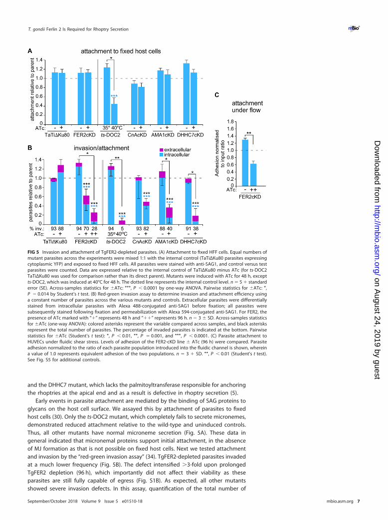
Early events in parasite attachment are mediated by the binding of SAG proteins toglycans on the host cell surface. We assayed this by attachment of parasites to fixedhost cells (30). Only the ts-DOC2 mutant, which completely fails to secrete micronemes,demonstrated reduced attachment relative to the wild-type and uninduced controls.Thus, all other mutants have normal microneme secretion (Fig. 5A). These data ingeneral indicated that micronemal proteins support initial attachment, in the absenceof MJ formation as that is not possible on fixed host cells. Next we tested attachmentand invasion by the “red-green invasion assay” (34). TgFER2-depleted parasites invadedat a much lower frequency (Fig. 5B). The defect intensified �3-fold upon prolongedTgFER2 depletion (96 h), which importantly did not affect their viability as theseparasites are still fully capable of egress (Fig. S1B). As expected, all other mutantsshowed severe invasion defects. In this assay, quantification of the total number of
FIG 5 Invasion and attachment of TgFER2-depleted parasites. (A) Attachment to fixed HFF cells. Equal numbers ofmutant parasites across the experiments were mixed 1:1 with the internal control (TaTiΔKu80 parasites expressingcytoplasmic YFP) and exposed to fixed HFF cells. All parasites were stained with anti-SAG1, and control versus testparasites were counted. Data are expressed relative to the internal control of TaTiΔKu80 minus ATc (for ts-DOC2TaTiΔKu80 was used for comparison rather than its direct parent). Mutants were induced with ATc for 48 h, exceptts-DOC2, which was induced at 40°C for 48 h. The dotted line represents the internal control level. n � 5 � standarderror (SE). Across-samples statistics for �ATc: ***, P � 0.0001 by one-way ANOVA. Pairwise statistics for �ATc: *,P � 0.014 by Student’s t test. (B) Red-green invasion assay to determine invasion and attachment efficiency usinga constant number of parasites across the various mutants and controls. Extracellular parasites were differentiallystained from intracellular parasites with Alexa 488-conjugated anti-SAG1 before fixation; all parasites weresubsequently stained following fixation and permeabilization with Alexa 594-conjugated anti-SAG1. For FER2, thepresence of ATc marked with “�” represents 48 h and “��” represents 96 h. n � 3 � SD. Across-samples statisticsfor �ATc (one-way ANOVA): colored asterisks represent the variable compared across samples, and black asterisksrepresent the total number of parasites. The percentage of invaded parasites is indicated at the bottom. Pairwisestatistics for �ATc (Student’s t test): *, P � 0.01, **, P � 0.001, and ***, P � 0.0001. (C) Parasite attachment toHUVECs under fluidic shear stress. Levels of adhesion of the FER2-cKD line � ATc (96 h) were compared. Parasiteadhesion normalized to the ratio of each parasite population introduced into the fluidic channel is shown, whereina value of 1.0 represents equivalent adhesion of the two populations. n � 3 � SD. **, P � 0.01 (Student’s t test).See Fig. S5 for additional controls.
T. gondii Ferlin 2 Is Required for Rhoptry Secretion ®
September/October 2018 Volume 9 Issue 5 e01510-18 mbio.asm.org 7
on August 24, 2019 by guest
http://mbio.asm
.org/D
ownloaded from

parasites per field allows for an estimation of parasite attachment. By this metric, adefect in the attachment of TgFER2-depleted parasites to host cells was observed. By96 h of knockdown, the numbers of parasites attached to host cells dropped nearly4-fold (Fig. 5B). This approaches the levels observed in the ts-DOC2 mutant, whereattachment and invasion are both severely defective. The similarity between thets-DOC2 mutant, where the primary defect is in attachment, and the DHHC7 mutant,with a penetration defect, highlights that this assay is unable to distinguish betweenthese two interconnected phenotypes.
It has been observed that the motility and attachment dynamics of Toxoplasma aredifferent under conditions of shear stress (35). To investigate whether these conditionsmight better clarify the phenotype of the TgFER2 knockdown, we measured the abilityof FER2-cKD parasites � ATc to adhere to human vascular endothelial cells (HUVECs)under flow. Depletion of TgFER2 led to a significant decrease in the number of parasitesretained in the chamber (Fig. 5C; see Fig. S5 in the supplemental material), althoughattachment of TgFER2-depleted parasites was less compromised under flow relative tostatic conditions. This further underscores that TgFER2 is essential for invasion but doesnot pinpoint the nature of the defect. FER2-cKD parasites were then scrutinized for theirinteractions with host cells by video microscopy. Both wild-type and FER2-cKD parasiteswere able to glide across the host cells. In contrast to control parasites, TgFER2-depleted parasites were not able to invade host cells (Fig. 6; see Movie S1 in thesupplemental material). Surprisingly, FER2-cKD parasites still exhibited “impulse motil-ity” characteristic of invading parasites (36). This typical burst of forward motionimmediately preceding invasion is followed by a momentary pause when parasitessecrete the RONs and create the MJ before proceeding with invasion and subsequentparasitophorous vacuole formation. Both control and induced FER2-cKD parasitesdisplayed bursts of impulse motility followed by a pause. However, only in controlparasites was this pause followed by forward motion (invasion) at approximately halfthe original speed. In contrast, the velocity of induced parasites dropped essentially to0 �m/s, and they failed to invade. This observation suggests that TgFER2 functions inthe very late stages of invasion and indicates that the MJ either is not formed or is notof sufficient strength to support the force required for parasite penetration into cells.
TgFER2 is required for rhoptry secretion. As shown in Fig. 3 and Fig. S3 and S6in the supplemental material, the localization, morphology, and apical anchoring of therhoptries were not affected by TgFER2 depletion. Thus, FER2 is not required to tetherthe rhoptries at the apical end of the parasite. To test rhoptry function, we firstmonitored the release of rhoptry neck proteins by tracking RON4 distribution andassaying MJ formation (Fig. 7A). In the noninduced control, we readily observed MJ
FIG 6 Impulse motility and host cell invasion. (A) Still panels from movies collected in Movie S1 recorded with FER2-cKDparasites � ATc in the presence of host cells. The TgFER2-replete parasite marked with the asterisk invades the host cellat the arrowhead. Invasion is complete in 20 s. The TgFER2-depleted parasite marked with asterisk makes an impulse moveto the arrowhead and appears to deform the host cell. However, the parasite does not invade and disengages from thehost cell, reversing the deformation in the 40-s frame. (B) Velocity profiles of FER2-cKD parasites � ATc. The red arrowheadmarks the synchronized frame where the parasites minus ATc invade or the parasites plus ATc engage the host cell. Eachthin line represents a single parasite from a single movie; heavy lines represent mean values for all parasites in each groupincluded in the graph. Note that both sets of parasites show an impulse in motility right before the point of invasion/engagement, followed by an immediate pause, but that only the TgFER2-replete parasites maintain a positive velocityduring the actual host cell invasion (magnified in the inset).
Coleman et al. ®
September/October 2018 Volume 9 Issue 5 e01510-18 mbio.asm.org 8
on August 24, 2019 by guest
http://mbio.asm
.org/D
ownloaded from

formation, but no MJ formation was detected in the absence of TgFER2. These datasuggest that the RON proteins are either not secreted, or if they are, they fail toassemble into the MJ.
We next performed “evacuole” assays to determine the quantity and quality ofrhoptry protein secretion in TgFER2 mutants. Evacuoles, which are rhoptry proteinclusters injected in the host cell cytosol, were visualized with ROP1 antiserum andclassified as illustrated in Fig. 7B (33). We used the AMA1-cKD as control for partialrhoptry secretion and weak MJ strength (32, 33) and DHHC7-cKD parasites as a controlfor defective rhoptry secretion (5). To examine the strength of the overall parasite-hostcell interaction, the number of parasites per field was counted and differentiated bywhether they were associated with evacuoles (Fig. 7C). More specifically, the strengthof the MJ itself can be measured by assaying the number of evacuoles per field andwhether they are associated with parasites (Fig. 7D). Levels of rhoptry secretion weredifferentiated by the relative size of evacuole patterns (Fig. 7E): small punctate ROP1staining indicates less secretion than long trails or clusters. For FER2-cKD parasites, thenumber of parasites per field was consistent with the observations from the attachmentand invasion assays: depletion of TgFER2 decreased parasite attachment (Fig. 7C).Among the parasites that were attached, very few were associated with evacuoles,indicating that they have not secreted the rhoptries’ contents into the host cell. TheTgFER2 data are comparable with the results for the DHHC7 mutant. However, theydiffered from parasites that lack AMA1, where few parasites attach but the majority ofthe parasites have secreted rhoptries and generated evacuoles (32, 33). TgFER2-
FIG 7 Rhoptry secretion of TgFER2-depleted parasites is impaired. (A) Formation of the MJ. A constant number of parasites across the various mutants andcontrols were incubated with host cells for 10 min. MJ formation was visualized with RON4 antiserum under semipermeabilizing conditions by 0.02% saponin.SAG1 stains the extracellular portion of the parasites. Arrowheads mark successfully invaded parasites that are not accessible to the SAG1 antibodies. Brightnessand contrast adjustments were made identical for both conditions, and, thus, signals are directly comparable. (B) Representative examples of parasite andevacuole features scored in the evacuole assay represented in panels C to E. na, not applicable. (C to E) Evacuole assay to monitor rhoptry bulb secretion andassess stability of the MJ attachment. Parasites as indicated were grown under ATc for 48 h (�) or 96 h (��) and incubated with host cells for 10 min. Evacuoleformation was visualized using ROP1 antiserum following paraformaldehyde fixation. n � 3 � SD. For across-samples statistics for �ATc (one-way ANOVA),correction is marked above the bar. Pairwise statistics for �ATc (Student’s t test) are marked above the connector line. *, P � 0.01; **, P � 0.001; ***, P � 0.0001.Asterisk color corresponds with the variable compared across samples; black asterisks correspond with analysis of the total number of parasites. Indicated belowthe graphs in panels C, D, and E are the percentages of the total number of events recorded that represent association with the evacuole (evac.) or parasite(par.) or display trails/groups of evacuoles, respectively. The data presented in these panels are derived from the same experiments.
T. gondii Ferlin 2 Is Required for Rhoptry Secretion ®
September/October 2018 Volume 9 Issue 5 e01510-18 mbio.asm.org 9
on August 24, 2019 by guest
http://mbio.asm
.org/D
ownloaded from

depleted parasites, like the DHHC7 mutant, appear to secrete very few, if any, rhoptries.When we assess how many of the observed evacuoles are associated with parasites, itbecomes clear that TgFER2 depletion is much more similar to DHHC7 depletion than toparasites lacking AMA1 (Fig. 7D). Finally, we observed relatively few extensive evacuolepatterns for both TgFER2- and DHHC7-depleted parasites (Fig. 7E) and conclude that inthe rare events of rhoptry secretion, very little material was released. Overall, weconclude that TgFER2 is required for the secretion of the rhoptries, which is necessaryto invade host cells.
DISCUSSION
Micronemes and rhoptries are essential to the invasion of apicomplexan parasites.These fascinating cellular structures are likely derived from ancestral organelles thatpersist in modern predatory protozoa and were adapted during the evolution of theApicomplexa’s intracellular, parasitic lifestyle (37). While the molecular details of theinitiation of microneme secretion are incompletely understood, the critical role ofintracellular Ca2� fluxes has been known for decades. Far less is known about either themechanisms of rhoptry secretion or the trafficking of their contents.
It has long been hypothesized that secretion from both micronemes and/or rhop-tries requires a membrane fusion event, but evidence for a canonical secretion ma-chinery has been elusive. Using C2 domains as the anchor for a bioinformatic search forpotential components of this pathway, we were unable to find homologs for eithersynaptotagmins or the canonical DOC2 family of Ca2� sensors that function in mam-malian neurotransmitter release (18, 38). We did find orthologs of the ferlin family ofCa2�-sensing membrane fusion proteins. TgFER1 and TgFER2 are widely conservedacross the Apicomplexa, whereas the degenerate TgFER3 is found only in the Coccidiaand a single Chromerid species, illustrative of the ancient history of these processes.
Detailed studies of Toxoplasma FER2 demonstrated its requirement for secretionfrom the rhoptries. This finding provides one of the first mechanistic insights intorhoptry secretion, firmly linking it to the activity of this C2 domain-containing protein.It is generally accepted that rhoptry secretion must be preceded by micronemesecretion and requires contact with an appropriate host cell (4, 39). However, neitherthe transduction of this attachment signal nor the process by which it leads to secretionof the organelle’s contents has been clarified. Although it is known that Mic8 is requiredfor rhoptry release and has been postulated to be key in a signal transduction pathway(40), there are no experimental data supporting this model. Furthermore, AMA1 (33)and RON5 (41) also appear to be involved in rhoptry secretion, but these mechanismsare equally unknown. Our finding that the Ca2� sensor TgFER2 is required for rhoptrysecretion provides a tantalizing hint at the molecular mechanism. The presence of anAsp residues constellation consistent with Ca2�-binding capacity in TgFER2’s C2Fdomain supports this model (see Fig. S7 in the supplemental material). This is consis-tent with Ca2� binding being restricted to the C2E and C2F domains in mammalianferlins (21). We have as yet been unable to definitively demonstrate a role for Ca2� inTgFER2 function specifically. However, ferlins are Ca2�-sensing proteins, and it is wellestablished that a rise in [Ca2�]i accompanies host cell invasion (2). Thus, while theconventional belief has been that this fluctuation acted only on activation of motility,conoid extrusion, and microneme secretion, our collective evidence provides a hint thatrhoptry secretion may be similarly dependent on variations in [Ca2�]i. The relativeimportance of the protein’s individual C2 domains in this process and their relativeCa2�-binding abilities are exciting open questions to be experimentally determined.
Of the mammalian ferlins, otoferlin is currently the best studied, yet its mechanismof action remains poorly understood (42). Otoferlin is expressed in many tissues, but incochlear hair cells (CHCs) it controls the release of neurotransmitter upon an increasein [Ca2�]i (43, 44). A rise in [Ca2�]i leads otoferlin to interact with phospholipids (42) andSNARE proteins in vitro (45), although SNAREs have been debated to be absent from thesite of secretion in CHCs (46). This highlights the potential for ferlin proteins to facilitate
Coleman et al. ®
September/October 2018 Volume 9 Issue 5 e01510-18 mbio.asm.org 10
on August 24, 2019 by guest
http://mbio.asm
.org/D
ownloaded from

membrane fusion in the absence of SNAREs, an important parallel to Toxoplasma, inwhich there is currently no evidence for either rhoptry- or microneme-resident SNAREs.
As part of this study, we compared different invasion and egress mutants acrossseveral commonly used assays, which allowed for several important observations. First,the fixed host cell attachment data (MJ absent) demonstrate that microneme proteinscontribute a large portion to the attachment strength. Somewhat unexpectedly, thered-green invasion assay did not differentiate the various mutants very well, with theexception of the partial attachment defect previously reported for the CnA mutant (9).Thus, this assay is not capable of specifically attributing individual phenotypes todefects in attachment versus invasion. In contrast, the evacuole assay was very pow-erful in differentiating different aspects of MJ formation and rhoptry secretion.
Overall, our findings support two interesting hypotheses. First, if ferlins act as Ca2�
sensors during Ca2�-dependent secretion in the Apicomplexa, TgFER2 may representthe link between the previously observed Ca2� fluctuations during invasion and thewell-described mechanics of MJ formation. If on the other hand, the essential role ofTgFER2 during rhoptry secretion is calcium independent, this would signify a fascinat-ing evolutionary divergence from the canonical function of ferlins as Ca2� sensors.While additional work will be required to distinguish between these models, the workpresented here is a critical step in our understanding of these critical virulenceprocesses.
MATERIALS AND METHODSParasites and mammalian cell lines. Transgenic derivatives of the RH strain were maintained in
human foreskin fibroblasts (HFFs) as previously described (47). For the attachment assay under fluidicshear stress, HUVECs were cultured in EGM-2 medium containing EGM-2 SingleQuot supplements andgrowth factors (Lonza, Allendale, NJ). TgFER2 CDS was amplified using primers YFP-FER2-F/R andNheI/EcoRV cloned into tub-YFPYFP(MCS)/sagCAT (48) to generate ptub-YFP-FER2/sagCAT, which wasused for Sanger sequencing validation of the gene model. FER2-cKD parasites were generated byBglII/NotI cloning of PCR-amplified FER2 sequence (primers BamHI-FER2-F/NotI-FER2-R) into N-terminalMyc epitope-tagged plasmid derived from dihydrofolate reductase (DHFR)-TetO7sag4-Nt-GOI (WassimDaher, Université de Montpellier) and linearized by XbaI prior to transfection. ts-DOC2 parasites weregenerated by first 5xTY tagging the DOC2 locus using the PCR amplicon (primers 5xTy_upstream_F/5xTy_PlusLink_R) from plasmid pLIC-5xTY-DD24/HX (Chris Tonkin, Walter and Eliza Hall Institute) andBglII/EcoRV cloning into tub-YFPYFP(MCS)/sagCAT. The tub promoter was PmeI/BglII replaced with the3= DOC2 homologous region PCR amplified from genomic DNA (gDNA [primers DOC2_3-target_F/R]).The CAT cassette was PmeI/NotI replaced with a DHFR minigene cassette and plasmid NheI linearizedprior to transfection. A CRISPR/Cas9 (clustered regularly interspaced short palindromic repeats with Cas9)plasmid was generated to mutate DOC2 F124 to S124 using primers DOC2_proto_F/R (49) and cotrans-fected with hybridized oligonucleotides DOC2_FM�SV_F/R in RHΔKu80ΔHX-DOC2-5xTY parasites. Allprimer sequences are provided in Table S1 in the supplemental material.
Imaging. The following antisera were used: anti-Myc monoclonal antibody (MAb) 9E10, anti-SAG1MAb DG52 (50), anti-Mic2 MAb 6D10 (51), mouse anti-AMA1 (33), rabbit anti-Mic3 (30), rabbit anti-Mic5(52), rabbit anti-Mic8 (53), and mouse anti-ROP1 (54). Alexa 488- or 594-conjugated secondary antibodieswere used (Invitrogen). Images were collected on a Zeiss Axiovert 200 M wide-field fluorescencemicroscope, and images were deconvolved and adjusted for phase contrast using Volocity software(Perkin Elmer).
Egress assay. The egress assay was performed as described previously (9, 14). Freshly lysed parasites,pretreated � ATc for 24 h, were inoculated into HFF cells and incubated � ATc for an additional 24 h.For 96 h, parasites treated � ATc for 68 h were inoculated and incubated � ATc for an additional 30 h.Egress was triggered by treatment with 2 �M A23187 or dimethyl sulfoxide (DMSO) at 37°C for 5 min,followed by IFA with rat anti-IMC3 (48). Intact vacuoles were counted for each sample in at least 10 fields,and the percentage of egress was calculated relative to the DMSO control.
Attachment and invasion. The combined attachment/invasion assay was performed as previouslypublished (14, 34) with modifications described in reference 9. Tachyzoites treated � ATc for the hourindicated (ts-DOC2 parasites incubated at 35 and 40°C) were added to host cells in a 96-well plate,centrifuged (28 � g, 3 min, room temperature), and allowed to invade for 1 h at 37°C. Noninvadedextracellular parasites were detected using Alexa 594-conjugated anti-SAG1 T41E5 (55). Followingfixation and permeabilization, all parasites were visualized with Alexa 488-conjugated anti-SAG1 T41E5.At least 300 parasites were counted per sample.
Attachment to fixed host cells. Assay was performed as previously described (33). HFF confluent96-well optical bottom plates were fixed with 3% formaldehyde (PFA) plus 0.06% glutaraldehyde for5 min at 4°C, followed by overnight 0.16 M ethanolamine quenching at 4°C. Wells were prerinsed with0.2% bovine serum albumin (BSA) in Dulbecco’s modified Eagle’s medium (DMEM). Cytoplasmic yellowfluorescent protein (YFP)-expressing TATiΔKu80 parasites mixed in a 1:1 ratio were used as an internalcontrol (9), centrifuged (28 � g, 5 min, 20°C) on the monolayer, and incubated for 30 min at 37°C. Wells
T. gondii Ferlin 2 Is Required for Rhoptry Secretion ®
September/October 2018 Volume 9 Issue 5 e01510-18 mbio.asm.org 11
on August 24, 2019 by guest
http://mbio.asm
.org/D
ownloaded from

were rinsed 3 times with phosphate-buffered saline (PBS), fixed with 4% PFA for 30 min at 4°C, andpermeabilized with 0.25% Triton X-100 for 10 min. After blocking with 1% BSA in PBS, the parasites wereprobed with rabbit anti-green fluorescent protein (anti-GFP [Torrey Pines Biolabs]), and mouse anti-SAG1DG52. Three random fields in 3 independent wells were counted.
Attachment under fluidic shear stress. Attachment under fluidic shear stress was performed asdescribed previously (35, 56). Microfluidic channels containing fibronectin were coated overnight withHUVECs. Freshly lysed parasites treated � ATc for 48 or 96 h were either stained with CMTPX CellTrackerred or carboxyfluorescein succinimidyl ester (CFSE [Life Technologies]), counted, and combined 1:1. Ineach replicate experiment, the dyes were switched on the parental and knockdown parasite lines.Parasites were flowed at a shear force of 0.5 dyne/cm2 for 10 min at 37°C and were fixed under flowconditions with 4% PFA for 30 min, followed by imaging on a Nikon Eclipse Ti microscope.
Conoid extrusion assay. The conoid extrusion assay was performed as published (27). Freshly lysedparasites grown � ATc for 48 h were resuspended in 10% fetal bovine serum (FBS) in HS buffer. Conoidextrusion was induced using 0.5 M ethanol or 5 �M A23187 for 30 s. Parasites were fixed and scored forconoid extrusion by phase-contrast microscopy. Samples were counted blindly, scoring more than 350parasites per sample.
Microneme Mic2, Mic8, and Mic10 secretion by Western blotting. Microneme Mic2, Mic8, andMic10 secretion by Western blot was performed as published (31). Freshly lysed parasites treated � ATcfor 48 h were resuspended in DMEM/FBS and added to a 96-well round-bottom plate, and secretion wasinduced by 1 �M A23187 or DMSO for 5 min at 37°C. For constitutive microneme secretion, there wasno stimulation at 37°C for 60 min. Supernatants were probed by Western blotting using MAb 6D10anti-Mic2 (51), rabbit anti-Mic8 (53), rabbit anti-Mic10 (57), and MAb anti-Gra1 (58). Signals werequantified using a densitometer.
Microneme Mic3, Mic5, and Mic8 secretion by IFA. Mic3 (30), Mic5 (52), or Mic8 (53) IFA onparasites exposed to a host cell monolayer was performed as published (31). Parasites resuspended inEndo buffer were spun onto HFF cells in a 6-well plate (28 � g, 5 min, room temperature) and incubatedat 37°C for 20 min. Endo buffer was replaced with a mixture of DMEM, 3% FBS, and 10 mM HEPES (pH7.2) and incubated at 37°C for 5 min. PBS-washed coverslips were fixed with 4% formaldehyde– 0.02%glutaraldehyde followed by IFA in the presence of 0.02% saponin.
Motility assessments. Motility was analyzed by video microscopy essentially as described previously(14). Intracellular tachyzoites grown for 96 h � ATc were physically harvested, resuspended in modifiedRinger’s medium, and added to HFF confluent glass-bottom culture dishes (MatTek). The dish wasimaged using a 63� objective at 37°C. Videos were recorded with 1-s intervals. Velocities of individualinvasion events were analyzed using the ImageJ/FIJI Cell Counter plug-in.
Moving junction formation. Moving junction formation was determined as published (40) withpreviously described modifications (9). Parasites grown � ATc for 48 h were inoculated into a HFFconfluent 24-well plate by centrifugation (28 � g, 5 min, 20°C) and incubated at 37°C for 10 min. Wellswere rinsed twice with PBS, fixed with 4% PFA at 4°C, and partly permeabilized with 0.02% saponin. MJwas detected using rabbit anti-RON4 (7), and all parasites were detected following full permeabilizationwith MAb anti-SAG1 DG52.
Evacuole assay. Evacuoles were determined as described (59) with modifications. A total of 1 � 107
parasites grown � ATc for 48 h were inoculated into HFF confluent 24-well plates. The plate wascentrifuged (28 � g, 15 min, 23°C) and incubated at 37°C for 10 min. Wells were rinsed twice with PBS,fixed with 4% PFA at 4°C, and 0.25% Triton X-100 permeabilized. Evacuoles were detected by MAb Tg49anti-ROP1 (54). More than 100 events per sample per experiment were counted.
Immunoelectron microscopy. Following washing with PBS, overnight-infected HFF cells were fixedin 4% PFA in 0.25 M HEPES (pH 7.4) for 1 h at room temperature and then in 8% PFA in the same bufferovernight at 4°C. They were infiltrated, frozen, and sectioned as previously described (60). Sections wereimmunolabeled with anti-Myc 9E10 in 1% fish skin gelatin and then with goat anti-IgG antibodies,followed by 10-nm protein A-gold particles before examination with a Philips CM120 electron micro-scope under 80 kV.
Transmission electron microscopy. Parasites were fixed in 4% glutaraldehyde in 0.1 M phosphatebuffer (pH 7.4) and processed for routine electron microscopy (61). Briefly, cells were postfixed in osmiumtetroxide and treated with uranyl acetate prior to dehydration in ethanol, treatment with propyleneoxide, and embedding in Spurr’s epoxy resin. Thin sections were stained with uranyl acetate and leadcitrate prior to examination with a JEOL 1200EX electron microscope.
Statistics. Student’s paired t test and one-way analysis of variance (ANOVA) using post hoc Bonfer-roni correction were used where indicated against the TaTiΔKu80 line.
SUPPLEMENTAL MATERIALSupplemental material for this article may be found at https://doi.org/10.1128/mBio
.01510-18.FIG S1, TIF file, 0.1 MB.FIG S2, TIF file, 3.3 MB.FIG S3, TIF file, 2.8 MB.FIG S4, TIF file, 2 MB.FIG S5, TIF file, 0.1 MB.FIG S6, TIF file, 5.1 MB.
Coleman et al. ®
September/October 2018 Volume 9 Issue 5 e01510-18 mbio.asm.org 12
on August 24, 2019 by guest
http://mbio.asm
.org/D
ownloaded from

FIG S7, TIF file, 0.2 MB.TABLE S1, DOCX file, 0.1 MB.MOVIE S1, AVI file, 5.5 MB.
ACKNOWLEDGMENTSWe thank Jamie Henzy for assistance with phylogenetic analysis, Sander Groffen for
assistance with modeling, Amir Bayegan for assistance with statistics, Elizabeth C. Grayfor technical assistance, P. J. Bradley, J. C. Boothroyd, V. B. Carruthers, M. F. Cesbron-Delauw, W. Daher, C. de Graffenried, J. F. Dubremetz, J. Saeij, L. D. Sibley, D. Soldati, B.Striepen, C. Tonkin, and G. E. Ward for sharing reagents, and Manoj Duraisingh forcritically reading the manuscript. We also thank the technical competence of KimberleyZichichi from the Electron Microscopy Facility at Yale University.
This study was supported by NIH AI108251 (B.I.C.), AI060767 (I.C.), AI099658 (M.-J.G.),and AI122923 (M.-J.G.), Deutsche Forschungsgemeinschaft (K.E.), American CancerSociety 126688-RSG-14-202-01-MPC (M.B.L.), and American Cancer Society RSG-12-175-01-MPC (M.-J.G.). The funders had no role in study design, data collection and analysis,decision to publish, or preparation of the manuscript.
REFERENCES1. Kafsack BF, Pena JD, Coppens I, Ravindran S, Boothroyd JC, Carruthers
VB. 2009. Rapid membrane disruption by a perforin-like protein facili-tates parasite exit from host cells. Science 323:530 –533. https://doi.org/10.1126/science.1165740.
2. Wetzel DM, Chen LA, Ruiz FA, Moreno SN, Sibley LD. 2004. Calcium-mediated protein secretion potentiates motility in Toxoplasma gondii. JCell Sci 117:5739 –5748. https://doi.org/10.1242/jcs.01495.
3. Carruthers VB, Tomley FM. 2008. Microneme proteins in apicomplex-ans. Subcell Biochem 47:33–45. https://doi.org/10.1007/978-0-387-78267-6_2.
4. Carruthers V, Boothroyd JC. 2007. Pulling together: an integrated modelof toxoplasma cell invasion. Curr Opin Microbiol 10:83– 89. https://doi.org/10.1016/j.mib.2006.06.017.
5. Beck JR, Fung C, Straub KW, Coppens I, Vashisht AA, Wohlschlegel JA,Bradley PJ. 2013. A Toxoplasma palmitoyl acyl transferase and thepalmitoylated armadillo repeat protein TgARO govern apical rhoptrytethering and reveal a critical role for the rhoptries in host cell invasionbut not egress. PLoS Pathog 9:e1003162. https://doi.org/10.1371/journal.ppat.1003162.
6. Bradley PJ, Sibley LD. 2007. Rhoptries: an arsenal of secreted virulencefactors. Curr Opin Microbiol 10:582–587. https://doi.org/10.1016/j.mib.2007.09.013.
7. Alexander DL, Mital J, Ward GE, Bradley P, Boothroyd JC. 2005. Identifi-cation of the moving junction complex of Toxoplasma gondii: a collab-oration between distinct secretory organelles. PLoS Pathog 1:e17.https://doi.org/10.1371/journal.ppat.0010017.
8. Mercier C, Cesbron-Delauw MF. 2015. Toxoplasma secretory granules:one population or more? Trends Parasitol 31:604. https://doi.org/10.1016/j.pt.2015.02.002.
9. Paul AS, Saha S, Engelberg K, Jiang RH, Coleman BI, Kosber AL, Chen CT,Ganter M, Espy N, Gilberger TW, Gubbels MJ, Duraisingh MT. 2015.Parasite calcineurin regulates host cell recognition and attachment byapicomplexans. Cell Host Microbe 18:49 – 60. https://doi.org/10.1016/j.chom.2015.06.003.
10. Lourido S, Shuman J, Zhang C, Shokat KM, Hui R, Sibley LD. 2010. Calcium-dependent protein kinase 1 is an essential regulator of exocytosis in Tox-oplasma. Nature 465:359–362. https://doi.org/10.1038/nature09022.
11. Garrison E, Treeck M, Ehret E, Butz H, Garbuz T, Oswald BP, Settles M,Boothroyd J, Arrizabalaga G. 2012. A forward genetic screen reveals thatcalcium-dependent protein kinase 3 regulates egress in Toxoplasma.PLoS Pathog 8:e1003049. https://doi.org/10.1371/journal.ppat.1003049.
12. Lourido S, Tang K, Sibley LD. 2012. Distinct signalling pathways controlToxoplasma egress and host-cell invasion. EMBO J 31:4524–4534. https://doi.org/10.1038/emboj.2012.299.
13. McCoy JM, Whitehead L, van Dooren GG, Tonkin CJ. 2012. TgCDPK3regulates calcium-dependent egress of Toxoplasma gondii from hostcells. PLoS Pathog 8:e1003066. https://doi.org/10.1371/journal.ppat.1003066.
14. Farrell A, Thirugnanam S, Lorestani A, Dvorin JD, Eidell KP, Ferguson DJ,Anderson-White BR, Duraisingh MT, Marth GT, Gubbels MJ. 2012. ADOC2 protein identified by mutational profiling is essential for apicom-plexan parasite exocytosis. Science 335:218 –221. https://doi.org/10.1126/science.1210829.
15. Martens S. 2010. Role of C2 domain proteins during synaptic vesicleexocytosis. Biochem Soc Trans 38:213–216. https://doi.org/10.1042/BST0380213.
16. Pang ZP, Sudhof TC. 2010. Cell biology of Ca2�-triggered exocytosis.Curr Opin Cell Biol 22:496 –505. https://doi.org/10.1016/j.ceb.2010.05.001.
17. Sudhof TC. 2013. A molecular machine for neurotransmitter release:synaptotagmin and beyond. Nat Med 19:1227–1231. https://doi.org/10.1038/nm.3338.
18. Walter AM, Groffen AJ, Sorensen JB, Verhage M. 2011. Multiple Ca2�
sensors in secretion: teammates, competitors or autocrats? Trends Neu-rosci 34:487– 497. https://doi.org/10.1016/j.tins.2011.07.003.
19. Martens S, McMahon HT. 2008. Mechanisms of membrane fusion: dis-parate players and common principles. Nat Rev Mol Cell Biol 9:543–556.https://doi.org/10.1038/nrm2417.
20. Lek A, Lek M, North KN, Cooper ST. 2010. Phylogenetic analysis of ferlingenes reveals ancient eukaryotic origins. BMC Evol Biol 10:231. https://doi.org/10.1186/1471-2148-10-231.
21. Lek A, Evesson FJ, Sutton RB, North KN, Cooper ST. 2012. Ferlins:regulators of vesicle fusion for auditory neurotransmission, receptortrafficking and membrane repair. Traffic 13:185–194. https://doi.org/10.1111/j.1600-0854.2011.01267.x.
22. Woo YH, Ansari H, Otto TD, Klinger CM, Kolisko M, Michálek J, Saxena A,Shanmugam D, Tayyrov A, Veluchamy A, Ali S, Bernal A, del Campo J,Cihlár J, Flegontov P, Gornik SG, Hajdušková E, Horák A, Janouškovec J,Katris NJ, Mast FD, Miranda-Saavedra D, Mourier T, Naeem R, Nair M,Panigrahi AK, Rawlings ND, Padron-Regalado E, Ramaprasad A, Samad N,Tomcala A, Wilkes J, Neafsey DE, Doerig C, Bowler C, Keeling PJ, Roos DS,Dacks JB, Templeton TJ, Waller RF, Lukeš J, Oborník M, Pain A. 2015.Chromerid genomes reveal the evolutionary path from photosyntheticalgae to obligate intracellular parasites. elife 4 https://doi.org/10.7554/eLife.06974.
23. Meissner M, Schluter D, Soldati D. 2002. Role of Toxoplasma gondiimyosin A in powering parasite gliding and host cell invasion. Science298:837– 840. https://doi.org/10.1126/science.1074553.
24. Ahras M, Otto GP, Tooze SA. 2006. Synaptotagmin IV is necessary for thematuration of secretory granules in PC12 cells. J Cell Physiol 173:241–251. https://doi.org/10.1083/jcb.200506163.
25. Ibata K, Fukuda M, Hamada T, Kabayama H, Mikoshiba K. 2000. Synap-totagmin IV is present at the Golgi and distal parts of neurites. JNeurochem 74:518 –526.
26. Mori Y, Fukuda M. 2011. Synaptotagmin IV acts as a multi-functional
T. gondii Ferlin 2 Is Required for Rhoptry Secretion ®
September/October 2018 Volume 9 Issue 5 e01510-18 mbio.asm.org 13
on August 24, 2019 by guest
http://mbio.asm
.org/D
ownloaded from

regulator of Ca2�-dependent exocytosis. Neurochem Res 36:1222–1227.https://doi.org/10.1007/s11064-010-0352-7.
27. Mondragon R, Frixione E. 1996. Ca(2�)-dependence of conoid extrusionin Toxoplasma gondii tachyzoites. J Eukaryot Microbiol 43:120 –127.https://doi.org/10.1111/j.1550-7408.1996.tb04491.x.
28. Carruthers VB, Moreno SN, Sibley LD. 1999. Ethanol and acetaldehydeelevate intracellular [Ca2�] and stimulate microneme discharge in Tox-oplasma gondii. Biochem J 342:379 –386. https://doi.org/10.1042/bj3420379.
29. Kremer K, Kamin D, Rittweger E, Wilkes J, Flammer H, Mahler S, Heng J,Tonkin CJ, Langsley G, Hell SW, Carruthers VB, Ferguson DJ, Meissner M.2013. An overexpression screen of Toxoplasma gondii Rab-GTPasesreveals distinct transport routes to the micronemes. PLoS Pathog9:e1003213. https://doi.org/10.1371/journal.ppat.1003213.
30. Garcia-Reguet N, Lebrun M, Fourmaux MN, Mercereau-Puijalon O, MannT, Beckers CJ, Samyn B, Van Beeumen J, Bout D, Dubremetz JF. 2000. Themicroneme protein MIC3 of Toxoplasma gondii is a secretory adhesinthat binds to both the surface of the host cells and the surface of theparasite. Cell Microbiol 2:353–364. https://doi.org/10.1046/j.1462-5822.2000.00064.x.
31. Carruthers VB, Sibley LD. 1999. Mobilization of intracellular calciumstimulates microneme discharge in Toxoplasma gondii. Mol Microbiol31:421– 428. https://doi.org/10.1046/j.1365-2958.1999.01174.x.
32. Lamarque MH, Roques M, Kong-Hap M, Tonkin ML, Rugarabamu G, MarqJB, Penarete-Vargas DM, Boulanger MJ, Soldati-Favre D, Lebrun M. 2014.Plasticity and redundancy among AMA-RON pairs ensure host cell entryof toxoplasma parasites. Nat Commun 5:4098. https://doi.org/10.1038/ncomms5098.
33. Mital J, Meissner M, Soldati D, Ward GE. 2005. Conditional expression ofToxoplasma gondii apical membrane antigen-1 (TgAMA1) demonstratesthat TgAMA1 plays a critical role in host cell invasion. Mol Biol Cell16:4341– 4349. https://doi.org/10.1091/mbc.e05-04-0281.
34. Carey KL, Westwood NJ, Mitchison TJ, Ward GE. 2004. A small-moleculeapproach to studying invasive mechanisms of Toxoplasma gondii. ProcNatl Acad Sci U S A 101:7433–7438. https://doi.org/10.1073/pnas.0307769101.
35. Harker KS, Jivan E, McWhorter FY, Liu WF, Lodoen MB. 2014. Shear forcesenhance Toxoplasma gondii tachyzoite motility on vascular endothe-lium. mBio 5:e01111-13. https://doi.org/10.1128/mBio.01111-13.
36. Bichet M, Joly C, Hadj Henni A, Guilbert T, Xemard M, Tafani V, Lagal V,Charras G, Tardieux I. 2014. The Toxoplasma-host cell junction is an-chored to the cell cortex to sustain parasite invasive force. BMC Biol12:773. https://doi.org/10.1186/s12915-014-0108-y.
37. Gubbels MJ, Duraisingh MT. 2012. Evolution of apicomplexan secretoryorganelles. Int J Parasitol 42:1071–1081. https://doi.org/10.1016/j.ijpara.2012.09.009.
38. Shin OH. 2014. Exocytosis and synaptic vesicle fusion. Compr Physiol4:149 –175. https://doi.org/10.1002/cphy.c130021.
39. Carruthers VB, Sibley LD. 1997. Sequential protein secretion from threedistinct organelles of Toxoplasma gondii accompanies invasion of hu-man fibroblasts. Eur J Cell Physiol 73:114 –123.
40. Kessler H, Herm-Gotz A, Hegge S, Rauch M, Soldati-Favre D, FrischknechtF, Meissner M. 2008. Microneme protein 8 —a new essential invasionfactor in Toxoplasma gondii. J Cell Sci 121:947–956. https://doi.org/10.1242/jcs.022350.
41. Beck JR, Chen AL, Kim EW, Bradley PJ. 2014. RON5 is critical for organi-zation and function of the Toxoplasma moving junction complex. PLoSPathog 10:e1004025. https://doi.org/10.1371/journal.ppat.1004025.
42. Pangrsic T, Reisinger E, Moser T. 2012. Otoferlin: a multi-C2 domainprotein essential for hearing. Trends Neurosci 35:671– 680. https://doi.org/10.1016/j.tins.2012.08.002.
43. Roux I, Safieddine S, Nouvian R, Grati M, Simmler MC, Bahloul A, Perfet-tini I, Le Gall M, Rostaing P, Hamard G, Triller A, Avan P, Moser T, Petit C.2006. Otoferlin, defective in a human deafness form, is essential forexocytosis at the auditory ribbon synapse. Cell 127:277–289. https://doi.org/10.1016/j.cell.2006.08.040.
44. Pangrsic T, Lasarow L, Reuter K, Takago H, Schwander M, Riedel D, FrankT, Tarantino LM, Bailey JS, Strenzke N, Brose N, Muller U, Reisinger E,Moser T. 2010. Hearing requires otoferlin-dependent efficient replenish-ment of synaptic vesicles in hair cells. Nat Neurosci 13:869 – 876. https://doi.org/10.1038/nn.2578.
45. Johnson CP, Chapman ER. 2010. Otoferlin is a calcium sensor that
directly regulates SNARE-mediated membrane fusion. J Cell Physiol191:187–197. https://doi.org/10.1083/jcb.201002089.
46. Nouvian R, Neef J, Bulankina AV, Reisinger E, Pangrsic T, Frank T, SikorraS, Brose N, Binz T, Moser T. 2011. Exocytosis at the hair cell ribbonsynapse apparently operates without neuronal SNARE proteins. NatNeurosci 14:411– 413. https://doi.org/10.1038/nn.2774.
47. Roos DS, Donald RG, Morrissette NS, Moulton AL. 1994. Molecular toolsfor genetic dissection of the protozoan parasite Toxoplasma gondii.Methods Cell Biol 45:27– 63.
48. Anderson-White BR, Ivey FD, Cheng K, Szatanek T, Lorestani A, BeckersCJ, Ferguson DJ, Sahoo N, Gubbels MJ. 2011. A family of intermediatefilament-like proteins is sequentially assembled into the cytoskeleton ofToxoplasma gondii. Cell Microbiol 13:18 –31. https://doi.org/10.1111/j.1462-5822.2010.01514.x.
49. Sidik SM, Hackett CG, Tran F, Westwood NJ, Lourido S. 2014. Efficientgenome engineering of Toxoplasma gondii using CRISPR/Cas9. PLoSOne 9:e100450. https://doi.org/10.1371/journal.pone.0100450.
50. Burg JL, Perelman D, Kasper LH, Ware PL, Boothroyd JC. 1988. Molecularanalysis of the gene encoding the major surface antigen of Toxoplasmagondii. J Immunol 141:3584 –3591.
51. Wan KL, Carruthers VB, Sibley LD, Ajioka JW. 1997. Molecular character-isation of an expressed sequence tag locus of Toxoplasma gondii en-coding the micronemal protein MIC2. Mol Biochem Parasitol 84:203–214. https://doi.org/10.1016/S0166-6851(96)02796-X.
52. Brydges SD, Zhou XW, Huynh MH, Harper JM, Mital J, Adjogble KD,Daubener W, Ward GE, Carruthers VB. 2006. Targeted deletion of MIC5enhances trimming proteolysis of Toxoplasma invasion proteins. Eu-karyot Cell 5:2174 –2183. https://doi.org/10.1128/EC.00163-06.
53. Meissner M, Reiss M, Viebig N, Carruthers VB, Toursel C, Tomavo S, AjiokaJW, Soldati D. 2002. A family of transmembrane microneme proteins ofToxoplasma gondii contain EGF-like domains and function as escorters.J Cell Sci 115:563–574.
54. Saffer LD, Mercereau-Puijalon O, Dubremetz JF, Schwartzman JD. 1992.Localization of a Toxoplasma gondii rhoptry protein by immunoelectronmicroscopy during and after host cell penetration. J Protozool 39:526 –530. https://doi.org/10.1111/j.1550-7408.1992.tb04844.x.
55. Couvreur G, Sadak A, Fortier B, Dubremetz JF. 1988. Surface antigens ofToxoplasma gondii. Parasitology 97:1–10. https://doi.org/10.1017/S0031182000066695.
56. Ueno N, Harker KS, Clarke EV, McWhorter FY, Liu WF, Tenner AJ, LodoenMB. 2014. Real-time imaging of Toxoplasma-infected human monocytesunder fluidic shear stress reveals rapid translocation of intracellularparasites across endothelial barriers. Cell Microbiol 16:580 –595. https://doi.org/10.1111/cmi.12239.
57. Hoff EF, Cook SH, Sherman GD, Harper JM, Ferguson DJ, Dubremetz JF,Carruthers VB. 2001. Toxoplasma gondii: molecular cloning and charac-terization of a novel 18-kDa secretory antigen, TgMIC10. Exp Parasitol97:77– 88. https://doi.org/10.1006/expr.2000.4585.
58. Cesbron-Delauw MF, Guy B, Torpier G, Pierce RJ, Lenzen G, Cesbron JY,Charif H, Lepage P, Darcy F, Lecocq JP. 1989. Molecular characterization ofa 23-kilodalton major antigen secreted by Toxoplasma gondii. Proc NatlAcad Sci U S A 86:7537–7541. https://doi.org/10.1073/pnas.86.19.7537.
59. Hakansson S, Charron AJ, Sibley LD. 2001. Toxoplasma evacuoles: a two-step process of secretion and fusion forms the parasitophorous vacuole.EMBO J 20:3132–3144. https://doi.org/10.1093/emboj/20.12.3132.
60. Folsch H, Pypaert M, Schu P, Mellman I. 2001. Distribution and functionof AP-1 clathrin adaptor complexes in polarized epithelial cells. J CellPhysiol 152:595– 606.
61. Ferguson DJ, Cesbron-Delauw MF, Dubremetz JF, Sibley LD, Joiner KA,Wright S. 1999. The expression and distribution of dense granule pro-teins in the enteric (coccidian) forms of Toxoplasma gondii in the smallintestine of the cat. Exp Parasitol 91:203–211. https://doi.org/10.1006/expr.1998.4384.
62. Kearse M, Moir R, Wilson A, Stones-Havas S, Cheung M, Sturrock S,Buxton S, Cooper A, Markowitz S, Duran C, Thierer T, Ashton B,Meintjes P, Drummond A. 2012. Geneious basic: an integrated andextendable desktop software platform for the organization and anal-ysis of sequence data. Bioinformatics 28:1647–1649. https://doi.org/10.1093/bioinformatics/bts199.
63. Jimenez JL, Bashir R. 2007. In silico functional and structural characterisationof ferlin proteins by mapping disease-causing mutations and evolutionaryinformation onto three-dimensional models of their C2 domains. J NeurolSci 260:114–123. https://doi.org/10.1016/j.jns.2007.04.016.
Coleman et al. ®
September/October 2018 Volume 9 Issue 5 e01510-18 mbio.asm.org 14
on August 24, 2019 by guest
http://mbio.asm
.org/D
ownloaded from