a modelling approach for investigating opto- mechanical...
TRANSCRIPT
This work is licensed under a Creative Commons Attribution 4.0 License. For more information, see https://creativecommons.org/licenses/by/4.0/.
This article has been accepted for publication in a future issue of this journal, but has not been fully edited. Content may change prior to final publication. Citation information: DOI 10.1109/TBME.2019.2927390, IEEETransactions on Biomedical Engineering
> TBME-00519-2019<
1
Abstract—Objective: The human visual system alters its focus by
a shape change of the eye lens. The extent to which the lens can
adjust ocular refractive power is dependent to a significant extent
on its material properties. Yet, this fundamental link between the
optics and mechanics of the lens has been relatively under
investigated. This study aims to investigate this opto-mechanical
link within the eye lens to gain insight into the processes of shape
alteration and their respective decline with age. Methods: Finite
Element models based on biological lenses were developed for five
ages: 16, 35, 40, 57 and 62 years by correlating in vivo
measurements of the longitudinal modulus using Brillouin
scattering with in vitro X-ray interferometric measurements of
refractive index and taking into account various directions of
zonular force. Results: A model with radial cortical Young’s
moduli provides the same amount of refractive power with less
change in thickness than a model with uniform cortical Young’s
modulus with a uniform stress distribution and no discontinuities
along the cortico-nuclear boundary. The direction of zonular
angles can significantly influence curvature change regardless of
the modulus distribution. Conclusions: The present paper
proposes a modelling approach for the human lens, coupling
optical and mechanical properties, which shows the effect of
parameter choice on model response. Significance: This advanced
modelling approach, considering the important interplay between
optical and mechanical properties, has potential for use in design
of accommodating implant lenses and for investigating non-
biological causes of pathological processes in the lens (e.g.
cataract).
Index Terms— Opto-Mechanical modelling, Finite Element
Analysis, Human eye lens, Accommodation, Radial cortical
Young’s moduli, Zonules.
I. INTRODUCTION
HE eye is a complex optical and neurological system for
refracting light to produce high quality images that undergo
processing at the retina and further processing, via the higher
visual pathways, in the visual cortex of the brain. The two
refractive elements in the eye are the cornea and the lens. The
cornea provides approximately two-thirds of the ocular
focusing power. The lens contributes the remainder and is
Copyright (c) 2017 IEEE. Personal use of this material is permitted.
However, permission to use this material for any other purposes must be
obtained from the IEEE by sending an email to [email protected].
This work was supported in part by Zeiss Meditec AG, Royal Society Grant
no IE160996 and beamtime grants at SPring-8 synchrotron (grant numbers: 2014A1710, 2015A1864 and 2016A1096).
responsible for adjusting the refractive power of the eye, via a
process called accommodation, to meet the visual demands over
a range of object distances. Accommodation decreases
gradually with age such that, by the sixth decade of life, the eye
can no longer focus on near objects [1] [2]. This age-related
process is known as presbyopia.
The lens, which is composed of a lamellar arrangement of
fibre cells and contained within the semi-elastic capsule, adjusts
the focus of the eye by altering its shape [3]. This is mediated
by a ring of suspensory ligaments, collectively called the zonule,
which is connected to the capsule around the equator of the lens
and transmits the forces that alter lens shape from the ciliary
body [3]. The anterior, equatorial and posterior sections of the
zonule originate from different locations on the ciliary body [4].
Yet a number of modelling approaches simplify these forces as
emanating from a single point [5], [6], [7], [8], [9]. Recently it
was shown that separating directions of zonular force across the
three sections makes a substantial difference to the shape
change and renders the modelled simulation closer to the
changes in shape seen in the biological lens [10]. This is
fundamental for understanding the mechanical behaviour of the
different zonular sections and for providing insights needed to
understand the accommodative process and its loss with age.
Such insights may resolve the conflicts between major
accommodative theories [11], [12], [13], [14]. Recent
modelling [10] and experimental studies on monkey lenses [15]
suggest that the equatorial zonular section is less effective when
compared to the anterior and posterior zonular sections in
altering central curvatures and optical power of the lens during
accommodation. This has been previously postulated [12], [16].
Two regions within the lens are broadly recognised: a central
nucleus that comprises approximately two-thirds of the total
lens from the perspective of radial distance, and the outer
cortical region [17], [18]. Whilst there is no biochemically
distinct cortico-nuclear boundary, the refractive index profile of
the human lens indicates a marked difference in magnitude and
variation in refractive index: there is an almost constant
refractive index over the central two-thirds of the lens and a
sharp gradient in the outer third (reviewed in [19]). The
K. Wang and *B.K. Pierscionek are with School of Science and Technology,
Nottingham Trent University, Clifton Campus, Clifton Lane, Nottingham,
NG11 8NS, UK. (correspondence email: [email protected]). D.T. Venetsanos is with School of Mechanical, Aerospace and Automotive
Engineering, Coventry University, Priory Street, Coventry, CV1 5FB, UK.
M. Hoshino, K. Uesugi and N. Yagi are with Japan Synchrotron Radiation Research Institute (Spring-8), 1-1-1, Kouto, Sayo-cho, Sayo-gun, Hyogo 679-
5198 Japan.
A Modelling Approach for Investigating Opto-
Mechanical Relationships in the Human Eye Lens
Kehao Wang, Demetrios T. Venetsanos, Masato Hoshino, Kentaro Uesugi, Naoto Yagi, and Barbara
K. Pierscionek*
T
This work is licensed under a Creative Commons Attribution 4.0 License. For more information, see https://creativecommons.org/licenses/by/4.0/.
This article has been accepted for publication in a future issue of this journal, but has not been fully edited. Content may change prior to final publication. Citation information: DOI 10.1109/TBME.2019.2927390, IEEETransactions on Biomedical Engineering
> TBME-00519-2019<
2
refractive index is linearly related to the concentrations of lens
proteins according to the Gladstone-Dale formula [20]
indicating that this gradient is also linearly related to that of the
protein concentration profile.
Mechanical properties in the living human eye lens have been
measured recently using Brillouin scattering analysis [21]. The
direct relationship between refractive index and elastic modulus
are not known as it has not been possible to measure both
properties in the same lens. However, profiles of longitudinal
elastic modulus, measured along the optic axis of the lens using
in vivo Brillouin scattering analysis [22], bear a close
resemblance to refractive index distributions from in vitro
samples measured using a phase contrast imaging modality: X-
ray Talbot interferometry [23]. Although light rays are utilized
by both measuring techniques, the methods of application are
different from one another. Brillouin scattering analysis relies
on the frequency shift between incident light and scattered light
caused by periodic modulations of refractive index by acoustic
phonons [22], [24], [25]. This Brillouin shift is dependent on
the propagation speed of the acoustic wave and can be
converted to longitudinal modulus using the ρ/n2 ratio (where ρ
is the density and n is the refractive index) of the sample. This
was found to be a constant value across the whole lens [22],
[25]. The X-ray Talbot grating interferometer consists of two
transmission gratings (a phase and an absorption grating) that
are used to create Moiré fringe patterns of X-ray beams after
traversing the sample [26]. Moiré fringes are used to determine
the spatially varying protein densities across the specimen from
which refractive indices are calculated using the Gladstone-
Dale formula [20], [27]. Both techniques have fine resolution:
60 μm for Brillouin analysis [22] and 5.5 μm for interferometric
analysis [26]. The similarity between distributions of refractive
index and longitudinal modulus provides a means of creating
optically relevant and mechanically viable models. Such
models, with gradient profiles, are needed to improve current
understanding of cataract, and to facilitate the design of
optically advanced, accommodating intraocular lenses.
Almost all previous modelling studies assumed uniform
distributions of material properties in the lens nucleus and
cortex, and used lens models based on limited ages [5], [6], [7],
[8], [9]. This study describes advanced models that correlate
distributions of material properties derived from in vitro optical
measurements of refractive index [23] with in vivo mechanical
analyses [22]. Finite Element lens models were created based
on human lenses from five different ages, covering the age
range from the second to the sixth decade of life. A parametric
analysis of 990 different combinations of zonular angles and
hence directions of force on the lens, was conducted for each
model using an exhaustive search scheme developed in a
previous study [10]. The resultant changes in lens thickness and
stress field distributions were analysed to investigate the opto-
mechanical relationship and how this may alter with age.
II. METHOD
Lens models were developed based on human lenses aged:
16, 35, 40, 57 and 62 years and subjected to X-ray Talbot
interferometric analysis to obtain refractive index and lens
shape [23]. Two distributions of cortical Young’s moduli were
modelled for each age: (a) a uniform distribution and (b) a radial
linear nodal distribution calculated from the longitudinal
modulus measured using in vivo Brillouin scattering analysis
[22] on lenses that covered a similar age range as in the optical
study [23].
A. Geometry of models
Boundaries of the lens outer shape were taken from iso-
indicial contours of refractive index reported by Pierscionek et
al. [23]. The contour corresponding to a minimum magnitude
of refractive index, approximately 1.35 [23] was taken as the
outer lens shape.
For each uniform model, the contour corresponding to the
central plateau region, shown on the index profile of each lens
in the sagittal plane along the central optical axes [23], was
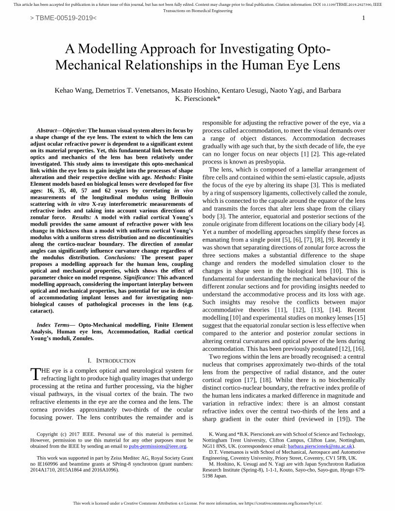
treated as the cortico-nuclear boundary. Fig. 1a shows an
example of how these geometric parameters were extracted for
a 40-year-old lens from X-ray interferometric analysis of the
refractive index gradient.
For models with radially varying Young’s moduli in the
cortex, the nuclear boundary for each model was created by
scaling the boundary of the outer lens using an age-related
scaling ratio that was determined from Besner et al. [22]. A
representative example for determining the nuclear shape by
scaling is shown in Fig. 1b for the 40-year-old lens [23].
B. Analysis of mechanical and optical data
The findings reported by Besner et al. [22] include detailed
parameters describing the profile shapes of longitudinal moduli
measured along the optical axis of each lens. For all 56
measured lenses, the Total Thickness (TT) and Nuclear
Thickness (NT) can be determined using the following
parameters:
𝑁𝑇 = 𝑥𝑐𝑝𝑜𝑠 − 𝑥𝑐𝑎𝑛𝑡 (1)
𝑇𝑇 = 𝐿𝑎𝑛𝑡 + 𝐿𝑝𝑜𝑠 + 𝑁𝑇 (2)
where 𝑥𝑐𝑝𝑜𝑠 , 𝑥𝑐𝑎𝑛𝑡 , 𝐿𝑎𝑛𝑡 and 𝐿𝑝𝑜𝑠 are parameters fully
defined in [22]. The ratio (NT/TT) describes the nuclear to total
lens thickness along the optic axis and is shown plotted against
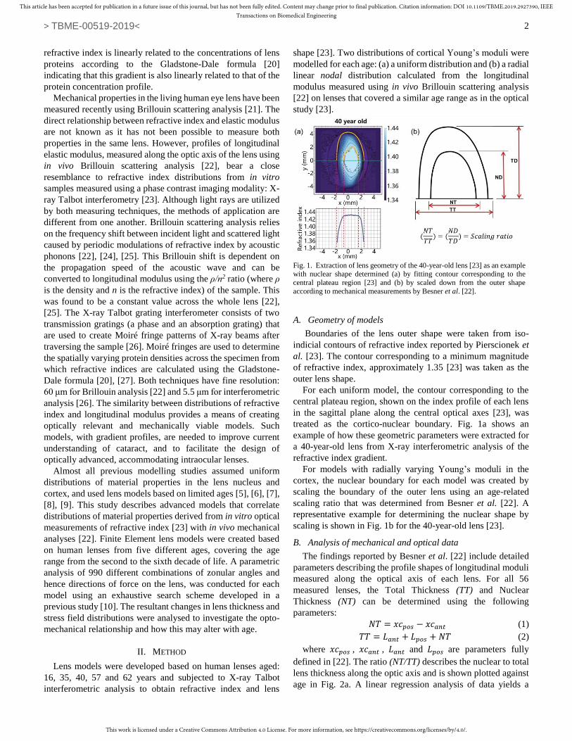
age in Fig. 2a. A linear regression analysis of data yields a
Fig. 1. Extraction of lens geometry of the 40-year-old lens [23] as an example with nuclear shape determined (a) by fitting contour corresponding to the
central plateau region [23] and (b) by scaled down from the outer shape
according to mechanical measurements by Besner et al. [22].
This work is licensed under a Creative Commons Attribution 4.0 License. For more information, see https://creativecommons.org/licenses/by/4.0/.
This article has been accepted for publication in a future issue of this journal, but has not been fully edited. Content may change prior to final publication. Citation information: DOI 10.1109/TBME.2019.2927390, IEEETransactions on Biomedical Engineering
> TBME-00519-2019<
3
relationship of y=0.0031x+0.4712 between age and the (NT/TT)
ratio, where x stands for age and y stands for the (NT/TT) ratio.
The scaling ratio used to determine shapes of lens nuclei for
radial models at each age was calculated from this equation and
is given in Table I.
The geometries of lenses used to measure refractive index by
Pierscionek et al. [23], include profiles both along the optic axis
and along the equatorial plane. The nuclear half-diameter (ND)
and the total half-diameter (TD) for each lens were determined
by measuring the central contour which corresponds to the
plateau region of each refractive index profile (seen in Fig. 1a).
The ratios (NT/TT) and (ND/DD) obtained from lens geometries
for lenses up to 70 years of age are plotted against age in Fig.
2b.
C. Material properties and opto-mechanical coupling
According to the profiles of longitudinal modulus [22], the
magnitude of the central plateau region for 56 lenses is within
the range of 3.278 ± 0.081 GPa with no age dependency. This
value decreases continuously from the central plateau region
toward the anterior and posterior pole of each lens to a
minimum value within the range of 2.498 ± 0.139 GPa [22].
Given that no age-related trend was observed, the average
values at the central plateau and at the lens poles, 3.286 GPa
and 2.471 GPa respectively, were used to construct the models.
Recent studies [25], [28] have derived an empirical relationship
between longitudinal modulus M and conventional low
frequency modulus, i.e. Young’s modulus E or shear modulus
G. This log-log linear equation [25] is described as:
log(𝑀) = 𝑎 log(𝐺) + 𝑏 (3)
where a and b are material dependent coefficients that were
determined for porcine lenses: a=0.093 and b=9.29 [25] which
have similar elastic shear moduli to young human lenses [29],
[reviewed in 30]. Taking the eye lens as nearly incompressible
[31], [32] Young’s and shear modulus can be linearly related:
E=3G [10], [30]. The calculated Young’s moduli E at the
central plateau and at the lens poles are 0.82 kPa and 0.04 kPa
respectively.
Consistent with the findings by Besner et al. [22], no age-
related variations in material properties were considered in the
present study. For both sets of models: those with uniform and
those with a radial distribution of cortical Young’s moduli, a
value of 0.82 kPa was used in the lens nucleus. Models with a
uniform cortical Young’s modulus were given an average value,
0.43 kPa, of the maximum and minimum for each age. For
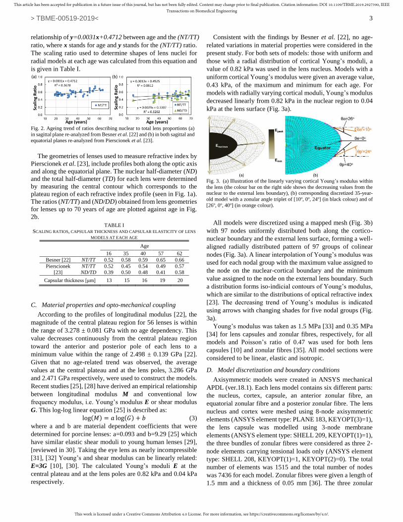
models with radially varying cortical moduli, Young’s modulus
decreased linearly from 0.82 kPa in the nuclear region to 0.04
kPa at the lens surface (Fig. 3a).
All models were discretized using a mapped mesh (Fig. 3b)
with 97 nodes uniformly distributed both along the cortico-
nuclear boundary and the external lens surface, forming a well-
aligned radially distributed pattern of 97 groups of colinear
nodes (Fig. 3a). A linear interpolation of Young’s modulus was
used for each nodal group with the maximum value assigned to
the node on the nuclear-cortical boundary and the minimum
value assigned to the node on the external lens boundary. Such
a distribution forms iso-indicial contours of Young’s modulus,
which are similar to the distributions of optical refractive index
[23]. The decreasing trend of Young’s modulus is indicated
using arrows with changing shades for five nodal groups (Fig.
3a).
Young’s modulus was taken as 1.5 MPa [33] and 0.35 MPa
[34] for lens capsules and zonular fibres, respectively, for all
models and Poisson’s ratio of 0.47 was used for both lens
capsules [10] and zonular fibres [35]. All model sections were
considered to be linear, elastic and isotropic.
D. Model discretization and boundary conditions
Axisymmetric models were created in ANSYS mechanical
APDL (ver.18.1). Each lens model contains six different parts:
the nucleus, cortex, capsule, an anterior zonular fibre, an
equatorial zonular fibre and a posterior zonular fibre. The lens
nucleus and cortex were meshed using 8-node axisymmetric
elements (ANSYS element type: PLANE 183, KEYOPT(3)=1),
the lens capsule was modelled using 3-node membrane
elements (ANSYS element type: SHELL 209, KEYOPT(1)=1),
the three bundles of zonular fibres were considered as three 2-
node elements carrying tensional loads only (ANSYS element
type: SHELL 208, KEYOPT(1)=1, KEYOPT(2)=0). The total
number of elements was 1515 and the total number of nodes
was 7436 for each model. Zonular fibres were given a length of
1.5 mm and a thickness of 0.05 mm [36]. The three zonular
Fig. 2. Ageing trend of ratios describing nuclear to total lens proportions (a)
in sagittal plane re-analyzed from Besner et al. [22] and (b) in both sagittal and
equatorial planes re-analyzed from Pierscionek et al. [23].
TABLE I
SCALING RATIOS, CAPSULAR THICKNESS AND CAPSULAR ELASTICITY OF LENS
MODELS AT EACH AGE
Age
16 35 40 57 62
Besner [22] NT/TT 0.52 0.58 0.59 0.65 0.66
Pierscionek
[23]
NT/TT 0.52 0.45 0.54 0.49 0.57
ND/TD 0.39 0.50 0.48 0.41 0.58
Capsular thickness [μm] 13 15 16 19 20
Fig. 3. (a) Illustration of the linearly varying cortical Young’s modulus within
the lens (the colour bar on the right side shows the decreasing values from the nuclear to the external lens boundary), (b) corresponding discretized 35-year-
old model with a zonular angle triplet of [10o, 0o, 24o] (in black colour) and of
[26o, 0o, 40o] (in orange colour).

This work is licensed under a Creative Commons Attribution 4.0 License. For more information, see https://creativecommons.org/licenses/by/4.0/.
This article has been accepted for publication in a future issue of this journal, but has not been fully edited. Content may change prior to final publication. Citation information: DOI 10.1109/TBME.2019.2927390, IEEETransactions on Biomedical Engineering
> TBME-00519-2019<
4
sections were modelled such that their free endpoints were
decoupled permitting movement in different directions. The
coupling mechanism of the zonular-capsular anchoring points
with surrounding nodes, shown in Fig. 3b, was the same as that
described previously [10].
The nodes on the central axis were constrained in the
horizontal direction and allowed a vertical translational degree
of freedom. A total displacement of 0.5 mm [37], introduced in
six even increments, was imposed on all models at the free
endpoints of all zonular fibres and in the direction indicated by
the orientation of a given fibre. The free endpoint of each
zonular fibre had in-plane translational degrees of freedom.
E. Applied procedure of exhaustive search
The present study conducted an exhaustive search scheme
introduced in a previous study [10] using two joint codes
developed in MatLab (ver. 2017b) and in ANSYS Mechanical
APDL (ver.18.1). With the MatLab code, three angles for the
anterior, equatorial and posterior bundles of zonular fibres were
generated and ANSYS was used, as an external FE solver, to
run in batch mode. The ANSYS code was then applied to read
the three zonular angles and build corresponding lens models
using predefined information, i.e., the lens geometry, material
properties, element types, meshing strategy and boundary
conditions. This information was stored in an input file. Once
the FE simulation with ANSYS was finished, the MatLab code
was used to retrieve results from the FE analysis and perform
post-processing analysis. The changes in the central radius of
curvature of the lens along the external boundary of the lens
(taken within a central 3mm diameter zone), the Central Optical
Power (COP), the sagittal thickness of the nucleus and of the
whole lens were calculated during the post-processing analysis
and stored in an output file. The COP was calculated using the
thick lens formula assuming an equivalent refractive index of
1.42 for each lens [19], [38]. This task indicated the successful
completion of one cycle of the exhaustive search. The MatLab
code then generated another set of zonular angles and entered
the next cycle of the exhaustive search. The domain of the
anterior zonular angle θa (as seen in Fig. 3b) was between 10o
to 28o towards the posterior of the eye (represented as [10o,
28o]); that of the equatorial zonular angle θe (Fig. 3b) was [-10o,
10o] (the negative sign denoting the posterior direction and the
positive sign denoting the anterior direction for θe only) and that
of posterior zonular angle θp (Fig. 3b) was [24o, 40o] towards
the anterior of the eye. The selection of the three angular
domains was such that the zonules would not merge with the
lens body during simulations for all the examined models.
Considering the computation resources and time required to
conduct the exhaustive search within the defined domains, a
step size of 2 degrees was used for each zonular angle. This
resulted in 990 combinations of zonular angles included in the
search procedure for each examined model.
III. RESULTS
For each model, the changes in thickness along the optical
axis as a percentage of the total lens thickness and as a
percentage of the nuclear thickness were calculated for all
simulated combinations of zonular angles. The values were
further averaged across the 990 angular combinations and
plotted against age in Fig. 4. For models with radially varying
cortical Young’s moduli, the nuclei are stretched to a greater
degree than the total lens for all ages (Fig. 4a). Models with a
uniform cortical Young’s modulus show a higher percentage of
change in thicknesses of both the nucleus and the total lens (Fig.
4b). The youngest lens model has a greater change in thickness
(Figs. 4a, b), but the difference is only slight for the set of
models with a uniform cortical Young’s modulus (Fig. 4b).
Fig. 5 shows the change in COP versus the stretching
increment, for each set of models. The curves correspond to
models with a zonular angle triplet of [10o, 0o, 24o], i.e., the
anterior, equatorial and posterior zonular angles, respectively.
The selection of this combination of zonular angles provided
the maximum change in COP amongst all 990 tested
combinations for all ages.
The 16-year-old model stands out from the others with a
substantially greater change in COP for every increment of
stretch and shows more variation with stretch than any of the
other models (Fig. 5). This applies whether the cortex is
modelled with radially varying Young’s moduli (Fig. 5a) or
with a uniform Young’s modulus (Fig. 5b). For older aged
models: 35, 40, 57 and 62 years old, there is a general decrease
in COP with stretching. Notably, for both sets of models, the
first stretching increment results in an increase in COP for those
aged 35, 40 and 62 years, with the latter showing the most
marked increase of 1.2 dioptres (Fig. 5a, b).
Fig. 6 shows the COPs of five selected zonular angle triplets
plotted against stretching for the 16-year-old model. With
stretching, the COP undergoes less change with more
convergent zonular angles than for less convergent zonular
angles and this occurs for models with both distributions of
cortical Young’s moduli (Fig. 6a, b). Indeed, there is negligible
difference in COP change between models with a uniform or a
radially varying cortical Young’s moduli. The influence of the
Fig. 4. Changes in lens thickness along the optic axis as percentages of the total and of the nuclear thickness plotted against age for all five aged models with
(a) radial distribution of cortical Young’s moduli and (b) uniform distribution
of cortical Young’s modulus.
Fig. 5. Changes in Central Optical Power (COP) versus stretching for both models with a zonular angle triplet of [10o, 0o, 24o] and with both (a) radial
distribution of cortical Young’s modulus and (b) uniform distribution of cortical Young’s modulus at all five ages.
This work is licensed under a Creative Commons Attribution 4.0 License. For more information, see https://creativecommons.org/licenses/by/4.0/.
This article has been accepted for publication in a future issue of this journal, but has not been fully edited. Content may change prior to final publication. Citation information: DOI 10.1109/TBME.2019.2927390, IEEETransactions on Biomedical Engineering
> TBME-00519-2019<
5
equatorial zonular angle was not included; this part of the
zonule has little effect on the curvature change. This is
demonstrated in Tables II and III in the Appendix.
Comparisons of changes in radii of curvature, COP and
internal stress distributions between models with all three sets
of zonular bundles (triplet) [10o, 0o, 24o] and with only anterior
and posterior zonular bundles (doublet) [10o, 24o] are shown in
Fig. 7 for the 16-year-old model with radial distribution of
cortical Young’s moduli. The models show the same changes
in central anterior and posterior radii of curvature (Fig. 7a, b) as
well as in COP (Fig. 7c) whether a triplet or doublet zonule is
used. However, the model with the triplet zonule (Fig. 7d) has
a greater displacement along the lens equator of 0.503 mm (the
model with doublet zonule has a displacement of 0.305 mm)
and higher stresses than the model with the doublet zonule (Fig.
7e) for the same degree of simulated stretch.
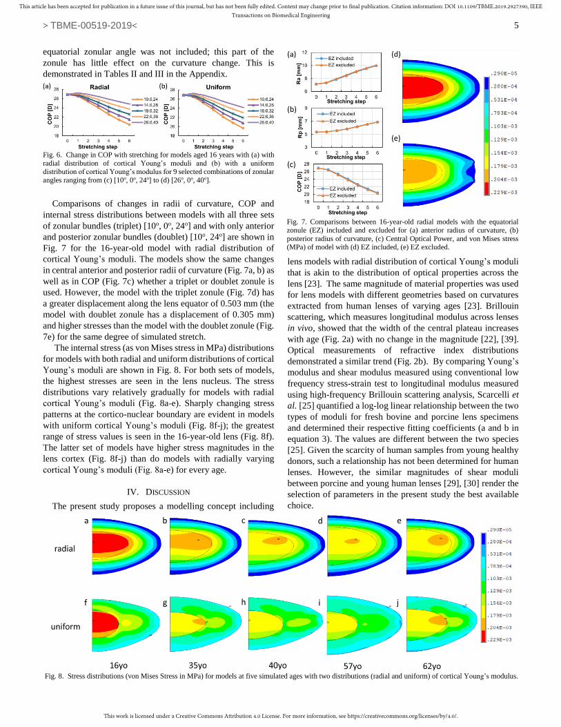
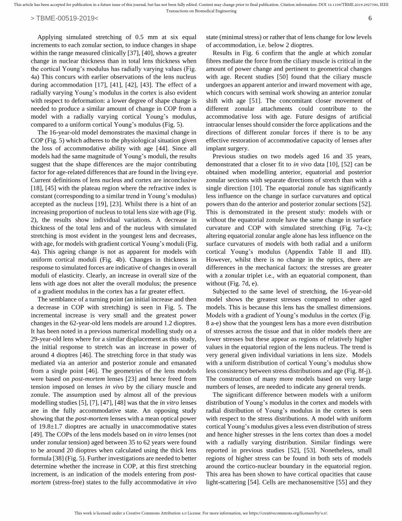
The internal stress (as von Mises stress in MPa) distributions
for models with both radial and uniform distributions of cortical
Young’s moduli are shown in Fig. 8. For both sets of models,
the highest stresses are seen in the lens nucleus. The stress
distributions vary relatively gradually for models with radial
cortical Young’s moduli (Fig. 8a-e). Sharply changing stress
patterns at the cortico-nuclear boundary are evident in models
with uniform cortical Young’s moduli (Fig. 8f-j); the greatest
range of stress values is seen in the 16-year-old lens (Fig. 8f).
The latter set of models have higher stress magnitudes in the
lens cortex (Fig. 8f-j) than do models with radially varying
cortical Young’s moduli (Fig. 8a-e) for every age.
IV. DISCUSSION
The present study proposes a modelling concept including
lens models with radial distribution of cortical Young’s moduli
that is akin to the distribution of optical properties across the
lens [23]. The same magnitude of material properties was used
for lens models with different geometries based on curvatures
extracted from human lenses of varying ages [23]. Brillouin
scattering, which measures longitudinal modulus across lenses
in vivo, showed that the width of the central plateau increases
with age (Fig. 2a) with no change in the magnitude [22], [39].
Optical measurements of refractive index distributions
demonstrated a similar trend (Fig. 2b). By comparing Young’s
modulus and shear modulus measured using conventional low
frequency stress-strain test to longitudinal modulus measured
using high-frequency Brillouin scattering analysis, Scarcelli et
al. [25] quantified a log-log linear relationship between the two
types of moduli for fresh bovine and porcine lens specimens
and determined their respective fitting coefficients (a and b in
equation 3). The values are different between the two species
[25]. Given the scarcity of human samples from young healthy
donors, such a relationship has not been determined for human
lenses. However, the similar magnitudes of shear moduli
between porcine and young human lenses [29], [30] render the
selection of parameters in the present study the best available
choice.
Fig. 8. Stress distributions (von Mises Stress in MPa) for models at five simulated ages with two distributions (radial and uniform) of cortical Young’s modulus.
Fig. 7. Comparisons between 16-year-old radial models with the equatorial
zonule (EZ) included and excluded for (a) anterior radius of curvature, (b)
posterior radius of curvature, (c) Central Optical Power, and von Mises stress (MPa) of model with (d) EZ included, (e) EZ excluded.
Fig. 6. Change in COP with stretching for models aged 16 years with (a) with
radial distribution of cortical Young’s moduli and (b) with a uniform distribution of cortical Young’s modulus for 9 selected combinations of zonular
angles ranging from (c) [10o, 0o, 24o] to (d) [26o, 0o, 40o].
This work is licensed under a Creative Commons Attribution 4.0 License. For more information, see https://creativecommons.org/licenses/by/4.0/.
This article has been accepted for publication in a future issue of this journal, but has not been fully edited. Content may change prior to final publication. Citation information: DOI 10.1109/TBME.2019.2927390, IEEETransactions on Biomedical Engineering
> TBME-00519-2019<
6
Applying simulated stretching of 0.5 mm at six equal
increments to each zonular section, to induce changes in shape
within the range measured clinically [37], [40], shows a greater
change in nuclear thickness than in total lens thickness when
the cortical Young’s modulus has radially varying values (Fig.
4a) This concurs with earlier observations of the lens nucleus
during accommodation [17], [41], [42], [43]. The effect of a
radially varying Young’s modulus in the cortex is also evident
with respect to deformation: a lower degree of shape change is
needed to produce a similar amount of change in COP from a
model with a radially varying cortical Young’s modulus,
compared to a uniform cortical Young’s modulus (Fig. 5).
The 16-year-old model demonstrates the maximal change in
COP (Fig. 5) which adheres to the physiological situation given
the loss of accommodative ability with age [44]. Since all
models had the same magnitude of Young’s moduli, the results
suggest that the shape differences are the major contributing
factor for age-related differences that are found in the living eye.
Current definitions of lens nucleus and cortex are inconclusive
[18], [45] with the plateau region where the refractive index is
constant (corresponding to a similar trend in Young’s modulus)
accepted as the nucleus [19], [23]. Whilst there is a hint of an
increasing proportion of nucleus to total lens size with age (Fig.
2), the results show individual variations. A decrease in
thickness of the total lens and of the nucleus with simulated
stretching is most evident in the youngest lens and decreases,
with age, for models with gradient cortical Young’s moduli (Fig.
4a). This ageing change is not as apparent for models with
uniform cortical moduli (Fig. 4b). Changes in thickness in
response to simulated forces are indicative of changes in overall
moduli of elasticity. Clearly, an increase in overall size of the
lens with age does not alter the overall modulus; the presence
of a gradient modulus in the cortex has a far greater effect.
The semblance of a turning point (an initial increase and then
a decrease in COP with stretching) is seen in Fig. 5. The
incremental increase is very small and the greatest power
changes in the 62-year-old lens models are around 1.2 dioptres.
It has been noted in a previous numerical modelling study on a
29-year-old lens where for a similar displacement as this study,
the initial response to stretch was an increase in power of
around 4 dioptres [46]. The stretching force in that study was
mediated via an anterior and posterior zonule and emanated
from a single point [46]. The geometries of the lens models
were based on post-mortem lenses [23] and hence freed from
tension imposed on lenses in vivo by the ciliary muscle and
zonule. The assumption used by almost all of the previous
modelling studies [5], [7], [47], [48] was that the in vitro lenses
are in the fully accommodative state. An opposing study
showing that the post-mortem lenses with a mean optical power
of 19.8±1.7 dioptres are actually in unaccommodative states
[49]. The COPs of the lens models based on in vitro lenses (not
under zonular tension) aged between 35 to 62 years were found
to be around 20 dioptres when calculated using the thick lens
formula [38] (Fig. 5). Further investigations are needed to better
determine whether the increase in COP, at this first stretching
increment, is an indication of the models entering from post-
mortem (stress-free) states to the fully accommodative in vivo
state (minimal stress) or rather that of lens change for low levels
of accommodation, i.e. below 2 dioptres.
Results in Fig. 6 confirm that the angle at which zonular
fibres mediate the force from the ciliary muscle is critical in the
amount of power change and pertinent to geometrical changes
with age. Recent studies [50] found that the ciliary muscle
undergoes an apparent anterior and inward movement with age,
which concurs with seminal work showing an anterior zonular
shift with age [51]. The concomitant closer movement of
different zonular attachments could contribute to the
accommodative loss with age. Future designs of artificial
intraocular lenses should consider the force applications and the
directions of different zonular forces if there is to be any
effective restoration of accommodative capacity of lenses after
implant surgery.
Previous studies on two models aged 16 and 35 years,
demonstrated that a closer fit to in vivo data [10], [52] can be
obtained when modelling anterior, equatorial and posterior
zonular sections with separate directions of stretch than with a
single direction [10]. The equatorial zonule has significantly
less influence on the change in surface curvatures and optical
powers than do the anterior and posterior zonular sections [52].
This is demonstrated in the present study: models with or
without the equatorial zonule have the same change in surface
curvature and COP with simulated stretching (Fig. 7a-c);
altering equatorial zonular angle alone has less influence on the
surface curvatures of models with both radial and a uniform
cortical Young’s modulus (Appendix Table II and III).
However, whilst there is no change in the optics, there are
differences in the mechanical factors: the stresses are greater
with a zonular triplet i.e., with an equatorial component, than
without (Fig. 7d, e).
Subjected to the same level of stretching, the 16-year-old
model shows the greatest stresses compared to other aged
models. This is because this lens has the smallest dimensions.
Models with a gradient of Young’s modulus in the cortex (Fig.
8 a-e) show that the youngest lens has a more even distribution
of stresses across the tissue and that in older models there are
lower stresses but these appear as regions of relatively higher
values in the equatorial region of the lens nucleus. The trend is
very general given individual variations in lens size. Models
with a uniform distribution of cortical Young’s modulus show
less consistency between stress distributions and age (Fig. 8f-j).
The construction of many more models based on very large
numbers of lenses, are needed to indicate any general trends.
The significant difference between models with a uniform
distribution of Young’s modulus in the cortex and models with
radial distribution of Young’s modulus in the cortex is seen
with respect to the stress distributions. A model with uniform
cortical Young’s modulus gives a less even distribution of stress
and hence higher stresses in the lens cortex than does a model
with a radially varying distribution. Similar findings were
reported in previous studies [52], [53]. Nonetheless, small
regions of higher stress can be found in both sets of models
around the cortico-nuclear boundary in the equatorial region.
This area has been shown to have cortical opacities that cause
light-scattering [54]. Cells are mechanosensitive [55] and they
This work is licensed under a Creative Commons Attribution 4.0 License. For more information, see https://creativecommons.org/licenses/by/4.0/.
This article has been accepted for publication in a future issue of this journal, but has not been fully edited. Content may change prior to final publication. Citation information: DOI 10.1109/TBME.2019.2927390, IEEETransactions on Biomedical Engineering
> TBME-00519-2019<
7
will collectively respond to regional perturbations to their
environment, whether these are caused by physiological,
biochemical or physical (mechanical) effects. Unlike nuclear
cataract, which is a homogenous process associated with
protein aggregation, cortical cataract has been related to stress
damage occurring as a result of continued accommodative
effort [54], [56]. The variations in cortical Young’s modulus
seen in vivo [22] and the effect of the equatorial zonule, albeit
playing a lesser role in optical change with accommodation than
other zonular sections, may be a biological means of protecting
the lens from what would otherwise be more frequently
occurring mechanically induced cataract.
V. CONCLUSIONS
We propose an approach that couples mechanical and optical
properties in the construction of human lens models
representing different ages and accommodative capacities.
Models with cortical Young’s moduli have a more uniform
stress distribution and require less thickness change to produce
similar refractive change than models with uniform cortical
Young’s moduli. Age-related changes in lens geometry are a
major contributing factor to accommodative loss but do not
completely explain the development of presbyopia. Simulations
for a range of zonular angle combinations, indicate that
attachment locations of different zonular sections to the ciliary
muscle are crucial for predicting lens accommodative capacity.
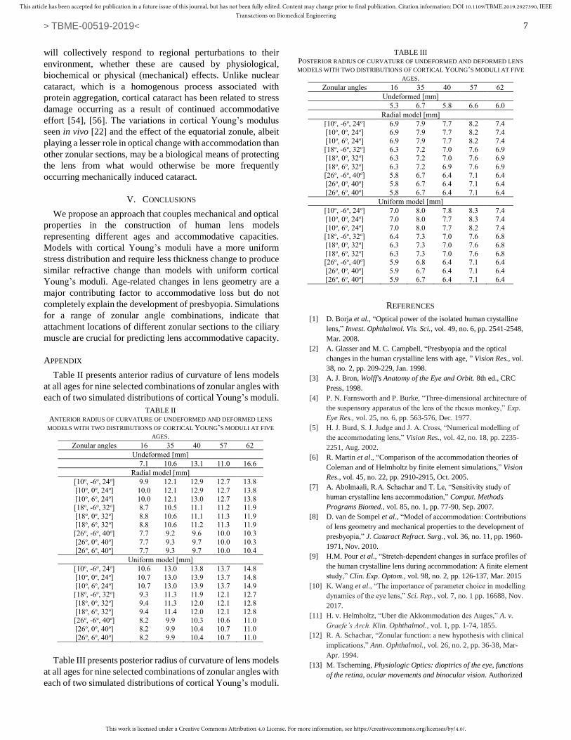
APPENDIX
Table II presents anterior radius of curvature of lens models
at all ages for nine selected combinations of zonular angles with
each of two simulated distributions of cortical Young’s moduli.
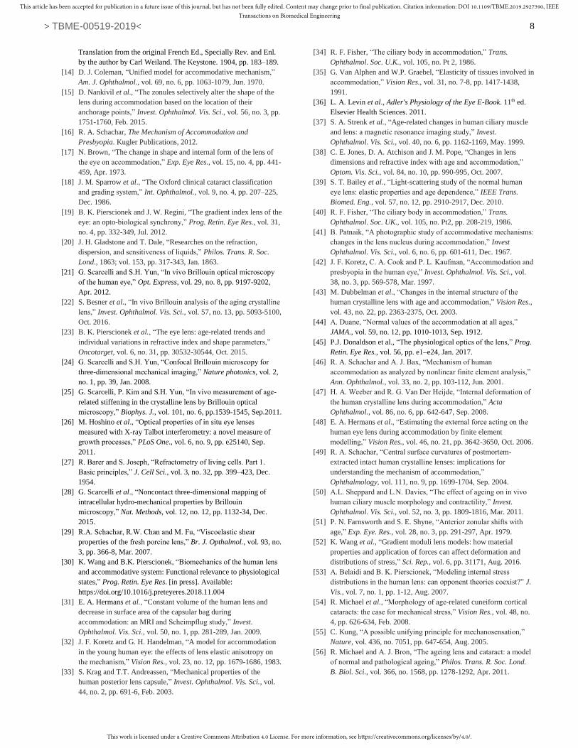
Table III presents posterior radius of curvature of lens models
at all ages for nine selected combinations of zonular angles with
each of two simulated distributions of cortical Young’s moduli.
REFERENCES
[1] D. Borja et al., “Optical power of the isolated human crystalline
lens,” Invest. Ophthalmol. Vis. Sci., vol. 49, no. 6, pp. 2541-2548,
Mar. 2008.
[2] A. Glasser and M. C. Campbell, “Presbyopia and the optical
changes in the human crystalline lens with age, ” Vision Res., vol.
38, no. 2, pp. 209-229, Jan. 1998.
[3] A. J. Bron, Wolff's Anatomy of the Eye and Orbit. 8th ed., CRC
Press, 1998.
[4] P. N. Farnsworth and P. Burke, “Three-dimensional architecture of
the suspensory apparatus of the lens of the rhesus monkey,” Exp.
Eye Res., vol. 25, no. 6, pp. 563-576, Dec. 1977.
[5] H. J. Burd, S. J. Judge and J. A. Cross, “Numerical modelling of
the accommodating lens,” Vision Res., vol. 42, no. 18, pp. 2235-
2251, Aug. 2002.
[6] R. Martin et al., “Comparison of the accommodation theories of
Coleman and of Helmholtz by finite element simulations,” Vision
Res., vol. 45, no. 22, pp. 2910-2915, Oct. 2005.
[7] A. Abolmaali, R.A. Schachar and T. Le, “Sensitivity study of
human crystalline lens accommodation,” Comput. Methods
Programs Biomed., vol. 85, no. 1, pp. 77-90, Sep. 2007.
[8] D. van de Sompel et al., “Model of accommodation: Contributions
of lens geometry and mechanical properties to the development of
presbyopia,” J. Cataract Refract. Surg., vol. 36, no. 11, pp. 1960-
1971, Nov. 2010.
[9] H.M. Pour et al., “Stretch‐dependent changes in surface profiles of
the human crystalline lens during accommodation: A finite element
study,” Clin. Exp. Optom., vol. 98, no. 2, pp. 126-137, Mar. 2015
[10] K. Wang et al., “The importance of parameter choice in modelling
dynamics of the eye lens,” Sci. Rep., vol. 7, no. 1 pp. 16688, Nov.
2017.
[11] H. v. Helmholtz, “Uber die Akkommodation des Auges,” A. v.
Graefe’s Arch. Klin. Ophthalmol., vol. 1, pp. 1-74, 1855.
[12] R. A. Schachar, “Zonular function: a new hypothesis with clinical
implications,” Ann. Ophthalmol., vol. 26, no. 2, pp. 36-38, Mar-
Apr. 1994.
[13] M. Tscherning, Physiologic Optics: dioptrics of the eye, functions
of the retina, ocular movements and binocular vision. Authorized
TABLE II
ANTERIOR RADIUS OF CURVATURE OF UNDEFORMED AND DEFORMED LENS
MODELS WITH TWO DISTRIBUTIONS OF CORTICAL YOUNG’S MODULI AT FIVE
AGES.
Zonular angles 16 35 40 57 62
Undeformed [mm]
7.1 10.6 13.1 11.0 16.6
Radial model [mm]
[10o, -6o, 24o] 9.9 12.1 12.9 12.7 13.8
[10o, 0o, 24o] 10.0 12.1 12.9 12.7 13.8
[10o, 6o, 24o] 10.0 12.1 13.0 12.7 13.8 [18o, -6o, 32o] 8.7 10.5 11.1 11.2 11.9
[18o, 0o, 32o] 8.8 10.6 11.1 11.3 11.9
[18o, 6o, 32o] 8.8 10.6 11.2 11.3 11.9 [26o, -6o, 40o] 7.7 9.2 9.6 10.0 10.3
[26o, 0o, 40o] 7.7 9.3 9.7 10.0 10.3
[26o, 6o, 40o] 7.7 9.3 9.7 10.0 10.4
Uniform model [mm]
[10o, -6o, 24o] 10.6 13.0 13.8 13.7 14.8
[10o, 0o, 24o] 10.7 13.0 13.9 13.7 14.8
[10o, 6o, 24o] 10.7 13.0 13.9 13.7 14.9 [18o, -6o, 32o] 9.3 11.3 11.9 12.1 12.7
[18o, 0o, 32o] 9.4 11.3 12.0 12.1 12.8
[18o, 6o, 32o] 9.4 11.4 12.0 12.1 12.8 [26o, -6o, 40o] 8.2 9.9 10.3 10.6 11.0
[26o, 0o, 40o] 8.2 9.9 10.4 10.7 11.0
[26o, 6o, 40o] 8.2 9.9 10.4 10.7 11.0
TABLE III
POSTERIOR RADIUS OF CURVATURE OF UNDEFORMED AND DEFORMED LENS
MODELS WITH TWO DISTRIBUTIONS OF CORTICAL YOUNG’S MODULI AT FIVE
AGES.
Zonular angles 16 35 40 57 62
Undeformed [mm]
5.3 6.7 5.8 6.6 6.0
Radial model [mm]
[10o, -6o, 24o] 6.9 7.9 7.7 8.2 7.4
[10o, 0o, 24o] 6.9 7.9 7.7 8.2 7.4
[10o, 6o, 24o] 6.9 7.9 7.7 8.2 7.4 [18o, -6o, 32o] 6.3 7.2 7.0 7.6 6.9
[18o, 0o, 32o] 6.3 7.2 7.0 7.6 6.9
[18o, 6o, 32o] 6.3 7.2 6.9 7.6 6.9 [26o, -6o, 40o] 5.8 6.7 6.4 7.1 6.4
[26o, 0o, 40o] 5.8 6.7 6.4 7.1 6.4
[26o, 6o, 40o] 5.8 6.7 6.4 7.1 6.4
Uniform model [mm]
[10o, -6o, 24o] 7.0 8.0 7.8 8.3 7.4
[10o, 0o, 24o] 7.0 8.0 7.7 8.3 7.4
[10o, 6o, 24o] 7.0 8.0 7.7 8.2 7.4 [18o, -6o, 32o] 6.4 7.3 7.0 7.6 6.8
[18o, 0o, 32o] 6.3 7.3 7.0 7.6 6.8
[18o, 6o, 32o] 6.3 7.3 7.0 7.6 6.8 [26o, -6o, 40o] 5.9 6.8 6.4 7.1 6.4
[26o, 0o, 40o] 5.9 6.7 6.4 7.1 6.4
[26o, 6o, 40o] 5.9 6.7 6.4 7.1 6.4
This work is licensed under a Creative Commons Attribution 4.0 License. For more information, see https://creativecommons.org/licenses/by/4.0/.
This article has been accepted for publication in a future issue of this journal, but has not been fully edited. Content may change prior to final publication. Citation information: DOI 10.1109/TBME.2019.2927390, IEEETransactions on Biomedical Engineering
> TBME-00519-2019<
8
Translation from the original French Ed., Specially Rev. and Enl.
by the author by Carl Weiland. The Keystone. 1904, pp. 183–189.
[14] D. J. Coleman, “Unified model for accommodative mechanism,”
Am. J. Ophthalmol., vol. 69, no. 6, pp. 1063-1079, Jun. 1970.
[15] D. Nankivil et al., “The zonules selectively alter the shape of the
lens during accommodation based on the location of their
anchorage points,” Invest. Ophthalmol. Vis. Sci., vol. 56, no. 3, pp.
1751-1760, Feb. 2015.
[16] R. A. Schachar, The Mechanism of Accommodation and
Presbyopia. Kugler Publications, 2012.
[17] N. Brown, “The change in shape and internal form of the lens of
the eye on accommodation,” Exp. Eye Res., vol. 15, no. 4, pp. 441-
459, Apr. 1973.
[18] J. M. Sparrow et al., “The Oxford clinical cataract classification
and grading system,” Int. Ophthalmol., vol. 9, no. 4, pp. 207–225,
Dec. 1986.
[19] B. K. Pierscionek and J. W. Regini, “The gradient index lens of the
eye: an opto-biological synchrony,” Prog. Retin. Eye Res., vol. 31,
no. 4, pp. 332-349, Jul. 2012.
[20] J. H. Gladstone and T. Dale, “Researches on the refraction,
dispersion, and sensitiveness of liquids,” Philos. Trans. R. Soc.
Lond., 1863; vol. 153, pp. 317-343, Jan. 1863.
[21] G. Scarcelli and S.H. Yun, “In vivo Brillouin optical microscopy
of the human eye,” Opt. Express, vol. 29, no. 8, pp. 9197-9202,
Apr. 2012.
[22] S. Besner et al., “In vivo Brillouin analysis of the aging crystalline
lens,” Invest. Ophthalmol. Vis. Sci., vol. 57, no. 13, pp. 5093-5100,
Oct. 2016.
[23] B. K. Pierscionek et al., “The eye lens: age-related trends and
individual variations in refractive index and shape parameters,”
Oncotarget, vol. 6, no. 31, pp. 30532-30544, Oct. 2015.
[24] G. Scarcelli and S.H. Yun, “Confocal Brillouin microscopy for
three-dimensional mechanical imaging,” Nature photonics, vol. 2,
no. 1, pp. 39, Jan. 2008.
[25] G. Scarcelli, P. Kim and S.H. Yun, “In vivo measurement of age-
related stiffening in the crystalline lens by Brillouin optical
microscopy,” Biophys. J., vol. 101, no. 6, pp.1539-1545, Sep.2011.
[26] M. Hoshino et al., “Optical properties of in situ eye lenses
measured with X-ray Talbot interferometry: a novel measure of
growth processes,” PLoS One., vol. 6, no. 9, pp. e25140, Sep.
2011.
[27] R. Barer and S. Joseph, “Refractometry of living cells. Part 1.
Basic principles,” J. Cell Sci., vol. 3, no. 32, pp. 399–423, Dec.
1954.
[28] G. Scarcelli et al., “Noncontact three-dimensional mapping of
intracellular hydro-mechanical properties by Brillouin
microscopy,” Nat. Methods, vol. 12, no. 12, pp. 1132-34, Dec.
2015.
[29] R.A. Schachar, R.W. Chan and M. Fu, “Viscoelastic shear
properties of the fresh porcine lens,” Br. J. Opthalmol., vol. 93, no.
3, pp. 366-8, Mar. 2007.
[30] K. Wang and B.K. Pierscionek, “Biomechanics of the human lens
and accommodative system: Functional relevance to physiological
states,” Prog. Retin. Eye Res. [in press]. Available:
https://doi.org/10.1016/j.preteyeres.2018.11.004
[31] E. A. Hermans et al., “Constant volume of the human lens and
decrease in surface area of the capsular bag during
accommodation: an MRI and Scheimpflug study,” Invest.
Ophthalmol. Vis. Sci., vol. 50, no. 1, pp. 281-289, Jan. 2009.
[32] J. F. Koretz and G. H. Handelman, “A model for accommodation
in the young human eye: the effects of lens elastic anisotropy on
the mechanism,” Vision Res., vol. 23, no. 12, pp. 1679-1686, 1983.
[33] S. Krag and T.T. Andreassen, “Mechanical properties of the
human posterior lens capsule,” Invest. Ophthalmol. Vis. Sci., vol.
44, no. 2, pp. 691-6, Feb. 2003.
[34] R. F. Fisher, “The ciliary body in accommodation,” Trans.
Ophthalmol. Soc. U.K., vol. 105, no. Pt 2, 1986.
[35] G. Van Alphen and W.P. Graebel, “Elasticity of tissues involved in
accommodation,” Vision Res., vol. 31, no. 7-8, pp. 1417-1438,
1991.
[36] L. A. Levin et al., Adler's Physiology of the Eye E-Book. 11th ed.
Elsevier Health Sciences. 2011.
[37] S. A. Strenk et al., “Age-related changes in human ciliary muscle
and lens: a magnetic resonance imaging study,” Invest.
Ophthalmol. Vis. Sci., vol. 40, no. 6, pp. 1162-1169, May. 1999.
[38] C. E. Jones, D. A. Atchison and J. M. Pope, “Changes in lens
dimensions and refractive index with age and accommodation,”
Optom. Vis. Sci., vol. 84, no. 10, pp. 990-995, Oct. 2007.
[39] S. T. Bailey et al., “Light-scattering study of the normal human
eye lens: elastic properties and age dependence,” IEEE Trans.
Biomed. Eng., vol. 57, no. 12, pp. 2910-2917, Dec. 2010.
[40] R. F. Fisher, “The ciliary body in accommodation,” Trans.
Ophthalmol. Soc. UK., vol. 105, no. Pt2, pp. 208-219, 1986.
[41] B. Patnaik, “A photographic study of accommodative mechanisms:
changes in the lens nucleus during accommodation,” Invest
Ophthalmol. Vis. Sci., vol. 6, no. 6, pp. 601-611, Dec. 1967.
[42] J. F. Koretz, C. A. Cook and P. L. Kaufman, “Accommodation and
presbyopia in the human eye,” Invest. Ophthalmol. Vis. Sci., vol.
38, no. 3, pp. 569-578, Mar. 1997.
[43] M. Dubbelman et al., “Changes in the internal structure of the
human crystalline lens with age and accommodation,” Vision Res.,
vol. 43, no. 22, pp. 2363-2375, Oct. 2003.
[44] A. Duane, “Normal values of the accommodation at all ages,”
JAMA., vol. 59, no. 12, pp. 1010-1013, Sep. 1912.
[45] P.J. Donaldson et al., “The physiological optics of the lens,” Prog.
Retin. Eye Res., vol. 56, pp. e1–e24, Jan. 2017.
[46] R. A. Schachar and A. J. Bax, “Mechanism of human
accommodation as analyzed by nonlinear finite element analysis,”
Ann. Ophthalmol., vol. 33, no. 2, pp. 103-112, Jun. 2001.
[47] H. A. Weeber and R. G. Van Der Heijde, “Internal deformation of
the human crystalline lens during accommodation,” Acta
Ophthalmol., vol. 86, no. 6, pp. 642-647, Sep. 2008.
[48] E. A. Hermans et al., “Estimating the external force acting on the
human eye lens during accommodation by finite element
modelling,” Vision Res., vol. 46, no. 21, pp. 3642-3650, Oct. 2006.
[49] R. A. Schachar, “Central surface curvatures of postmortem-
extracted intact human crystalline lenses: implications for
understanding the mechanism of accommodation,”
Ophthalmology, vol. 111, no. 9, pp. 1699-1704, Sep. 2004.
[50] A.L. Sheppard and L.N. Davies, “The effect of ageing on in vivo
human ciliary muscle morphology and contractility,” Invest.
Ophthalmol. Vis. Sci., vol. 52, no. 3, pp. 1809-1816, Mar. 2011.
[51] P. N. Farnsworth and S. E. Shyne, “Anterior zonular shifts with
age,” Exp. Eye. Res., vol. 28, no. 3, pp. 291-297, Apr. 1979.
[52] K. Wang et al., “Gradient moduli lens models: how material
properties and application of forces can affect deformation and
distributions of stress,” Sci. Rep., vol. 6, pp. 31171, Aug. 2016.
[53] A. Belaidi and B. K. Pierscionek, “Modeling internal stress
distributions in the human lens: can opponent theories coexist?” J.
Vis., vol. 7, no. 1, pp. 1-12, Aug. 2007.
[54] R. Michael et al., “Morphology of age-related cuneiform cortical
cataracts: the case for mechanical stress,” Vision Res., vol. 48, no.
4, pp. 626-634, Feb. 2008.
[55] C. Kung, “A possible unifying principle for mechanosensation,”
Nature, vol. 436, no. 7051, pp. 647-654, Aug. 2005.
[56] R. Michael and A. J. Bron, “The ageing lens and cataract: a model
of normal and pathological ageing,” Philos. Trans. R. Soc. Lond.
B. Biol. Sci., vol. 366, no. 1568, pp. 1278-1292, Apr. 2011.