a modular surgical robotic system for image guided ...rht/rht papers/1998/modular surgical robotic...
TRANSCRIPT
A Modular Surgical Robotic Systemfor Image Guided Percutaneous Procedures
Dan Stoianovici Ph.D.1,2, Louis L. Whitcomb Ph.D.2,James H. Anderson Ph.D.4, Russell H. Taylor Ph.D.3, and
Louis R. Kavoussi M.D.1
1 James Buchanan Brady Urological Institute, Johns Hopkins Medical Institutions2 Department of Mechanical Engineering, Whiting School of Engineering
3 Department of Computer Science, Whiting School of Engineering4 Department of Radiology, Johns Hopkins Medical Institutions
- Johns Hopkins University -
Abstract. This paper presents a robotic system for precise needle ins-ertion under radiological guidance for surgical interventions and for deli-very of therapy. It is extremely compact and is compatible with portableX-ray units and computer tomography scanners. The system presents amodular structure comprising a global positioning module, a miniaturerobotic module, and a radiolucent needle driver module. This system isthe newest member of a growing family of modular surgical robots underdevelopment. The system may be operated stand-alone under joystickcontrol making it readily adaptable to any operating room, or under fullimage guided computer control.
1 Introduction
Needle access required for percutaneous surgery is presently preformed in theoperating room by manually inserting the needle under single-view fluoroscopicradiological guidance. This procedure is challenging; it requires extensive expe-rience due to the lack of three-dimensional information of the inter-operativeX-ray imager.
To overcome this problem several researchers investigated the use of roboticsystems to assist in needle placement. Potamianos and Davies [3], [4] proposeda stereo-pair of two x-ray views registered to a common fiducial system with afive degree of freedom (DOF) passive linkage equipped with position encodersto position a passive needle guide. Bzostek et al. [1] used an active robot (LARS[8]) for similar purposes. These systems successfully addressed difficult issues ofimage-to-robot registration and provided convenient means for defining targetanatomy. In their present state of development, however, these robotic systemsare expensive and their size makes them cumbersome for routine use in theoperating room.
In contrast, our group recently reported the development of a simple, non-computerized system, PAKY [5], which based on a minimal approach offered
W.M. Wells et al. (Eds.): MICCAI’98, LNCS 1496, pp. 404–410, 1998.c© Springer-Verlag Berlin Heidelberg 1998
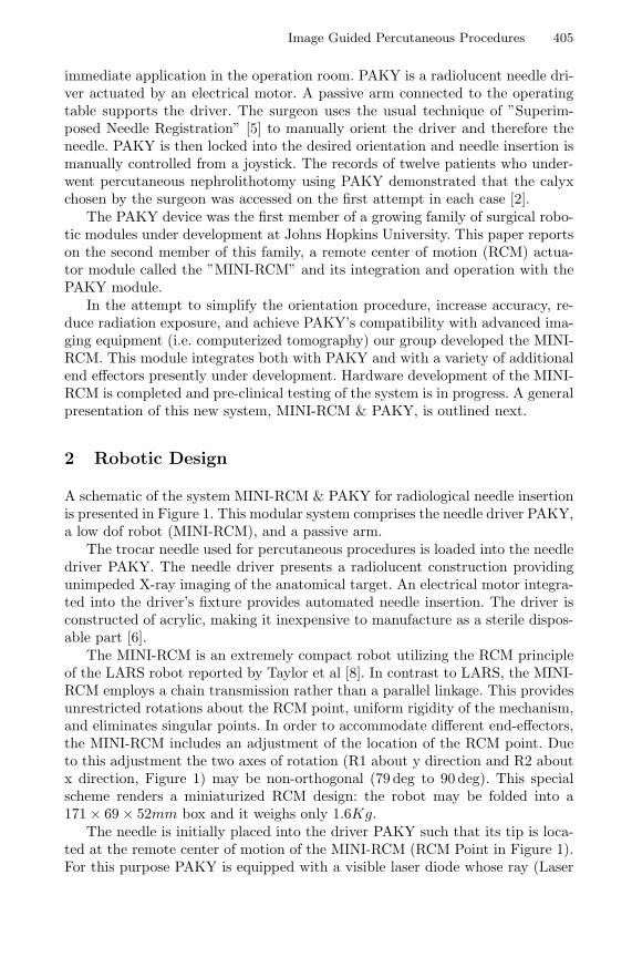
Image Guided Percutaneous Procedures 405
immediate application in the operation room. PAKY is a radiolucent needle dri-ver actuated by an electrical motor. A passive arm connected to the operatingtable supports the driver. The surgeon uses the usual technique of ”Superim-posed Needle Registration” [5] to manually orient the driver and therefore theneedle. PAKY is then locked into the desired orientation and needle insertion ismanually controlled from a joystick. The records of twelve patients who under-went percutaneous nephrolithotomy using PAKY demonstrated that the calyxchosen by the surgeon was accessed on the first attempt in each case [2].
The PAKY device was the first member of a growing family of surgical robo-tic modules under development at Johns Hopkins University. This paper reportson the second member of this family, a remote center of motion (RCM) actua-tor module called the ”MINI-RCM” and its integration and operation with thePAKY module.
In the attempt to simplify the orientation procedure, increase accuracy, re-duce radiation exposure, and achieve PAKY’s compatibility with advanced ima-ging equipment (i.e. computerized tomography) our group developed the MINI-RCM. This module integrates both with PAKY and with a variety of additionalend effectors presently under development. Hardware development of the MINI-RCM is completed and pre-clinical testing of the system is in progress. A generalpresentation of this new system, MINI-RCM & PAKY, is outlined next.
2 Robotic Design
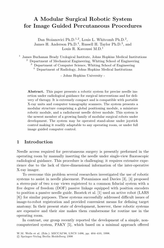
A schematic of the system MINI-RCM & PAKY for radiological needle insertionis presented in Figure 1. This modular system comprises the needle driver PAKY,a low dof robot (MINI-RCM), and a passive arm.
The trocar needle used for percutaneous procedures is loaded into the needledriver PAKY. The needle driver presents a radiolucent construction providingunimpeded X-ray imaging of the anatomical target. An electrical motor integra-ted into the driver’s fixture provides automated needle insertion. The driver isconstructed of acrylic, making it inexpensive to manufacture as a sterile dispos-able part [6].
The MINI-RCM is an extremely compact robot utilizing the RCM principleof the LARS robot reported by Taylor et al [8]. In contrast to LARS, the MINI-RCM employs a chain transmission rather than a parallel linkage. This providesunrestricted rotations about the RCM point, uniform rigidity of the mechanism,and eliminates singular points. In order to accommodate different end-effectors,the MINI-RCM includes an adjustment of the location of the RCM point. Dueto this adjustment the two axes of rotation (R1 about y direction and R2 aboutx direction, Figure 1) may be non-orthogonal (79 deg to 90 deg). This specialscheme renders a miniaturized RCM design: the robot may be folded into a171 × 69 × 52mm box and it weighs only 1.6Kg.
The needle is initially placed into the driver PAKY such that its tip is loca-ted at the remote center of motion of the MINI-RCM (RCM Point in Figure 1).For this purpose PAKY is equipped with a visible laser diode whose ray (Laser
406 D. Stoianovici et al.
Fig. 1. Schematic of the robotic system
Beam in Figure 1) intersects the needle at the RCM point, thus helping the sur-geon position the needle into the driver. The robot presents two motorized DOFimplementing the rotations R1 and R2 about the RCM point (needlepoint).Atpresent, the PAKY & MINI-RCM robotic assembly is supported by a passive7 DOF passive arm which may be locked at the desired position by depressinga lever. A custom rigid rail is mounted on the side of the operating room flu-oroscopic table (Figure 1) to provide a sturdy base for the robotic arm. This iscritical in order to provide a fixed reference frame for the robotic system andmaintain needle trajectory under the insertion force.
A photograph of this modular system presenting the PAKY needle driversupported into the MINI-RCM robot is presented in Figure 2. The laser beamused for needle positioning may be observed at its point.
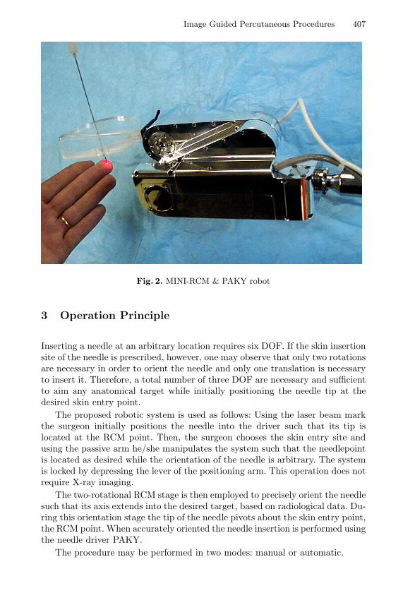
Image Guided Percutaneous Procedures 407
Fig. 2. MINI-RCM & PAKY robot
3 Operation Principle
Inserting a needle at an arbitrary location requires six DOF. If the skin insertionsite of the needle is prescribed, however, one may observe that only two rotationsare necessary in order to orient the needle and only one translation is necessaryto insert it. Therefore, a total number of three DOF are necessary and sufficientto aim any anatomical target while initially positioning the needle tip at thedesired skin entry point.
The proposed robotic system is used as follows: Using the laser beam markthe surgeon initially positions the needle into the driver such that its tip islocated at the RCM point. Then, the surgeon chooses the skin entry site andusing the passive arm he/she manipulates the system such that the needlepointis located as desired while the orientation of the needle is arbitrary. The systemis locked by depressing the lever of the positioning arm. This operation does notrequire X-ray imaging.
The two-rotational RCM stage is then employed to precisely orient the needlesuch that its axis extends into the desired target, based on radiological data. Du-ring this orientation stage the tip of the needle pivots about the skin entry point,the RCM point. When accurately oriented the needle insertion is performed usingthe needle driver PAKY.
The procedure may be performed in two modes: manual or automatic.
408 D. Stoianovici et al.
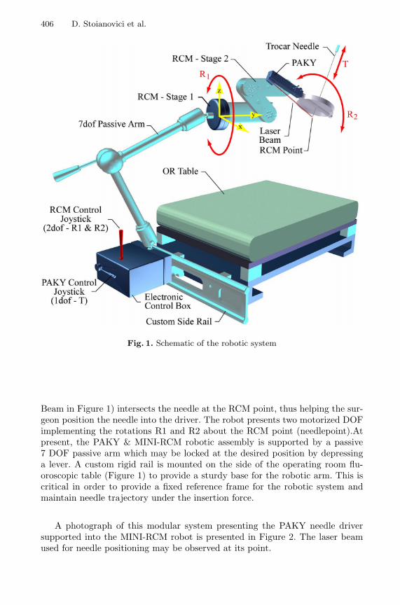
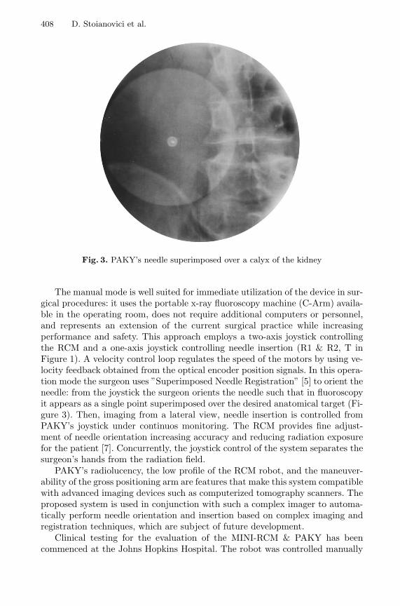
Fig. 3. PAKY’s needle superimposed over a calyx of the kidney
The manual mode is well suited for immediate utilization of the device in sur-gical procedures: it uses the portable x-ray fluoroscopy machine (C-Arm) availa-ble in the operating room, does not require additional computers or personnel,and represents an extension of the current surgical practice while increasingperformance and safety. This approach employs a two-axis joystick controllingthe RCM and a one-axis joystick controlling needle insertion (R1 & R2, T inFigure 1). A velocity control loop regulates the speed of the motors by using ve-locity feedback obtained from the optical encoder position signals. In this opera-tion mode the surgeon uses ”Superimposed Needle Registration” [5] to orient theneedle: from the joystick the surgeon orients the needle such that in fluoroscopyit appears as a single point superimposed over the desired anatomical target (Fi-gure 3). Then, imaging from a lateral view, needle insertion is controlled fromPAKY’s joystick under continuos monitoring. The RCM provides fine adjust-ment of needle orientation increasing accuracy and reducing radiation exposurefor the patient [7]. Concurrently, the joystick control of the system separates thesurgeon’s hands from the radiation field.
PAKY’s radiolucency, the low profile of the RCM robot, and the maneuver-ability of the gross positioning arm are features that make this system compatiblewith advanced imaging devices such as computerized tomography scanners. Theproposed system is used in conjunction with such a complex imager to automa-tically perform needle orientation and insertion based on complex imaging andregistration techniques, which are subject of future development.
Clinical testing for the evaluation of the MINI-RCM & PAKY has beencommenced at the Johns Hopkins Hospital. The robot was controlled manually
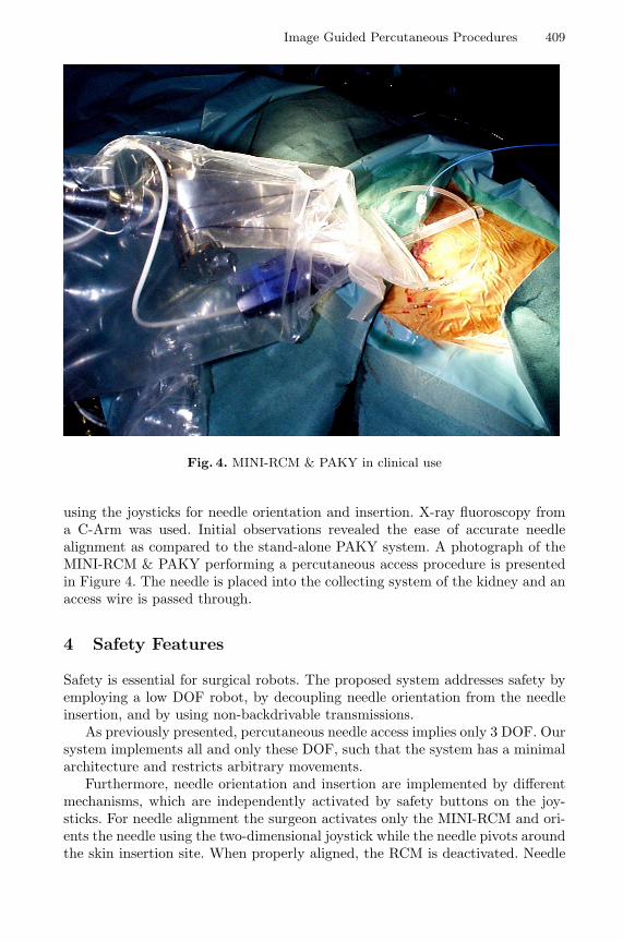
Image Guided Percutaneous Procedures 409
Fig. 4. MINI-RCM & PAKY in clinical use
using the joysticks for needle orientation and insertion. X-ray fluoroscopy froma C-Arm was used. Initial observations revealed the ease of accurate needlealignment as compared to the stand-alone PAKY system. A photograph of theMINI-RCM & PAKY performing a percutaneous access procedure is presentedin Figure 4. The needle is placed into the collecting system of the kidney and anaccess wire is passed through.
4 Safety Features
Safety is essential for surgical robots. The proposed system addresses safety byemploying a low DOF robot, by decoupling needle orientation from the needleinsertion, and by using non-backdrivable transmissions.
As previously presented, percutaneous needle access implies only 3 DOF. Oursystem implements all and only these DOF, such that the system has a minimalarchitecture and restricts arbitrary movements.
Furthermore, needle orientation and insertion are implemented by differentmechanisms, which are independently activated by safety buttons on the joy-sticks. For needle alignment the surgeon activates only the MINI-RCM and ori-ents the needle using the two-dimensional joystick while the needle pivots aroundthe skin insertion site. When properly aligned, the RCM is deactivated. Needle
410 D. Stoianovici et al.
insertion is then enabled by activating PAKY. Using this scheme, the systemprevents the needle to be inserted before being properly aligned and preventschanges of orientation while inserting it.
In addition, the robot uses worm transmissions rendering a non-backdrivablemechanism. This preserves robot’s configuration when deactivated or in the eventof a power failure.
5 Conclusion
The proposed system extends PAKY’s performance and capabilities for perfor-ming image-guided needle access by employing a low DOF RCM robot. The par-ticular design of the RCM robot renders miniaturization and versatility makingthis system fully compatible with complex imaging equipment. Concurrently,the system integrates a manual operation mode such that the system may berapidly transferred to the surgical setting.
References
1. Bzostek, A., Schreiner, S., Barnes, A.C., Cadeddu, J.A., Roberts, W., Anderson,J.H., Taylor, R.H., Kavoussi, L.R.: An automated system for precise percutaneousaccess of the renal collecting system. Lecture Notes in Computer Science, Vol. 1205.Springer-Verlag, Berlin Heidelberg New York (1997) 299–308
2. Cadeddu J.A., Stoianovici D., Chen R.N., Moore R.G., Kavoussi L.R.: Stereotacticmechanical percutaneous renal access. Journal of Endourology, Vol. 12, No. 2, (1998)121–126
3. Potamianos, P., Davies, B.L., and Hibberd, R.D.: Intra-operative imaging guidancefor keyhole surgery methodology and calibration. Proc. First Int. Symposium onMedical Robotics and Computer Assisted Surgery, Pittsburgh, PA. (1994) 98–104
4. Potamianos, P., Davies, B.L., and Hibberd, R.D.: Intra-operative registration forpercutaneous surgery. Proc. First Int. Symposium on Medical Robotics and Com-puter Assisted Surgery, Baltimore, MD. (1995) 156–164
5. Stoianovici, D., Cadeddu, J., A., Demaree, R., D., Basile, H., A., Taylor, R., H.,Whitcomb, L., L., Sharpe, W. N. Jr., Kavoussi, L., R.: An Efficient Needle InjectionTechnique and Radiological Guidance Method for Percutaneous Procedures. LectureNotes in Computer Science, Vol. 1205. Springer-Verlag, Berlin Heidelberg New York(1997) 295–298
6. Stoianovici, D., Cadeddu, J., A., Demaree, R., D., Basile, H., A., Taylor, R., H.,Whitcomb, L., L., Kavoussi, L., R.: A Novel Mechanical Transmission Applied toPercutaneous Renal Access. Proceedings of the ASME Dynamic Systems and Con-trol Division, DSC, Vol. 61 (1997) 401–406
7. Stoianovici, D., Cadeddu, J., A., Whitcomb, L., L., Taylor, R., H., Kavoussi, L.,R.: A Robotic System for Precise Percutaneous Needle Insertion. Thirteen AnnualMeeting of the Society for Urology and Engineering, May 1998, San Diego, CA(1998) 5–6
8. Taylor R.H., Funda J., Eldridge B., Gruben K., LaRose D., Gomory S., TalaminiM., Kavoussi L.R., Anderson J.: A Telerobotic Assistant for Laparoscopic Surgery.IEEE Engineering in Medicine and Biology Magazine, Vol. 14, (1995) 279–287