a mouse serine protease tesp5 is selectively included into lipid
TRANSCRIPT

Mouse Sperm TESP5 1
A Mouse Serine Protease TESP5 Is Selectively Included into Lipid Rafts of Sperm
Membrane Presumably as a GPI-Anchored Protein*
Arata Honda, Kazuo Yamagata§, Shin Sugiura, Katsuto Watanabe, and Tadashi Baba‡
From the Institute of Applied Biochemistry, University of Tsukuba, Tsukuba Science City,
Ibaraki 305-8572, Japan
Mailing address: Tadashi Baba, Ph.D.
Institute of Applied Biochemistry
University of Tsukuba
Tsukuba Science City
Ibaraki 305-8572
Japan
TEL/FAX: +81-298-53-6632 or 6599
E-mail: [email protected]
by guest on February 22, 2018http://w
ww
.jbc.org/D
ownloaded from

Mouse Sperm TESP5 2
RUNNING TITLE: Mouse Sperm TESP5
by guest on February 22, 2018http://w
ww
.jbc.org/D
ownloaded from
Mouse Sperm TESP5 3
(SUMMARY)
We have previously indicated that at least in mouse, sperm serine protease(s) other than
acrosin probably act on the limited proteolysis of egg zona pellucida to create a penetration
pathway for motile sperm, although the participation of acrosin can not be ruled out
completely. A 42-kDa gelatin-hydrolyzing serine protease present in mouse sperm is a
candidate enzyme involved in the sperm penetration of the zona pellucida. In this study, we
have PCR-amplified an EST clone encoding a testicular serine protease, termed TESP5, and
then screened a mouse genomic DNA library using the DNA fragment as a probe. The DNA
sequence of the isolated genomic clones indicated that the TESP5 gene is identical to the
genes coding for testicular testisin and eosinophilic esp-1. Immunochemical analysis using
affinity-purified anti-TESP5 antibody revealed that 42- and 41-kDa forms of TESP5 with the
isoelectric points of 5.0 to 5.5 are localized in the head, cytoplasmic droplet, and midpiece of
cauda epididymal sperm probably as a membranous protein. Moreover, these two forms of
TESP5 were selectively included into Triton X-100-insoluble microdomains, lipid rafts, of the
sperm membranes. These results show the identity between TESP5/testisin/esp-1 and the
42-kDa sperm serine protease. When HEK293 cells were transformed by an expression
plasmid carrying the entire protein-coding region of TESP5, the recombinant protein
produced was released from the cell membrane by treatment with Bacillus cereus
phosphatidylinositol-specific phospholipase C, indicating that TESP5 is
glycosylphosphatidylinositol-anchored on the cell surface. Enzymatic properties of
recombinant TESP5 was similar to but distinguished from those of rat acrosin and pancreatic
trypsin by the substrate specificity and inhibitory effects of serine protease inhibitors.
by guest on February 22, 2018http://w
ww
.jbc.org/D
ownloaded from

Mouse Sperm TESP5 4
(INTRODUCTION)
Mammalian fertilization involves a complex set of molecular events, including
adhesion and binding of sperm to the zona pellucida (ZP), an extracellular glycoprotein
matrix surrounding the egg, acrosome reaction, penetration of sperm through the ZP, and
fusion between sperm and egg (for reviews see Refs. 1-4). Of these events, the acrosome
reaction is a fusion between the outer acrosomal and plasma membranes at the anterior region
of sperm head. Consequently, the acrosomal components are released and interact with the
ZP. The sperm penetration of the ZP is believed to require both sperm motility and
enzymatic hydrolysis by acrosomal protease(s) (1, 5).
A sperm serine protease, acrosin, is localized in the acrosomal matrix as an
enzymatically inactive zymogen, proacrosin, which is then converted into the active form
during the acrosome reaction (6, 7). The role of acrosin in fertilization has long been
considered to participate in the limited proteolysis of ZP, which enables sperm to penetrate the
ZP. However, our previous work (8) using acrosin-deficient mutant mice conclusively
showed that acrosin is not essential both for the sperm penetration of the egg ZP and for
fertilization. The deficiency of acrosin causes a delay in the dispersal of acrosomal proteins
during the acrosome reaction (9), which results in the delayed sperm penetration of the ZP at
the early stages of fertilization in vitro (IVF) after insemination (8). It has been reported that
various trypsin inhibitors prevent sperm from penetrating the ZP (10-14). Since
p-aminobenzamidine (pAB), a competitive inhibitor for trypsin-like serine proteases,
effectively blocks the penetration of acrosin-deficient mouse sperm through the ZP (14),
pAB-sensitive protease other than acrosin likely functions in the penetration step of mouse
sperm. We (8, 14, 15) have demonstrated that two gelatin-hydrolyzing proteins with sizes of
42 and 41 kDa are present in the extracts of wild-type mouse sperm, whereas
acrosin-deficient mouse sperm contain the 42-kDa protein and apparently lack the 41-kDa
protein. The inhibition profiles toward serine protease inhibitors indicate that these two
by guest on February 22, 2018http://w
ww
.jbc.org/D
ownloaded from

Mouse Sperm TESP5 5
gelatin-hydrolyzing proteins belong to the superfamily of trypsin-like serine proteases (14).
Thus, the 42-kDa protease is a candidate enzyme involved in the sperm penetration of egg ZP
at least in mouse.
As described above, only a 42-kDa serine protease exhibiting gelatin-hydrolyzing
activity is found in sperm extracts from wild-type and acrosin-deficient mice (8, 14, 15).
Production of the active 42- and 41-kDa proteases is accelerated by incubation of the sperm
extracts at pH 8.5, and by addition of exogenous pancreatic trypsin to the extracts. The
gelatin-hydrolyzing activity of the 42-kDa protease in the acrosin-deficient mouse remains
constant during the pH-8.5 incubation (8, 14), and the active 41-kDa protease is not found in
the sperm extracts without addition of exogenous trypsin (15). These data imply that the
42-kDa protease as well as acrosin may be present in acrosome-intact sperm as an
enzymatically inactive pro-protein (zymogen), a part of which is already converted into the
active enzyme by a processing enzyme(s) with a trypsin-like cleavage specificity, including
acrosin, and/or by autoactivation (14, 15). It is also possible that the zymogens of the 42-
and 41-kDa proteases are essentially different molecules, or that the 41-kDa protease is
produced from the 42-kDa protease by proteolytic processing. To prove these possibilities,
the molecular basis of the pro- and mature forms of the 42-kDa serine protease needs to be
clarified.
cDNA clones encoding four different serine proteases, TESP1 (testicular serine
protease 1), TESP2, TESP3, and TESP4, have been identified as candidates for 42-kDa
gelatin-hydrolyzing enzyme in mouse sperm (16, 17). Although TESP1, TESP2, and TESP4
are all present in the acrosome of mouse sperm, Western blot analysis of sperm protein
extracts indicates that the three proteases differ from the 42-kDa gelatin-hydrolyzing enzyme
in molecular size. In addition, TESP3 is localized solely in spermatogenic cells of the testis
when antibody raised against the N-terminal 6-residue peptide of TESP3 is used (Sato, S. and
Baba, T., unpublished data). These data imply that none of the four TESPs is identical to the
42-kDa gelatin-hydrolyzing protease. Thus, further experiments are required to elucidate the
by guest on February 22, 2018http://w
ww
.jbc.org/D
ownloaded from

Mouse Sperm TESP5 6
mechanism of sperm penetration through the ZP in mouse.
In this study, we have isolated genomic clones encoding a testicular serine protease,
termed TESP5, from a mouse genomic DNA library. The nucleotide sequence of the TESP5
gene demonstrates the identity of this gene with the testisin (18) and esp-1 (19) genes
previously reported. TESP5/testisin/esp-1 is localized on the sperm membrane probably as a
glycosylphosphatidylinositol (GPI)-anchored protein, and corresponds to the 42- and 41-kDa
gelatin-hydrolyzing enzymes. On the basis of biochemical data regarding TESP5, a possible
role of TESP5 in the sperm/egg interaction is discussed.
by guest on February 22, 2018http://w
ww
.jbc.org/D
ownloaded from

Mouse Sperm TESP5 7
EXPERIMENTAL PROCEDURES
Materials — Triton X-100 (TX-100), Nonidet P-40 (NP-40), calcium ionophore
A23187, bovine pancreatic trypsin (type III, T-8253), Bacillus cereus
phosphatidylinositol-specific phospholipase C (PI-PLC), and protease inhibitors,
p-aminobenzamidine (pAB), diisopropyl fluorophosphate (DFP), Nα-tosyl-L-lysine
chloromethyl ketone (TLCK), phenylmethanesulfonyl fluoride (PMSF), and
N-tosyl-L-phenylalanine chloromethyl ketone (TPCK), were purchased from Sigma.
Ampholine (pH 3.5-10) and Immobiline DryStrip (pH 3-10, 7 cm) were purchased from
Amersham Pharmacia Biotech. Monoclonal antibodies against acrosomal proteins of mouse
sperm, MN7 (20) and MC101 (21), were provided by Dr. K. Toshimori. Rabbit anti-mouse
AKAP82 antiserum (22) was a gift from Dr. S. B. Moss. Protease substrates,
t-butyloxycarbonyl (Boc)-, N-succinyl (Suc)-, or carbobenzoxy
(Z)-peptidyl-4-methylcoumaryl-7-amide (MCA), were purchased from Peptide Institute, Inc.
(Osaka, Japan). Rat acrosin was purified from cauda epididymal sperm, as described
previously (15). Experimental animals, ICR mice, Wistar rats, and New Zealand White
rabbits, were obtained from Japan SLC Inc. (Shizuoka, Japan).
Reverse Transcriptase-Polymerase Chain Reaction (RT-PCR) — RT-PCR was carried
out using a 3’-Full RACE kit (Takara Shuzo, Shiga, Japan) according to the manufacturer’s
protocol. First-strand cDNA was synthesized from total cellular RNAs of various tissues
and male germ cells by AMV RT XL using an oligo dT-3sites adaptor primer. PCR was
carried out in a mixture (25 µl) containing 10 mM Tris/HCl, pH 8.8, 50 mM KCl, 1.5 mM
MgCl2, 0.1% TX-100, 0.2 mM each of dATP, dCTP, dGTP, and dTTP, 1 µM each of the
primers, the template DNA, and 2.5 units of Taq DNA polymerase (Wako, Osaka, Japan).
The PCR products were purified by polyacrylamide gel electrophoresis (PAGE), and then
introduced into a pUC19 vector.
Isolation of Genomic Clones — An expressed sequence tag (EST) clone, AA144961,
by guest on February 22, 2018http://w
ww
.jbc.org/D
ownloaded from

Mouse Sperm TESP5 8
was amplified from a mouse testis cDNA library by PCR using T6-1,
5’-CCTGCGGTCACAGGACCATCC-3’, and T6-2,
5’-ACAGCTTCAGCAGGGCTATGTCA-3’, as primers. The DNA fragment amplified was
labeled with [α-32P]dCTP (Amersham Pharmacia Biotech), and used as a probe to screen
approximately 9.0 x 10⁵ plaques from a mouse 129/SvJ genomic DNA library in λFIXII
(Stratagene), as described previously (23). Phage DNAs were prepared from the positive
clones, digested by various restriction enzymes, and introduced into pUC19 for further
characterization. Nucleotide sequence analysis was carried out using an ABI Prism 310
genetic analyzer.
Southern and Northern Blot Analysis — DNAs and RNAs were separated by agarose
gel electrophoresis and transferred onto Hybond-N+ nylon membranes (Amersham Pharmacia
Biotech), as described previously (23). The blots were probed by 32P-labeled DNA
fragments, and analyzed by a BAS-1800II Bio-Image analyzer (Fuji Photo Film, Tokyo). To
remove poly(A) tails of mRNAs, RNase H digestion of total RNAs, which had been annealed
with oligo(dT)15, was carried out as described (24). The RNase H-digested RNA samples
were subjected to Northern blot analysis, as mentioned above.
Preparation of Crude Protein Extracts — Mouse testes were homogenized in 3 ml of
phosphate-buffered saline (PBS) containing 1 mM EDTA, 1 mM benzamidine/HCl, 1 mM
PMSF, leupeptin (1 µg/ml), and pepstatin A (1 µg/ml) using a Teflon-glass homogenizer at
900 rpm (5 strokes/min). The homogenate was centrifuged in a Tomy SRX-201 centrifuge
using a TA-24BH rotor (Tokyo, Japan) at 19,000 x g for 10 min. The supernatant solution
was further centrifuged at 19,000 x g for 10 min, and the resulting supernatant was used as
“soluble protein extracts”. The precipitate obtained by first centrifugation was extracted in
PBS containing 1% SDS at room temperature for 1 h, and then centrifuged at 16,000 x g for
10 min. The supernatant was used as “insoluble protein extracts”. Fresh cauda epididymal
sperm in a modified Krebs-Ringer bicarbonate solution (TYH medium, see Ref. 8) free of
bovine serum albumin (BSA) were incubated at 37°C for 30 min under 5% CO2 in air. The
by guest on February 22, 2018http://w
ww
.jbc.org/D
ownloaded from

Mouse Sperm TESP5 9
dispersed sperm suspension was centrifuged in an Eppendorf 5415D centrifuge at 800 x g for
10 min, and the supernatant was discarded. The pellet was washed, extracted in 1mM HCl
containing 1% SDS or 1% TX-100 at room temperature for 1 h, and centrifuged in the above
Tomy centrifuge at 16,000 x g for 10 min. The supernatant was subjected to SDS-PAGE and
Western blot analysis. Protein concentration was determined using a BCA protein assay
reagent (Pierce).
Subcellular Fractionation of Sperm — Subcellular components of cauda epididymal
sperm were prepared as described previously (15, 25) with minor modifications. Sperm
suspension was suspended in the TYH medium containing calcium ionophore A23187 (10
µg/ml), incubated for 60 min at 37°C under 5% CO2 in air, and centrifuged twice in an
Eppendorf 5415D centrifuge at 800 x g for 10 min to remove sperm. The supernatant was
further separated into the precipitate and supernatant fractions by centrifugation in a Beckman
L8-70M ultracentrifuge using an SW41 rotor at 100,000 x g for 90 min at 4°C. The
precipitate was washed with PBS, centrifuged at 100,000 x g for 60 min, and resuspended in
PBS (Fraction A). The supernatant solution after first ultracentrifugation was dialyzed
against PBS, and centrifuged in the above Eppendorf centrifuge at 13,000 x g for 10 min
(Fraction B). Fractions A and B were used as samples enriched by plasma and
outer-acrosomal membranes, and soluble proteins released by the A23187-induced acrosome
reaction, including acrosomal components, respectively. Acrosome-reacted sperm were
washed three times with PBS, and sonicated as described (25). To the 2.5-ml suspension an
equal volume of 1.8 M sucrose was added, and the mixture was put onto a discontinuous
sucrose gradient solution containing 2.5 ml each of 2.20 and 2.05 M sucrose, and centrifuged
in the above Beckman L8-70M ultracentrifuge at 100,000 x g for 16 h at 4°C. Fractions C,
D, and E corresponding to acrosome-reacted sperm heads, tails, and cytoplasmic droplets
formed a pellet at the bottom of the centrifuge tube, a band at the middle of the 2.05 M
sucrose layer, and a band at the interface between the 0.9 and 2.05 M sucrose layers,
respectively. Each of Fractions D and E was precipitated by centrifugation at 100,000 x g.
by guest on February 22, 2018http://w
ww
.jbc.org/D
ownloaded from

Mouse Sperm TESP5 10
The purities of these fractions were microscopically more than 95%. The samples were
suspended in PBS, mixed with an SDS sample buffer, and then subjected to Western blot
analysis.
Preparation of Low Density TX-100-Insoluble Membrane Fractions — Low density
membrane fractions insoluble in TX-100 were prepared from cauda epididymal sperm by the
established method (26) with minor modifications. Sperm (3.0 x 10⁸ sperm/ml) were
suspended in 10 mM Tris-HCl, pH 7.5, 0.15 M NaCl, and 5 mM EDTA (TNE) containing 75
units/ml aprotinin and 1% TX-100, and put on ice for 20 min. The suspension was
homogenized by a Dounce homogenizer (5 strokes), and centrifuged in an Eppendorf 5415D
centrifuge at 2,000 x g for 5 min to remove nuclei and cell debris. The supernatant solution
was brought to 40% sucrose using 80% sucrose in TNE, placed at the bottom of a Beckman
Ultra Clear centrifuge tube, and overlaid with 30% sucrose (6 ml) and 5% sucrose (3.5 ml) in
TNE. After centrifugation in a Beckman L8-70M ultracentrifuge using an SW41 rotor at
200,000 x g for 18 h, fractions (1 ml) were collected from the top to the bottom of the
gradient, and then subjected to SDS-PAGE in the presence of 0.1% gelatin.
Production of Recombinant Proteins — An 871-bp DNA fragment encoding the pro-
and catalytic domains of TESP5 was PCR-amplified from a mouse testis cDNA library, using
MTP4, 5’-TGCGGCCATGGCCTTACA-3’, and MTP5,
5’-AACTCGAGTTAGTCAGGCCTGAGCAGCC-3’, as primers. The PCR product was
introduced into a pET-23d vector (Novagen, Madison, WI) at the NcoI and XhoI sites for
expression in Escherichia coli BL21 (DE3). A single colony of the transformants was
cultured at 37°C overnight in Luria broth containing 0.1 mg/ml ampicillin (5 ml, LA) with
constant shaking. A portion (3 ml) of the bacterial culture was added to fresh LA (120 ml),
and incubated at 37°C for 2 h with shaking. Production of the recombinant proteins was
induced by addition of 0.3 ml of 0.1 M isopropyl-β-D-thio-galactopyranoside, and the cell
growth was continued at 20°C overnight. E. coli cells were collected by centrifugation in a
Tomy RL-131 centrifuge using a TS-7 rotor at 1,500 x g for 10 min, and suspended in 10 mM
by guest on February 22, 2018http://w
ww
.jbc.org/D
ownloaded from

Mouse Sperm TESP5 11
sodium phosphate, pH 7.4, containing 30 mM NaCl, 10 mM 2-mercaptoethanol, 10 mM
EDTA, and 0.25% Tween 20. The suspension was frozen at -80°C, sonicated, and then
centrifuged in a Tomy SRX-201 centrifuge using a TA-24BH rotor at 10,000 x g for 10 min at
4°C. Recombinant TESP5 was found as an enzymatically inactive protein solely in the
precipitate.
For production of recombinant TESP5 in a mammalian cell line, HEK293, an
expression plasmid carrying the entire protein-coding region in the TESP5 cDNA sequence
was constructed by PCR-directed mutagenesis using a set of oligonucleotide primers, MTP2,
5’-TGAATTCACAGGTGTGACGTACAC-3’, and MTP3,
5’-TTGAATTCGAGAGGTGGCCATGGGC-3’. The amplified DNA fragment was
introduced into a pCXN2 (27) vector at the EcoRI site. HEK293 cells (4 x 10⁶ cells), which
were maintained in Dulbecco’s modified Eagle’s medium (DMEM, Sigma) supplemented
with 10% fetal bovine serum, 100 units/ml penicillin, and 0.1 mg/ml streptomycin at 37°C
under 5% CO2 in air, were electroporated at 500 µF with the expression plasmid (10 µg) in
PBS, and viable cells were plated in 35-mm culture dishes. After 24 h, cells were treated
with trypsin and grown in the above medium containing 0.5 mg/ml G418 to obtain stable
transformants.
Preparation of Antibody — Recombinant mouse TESP5 (0.5 mg) produced in E. coli
was emulsified by sonication in an equal volume of Freund’s complete adjuvant (Difco
Laboratories), and injected intradermally into female rabbits. Rabbit anti-TESP5 antibody
was purified by fractionation with ammonium sulfate (0-40% saturation) followed by
immunoaffinity chromatography on a column of Sepharose 4B that had been substituted with
the recombinant protein by the cyanogen bromide procedure (28), as described (23).
SDS-PAGE and Western Blot Analysis — Proteins were separated by SDS-PAGE under
non-reducing conditions and transferred onto Immobilon-P polyvinylidene difluoride
membranes (Millipore). After blocking with 2% skim milk, the blots were incubated with
affinity-purified anti-TESP5 antibody at room temperature for 2 h, and then incubated with
by guest on February 22, 2018http://w
ww
.jbc.org/D
ownloaded from

Mouse Sperm TESP5 12
horseradish peroxidase-conjugated goat anti-rabbit IgG (Jackson Immunoresearch
Laboratories) for 1 h. The immunoreactive proteins were detected by an ECL Western
blotting detection kit (Amersham Pharmacia Biotech). To detect proteins exhibiting
gelatin-hydrolyzing activities, SDS-PAGE in the presence of 0.1% gelatin was carried out as
described previously (15).
Two-Dimensional (2D) PAGE — Proteins (0.2 mg) were pre-incubated in 50 mM
Tris-HCl, pH 8.5, at room temperature for 3 h, lyophilized, dissolved in 8 M urea (120 µl)
containing 0.5% TX-100 and 0.5% Ampholine (pH 3.5-10), and placed onto polyacrylamide
gel strips with an immobilized pH gradient (Immobiline DryStrips) at 20°C for 10 h.
Isoelectric focusing was carried out at 20°C using an IPGphor instrument (Amersham
Pharmacia Biotech) according to the following program: a linear-gradient run at 0 to 500 V
for 60 min, 500 to 1,000 V for 60 min, and 1,000 to 8,000 V for 60 min, and a constant run at
8,000 V for 3 h. After electrophoresis, the gel strips were soaked in 50 mM Tris-HCl, pH
6.8, containing 6 M urea, 30% glycerol, and 1% SDS, and then subjected to SDS-PAGE.
Immunoprecipitation — Affinity-purified anti-TESP5 antibody (30 µl) was incubated
at 4°C for 1 h in 0.5 ml of PBS containing 0.5% NP-40 and 6 µl of protein A immobilized on
agarose beads (Pierce). The agarose beads were washed with PBS by centrifugation to
remove the unbound antibodies, mixed with sperm proteins (0.2 mg) in 0.5 ml of PBS
containing 0.5% NP-40, incubated at 4°C overnight, and centrifuged. The pellet was washed
three times with PBS containing 0.5% NP-40, dissolved in 8 M urea, and then subjected to
SDS-PAGE.
Immunostaining of Testicular Sections and Sperm — Testicular tissues from adult mice
were fixed in a solution containing 50 mM sodium phosphate, pH 7.4, 0.1 M lysine
hydrochloride, and 2% paraformaldehyde (PLP) at 4°C for 3 h, snap-frozen, and embedded in
a Tissue-TekTM O.C.T. compound (Sakura Finetechnical Co., Tokyo). Sections (7 µm) were
prepared in a Leica CM3000 cryostat, mounted on silanized glass slides, air-dried at room
temperature, and washed with PBS. Each slide for the testicular sections was blocked with
by guest on February 22, 2018http://w
ww
.jbc.org/D
ownloaded from

Mouse Sperm TESP5 13
2% normal goat serum in PBS, and incubated with affinity-purified anti-TESP5 antibody in
PBS containing 0.05% BSA at 4°C overnight. After washing with PBS, the slides were
treated with 0.3% hydrogen peroxide to remove endogenous peroxidase activity, washed with
PBS, and incubated with horseradish peroxidase-conjugated goat anti-rabbit IgG. The
samples on the slides were stained with 3,3’-diaminobenzidine as a chromogen,
counterstained with 2.5% methyl green, and viewed under an Olympus BX50 microscope.
Indirect immunofluorescence microscopy of cauda epididymal sperm was carried out as
described previously (9). Briefly, sperm samples on slides, which had been fixed in PLP,
were incubated with the primary antibodies overnight, washed with PBS, and treated with
fluorescein isothiocyanate-conjugated goat anti-rabbit IgG (Jackson Immunoresearch
Laboratories) for 4 h. After washing with PBS, the slides were observed using an Olympus
BX50 fluoromicroscope.
Measurement of Enzyme Activity — Enzyme activity was measured using various Boc-,
Suc-, or Z-peptidyl-MCAs as substrates, as described previously (8). The reaction mixture
(0.5 ml) consisted of 50 mM Tris-HCl, pH 8.0, 10 mM CaCl2, 40 µM substrate, and an
appropriate amount of enzyme. Following incubation at 30°C for 30 min, the reaction was
terminated by addition of 0.1 M acetate buffer, pH 4.3 (1.5 ml). The amounts of
7-amino-4-methylcoumarin formed were fluorometrically determined using excitation at 380
nm and emission at 460 nm.
by guest on February 22, 2018http://w
ww
.jbc.org/D
ownloaded from

Mouse Sperm TESP5 14
RESULTS
To identify genes encoding novel serine proteases present in mouse sperm, we initially
searched the EST database derived from the mouse testis. An EST clone, GenBank
accession number AA144961, was found to code for a serine protease, termed TESP5, that
did not correspond to acrosin and four TESPs. Thus, a 326-bp DNA fragment in the TESP5
cDNA sequence was amplified by RT-PCR using mouse testicular RNA as a template, and
used as a probe to screen a mouse 129/SvJ genomic DNA library. Two positive clones,
HGC3 and HGC14, which overlap to each other, have been identified (Fig. 1A). The DNA
sequence indicated that the mouse TESP5 gene is approximately 4.5 kbp in length, and
consists of six exons interrupted by five introns. Southern blot analysis also demonstrated
that this gene is a single copy gene on the mouse genome (Fig. 1B).
A computer search for known genes deposited in the GenBank Data Bank revealed that
the mouse TESP5 gene (data not shown, see GenBank accession number AB059414) was
98.5 and 99.9% homologous to the testisin (AF304012, see Ref. 18) and esp-1 (AB041645,
see Ref. 19) genes, respectively, the former of which was most recently characterized as a
possible suppressor gene for non-classical type II tumor (29). The differences of the DNA
sequences among these three genes were present only in the 5’-flanking region, second, fourth,
and fifth introns, and 3’-untranslated region in sixth exon (data not shown). Indeed, the
composite sequence of six exons in the TESP5 gene perfectly matched with the testisin cDNA
sequence (AY005145). Thus, we conclude that these three genes are the same.
The DNA-derived amino acid sequence indicated that mouse TESP5/testisin/esp-1 is
initially synthesized as a single-chain 324-residue polypeptide with a calculated molecular
mass of 36,175 Da (Fig. 2A). The N-terminal 21-residue sequence (DI in Figs. 1A and 2A)
of the TESP5/testisin/esp-1 prepro-protein was predicted to be a signal peptide for a nascent
secretory protein because of the hydrophobic profile (data not shown), suggesting that the
pro-form (zymogen) of TESP5 may start with Leu at residue 22. Alignment of the entire
by guest on February 22, 2018http://w
ww
.jbc.org/D
ownloaded from

Mouse Sperm TESP5 15
sequence of mouse TESP5 with those of human testisin, human chymotrypsin, and mouse
trypsin indicated the conservation of Cys residues, three active-site residues, His, Asp, and
Ser, required for the proteolytic activity of serine proteases (30), and a substrate recognition
residue, Asp, for the Arg/Lys-Xaa bond cleavage (31). Moreover, TESP5 contained the
Cys-Gly-His-Arg-Thr-Ile-Pro-Ser-Arg (residues 46-54) and Ile-Val-Gly-Gly sequences
(residues 55-58) in DII and DIII (Figs. 1A and 2A), which are highly similar or identical to
the typical sequences of a pro-enzyme segment (activation peptide) of serine protease
zymogens, including chymotrypsinogen, and of an activated serine protease at the N-terminus
(30), respectively. Thus, the TESP5 zymogen appears to be a single-chain polypeptide of
303 amino acids containing a possible 33-residue pro-part (DII) of the zymogen at the
N-terminus. A highly hydrophobic sequence of approximately 20 residues, which may
function as a direct anchor to the plasma membrane or as a signal sequence for attachment to
GPI, was present at the C-terminus of TESP5 (Fig. 2B).
Two mRNA signals with sizes of 1.5 and 1.3 kb were found by Northern blot analysis
exclusively in the testis among the mouse tissues tested (Fig. 3A), and in pachytene
spermatocytes and round spermatids (Fig. 3B). Only a single 1.2-kb mRNA signal was
detected when the RNA samples were annealed with oligo(dT)15 and digested with RNase H.
Thus, the difference of the two mRNAs is most likely due to the length of the poly(A) tail.
RT-PCR analysis, using a set of oligonucleotides, T6-1 and T6-2, as primers, revealed the
initial transcription of the TESP5 gene in the testis at 18th day after birth (data not shown).
These data demonstrate specific expression of the mouse TESP5 gene in pachytene
spermatocytes and round spermatids.
Little is known of the functions of testicular testisin (18, 29) and eosinophilic esp-1
(19), because the experimental data have been still restricted to the structure, organization,
and chromosomal assignment of the gene, and the protein localization in testicular cells.
Thus, we first carried out Western blot analysis of soluble and insoluble proteins from
testicular tissues, and of detergent-soluble proteins from cauda epididymal sperm, using
by guest on February 22, 2018http://w
ww
.jbc.org/D
ownloaded from

Mouse Sperm TESP5 16
affinity-purified anti-TESP5 antibody as a probe (Fig. 4A). Two immunoreactive proteins
with sizes of 43 and 42 kDa were detected only in the insoluble protein extracts of testis,
whereas the sperm extracts gave two bands corresponding to the 42- and 41-kDa proteins.
In addition, 2D-PAGE of the sperm TX-100 extracts indicated that the 42- and 41-kDa forms
of TESP5 with the isoelectric points (pI) of 5.0 to 5.5 exhibit gelatin-hydrolyzing activity (Fig.
4B). These results imply the presence of TESP5 on the membranes of sperm as well as of
testicular germ cells. Most importantly, the molecular sizes and pI values of the two forms
of TESP5 are consistent with those of 42- and 41-kDa serine proteases that was identified as
candidate proteins necessary for sperm penetration of the egg ZP in mouse (8, 14, 15).
To ascertain whether TESP5 is identical to the 42- and 41-kDa sperm serine proteases
(8, 14, 15), proteins in sperm TX-100 extracts immunoprecipitated with affinity-purified
anti-TESP5 antibody, and then subjected to SDS-PAGE in the presence of gelatin (Fig. 5).
Two gelatin-hydrolyzing proteins with molecular masses of 42 and 41 kDa were clearly found
in the immunoprecipitates. No significant band was detectable when affinity-purified
anti-mouse sp32 antibody (23) was used for the immunoprecipitation as a control. Thus,
these data demonstrate the identity between TESP5/testisin/esp-1 and the 42- and 41-kDa
sperm serine proteases.
When sections of adult mouse testis were immunohistochemically analyzed using the
affinity-purified anti-TESP5 antibody, the signals were observed in round and elongating
spermatids in the seminiferous tubules (panels a and b in Fig. 6A). In particular, the granular
signals with a great intensity were found in elongating spermatids. Spermatogonia,
pachytene spermatocytes, and Sertoli cells gave no signal at detectable level. Indirect
immunofluorescence assay of acrosome-intact sperm revealed the presence of strong and
weak signals in the cytoplasmic droplet, and in the head and midpiece of sperm, respectively
(panels c and d in Fig. 6A). The immunofluorescence signals disappeared in the presence of
the recombinant TESP5 protein (panels e and f). To verify the localization of TESP5,
proteins in five subcellular fractions (Fractions A to E) of mouse sperm (see Experimental
by guest on February 22, 2018http://w
ww
.jbc.org/D
ownloaded from

Mouse Sperm TESP5 17
Procedures) were examined by Western blot analysis using antibodies against an acrosomal
proacrosin-binding protein, sp32 (23), a 90-kDa intra-acrosomal protein, MN7 (20), a
155-kDa intra-acrosomal protein, MC101 (21), and a protein kinase A anchor protein,
AKAP82 (22), in the flagellum fibrous sheath, as markers (Fig. 6B). The 42- and 41-kDa
forms of TESP5 were found in all five fractions, including Fractions A (plasma and
outer-acrosomal membranes), B (soluble proteins released by acrosome reaction including
acrosomal components), and C (acrosome-reacted sperm heads). Thus, these results
demonstrate that TESP5 is localized in the sperm head, cytoplasmic droplet, and midpiece
probably as a membranous protein. However, it is unclear whether TESP5 is partly present
as a soluble protein in the sperm acrosome.
Preferential clustering of sphingolipids and cholesterol in the lipid bilayers of cell
membranes is known to form organized compositional microdomains, “rafts”, that move
within the fluid bilayer (32, 33). GPI-anchored, transmembrane, and lipid-linked proteins
can be selectively included into lipid rafts that are insoluble in non-ionic detergents such as
TX-100. Western blot analysis indicated the presence of 42- and 41-kDa forms of TESP5
exhibiting gelatin-hydrolyzing activity in the TX-100-insoluble, low-density membrane
fractions (lanes 7 and 8) from mouse sperm (Fig. 7). Judging by the intensities of the
immunoreactivities with affinity-purified anti-TESP5 antibody, only a small amount of
TESP5 was found in the TX-100-soluble fractions (lanes 1 and 2). Thus, TESP5 is mostly
included into lipid rafts of the sperm membrane.
To prepare enzymatically active TESP5, HEK293 cells were transformed by
introducing an expression plasmid carrying the entire protein-coding region of TESP5. The
recombinant protein produced was effectively released from the cell membrane by treatment
with exogenous bacterial PI-PLC, and exhibited a relatively strong activity for gelatin
hydrolysis (Fig. 8). However, the molecular size of the recombinant protein was
approximately 4 kDa larger than those of the native proteins in the testis and sperm (Fig. 4A).
This discrepancy may be explained by the structural and/or numerical differences of
by guest on February 22, 2018http://w
ww
.jbc.org/D
ownloaded from

Mouse Sperm TESP5 18
carbohydrate side chains between the recombinant and native proteins. At any rate, these
data demonstrate that TESP5 is GPI-anchored on the membrane of at least HEK293 cells.
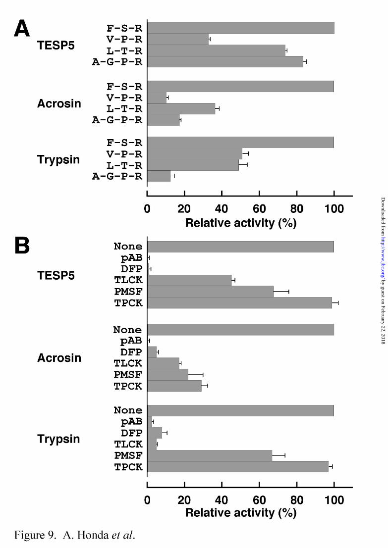
Proteolytic activity of recombinant TESP5 released by PI-PLC treatment was measured
using various peptidyl-MCAs as substrates (Fig. 9A). For comparison, rat sperm acrosin and
bovine pancreatic trypsin were also used. These three proteins exhibited the maximum
activity toward Boc-Phe-Ser-Arg-MCA among the substrates tested. TESP5 was
distinguished from acrosin and trypsin by the substrate specificity; TESP5 was capable of
hydrolyzing Boc-Leu-Thr-Arg-MCA and Boc-Ala-Gly-Pro-Arg-MCA (typical substrates for
factor VIIa-Tf and processing enzyme of atrial natriuretic peptide precursor, respectively) as
effectively as Boc-Phe-Ser-Arg-MCA. No enzyme activity was detected in these three
proteases when Suc-Ala-Ala-Ala-MCA, Suc-Ile-Ile-Trp-MCA, Suc-Leu-Leu-Val-Tyr-MCA,
and Z-Val-Lys-Met-MCA, typical substrates for elastase, endothelin, chymotrypsin, and
amyloid A4 generating enzyme, respectively, were used (data not shown). Moreover, the
inhibition profile of TESP5 was similar to those of acrosin and trypsin, except that the
proteolytic activity of TESP5 was inhibited by TLCK to a relatively low extent (Fig. 9B).
However, the inhibitory effects of pAB, DFP, TLCK, and PMSF on the TESP5 activity were
consistent with our previous results obtained by SDS-PAGE in the presence of gelatin (14).
by guest on February 22, 2018http://w
ww
.jbc.org/D
ownloaded from

Mouse Sperm TESP5 19
DISCUSSION
This study describes the identity of TESP5 with testicular testisin and eosinophilic
esp-1 (Figs. 1 and 2), and with 42- and 41-kDa sperm serine proteases (Figs. 4 and 5) that
may participate in the limited proteolysis of the egg ZP in fertilization, instead of and/or in
cooperation partly with acrosin (8, 9, 14, 15). TESP5 is localized on the membranes of
testicular germ cells and cauda epididymal sperm (Figs. 4 and 6). Particularly, the 42- and
41-kDa forms of TESP5 are selectively included into lipid rafts of sperm membranes
presumably as a GPI-anchored protein (Fig. 7). Since lipid rafts are thought to function in
signal transduction at the cell surface in response to intra- and extracellular stimuli (32-37), it
is conceivable that TESP5 plays an important role(s) in the sperm/egg interactions, including
the sperm penetration of the ZP.
Human testisin was originally identified as a candidate for non-classical type II tumor
suppressor, since expression of the testisin gene is completely lost in the testicular tumors (29).
A possible function of both human and mouse testisins has been speculated to participate in
proteolytic cleavage and release of biologically active molecules required for spermatogenesis,
including the migration of maturing germ cells in the seminiferous tubules (18, 29).
However, the expression pattern during spermatogenesis is highly divergent between these
two genes (proteins); human testisin is localized only in pre-meiotic spermatocytes (29),
whereas the occurrence of the mouse protein is specific for haploid germ cells (18). These
data imply that the function of mouse testisin in germ cells may be essentially different from
that of the human protein. The roles of human and mouse testisins in fertilization are
unknown at all, since the presence of the proteins in sperm has not been examined minutely.
In the present study, our data concerning the localization of TESP5 in lipid rafts of the sperm
membranes suggest that at least in the mouse, TESP5/testisin is probably implicated rather in
fertilization than in spermatogenesis.
Although 42- and 41-kDa gelatin-hydrolyzing serine proteases have been supposed to
by guest on February 22, 2018http://w
ww
.jbc.org/D
ownloaded from

Mouse Sperm TESP5 20
be different proteins (8, 14, 15), Western blot analysis indicates that the 41-kDa form of
TESP5 is likely to be a processed form of the 42-kDa protein (Figs. 4 and 5). However, it is
unclear at present whether the 42-kDa form corresponds to the TESP5 zymogen, and whether
the zymogen itself is converted by autoactivation into enzymatically active form(s). As
shown in Fig. 4, two 43- and 42-kDa proteins, which immunoreact with affinity-purified
anti-TESP5 antibody, are present in the testicular extracts. Our preliminary experiments
indicate that the two testicular proteins exhibit no gelatin-hydrolyzing activity (Honda, A. and
Baba, T., unpublished data). These results suggest that the zymogen(s) of TESP5 may
correspond to the 43- or 42-kDa protein or both present in the testis, and may barely have
ability to autoactivate itself. Acrosin perhaps functions partly as a processing enzyme to
convert the TESP5 zymogen(s) into the active 42- and 41-kDa proteins. If so, the
insufficient ability of the TESP5 zymogen(s) in conversion into the active enzymes in the
absence of acrosin (14) seems to explain the fact that the sperm penetration of the egg ZP is
delayed in the acrosin-deficient mouse (8).
Regardless of treatment with exogenous pancreatic trypsin, extracts of rat and hamster
sperm contain no gelatin-hydrolyzing protein corresponding to mouse TESP5 (15). When
the rat sperm extracts were analyzed by Western blotting, only a single protein with a
molecular mass of 43 kDa and a pI value of 6.0 to 6.5 indeed immunoreacted with
affinity-purified anti-mouse TESP5 antibody (Honda, A. and Baba, T., unpublished data).
However, no gelatin-hydrolyzing activity of the rat 43-kDa protein was found even if the rat
sperm extracts were treated with exogenous pancreatic trypsin. These data are entirely
consistent with the previous observation (15) that the 42- and 41-kDa gelatin-hydrolyzing
proteases (TESP5/testisin) are present specifically in mouse sperm, and emphasize that the
system of sperm serine proteases is highly different between mouse and other animals,
including rat and hamster.
Recombinant TESP5 produced in HEK293 cells is readily released from the cell
surface by treatment with bacterial PI-PLC (Fig. 8), suggesting that the C-terminal
by guest on February 22, 2018http://w
ww
.jbc.org/D
ownloaded from

Mouse Sperm TESP5 21
hydrophobic sequence of TESP5 (Fig. 2) functions as the signal sequence required for
attachment to GPI at least in HEK293 cells. To ascertain whether TESP5 is GPI-anchored
on the sperm membrane, mouse cauda epididymal sperm were treated with bacterial PI-PLC,
and the released proteins were analyzed by Western blotting. Sperm TESP5 was resistant to
the PI-PLC treatment, as reported in mouse TESP1 and TESP2 (16), and rat 2B1 glycoprotein
(PH20, see Ref. 38). The reason for this discrepancy may be due to a possible
modification(s) of GPI anchors on the sperm membrane, including the attachment of an extra
fatty acid to the inositol ring, as described previously (39, 40). The resistance of some
sperm GPI-anchored proteins to PI-PLC possibly implies the protection of the proteins from
the attack of PI-PLC-like enzymes because of the physiological importance in the sperm/egg
interactions.
The mechanism of sperm penetration through the egg ZP is not completely elucidated
yet. Since TESP5 (42- and 41-kDa serine proteases) is the most predominant serine protease
in mouse sperm, further characterization of TESP5 will be necessary to understand the
molecular events in the sperm/egg interactions, including the sperm penetration through the
ZP.
Acknowledgments — We thank Drs. K. Toshimori and S. B. Moss for kind gifts of
monoclonal antibodies MN7 and MC101, and rabbit anti-AKAP82 antiserum, respectively.
by guest on February 22, 2018http://w
ww
.jbc.org/D
ownloaded from

Mouse Sperm TESP5 22
REFERENCES
1. Yanagimachi, R. (1994) in The Physiology of Reproduction (Knobil, E. and Neill, J.,
eds), pp. 189-317, Raven Press, New York
2. Wassarman, P. M. (1999) Cell 96, 175-183
3. Wassarman, P. M., Jovine, L., and Litscher, E. S. (2001) Nature Cell Biol. 3, E59-E64
4. Ward, C. R. and Kopf, G. R. (1993) Dev. Biol. 158, 9-34
5. Bedford, J. M. (1998) Biol. Reprod. 59, 1275-1287
6. Baba, T., Kashiwabara, S., Watanabe, K., Itoh, H., Michikawa, Y., Kimura, K., Takada,
M., Fukamizu, A., and Arai, Y. (1989) J. Biol. Chem. 264, 11920-11927
7. Klemm, U., Müller-Esterl, W., and Engel, W. (1991) Hum. Genet. 87, 635-641
8. Baba, T., Azuma, S., Kashiwabara, S., and Toyoda, Y. (1994) J. Biol. Chem. 269,
31845-31849
9. Yamagata, K., Murayama, K., Okabe, M., Toshimori, K., Nakanishi, T., Kashiwabara, S.,
and Baba, T. (1998) J. Biol. Chem. 273, 10470-10474
10. Stambaugh, R., Brackett, B. G., and Mastroianni, L. (1969) Biol. Reprod. 1, 223-227
11. Zaneveld, L. J. D., Polakoski, K. L., Robertson, R. T., and Williams, W. L. (1971)
Proceedings of the First International Conference on Proteinase Inhibitors, pp.
236-244, Walter de Gruyter, Berlin
12. Fraser, L. R. (1982) J. Reprod. Fertil. 65, 185-194
13. Miyamoto, H. and Chang, M. C. (1973) Biol. Reprod. 9, 533-537
14. Yamagata, K., Murayama, K., Kohno, N., Kashiwabara, S., and Baba, T. (1998) Zygote
6, 311-319
15. Yamagata, K., Honda, A., Kashiwabara, S., and Baba, T. (1999) Dev. Genet. 25, 115-122
16. Kohno, N., Yamagata, K., Yamada, S., Kashiwabara, S., Sakai, Y., and Baba, T. (1998)
Biochem. Biophys. Res. Commun. 245, 658-665
17. Ohmura, K., Kohno, N., Kobayashi, Y., Yamagata, K., Sato, S., Kashiwabara, S., and
by guest on February 22, 2018http://w
ww
.jbc.org/D
ownloaded from

Mouse Sperm TESP5 23
Baba, T. (1999) J. Biol. Chem. 274, 29426-29432
18. Scarman, A. L., Hooper, J. D., Boucaut, K. J., Sit, M. L., Webb, G. C., Normyle, J. F.,
and Antalis, T. M. (2001) Eur. J. Biochem. 268, 1250-1258
19. Inoue, M., Isobe, M., Itoyama, T., and Kido, H. (1999) Biochem. Biophys. Res. Commun.
266, 564-568
20. Tanii, I., Araki, K., and Toshimori, K. (1994) Cell Tissue Res. 277, 61-67
21. Toshimori, K., Tanii, I., and Araki, S. (1995) Mol. Reprod. Dev. 42, 72-79
22. Johnson, L. R., Foster, J. A., Haig-Ladewig, L., VanScoy, H., Rubin, C. S., Moss, S. B.,
and Gerton, G. L. (1997) Dev. Biol. 192, 340-350
23. Baba, T., Niida, Y., Michikawa, Y., Kashiwabara, S., Kodaira, K., Takenaka, M., Kohno,
N., Gerton, G. L., and Arai, Y. (1994) J. Biol. Chem. 269, 10133-10140
24. Kashiwabara, S., Zhuang, T., Yamagata, K., Noguchi, J., Fukamizu, A., and Baba, T.
(2000) Dev. Biol. 228, 106-115
25. Walensky, L. D. and Snyder, S. H. (1995) J. Cell Biol. 130, 857-869
26. Rodgers, W., and Rose, J. K. (1996) J. Cell Biol. 135, 1515-1523
27. Niwa, H., Yamamura, K., and Miyazaki, J. (1991) Gene 108, 193-199
28. Fuller, S. A., Takahashi, M., and Hurrell, F. G. R. (1991) in Current Protocols in
Molecular Biology (Ausubel, F. M., Brent, P., Kingston, R. E., Moore, D. D.,
Seidman, J. G., Smith, J. A., and Struhi, K., eds.), pp. 11.11.1-11.11.5, Greene
Publishing and Wiley-Interscience, New York
29. Hooper, J. D., Nicol, D. L., Dickinson, J. L., Eyre, H. J., Scarman, A. L., Normyle, J. F.,
Stuttgen, M. A., Douglas, M. L., Loveland, K. A., Sutherland, G. R., and Antalis, T.
M. (1999) Cancer Res. 59, 3199-3205
30. Davie, E. W., Fujikawa, K., Kurachi, K., and Kisiel, W. (1979) Adv. Enzymol. 48,
277-318
31. Perona, J. J. and Craik, C. S. (1995) Protein Sci. 4, 337-360
32. Simons, K. and Ikonen, E. (1997) Nature 387, 569-572
by guest on February 22, 2018http://w
ww
.jbc.org/D
ownloaded from

Mouse Sperm TESP5 24
33. Varma, R. and Mayor, S. (1998) Nature 394, 798-801
34. Harder, T. and Simons, K. (1997) Curr. Opin. Cell Biol. 9, 534-542
35. Jacobson, K. and Dietrich, C. (1999) Trends. Cell Biol. 9, 87-91
36. Ohta, K., Sato, C., Matsuda, T., Toriyama, M., Lennarz, W. J., and Kitajima, K. (1999)
Biochem. Biophys. Res. Commun. 258, 616-623
37. Nishimura, H., Cho, C., Branciforte, G. R., Myles, D. G., and Primakoff, P. (2001) Dev.
Biol. 233, 204-213
38. Seaton, G. J., Hall, L., and Jones, R. (2000) Biol. Reprod. 62, 1667-1676
39. Roberts, W. L., Myher, J. J., Kuksis, A., Low, M. G., and Rosenberry, T. L. (1988) J.
Biol. Chem. 263, 18766-18775
40. Guther, M. L., Dealmeida, M. L., Rosenberry, T. L., and Ferguson, M. A. (1994) Anal.
Biochem. 219, 249-255
by guest on February 22, 2018http://w
ww
.jbc.org/D
ownloaded from

Mouse Sperm TESP5 25
(FOOTNOTES)
*This work was supported in part by Grant-in-Aids for Scientific Research on Priority
Area (B), Scientific Research (A), and Exploratory Research from Japan Society for the
Promotion of Science (JSPS) and Ministry of Education, Culture, Sports, Science and
Technology in Japan (MEXT).
Nucleotide sequences reported in this paper have been submitted to the
DDBJ/GenBank™ Data Bank with accession numbers AB059414 and AB059415.
‡To whom correspondence should be addressed. Tel./Fax: +81-298-53-6632; E-mail:
§Present address: Genome Information Research Center, Osaka University, Yamadaoka
3-1, Suita, Osaka 565-0871, Japan.
The abbreviations used are: Boc, t-butyloxycarbonyl; BSA, bovine serum albumin;
DFP, diisopropyl fluorophosphate; GPI, glycosylphosphatidylinositol; IVF, in vitro
fertilization; MCA, 4-methylcoumaryl-7-amide; NP-40, Nonidet P-40; pAB,
p-aminobenzamidine; PAGE, polyacrylamide gel electrophoresis; PBS, phosphate-buffered
saline; PCR, polymerase chain reaction; PI-PLC, phosphatidylinositol-specific phospholipase
C; PMSF, phenylmethanesulfonyl fluoride; RT, reverse transcriptase; SDS, sodium dodecyl
sulfate; Suc, N-succinyl; TESP, testicular serine protease; TLCK, Nα-tosyl-L-lysine
chloromethyl ketone; TPCK, N-tosyl-L-phenylalanine chloromethyl ketone; 2D,
two-dimensional; TX-100, Triton X-100; Z, carbobenzoxy; ZP, zona pellucida.
by guest on February 22, 2018http://w
ww
.jbc.org/D
ownloaded from

Mouse Sperm TESP5 26
(FIGURE LEGENDS)
Fig. 1. Exon/intron organization of the mouse TESP5 gene, potential structure of
prepro-form of TESP5, and Southern blot analysis of mouse genomic DNA. A, Gene
and protein structures of mouse TESP5. Two genomic clones, HGC3 and HGC14, encoding
the TESP5 gene have been identified from a mouse genomic DNA library. The TESP5 gene
is approximately 4.5 kbp in length, and consists of six exons (closed boxes) interrupted by
five introns. Restriction enzyme sites are as follows: G, BglII; E, EcoRI; H, HindIII. The
prepro-form of mouse TESP5 is a single-chain 324-residue polypeptide containing three
domains (DI, DII, and DIII). The disulfide-bond arrangements (C-C) and positions of three
active-site residues, His, Asp, and Ser, are based on the sequence similarity of TESP5 with
other serine proteases. Potential N-glycosylation sites are indicated by Y symbols. B,
Southern blot analysis of mouse genomic DNA. Mouse genomic DNAs digested by BglII
(G), EcoRI (E), and HindIII (H) were separated by agarose gel electrophoresis and transferred
onto nylon membranes. The blots were probed by a 32P-labeled DNA fragment encoding a
part of sixth exon.
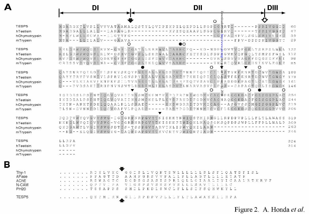
Fig. 2. Comparison of the amino acid sequence of mouse TESP5 with those of
other serine proteases and GPI-anchored proteins. A, Sequence alignment of TESP5
with human testisin, human chymotrypsin, and mouse trypsin. As described in Fig. 1A, the
prepro-form of mouse TESP5 is a 324-residue polypeptide containing three domains (DI, DII,
and DIII). Dashes represent gaps introduced to optimize the alignment. Identical residues
in the sequences between TESP5 and other proteases are shown by shaded boxes. Closed
and open arrows indicate the defined or putative cleavage sites during initial transfer of the
nascent protein to the endoplasmic reticulum and during activation of the serine protease
zymogens, respectively. The locations of Cys residues (open circles) and three active-site
residues (closed circles) as a serine protease are also represented above the sequence.
by guest on February 22, 2018http://w
ww
.jbc.org/D
ownloaded from

Mouse Sperm TESP5 27
Closed triangles indicate potential N-glycosylation sites. B, Sequence alignment of TESP5
with five GPI-anchored proteins. The C-terminal sequence of TESP5 is compared with
those of rat Thy-1, human alkaline phosphatase (APase), Drosophila acetylcholinesterase
(AChE), chicken N-CAM, and guinea pig PH20. Arrows represent possible or defined
GPI-attachment sites.
Fig. 3. Northern blot analysis of total cellular RNAs from various tissues and
purified populations of spermatogenic cells. A, Northern blot analysis of total RNAs (10
µg) from mouse testis (T), brain (B), lung (Lu), heart (H), liver (Li), kidney (K), ovary (O),
and uterus (U). The blots were first probed by a 32P-labeled DNA fragment encoding mouse
TESP5, and then re-probed by the DNA fragment coding for mouse
glyceraldehyde-3-phosphate dehydrogenase (GDH). B, Removal of mRNA poly(A) tails by
RNase H treatment. Total RNAs (12 µg) of pachytene spermatocytes (P), round spermatids
(R), and a mixture of elongating spermatids and residual bodies (E) from 60-day-old mouse
testes were annealed with oligo(dT)15, treated with RNase H, and then subjected to Northern
blot analysis. The blots were probed by a 32P-labeled DNA fragment encoding mouse
TESP5.
Fig. 4. Western blot analysis of protein extracts from mouse testis and sperm.
A, Western blot analysis of soluble and insoluble proteins (30 µg) from testicular tissues, and
of detergent-soluble proteins (20 µg) from cauda epididymal sperm, using affinity-purified
anti-TESP5 antibody as a probe. Proteins were separated by SDS-PAGE under non-reducing
conditions, and subjected to Western blot analysis using affinity-purified anti-TESP5 antibody.
Two immunoreactive proteins with sizes of 43 and 42 kDa were detected only in the insoluble
protein extracts of testis, whereas the sperm extracts solubilized with 1% SDS or 1% Triton
X-100 (TX-100) gave two bands corresponding to 42- and 41-kDa proteins. B, Western blot
analysis of TX-100-soluble proteins from cauda epididymal sperm, using affinity-purified
by guest on February 22, 2018http://w
ww
.jbc.org/D
ownloaded from

Mouse Sperm TESP5 28
anti-TESP5 antibody as a probe. Proteins (0.2 mg) were separated by 2D-PAGE under
non-reducing conditions, and subjected to Western blot analysis (left panel) or SDS-PAGE in
the presence of gelatin (right panel). Closed and open arrowheads indicate the locations of
the immunoreactive and gelatin-hydrolyzing proteins, respectively.
Fig. 5. Immunoprecipitation of TESP5 from protein extracts of mouse sperm.
Triton X-100 (TX-100)-soluble extracts (0.2 mg of proteins) of cauda epididymal sperm were
used for immunoprecipitation of TESP5 in the absence (lane 2) or presence of
affinity-purified antibodies against mouse sp32 (lane 3) and mouse TESP5 (lane 4). The
sperm protein extracts were also used as a positive control (lane 1). Aliquots of the
immunoprecipitates were separated by SDS-PAGE under non-reducing conditions, and
subjected to Western blot analysis using affinity-purified anti-TESP5 antibody (A) and
SDS-PAGE in the presence of gelatin (B). The immunoreactive proteins with sizes of 42
and 41 kDa exhibiting the gelatin-hydrolyzing activity were found only in lanes 1 and 4.
Fig. 6. Location of TESP5 in mouse testis and cauda epididymal sperm. A,
Immunohistochemical analysis of testicular sections (a and b) and indirect
immunofluorescence assay of epididymal sperm (c, d, e, and f). For immunohistochemistry,
sections of mouse testis were probed by affinity-purified anti-TESP5 antibody using
3,3-diaminobenzidine as a chromogen. The sections were counterstained with methyl green
(a, x 400; b, x 1,000). Red arrows indicate the location of granular signals with a great
intensity in elongating spermatids. Indirect immunofluorescence assay of acrosome-intact
sperm was also carried out in the absence (c and d) or presence (e and f) of recombinant
TESP5. The immunofluorescence signals in the sperm head (arrowheads), midpiece, and
cytoplasmic droplet (arrows) disappeared in the presence of the recombinant protein (f). The
phase-contrast (c and e) and immunofluorescence (d and f) views are represented. B,
Western blot analysis of proteins in subcellular components of cauda epididymal sperm.
by guest on February 22, 2018http://w
ww
.jbc.org/D
ownloaded from

Mouse Sperm TESP5 29
Proteins in the sperm subcellular components enriched by plasma and outer-acrosomal
membranes (Fraction A), soluble proteins released by the A23187-induced acrosome reaction,
including acrosomal components (Fraction B), acrosome-reacted sperm heads (Fraction C),
tails (Fraction D), and cytoplasmic droplets (Fraction E, see Experimental Procedures) were
analyzed by Western blotting using affinity-purified anti-TESP5 antibody. Antibodies
against an acrosomal proacrosin-binding protein, sp32, a 90-kDa intra-acrosomal protein,
MN7, a 155-kDa intra-acrosomal protein, MC101, and a protein kinase A anchor protein,
AKAP82, in the flagellum fibrous sheath, were also used as markers. Note that the 42- and
41-kDa forms of TESP5 are detected in all five fractions.
Fig. 7. TESP5 is selectively included into Triton X-100-insoluble, low-density
membrane fractions of mouse sperm. Low-density membrane fractions insoluble in Triton
X-100 were prepared from cauda epididymal sperm. An aliquot (10 µl) of the fractions was
analyzed by Western blotting using affinity-purified anti-TESP5 antibody (A) or SDS-PAGE
in the presence of gelatin (B). The 42- and 41-kDa forms of TESP5 exhibiting
gelatin-hydrolyzing activity are found in the Triton X-100-insoluble, low-density membrane
fractions (lanes 7 and 8).
Fig. 8. Release of recombinant TESP5 from the membrane of HEK293 cells by
Bacillus cereus phosphatidylinositol-specific phospholipase C (PI-PLC). Transformed
HEK293 cells (5 x 10⁶ cells in 0.1 ml of PBS) were incubated at 37°C for 1 h in the absence
or presence of bacterial PI-PLC (0.1 or 1.0 units/ml), and centrifuged. Aliquots (10 µl) of
the supernatants were subjected to SDS-PAGE in the presence of gelatin under non-reducing
conditions. Western blot analysis was carried out using affinity-purified anti-TESP5
antibody. M, mock transformants; T, TESP5 transformants.
Fig. 9. Substrate specificity of TESP5 and inhibitory effects of various serine
by guest on February 22, 2018http://w
ww
.jbc.org/D
ownloaded from

Mouse Sperm TESP5 30
protease inhibitors on the TESP5 activity. A, Substrate specificity of TESP5. The
enzyme activities of recombinant TESP5, rat acrosin, and bovine pancreatic trypsin were
measured using Boc-Phe-Ser-Arg-MCA (F-S-R), Boc-Val-Pro-Arg-MCA (V-P-R),
Boc-Leu-Thr-Arg-MCA (L-T-R), and Boc-Ala-Gly-Pro-Arg-MCA (A-G-P-R) as substrates.
The enzyme reactions were carried out at 30°C for 30 min, and terminated by addition of 0.1
M acetate buffer, pH 4.3. The enzyme activity toward Boc-Phe-Ser-Arg-MCA is indicated
as 100%. Data are expressed as the means ± S.D., where n > 3. B, Effects of serine
protease inhibitors on enzyme activity of TESP5. The enzyme activity was measured in the
presence of 1 mM each of p-aminobenzamidine (pAB), diisopropyl fluorophosphate (DFP),
Nα-tosyl-L-lysine chloromethyl ketone (TLCK), phenylmethanesulfonyl fluoride (PMSF), and
N-tosyl-L-phenylalanine chloromethyl ketone (TPCK) using Boc-Phe-Ser-Arg-MCA as a
substrate, as described above. The enzyme activity in the absence of any inhibitors (None) is
indicated as 100%. Data are expressed as the means ± S.D., where n > 3.
by guest on February 22, 2018http://w
ww
.jbc.org/D
ownloaded from


Arata Honda, Kazuo Yamagata, Shin Sugiura, Katsuto Watanabe and Tadashi Babamembrane presumably as a GPI-anchored protein
A mouse serine protease TESP5 is selectively included into lipid rafts of sperm
published online February 22, 2002J. Biol. Chem.
10.1074/jbc.M112470200Access the most updated version of this article at doi:
Alerts:
When a correction for this article is posted•
When this article is cited•
to choose from all of JBC's e-mail alertsClick here
by guest on February 22, 2018http://w
ww
.jbc.org/D
ownloaded from