a multidimensional manual therapy model for managing
TRANSCRIPT
M C Steffen
A thesis submitted in fulfillment of the requirements for the degree of
PHILOSOPHIAE DOCTOR
In the Department of Physiotherapy School of Health Care Sciences
Faculty of Health Sciences University of Pretoria
Supervisor : Dr C A Eksteen
A multidimensional manual therapy model for
managing patients with chronic non-specific low
back pain
©© UUnniivveerrssiittyy ooff PPrreettoorriiaa

ii
DECLARATION
I, Marjory Christine Steffen declare that the work:
‗A multidimensional manual therapy model for managing patients with chronic non-
specific low back‟
is my original work, and that it has not been submitted before for any degree or
examination at any other institution. All the sources that have been used or quoted
have been acknowledged by means of complete references in the text and
bibliography.
Researcher‘s Signature …………………………..
Date: ……………………….

iii
Acknowledgements
The author gratefully acknowledges the assistance received from many people in
completing this thesis. In particular the considerable contributions made by the
following people:
Dr Carina Eksteen, who has been my supervisor on this long journey. I greatly
appreciate her inspiration, dedication, guidance and endless energy and
driving force;
Lynne Thompson – I am eternally grateful to my colleague and friend Lynne
for her continuous support and encouragement during the many debates we
had in developing this thesis;
The many support groups I have relied upon at the University of Pretoria, for
their willing assistance in library services, graphics, and professional
coordination;
My family,who have assisted in every way possible to enable me to fulfil my
ambitions at their personal sacrifices; and
Most importantly, my motivation to complete this thesis is due to my
handicapped patients who have provided the inspiration for achieving our
ultimate objectives of health and a reward for normal life.

iv
Contents List of Tables .............................................................................................................. ix
List of Figures ............................................................................................................. x
Abstract .................................................................................................................... xiii
Background and rationale .......................................................................................... 1
1.1 Introduction .................................................................................................. 1
1.2 The mechanisms in the development of CNSLBP ....................................... 6
1.3 Management of patients with CNSLBP ...................................................... 10
1.3.1 The researcher‘s multidimensional manual therapy approach to the
management of patients with CNSLBP ................................................ 16
1.4 Problem statement ..................................................................................... 19
1.5 Research questions ................................................................................... 22
1.6 Research aims and objectives ................................................................... 22
1.7 Research approach .................................................................................... 23
1.8 The nature of this study.............................................................................. 26
1.9 Clarification of terminology ......................................................................... 32
1.9.1 Low back pain ....................................................................................... 32
1.9.2 Acute specific low back pain ................................................................. 33
1.9.3 Manual therapy ..................................................................................... 33
1.9.4 Multidisciplinary approach to management of CNSLBP ....................... 34
1.9.5 Multidimensional manual therapy for the management of CNSLBP ..... 35
1.9.6 Chronicity .............................................................................................. 35
1.9.7 Dysfunction and disability in patients with CNSLBP ............................. 35
1.9.8 Plasticity ............................................................................................... 36
1.9.9 Integrated spinal movement system (ISMS) ......................................... 37
1.10 Outline of the study .................................................................................... 37

v
Research methodology ............................................................................................ 39
2.1 Introduction ................................................................................................ 39
2.2 The frame of reference............................................................................... 40
2.2.1 The paradigm of this study ................................................................... 40
2.3 The research approach .............................................................................. 46
2.3.1 The hermeneutic process as knowledge-generating process ............... 48
2.3.2 Model development .............................................................................. 57
2.3.3 The role of literature in the development of a holistic manual therapy
model for managing patients with CNSLBP .......................................... 58
2.4 Trustworthiness of the conceptualisation of ISMS dysfunction and the
development of the multidimensional manual therapy model ..................... 58
2.5 Significance of the study ............................................................................ 59
2.6 Ethical considerations ................................................................................ 60
2.7 Summary of the chapter ............................................................................. 61
Mechanisms generating the development of a dysfunctional integrated spinal
movement system3.1 ................................................................................. Introduction
................................................................................................................... 62
3.2 Conceptualisation of the ISMS ................................................................... 63
3.2.1 The articular components of the ISMS ................................................. 64
3.2.2 The muscle system of the ISMS ........................................................... 69
3.2.3 The neural components of the spine .................................................... 78
3.2.4 The connective tissues in the trunk ...................................................... 83
3.3 Postural control of the ISMS ...................................................................... 86
3.4 Patho-physiological responses underlying the development of ISMS
dysfunction ................................................................................................. 90
3.4.1 The effect of muscle spasm, overuse and disuse on muscular tissue .. 90
3.4.2 The development of trigger points ........................................................ 91
3.4.3 The process of connective tissue stiffening in patients with CNSLBP .. 96
vi
3.4.4 Effects of nervous tension (stress) on the musculature of the body ..... 97
3.5 Development of integrated spinal movement system (ISMS) dysfunction.. 98
3.5.1 Musculoskeletal adaptation to unilateral abnormal spinal loading ...... 101
3.6 Soft tissue plasticity as an inherent process in the development of ISMS
dysfunction ............................................................................................... 113
3.6.1 Plasticity and postural control ............................................................. 115
3.7 Factors that can influence/adapt the typical pattern of ISMS dysfunction 116
3.7.1 Differences in response of the lower and upper lumbar motion segments
........................................................................................................... 116
3.7.2 Poor posture and postural control ....................................................... 116
3.7.3 Disuse and sedentary lifestyle ............................................................ 117
3.7.4 The process of spinal loading ............................................................. 117
3.7.5 Cervical and thoracic dysfunction ....................................................... 118
3.7.6 Association of chronic unilateral low back pain with disruption of tactile
input.................................................................................................... 119
3.7.7 Neural referred pain through torsioning of the biomechanical ISMS ... 119
3.7.8 The effect of stress on spinal dysfunction ........................................... 120
3.7.9 The influence of underlying degeneration in the synovial joints of the
spine ................................................................................................... 120
3.7.10 Previous history of back pain and response to health care management
........................................................................................................... 121
3.8 Pain processing as integral component driving the development of ISMS
dysfunction ............................................................................................... 121
3.8.1 The biomechanical origin of pain processing in the development of ISMS
dysfunction ......................................................................................... 122
3.8.2 The neuromatrix as part of the ISMS dysfunction ............................... 127
3.8.3 Characteristic adaptive behaviour in patients with ISMS dysfunction . 132
3.8.4 Pain modulation .................................................................................. 138

vii
3.9 Conclusion ............................................................................................... 141
The principles of a multidimensional assessment model for patients with chronic
nonspecific low back pain ....................................................................................... 147
4.1 Introduction .............................................................................................. 147
4.2 Typical clinical appearance of a patient with CNSLBP ............................. 149
4.2.1 History taking ...................................................................................... 151
4.3 A multidimensional model for the assessment of patients with CNSLBP . 173
4.4 Summary of the chapter and discussion of the holistic integrated model for
the assessment of patients with CNSLBP ................................................ 175
Principles of a multidimensional manual therapy approach to patients with chronic
non-specific low back pain ..................................................................................... 177
5.1 Introduction .............................................................................................. 177
5.2 Principles of pain modulation ................................................................... 181
5.2.1 The therapist as pain-inhibiting agent through a professional therapist-
patient relationship ............................................................................. 181
5.2.2 The role of cognitive behavioural therapy in the multidimensional manual
therapy of patients with CNSLBP ....................................................... 182
5.2.3 Pain modulation through manual therapy ........................................... 183
5.2.4 The role of pharmacology as part of the holistic approach to manual
therapy for patients with CNSLBP ...................................................... 196
5.2.5 Re-education of postural control ......................................................... 197
5.3 Principles of multidimensional pain modulation in patients with CNSLBP 200
5.4 Clinical principles for the treatment of patients with CNSLBP .................. 202
5.5 Risk factors to take cognisance of during manual therapy ....................... 204
5.6 A multidimensional manual therapy model for management of patients with
CNSLBP ................................................................................................... 208

viii
Conclusion, discussion, limitations and recommendations ..................................... 211
6.1 Introduction .............................................................................................. 211
6.2 Evaluation of the multidimensional manual therapy model for the treatment
of patients with CNSLBP .......................................................................... 213
6.2.1 Summary of the multidimensional manual therapy model .................. 213
6.2.2 Evaluation of the multidimensional manual therapy model against other
models used in the management of CNSLBP .................................... 214
6.3 Limitations of this study ............................................................................ 222
6.4 Recommendations ................................................................................... 223
6.4.1 Recommendations for further research .............................................. 223
6.5 Summary .................................................................................................. 225
References ............................................................................................................. 228

ix
List of Tables
Table 1.1: Summary of the components of the model 24
Table 3.1: The global stabilisers and mobilisers of the neck
and limb girdles (Comerford & Mottram, 2012) 71
Table 4.1: Predictors of chronic pain and disability
(Waddell, 2004 p 128) 153

x
List of Figures
Figure 1.1: Differentiation between specific and non-specific low back pain 3
Figure 1.2: Comparison of the key moments in the development of
manual therapy, diagnostic medicine, and neuro- and
orthopaedic surgery experienced by the researcher since 1970 16
Figure 1.3: Types of knowledge and internal influences on knowledge generation 27
Figure 1.4: The research, theory, practice cycle 30
Figure 2.1: Summary of the research approach and research methodology 47
Figure 2.2: Dialectical reasoning in the diagnosis and management of patients 54
Figure 2.3: The contributions of empirico-analytical and interpretive
reasoning paradigms to the formation of clinical knowledge 56
Figure 3.1: Degrees of motion of the motion segment 65
Figure 3.2(a): Intersegmental posterior spinal muscles 69
Figure 3.2(b): Multisegmental posterior spinal muscles 70
Figure 3.3: The interconnectedness between the spinal cord, the dorsal
and ventral rami of the spinal nerve and the sympathetic chain adjacent
to the vertebral column 79
Figure 3.4: Diagrammatic sketch of the (specialised) connective tissues
around the spinal cord and nerve roots 80
Figure 3.5(a) and 3.5(b): The visible areas of superficial and deeper
fascia on the posterior spine 85
Figure 3.6: The integrated spinal movement system 88
Figure 3.7: The activation of myofascial trigger points 93
Figure 3.8(a): Anatomy of the quadratus lumborum muscle trigger point 94
Figure 3.8(b): Area of referral of the quadratus lumborum muscle 94
Figure 3.8(c): Anatomy of trigger points of the psoas and iliacus muscles 94
Figure 3.8(d): Area of referred pain of psoas and iliacus muscles 94
Figure 3.8(e): Anatomy of the trigger point of the gluteus maximus muscle 95
Figure 3.8(f): Area of referred pain of the gluteus maximus muscle 95
Figure 3.8(g): Anatomy of the gluteus medius trigger muscle 95
Figure 3.9(h): Gluteus medius muscle point referral pain pattern 95
Figure 3.8(i): Anatomy of the gluteus minimus trigger muscle 96
Figure 3.8(j): Gluteus minimus point pain referral pattern 96
Figure 3.8(k): Anatomy of the piriformis muscle trigger point 96
Figure 3.8(l): The piriformis muscle pain referral pattern 96
xi
Figure 3.8(m): Anatomy of the trigger point in the iliocostalis
lumborum muscle 97
Figure 3.8(n): Pattern of pain referral of the iliocostalis lumborum muscle 97
Figure 3.9(a): A presentation of the somatic referred pain
from thoracic zygapophyseal joints 109
Figure 3.9(b): A presentation of the somatic referred pain
from lumbar zygapophyseal joints 109
Figure 3.10(a): The superficial thoracolumbar fascia 111
Figure 3.10(b): The deep thoraco-lumbar fascia 111
Figure 3.11: A schematic presentation of the circuits that mediate
physiological pain 124
Figure 3.12: Thalamus (Th), the amygdala (Amyg), the insula cortex (Insula),
the supplementary motor area (SMA), the posterior parietal cortex (PPC),
the prefrontal cortex (PFC), the cingulate cortex (ACC), the periaqueductal
grey (PAG), the basal ganglia and cerebellar cortex (not shown) and the
primary (S1) and secondary (S2, not shown) sensory cortex 125
Figure 3.13: Disease-induced functional and structural plasticity in neural
substrates of pain on molecular, synaptic, cellular and network levels 131
Figure 3.14: The fear avoidance model 134
Figure 3.15: Interrelated processes involved in generating and maintaining
ISMS dysfunction 137
Figure 3.16: Three stages in the development of chronic non-specific
low back pain 142
Figure 4.1: A Biopsychosocial model of CNSLBP 148
Figure 4.2: Diagnostic triage 151
Figure 4.3: An axial view of the position of the shoulder girdle relative
to the pelvis due to ISMS dysfunction (observation 1) 158
Figure 4.3(b): An anterior, posterior and lateral view of the patients posture
typical of ISMS dysfunction 158
Figure 4.4: An axial view of the position of the shoulder girdle relative
to the pelvis due to ISMS dysfunction (observation 2) 159
Figure 4.7: Fascia of the posterior aspect of the trunk 166
Figure 4.8: Model for the multidimensional assessment of a patient
with CNSLBP 173
Figure 5.1: Integration of the three driving factors integral to the
complexity of CNSLBP 177

xii
Figure 5.1: Lumbar rotation in sidelying (Maitland, 2004) 186
Figure 5.2: Myofascial release of the lateral oblique area 188
Figure 5.3: A schematic overview of the main circuits that mediate
physiological pain 192
Figure 5.4: Sympathetic pathways 194
Figure 5.5: Principles of multidimensional pain modulation in patients
with CNSLBP 201
Figure 5.6: A Multidimensional manual therapy model for management
of patients with CNSLBP 209
xiii
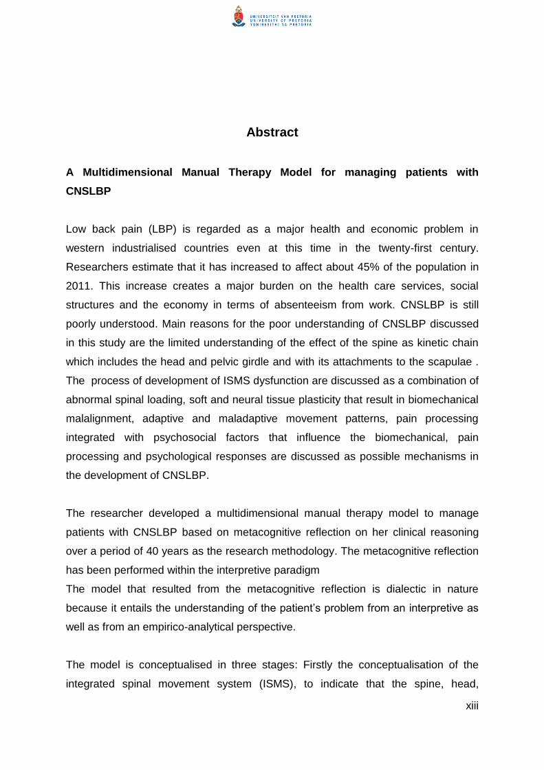
Abstract
A Multidimensional Manual Therapy Model for managing patients with
CNSLBP
Low back pain (LBP) is regarded as a major health and economic problem in
western industrialised countries even at this time in the twenty-first century.
Researchers estimate that it has increased to affect about 45% of the population in
2011. This increase creates a major burden on the health care services, social
structures and the economy in terms of absenteeism from work. CNSLBP is still
poorly understood. Main reasons for the poor understanding of CNSLBP discussed
in this study are the limited understanding of the effect of the spine as kinetic chain
which includes the head and pelvic girdle and with its attachments to the scapulae .
The process of development of ISMS dysfunction are discussed as a combination of
abnormal spinal loading, soft and neural tissue plasticity that result in biomechanical
malalignment, adaptive and maladaptive movement patterns, pain processing
integrated with psychosocial factors that influence the biomechanical, pain
processing and psychological responses are discussed as possible mechanisms in
the development of CNSLBP.
The researcher developed a multidimensional manual therapy model to manage
patients with CNSLBP based on metacognitive reflection on her clinical reasoning
over a period of 40 years as the research methodology. The metacognitive reflection
has been performed within the interpretive paradigm
The model that resulted from the metacognitive reflection is dialectic in nature
because it entails the understanding of the patient‘s problem from an interpretive as
well as from an empirico-analytical perspective.
The model is conceptualised in three stages: Firstly the conceptualisation of the
integrated spinal movement system (ISMS), to indicate that the spine, head,
xiv
shoulder and pelvic girdles function as a closed kinematic chain. Secondly the
process of the development of ISMS dysfunction as a major concept in the clinical
picture of patients with CNSLBP is based on functional anatomy of the ISMS and the
researcher‘s clinical observation in clinical practice. The researcher indicates how
the development of ISMS dysfunction and characteristic adaptive behaviour are
integrated components of the patient‘s complex heterogenic clinical picture. The
underlying process for the development of ISMS dysfunction as a possible
mechanism for CNSLBP is described as plasticity of soft and neural tissues
(including the brain) which result in chronicity over time.
Thirdly a multidimensional manual therapy model to manage patients with CNSLBP‘s
heterogenic condition is discussed. The model indicates how the mechanisms
underlying the development of ISMS dysfunction is addressed in a multidimensional
approach to patient management. Finally the multidimensional manual therapy
model is discussed in relation to other relevant intervention approaches. The model
finally serves as a point of departure for planning and conducting appropriate
research in basic and clinical sciences.
The multidimensional manual therapy model for the management of patients with
CNSLBP has been developed in clinical practice and is presented as a practice-
theory in the form of a model.
Key words: chronic non-specific low back pain, chronicity, plasticity, pain processing,
multidimensional manual therapy, clinical reasoning, holistic approach, spinal
dysfunction, hermeneutic research approach, practice-theory model.

1
CHAPTER 1
Background and rationale
1.1 Introduction
Low back pain (LBP) has affected human beings throughout recorded history (Allan
& Waddell, 1989). The biggest changes in the understanding of the pathophysiology,
diagnostics, and surgery that formed the basic management of patients with LBP
have taken place during the 20th Century.
From the 1900s, important milestones (Bucy, 1988) first showed that the pressure of
disc herniation could produce neurological symptoms.
In 1934 Mixter and Barr (White & Anderson, 1991) performed surgery on a patient
with low back pain and gave the first complete clinical, pathologic and surgical
description of disc prolapse as the cause of sciatica. Their paper in 1934 was
regarded by surgeons as a classic contribution to surgery and showed that surgery
for disc prolapse was possible.
In 1965 Melzack and Wall proposed the gate-control theory of pain (Melzack & Wall,
1996). The gate control theory proposes that pain is a multidimensional experience
produced by characteristic ‗neuro-signature‘ patterns of neuro-impulses generated
by a widely distributed neural network – the body-self neuromatrix in the brain
(Melzack & Wall, 1996).
Around 1970, the patho-physiology of lumbar degeneration, work done by Yong-Hing
and Kirkaldy-Willis (1984) resulted in a major breakthrough towards the
understanding of degenerative back disease and its effects on the components of
the vertebrae and soft tissues, resulting in changes within the biomechanics of the
patient‘s alignment and movements. This knowledge at that time revealed two ‗new‘
clinical entities, namely nerve root entrapment and spinal stenosis, which gave rise
to the conceptualisation of the ‗structural–anatomical–biomechanical‘ (SAB) or
2
‗disease model‘ (Alan & Waddell, 1989) for assessment and treatment of LBP. The
SAB model of management of LBP withstood the test of time, and it is still practiced
(Zusman, 2007).
The SAB era was followed by growing evidence that biological pain perception is
exacerbated by psychological as well as by social factors and may contribute to the
chronic pain and dysfunction in these patients Main & Watson (1999). Researchers
also found that pain may originate and be maintained by altered pathways in the
brain (Apkarian, Baliki & Geha 2009; Tracey and Bushnell, 2009; Kuner 2010).
Waddell (2004) concludes that LBP is a 20th Century medical disaster, and that
physiotherapists and medical practitioners together have failed in their attempts to
improve the manifestations of LBP. If properly managed by these professions
chronic back pain and disability should be reducing, but instead the opposite is true.
He also concludes that clinicians have lost sight of the basic principles to understand
pain and disability in their approach to management of CNSLBP.
The influence of the psychological and social factors on biological pain perception
contributed to the fact that the ‗biopsychosocial‘ (BPS) model was conceptualised for
more accurate multidimensional management of acute as well as chronic LBP
(McCarthy, Arnall, Strimpakos, Freemont, & Oldham, 2004).
Waddell‘s classification of LBP into specific and non-specific low back pain brought
much clarity on the understanding of the problem (Figure 1.1).

3
Figure 1.1: Differentiation between specific and non-specific low back pain
(Waddell, 2004; Adams, Bogduk, Burton, and Dolan 2002; Porterfield and DeRosa, 1990)
Acute specific LBP
~15% (requires
specific intervention)
Acute non-specific LBP ~ 85%
~70% ANSLBP
resolve after acute
episode with or
without treatment
~ 15% of the 85%
(some publications
see it as high as
~45-50%) of
patients start as
acute and continue
to develop into the
chronic phase
Predisposing
factors for the
development of
CNSLBP are:
Psychological
and social
influences
Genetic factors
Biomechanical
ISMS
dysfunction of
the spine
Tissue repair
Pain
processing
All low back pain sufferers can be classified as
acute specific or non-specific low back pain:

4
Henchoz and So (2008); Chanda, Alvin, Schnitzer and Apkarian (2011) estimate that
among the adult population between 60% and 85% of the general population suffers
from LBP at least once in their life time. Back pain affects at least 20% of people at
any time in their lives and about half the global population has had at least one
episode of LBP by the age of 30 (Docking, Fleming, Brayne, Zhao, Macfarlane &
Jones, 2011). Between 44% and 78% of people suffer relapses of LBP after an initial
episode of LBP. According to the European Guidelines for the Management of
Chronic Nonspecific Low Back Pain (CNSLBP) (2006) there is little scientific
evidence regarding the prevalence of CNSLBP: best estimates suggest that the
prevalence of people who are disabled by chronic low back pain is approximately
11% to 12% of the population.
More specifically, the problem of non-specific low back pain (NSLBP) is regarded as
the leading cause of disability among the population in developed countries (Bunzli,
Gilham & Esterman, 2011) and the risk of disabling back pain rises with ageing
(Docking et al., 2011). Chronic non-specific (or ‗idiopathic‘) back pain accounts for
the majority of patients treated in primary care because they make use of all the
health care resources available (Chanda et al., 2011). Any primary care intervention
has resulted in a disappointing outcome in terms of decreasing the burden of
suffering in patients with LBP (Pransky, Borkan, Young & Cherkin, 2011).
Pransky et al. (2011) report that despite the enormous increase in the number,
quality and variety of the research studies on LBP, since 1990, the progress in terms
of the impact it had on primary care resulted in a disappointed outcome. The
International Forum on Primary Care Research on Low Back Pain which is the
premier global conference on LBP concluded that few of the treatment approaches
for LBP could withstand the test of a randomised controlled trial. Pransky et al.
(2011) are of the opinion that the evidence-based guidelines and systematic reviews
that were carried out had little impact on primary care clinical practice for patients
with LBP.
In 2004 Bogduk published a clinical update on the management of chronic low back
pain in which he stated that 70% of patients with acute low back pain could expect to

5
become pain free with a recurrence rate of less that 25%. The management of
patients with CNSLBP is, however, a different situation because patients with this
condition have a complex clinical picture including physical disabilities and
psychological distress with a duration of more than three months. The management
approaches for these patients fell into three categories: monotherapy (analgesics,
non-steroidal anti-inflammatories, muscle relaxants anti-depressants, physiotherapy,
manipulative therapy and surgery) multidisciplinary therapy (intensive exercises) and
reductionism (pursuit of a patho-anatomical diagnosis in order to target specific
treatment). However treatment approaches in all three broad categories have shown
limited efficacy in the management of patients with CLBP (Croft, Papageorgiou &
McNally, 1997).
The impact of CNSLBP on the economy is described in terms of lost work-days due
to workers‘ absence from work and the increased number of medical visits as a
result of LBP which increases the demand on health care. It is estimated that after
an initial episode of LBP between 26% and 37% of people have relapses of absence
from work (Pransky et al., 2011; European Guidelines for the Management of
Chronic Nonspecific Low Back Pain, 2006).
According to Fourney Andersson, Arnold, Dettori, Cahana, Fehlings, Norvell,
Samartzis and Chapman (2011) the fact that CNSLBP affects the patients cognitive
and emotional status as a result of the involvement of the neuromatrix specifically
the frontal and parietal brain lobes makes CNSLBP a deleterious condition in which
around 5% of patients account for 75% of the health care cost and absenteeism from
work.
Based on a review of the literature by Fourney et al. (2011), the authors concluded
that (CNS)LBP is a multidimensional problem which affects people of all age groups
occupations, races and cultures. The authors confirm that CNSLBP is a condition
that is a problem to diagnose and manage despite the advances of modern
medicine. Fourney et al. (2011) also state that CNSLBP is a symptom but that the
ramifications of these symptoms manifests as a disease. The authors (Fourney et
al., 2011) confirm Waddell‘s (2004) opinion that for most people with (acute

6
nonspecific) LBP it may be a time-limited and harmless episode while for an
unpredictable 15% it can become a chronic life-changing phenomenon.
From the literature it is clear that a diagnosis of CNSLBP is based on a number of
characteristics namely, the fact that a diagnostic radiological investigation does not
show any specific origin of the patients‘ signs and symptoms. The duration of the
patients‘ symptoms is at least 12 weeks or longer. Patients present with physical
signs characteristic of impaired postural control (decreased range of motion, trunk
muscle strength, muscle imbalance and endurance, impaired tactile awareness and
spatial orientation) psychological behaviour such as fear avoidance,
catastrophisation, hypervigilance, depression. The symptoms are strongly influenced
by social stressors (Waddell, 2004; Dankaerts, O‘Sullivan, Burnet & Straker, 2006;
O‘Sullivan, Twomey, Allison, Sinclair, Miller & Knox, 1997).
1.2 The mechanisms in the development of CNSLBP
In Figure 1.1 Waddell‘s (2004) opinion on the transition from ANSLBP to CNSLBP is
illustrated. Acute non-specific LBP is defined as an acute episode of LBP with no
structural tissue damage that can be detected by radiological investigations which
can explain the patient‘s widespread combination of signs and symptoms (European
Clinical Practice Guidelines, for the Management of Chronic Non-Specific Low Back
Pain, 2005). Within this definition of CNSLBP there is thus no detectable patho-
anatomical cause underlying the condition (Fersum, 2010; O‘Sullivan, 2005).
Reasons why around 15% of the 85% of patients with acute non-specific LBP
(ANSLBP) develop sub-acute LBP and the fact that CNSLBP is only diagnosed after
12 weeks suggest that CNSLBP develops over time (Spitzer, Leblanc, Dupuis et al.,
1987) (refer to Figure 1.1). When and how it becomes chronic during this time is still
a researcher‘s and clinician‘s challenge (Fourney et al., 2011).
Social and psychosocial factors impact on patients‘ pain modulation by
hypersensitising the central nervous system due to hypervigilance and
catastrophising in vulnerable patients (Zusman, 2002; Waddell, 2004). Negative

7
thinking patterns, fear of pain and maladaptive coping strategies in addition to
stressful relationships in all or some spheres of life (family, friends or work-related
relationships), work-structure, support structures, cultural factors, medical advice,
compensation and socio-economic factors (Waddell, 2004) can in various
combinations become drivers of the patient‘s pain perception (O‘Sullivan, 2005)
CNSLBP is a condition that develops over approximately 12 weeks after an acute
incident of non-specific LBP (ANSLBP) (Waddell, 2004). Factors that can contribute
towards the development of chronicity of the LBP are numerous and most probably
according to the research heterogenetic.
The nature of the mechanism that is behind the development of CNSLBP is still
unclear but a number of factors which are thought to contribute towards the condition
are biological (Fourney et al., 2011) or physical factors, mechanical forces and
physiological processes in the neuromusculoskeletal systems. Fersum et al. (2012)
describe it in more detail by listing pain provocative postures and movement patterns
related to altered body schema, muscle guarding, pain behaviours and physical
deconditioning as the physical factors.
Reasons why it takes time to develop are unclear at present. Field (2009); is of the
opinion that some people may have a genetic predisposition towards the
development of CNSLBP while in others there is a ‗process of development‘ towards
the chronicity of the pain.
From a biomechanical perspective, Panjabi (2003) and O‘Sullivan (2005) describe
abnormal spinal loading as an originating factor to CNSLBP. Axial loading of the
spine involves impairment to the neutral zone of the disc resulting in adaptive
stiffening of the spine by the stabilising effect of the muscular component which over
time results in articular segmental spinal stiffness (Panjabi, 2003). Acute spinal
dysfunction that results in inflammation or swelling with or without accompanying
disease processes such as joint degeneration can be the origin of ongoing
nociceptive stimulation and as such over time, result in CNSLBP. Spinal loading as a
cause or contributing factor towards the development of CNSLBP can
8
characteristically be the cause of the initial and/or of the recurrent episodes of
(C)NSLBP (O‘Sullivan, 2005) and may exacerbate the condition. Patients with
CNSLBP have a typical history of recurrent episodes of ANSLBP or acute ANSLBP
episodes superimposed on CNSLBP (Croft et al., 1997). Van Korff, Deyo, Cherkin
and Barlow (1993) caution that the duration of each acute episode and its remission
may not give a true clinical picture of its outcome because the distinction between
acute and chronic pain may not be clear cut.
Sahrmann (2002) mentions that abnormal spinal loading can be caused by obesity,
poor posture which is also associated with muscle imbalance and endurance.
Abnormal motor control has also been identified by various other authors as a
contributing or characteristic factor of patients‘ clinical picture of CNSLBP (Fersum et
al., 2009; O‘Sullivan, 2005). However, O‘Sullivan (2005) is of the opinion that the
cause and effect of poor motor control are variable and unclear. Hodges and
Moseley (2003) and Van Dieen, Selen and Cholewicki (2003) are of the opinion that
abnormal motor control occurs secondary as a result of the pain.
Abnormal motor control is intimately associated with abnormal muscle recruitment
which is observable as changes in the quality of movement or abnormal movement
components during functional activities of daily life such as gait. Whether abnormal
motor control (for instance during gait) results in CNSLBP or is a result of the
patient‘s pain response is not clear (Richardson & Jull, 1995; O'Sullivan, Twomey,
Allison, Sinclair, Miller & Knox, 1997; O'Sullivan, 2000; Dankaerts et al., 2009).
With specific reference to patients diagnosed with CNSLBP who present with
abnormal motor (postural) control, O‘Sullivan (2005) distinguishes between patients
with CNSLBP in whom the pathophysiological process drives the pain which is
characterised by pain avoidance behaviour and a second group of patients in whom
psychological and/or social factors drive the pain. Patients in whom the
pathophysiological process drives the pain adapts to the nociceptive stimuli and their
movement is characteristic of adaption to the painful (pain avoidance behaviour).
Patients in whom pain is driven by psychological and/or social factors‘ movement

9
patterns characteristically display provocative movement strategies which are typical
of maladaptive coping strategies which become ongoing sources of pain.
Neurological pain processing is closely associated with these factors. Continuous
stimulation of the nociceptors results in peripheral and central sensitisation, and
adaptive pathways in the brain. Chronic pain is further associated with cortical
thinning or degeneration in the frontal and parietal grey matter in the brain. The
changes in the peripheral, autonomic and central nervous system as a result of
continuous nociceptor stimulation is the link between pain processing and
psychological factors (e.g. fear avoidance and guarded movements) (Kuner 2010).
However, depression is often present in patients diagnosed with CNSLBP (Kuner,
2010), which can be associated with pain processing. Pain processing per sé has
been identified as one of the driving factors of CNSLBP, specifically the cortical
thinning and altered pathways in the brain. These changes in the brain also have an
effect on cognitive and emotional functioning (Fourney et al., 2011)
The cognitive factors that are identified as playing a role in the development of
CNSLBP are negative beliefs, fear-avoidance behaviours, catastrophising,
hypervigilance, anxiety, depression, stress, poor pacing and maladaptive coping.
Stress, catastrophisation, anxiety, depression are also regarded as psychological
factors that influence CNSLBP (Fourney et al., 2011).
Social factors that influence CNSLBP are lifestyle and interpersonal interaction
(Fourney et al., 2011). Lifestyle factors associated with the condition include
sedentary behaviour, inactivity and sleep deficits (Fersum et al., 2012).
Socio-demographic factors that are associated with CNSLBP are gender, age (>50-
55), marital/family status (single parent/young children, partner retired or disabled),
health condition (mental health conditions musculoskeletal conditions,
comorbidities), occupational/educational level, time since last worked, and
occupational status (no longer employed or unemployed) (Waddell, 2004).

10
Fourney et al. (2011) describe CNSLBP as a heterogeneous condition characterised
by multidimensional interaction between various factors which interact with each
other. The condition can be driven by one or various combinations of factors and is
therefore considered as a complex multifaceted problem of which the underlying
mechanism is still unclear. Fourney et al. (2011) call it a heterogeneous problem
which poses a challenge for an evidence-based approach.
1.3 Management of patients with CNSLBP
In a systematic review Middelkoop, Rubinstein, Kuijpers, Verhagen, Ostelo, Koes et
al. (2010) assessed the effectiveness of single treatment modalities in the
management of CNSLBP. The result of the systematic review was that exercise
therapy compared with no exercise; back school/education; behavioural therapy in
the short- and long term; manual therapy/manipulation in the short- and long term
and different exercise interventions with each other showed low-quality evidence or
no statistically significant difference of the effect of the modalities on pain and
disability. A 12-week viniyoga programme compared to a 12-week conventional
exercise class showed that the viniyoga programme improved participants‘ back-
related function superiorly compared to the conventional exercise programme; motor
control proved to have slightly significantly better outcomes when compared to a
general exercise group after 12 weeks.
A statistically significant difference in the decrease of disability was found in favour of
exercise therapy when the effect of exercise and psychotherapy in favour of exercise
therapy. No difference was found between the groups in post-treatment pain
intensity and also not after six months.
When ‗back school‘ was compared to education/information, a significant difference
in the outcome of disability has been shown but there was no statistical improvement
in reduction of the patient‘s pain perception.
11
A statistically significant decrease in the number of days ‗sick leave‘ that participants
took during the four months after they received multidisciplinary treatment was found
in the exercise group compared to a control group who received no treatment.
In a systematic review on the effectiveness of behavioural treatment of patients with
CNSLBP, the authors (Ostelo, van Tulder, Vlaeyen, Linton, Morley, & Assendelft,
2000) showed that behavioural treatment has a small positive effect on behavioural
outcomes and a moderate positive effect on pain intensity.
From the systematic review by Middelkoop et al. (2010) it is clear that exercise is a
popular form of treatment for patients with CNSLBP, although there is no evidence
that one form of exercise is more effective than another (Liddle, Baxter & Gracey,
2004).
Fourney et al. (2011) confirm the disappointing results of single management
approaches for patients with CNSLBP and are of the opinion that the problem is still
treated from a homogenous (fragmented) perspective rather than from an integrated
heterogeneous perspective. Based on their research Fourney et al. (2011) clinically
categorise patients with CNSLBP into five treatment spheres: those who need
procedural-based specialities, those who need strength-based rehabilitation,
cognitive behavioural therapy, pain management and manipulative care. These
authors (Fourney et al., 2011) argue that C(NS)LBP is a heterogeneous condition
which requires a multidisciplinary intervention approach. The multidisciplinary team
these authors suggest include a spinal surgeon, anaesthesiologist, psychiatrists,
radiologist, physical therapist, rehabilitation psychologist, pain medicine practitioners,
chiropractors and osteopaths.
Fourney et al. (2011) also strongly suggest that converging the five categories of
intervention into a conjoined approach to the management of patients with CNSLBP
would be a major step towards research, knowledge and to address the various
facets with which patient‘s presents.
Wand and O‘Connell (2008) concluded that the disappointing results from the clinical
trials on the intervention for patients with CNSLBP could be ascribed to the fact that

12
patients might not have been appropriately selected for the research in the sense
that unsuitable (single) treatment protocols might have been selected to address the
heterogeneity of the patients problem; or clinical trials failed to capture the true
effectiveness of current practice. These authors also suggest that the current
approaches to the management of CNSLBP should be revisited (Wand & O‘Connell,
2008).
Fersum et al. (2012) also concluded that the possible reasons for the lack of
effective management of patients with CNSLBP could probably be found in the fact
that single treatment approaches do not address the complex heterogenetic nature
of CNSLBP. The heterogeneity of the conditions lies in the fact that in patients with
CNSLBP cognitive, physical and lifestyle factors could all or in varying combinations
be the provocative of driving factor(s) of the condition.
Manual therapy is commonly accepted as a treatment approach for patients with
CNSLBP. However, in a systematic review on the effectiveness of manual therapy
for patients with CNSLBP, the researchers concluded that manual therapy only
shows a minimal clinical meaningful effect compared to other treatment options
(European Guidelines for the Management of Chronic Nonspecific Low Back Pain,
2006; Assendelft, Morton, Suttorp & Shekelle, 2004; Dagenais et al., 2010)
Rubinstein, van Middelkoop, Assendelft, de Boer, & van Tulder et al., (2011) found
no clinical relevant difference between spinal manipulative therapy (SMT) and other
intervention to reduce pain.
From the systematic review by Middelkoop et al. (2010) it is clear that exercise is a
popular form of treatment for patients with CNSLBP, although there is no evidence
that one form of exercise is more effective than another (Liddle, Baxter & Gracey,
2004).
Ferreira, Ferreira, Latimer, Herbert, Hodges, Jennings Maher and Refshauge (2003)
and Kääpä, Frantsi, Sarna, and Malmivaara (2006) conducted a randomised clinical
trial to evaluate the effect of an intervention which consisted of individual
physiotherapy, ultrasound and light active exercise, and advice compared to a
multidisciplinary intervention program.

13
The individual physiotherapy consisted of (1) passive pain treatment which was a
combination of massage, spine traction and manual mobilisation of the spine, (2) the
exercise therapy part of the intervention consisted of muscle stretching, spine
mobilisation and deep trunk exercises and (3) advice to keep active with large
movement activities. The intervention was applied in a cognitive behavioural way.
The multidisciplinary rehabilitation programme (presented by a multidisciplinary
team), entailed group sessions consisting of cognitive behavioural stress
management methods, (rational emotive), applied relaxation session, back school
education, including occupational intervention, and a physical exercise programme.
The researchers found that the multidisciplinary rehabilitation programme does not
offer incremental benefits when compared to individual physiotherapy. The majority
of the participants (98%) were female.
The limited evidence for the effectiveness of manual therapy for ‗LBP‘ (CNSLBP,
LBP or acute LBP) may be contributed to the heterogeneity of the sample of patients
who participated in the RCTs to investigate the effectiveness of manual therapy.
Heterogeneous sample groups are known to reduce the likelihood of a significant
treatment effect especially if the sample-size of the RCTs is small (Slater et al.,
2012; Kent et al., 2005).
To overcome this problem of heterogeneity in RCTs, sub-grouping of the
heterogeneous population of CNSLBP has been suggested to compile homogenous
groups of patients with CNSLBP who are likely to respond similarly to manual
therapy (McCarthy et al., 2004; O‘Sullivan, 2005).
Sample selection based on reliable sub-classification strategies within a
biopsychosocial framework will result in more patient-centred targeted management
and reliable and valid outcomes (research results) on patients‘ with similar
underlying mechanisms driving the complex condition of CNSLBP (Fersum et al.,
2012).

14
The authors (Fersum et al., 2012) also state that the reason for clinicians‘ failure to
manage patients with CNSLBP in clinical practice is that patients are not managed
within a multidimensional biopsychosocial framework. Fersum et al. (2012) therefore
conducted a randomised controlled trial in which they compared classification based
‗cognitive functional therapy‘ (CBFT) with ‗manual therapy and exercise‘ (MT-EX).
The patients in the classification-based cognitive functional therapy demonstrated
superior outcomes on the Oswestery Disability Index (ODI), pain intensity (PINRS),
Hopkins Symptom Checklist (for anxiety and depression), fear avoidance in the
physical social environment and fear avoidance work environment, and the total
range of spinal motion 12 weeks and 12 months post-intervention.
In essence the researchers (Fersum et al., 2012) used a multidimensional approach
in the CB-CFT as well as the MT-EX to address the complex multidimensional
aspects. The authors (Fersum et al. (2012), found that CB-CFT resulted in superior
outcomes in reducing the patients‘ pain, disability, fear, beliefs, mood and sick leave
at the 12 month follow-up compared to MT-EX. They (Fersum et al., 2012)
concluded that it is unclear as to the exact basis for the superior outcomes of the
multidimensional nature of CB-CFT intervention. Their hypothesis indicates that the
mechanisms for change in the patient-centred body-mind behavioural approach most
likely addressed the heterogeneous nature of the condition by having an impact of
the cognitive factors known to have an effect on pain sensitivity and disability. These
cognitive factors include the generation of positive beliefs, control of pain, reducing
fear of pain, adaptive coping enhanced self-efficacy, confidence and improved mood
(Fersum et al., 2012 p 10). The authors achieved these effects by enhancing body
awareness relaxation of guarded muscles, normalising maladaptive movement
patterns, body schema retraining and extinguishing pain behaviours.
The study by Fersum et al. (2012) emphasises the importance of patients to receive
multidimensional intervention as was the case with the group who received CB-CFT.
It appears that the MT-EX group did not receive a multidimensional intervention to
the same extent as the group who received the CB-CFT.
15
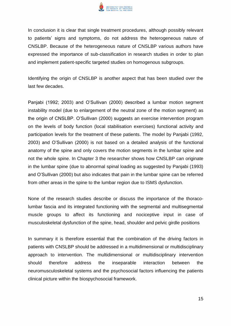
In conclusion it is clear that single treatment procedures, although possibly relevant
to patients‘ signs and symptoms, do not address the heterogeneous nature of
CNSLBP. Because of the heterogeneous nature of CNSLBP various authors have
expressed the importance of sub-classification in research studies in order to plan
and implement patient-specific targeted studies on homogenous subgroups.
Identifying the origin of CNSLBP is another aspect that has been studied over the
last few decades.
Panjabi (1992; 2003) and O‘Sullivan (2000) described a lumbar motion segment
instability model (due to enlargement of the neutral zone of the motion segment) as
the origin of CNSLBP. O‘Sullivan (2000) suggests an exercise intervention program
on the levels of body function (local stabilisation exercises) functional activity and
participation levels for the treatment of these patients. The model by Panjabi (1992,
2003) and O‘Sullivan (2000) is not based on a detailed analysis of the functional
anatomy of the spine and only covers the motion segments in the lumbar spine and
not the whole spine. In Chapter 3 the researcher shows how CNSLBP can originate
in the lumbar spine (due to abnormal spinal loading as suggested by Panjabi (1993)
and O‘Sullivan (2000) but also indicates that pain in the lumbar spine can be referred
from other areas in the spine to the lumbar region due to ISMS dysfunction.
None of the research studies describe or discuss the importance of the thoraco-
lumbar fascia and its integrated functioning with the segmental and multisegmental
muscle groups to affect its functioning and nociceptive input in case of
musculoskeletal dysfunction of the spine, head, shoulder and pelvic girdle positions
In summary it is therefore essential that the combination of the driving factors in
patients with CNSLBP should be addressed in a multidimensional or multidisciplinary
approach to intervention. The multidimensional or multidisciplinary intervention
should therefore address the inseparable interaction between the
neuromusculoskeletal systems and the psychosocial factors influencing the patients
clinical picture within the biospychosocial framework.

16
1.3.1 The researcher’s multidimensional manual therapy approach to the
management of patients with CNSLBP
The researcher has developed a multidimensional manual therapy approach to the
management of patients with CNSLBP over a period of 30 years. This period was
also characterised by the development of manual therapy on a timeline as shown in
Figure 1.2. As a clinician the researcher attended the relevant courses and
conferences on national and international level to keep abreast of the development
of manual therapy as it occurred. What is characteristic of the development of
manual therapy at the time is that it occurred in parallel with the development of
scientific knowledge in the basic sciences of especially Physiology, biomechanics
(functional Anatomy) and Pathology, and the clinical sciences in Medicine, e.g.
Orthopaedics and Radiology. Development in the basic and clinical sciences
contributed to the accuracy of diagnoses and understanding of underlying disease
processes which paved the way for the manual therapy researchers.
17
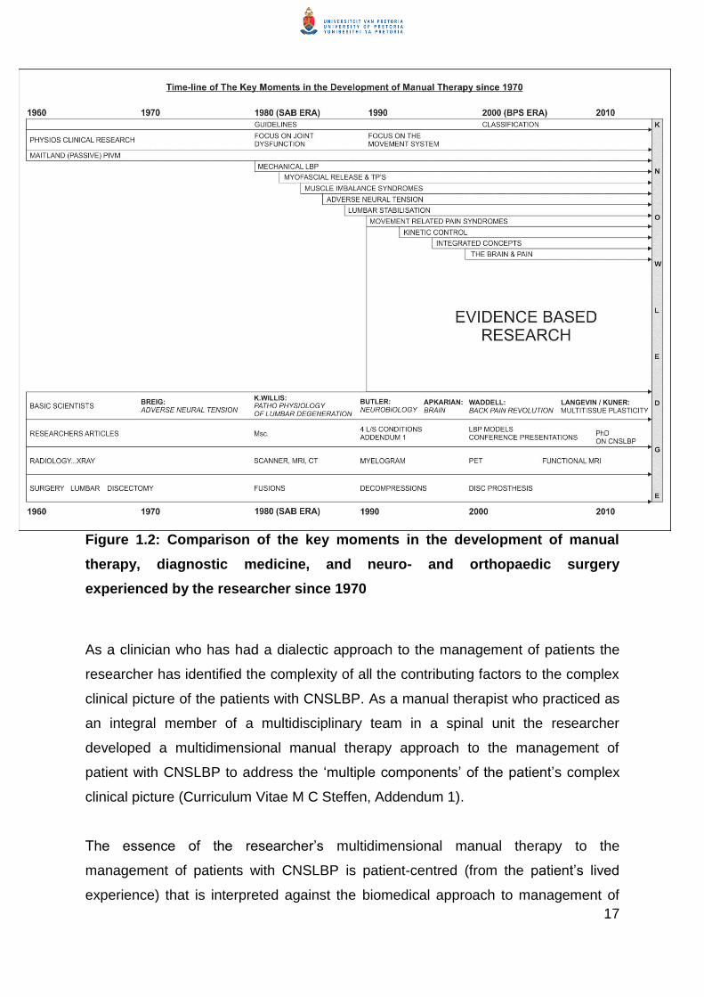
Figure 1.2: Comparison of the key moments in the development of manual
therapy, diagnostic medicine, and neuro- and orthopaedic surgery
experienced by the researcher since 1970
As a clinician who has had a dialectic approach to the management of patients the
researcher has identified the complexity of all the contributing factors to the complex
clinical picture of the patients with CNSLBP. As a manual therapist who practiced as
an integral member of a multidisciplinary team in a spinal unit the researcher
developed a multidimensional manual therapy approach to the management of
patient with CNSLBP to address the ‗multiple components‘ of the patient‘s complex
clinical picture (Curriculum Vitae M C Steffen, Addendum 1).
The essence of the researcher‘s multidimensional manual therapy to the
management of patients with CNSLBP is patient-centred (from the patient‘s lived
experience) that is interpreted against the biomedical approach to management of

18
these patients. The principles of the researcher‘s multidimensional manual therapy
had always been to manage the patients‘ pain through facilitation of endogenous
pain-inhibiting mechanisms which involve an integrated interaction between:
The therapist as a pain inhibiting agent in a professional therapist-patient
relationship. By putting the patient at ease and re-assuring him/her about the
nature and seriousness of his/her condition within a professional therapist-
patient relationship the researcher explained the patient‘s condition and
findings of her treatment to him/her as the treatment progressed.
Manual therapy is applied to patients based on the presentation of the
patient‘s signs and symptoms to
o Inhibit ascending pain modulation by releasing soft tissue and joint
restrictions on segmental and multisegmental levels and re-alignment
of the (integrated spinal movement system (ISMS which entails the
whole spine including the head, shoulder and pelvic girdles). (The
researcher discovered that patients with CNSLBP need manual
therapy to the ISMS because the ISMS (all spinal structures) were
affected: patients with CNSLBP often experience diffuse pain
simultaneously with LBP at various sites. By releasing muscle spasm,
and restoring soft tissue mobility muscles are prepared for better
recruitment.
o At the time the researcher also ‗discovered‘ that patients who received
feedback on where they experienced symptoms and how it responded
to her treatment, were more aware of their proprioceptive awareness
and responded better to specific exercises. This concept was only
described in 2012 by Moseley, Gallagher and Gallace as tactile
awareness and spatial orientation but has been used in principle by the
researcher over many years during manual therapy.
o Patients are encouraged to engage in normal activities of daily life
within the limits of their pain perception before they are given an
exercise program.
o The researcher found that manual therapy as she practised it
addressed the patient‘s pain and dysfunction including that of the
autonomic nervous system effectively.

19
Because the researcher practised as a member of the multidisciplinary team
pharmacology was introduced as an integral part of the multidimensional
manual therapy management.
Re-education of postural control and characterised adaptive behaviour had
always been part of the researcher‘s multidimensional manual therapy
management of patients to maintain mobility and alignment of the ISMS and
facilitate their functional restoration.
1.4 Problem statement
From the discussion in Section 1.3 it is clear that many studies have not addressed
the heterogenic nature of CNSLBP in patients with a multidisciplinary or a
multidimensional approach. Results from these studies have therefore either not
shown a statistically significant difference, or showed a small effect size, or only had
a short term effect on the patients signs and symptoms (Middelkoop et al, 2010;
Wand & O‘Connell, 2008).
From the systematic review by Middelkoop et al. (2010) it is also evident that
exercise is a popular form of treatment for patients with CNSLBP, although there is
no evidence that one form of exercise is more effective than another (Liddle, Baxter
& Gracey, 2004).
The heterogenic nature with which CNSLBP can present in patients entails the fact
that varying combinations of biopsychosocial factors may drive the condition which
may be the major contributing factor in the poor effect size observed in the
randomised clinical trials. Various authors have therefore investigated and
suggested ways in which patients with CNSLBP can be subgrouped (O‘Sullivan,
2005; McKenzie, 2003; Herbert, 2007; Cook, Gebski & Keech, 2004; McCarthy et al.,
2004) into more homogenic groups who have the same or similar driving factors so
that interventions can be more patient-specific within the biopsychosocial
nframework.
Despite this recommendation that subgrouping is a way to create homogeneous
subsets within the CNSLBP population, Fersum et al. (2010) found that the
application of a classification system to plan and implement RCTs to evaluate the

20
efficiency of manual therapy and exercise with other matched treatments are very
limited to non-existing. The alternative is that clinicians and researchers may not
understand the complexity of the underlying mechanism(s) that drive the patient with
CNSLBP‘s clinical picture and may not be able to select appropriate classification
systems; that clinicans have a perception that a classification system is not very
valuable; that clinicians use other methods to assess and implement targeted
patient-specific ingtervention, or that the classification systems do not integrate the
multidimensional nature of CNSLBP (Karayannis, Jull & Hodges, 2012)
Fersum et al. (2012) used the systematic review by applying a person-centered
‗mechanical behaviour‘ (O‘Sullivan, 2005) classification system to assess the effect
of ‗classification based cognitive functional therapy‘ (CB-CFT) versus a ‗manual
therapy and exercise‘ (MT-EX) approach to treatment of patients with CNSLBP.
The manual therapy section of the MT-EX group was administered to the spine OR
the pelvis to address patients‘ signs and symptoms of the patient‘s condition. The
exercise section of the MT-EX group was administered to isolated muscle
contraction such as abdominal muscles in different functional positions OR a home
exercise programme consisting of ‗general exercise‘ or ‗abdominal muscle
contraction‘.
The manual therapy in this RCT does not appear to address the soft tissue
shortening (muscles, fascia, ligaments and joint capsules) and realignment of the
biological (biomechanical stiffness and malalignment and physiological processes
involved in the development and clinical presentation of patients with CNSLBP)
heterogeneity typical in patients with CNSLBP. Neither does it mention the
importance of addressing the functioning of the spine as a closed kinetic chain, nor
the effect that the attachments of the head, shoulder and pelvic girdles to the spine
may have on pain or dysfunction in the lumbar region.
It is not clear from the publication whether the exercise section of the MT-EX group
have addressed ‗pain avoidance‘ or ‗pain provocation behaviour‘ (O‘Sullivan 2005). It
is therefore not unexpected that CB-CFT was shown to be superior to the MT-EX.
The manual therapy (MT-EX) applied as a treatment procedure in this RCT was left

21
to the discretion of experienced manual therapists and seem to differ from the way
manual therapy is practised in more specific detail by the researcher (Section 1.3.1).
Kääpä et al. (2006), who compared ‗individual therapy‘ which included passive
mobilisation and spinal traction, education and exercises, with multidisciplinary
rehabilitation that included education, exercises, relaxation, stress management and
advice, do not discuss the expected difference between the benefits of the two
management approaches. The passive mobilisation and spinal traction given to the
group who received the individual therapy was not applied based on specific
biomechanical or biological criteria or aims of treatment.
The authors that recommend a multidimensional or a multidisciplinary treatment
approach do not explain the expected structural and physiological and psychological
mechanisms that could have played a role in the explanation of the interventions that
they compared (Fersum et al., 2010; Fourney et al, 2011; Kääpä et al. 2006).
The limited understanding of the mechanisms underlying CNSLBP which should also
be the mechanisms that should be addressed in a multidicisplinary or
multidimensional intervention for these patients, create in the researcher‘s opinion a
limitation in the management of this heterogeneous condition.
None of the research studies describes or discusses the importance of the thoraco-
lumbar fascia and its integrated functioning with the segmental and multisegmental
muscle groups working in on the spine and its nociceptive input in the case of
musculoskeletal dysfunction of the spine as a kinetic chain (Middleditch and Oliver,
2005). Although many studies recognise the importance of addressing the single or
combination of driving factors in patients with CNSLBP, no study addressed the
inseparable biological interaction between the neuromusculoskeletal systems and
the psychological response of the patient as a result of the pain perception in the
brain.
The process of plasticity that play a major role in the development of soft and neural
tissue shortening and motion segment stiffness throughout the spine (integrated
spinal movement system (ISMS), and the remodelling of these tissues and
22
realignment of the motion segments throughout the spine, head, shoulder and pelvic
girdles (ISMS) (through the process of plasticity) is not addressed as part of the
research or treatment interventions. The process of neural plasticity in the
development as well as the ‗unlearning‘ of the altered pathways and changes in the
neuromatrix as a result of adaptive or maladaptive motor behaviour and pain
processing is not discussed as the mechanisms behind the cognitive and
psychosocial driving factors of the condition (Flor, Braun, Elbert, & Birbaumer, 1997;
Kuner, 2010). By understanding these complex neurophysiological processes,
clinicians and researchers can optimise intervention (education to understand the
condition, advice, exercise to address adaptive and maladaptive motor behaviour)
researchers will be able to explore the exceptional results of the CB-CFT achieved
by Fersum et al (2012).
1.5 Research questions
The research questions of this study were:
Can the concept of an ‗integrated spinal movement system‘ ISMS be
conceptualised based on the anatomy of the trunk?
What are the underlying systems, processes and influences that result in
ISMS dysfunction and contribute to the clinical picture of patients with
CNSLBP?
What contribution can the professional craft knowledge and the personal tacit
knowledge acquired by the researcher over many years of clinical practice,
make towards the declarative professional knowledge of manual therapy?
1.6 Research aims and objectives
The primary aim of this research was to develop a multidimensional manual therapy
model for patients with CNSLBP based on clinical observations, clinical reasoning,
professional craft knowledge and personal tacit knowledge
The process of model development requires the following sub-aims:

23
(1) To discuss the theoretical basis for the conceptualisation of the ISMS and
ISMS dysfunction as the focus of the multidimensional manual therapy model
regardless of whether the origin of the CNSLBP is more biomechanical or as
a result of increased muscle tone due to hypervigilance in the brain as a result
of social stressors.
(2) To discuss the underlying process involved in the development of ISMS
dysfunction and the possible reason for the variations in ISMS dysfunction
that can occur.
(3) To discuss the assessment of a patient with CNSLBP as part of the
multidimensional manual therapy model.
(4) To discuss the principles of a multidimensional manual therapy model for
managing patients with CNSLBP.
(5) To conceptualise a multidimensional manual therapy model for managing
patients with CNSLBP.
(6) To discuss the multidimensional manual therapy model in the context of other
relevant models for managing patients with CNSLBP.
1.7 Research approach
Model development based on a grounded theory development
Model development as a research design and as the outcome of this study was
chosen because in a model the relationship between the different components and
concepts related to the development and management of CNSLBP could be
illustrated instead of extensively described in terms of management processes,
guidelines, services to patients and the identification of new fields for further
research. When the relationships between the components and concepts are
illustrated they can be tested with empirico-analytical research (Higgs et al., 2010).
The basic function of a model is to promote, explain and define relationships,
structure, and linkages between concepts to enhance understanding of a
phenomenon: in other words it is ‗heuristic, i.e. discovering or “exposing” certain
relationships between concepts‟ (Mouton & Marais, 1990 p 60).

24
The process for developing a model (Polit & Beck, 2008 p 85) will therefore be the
same as the process for developing a theory. This process is described by Walker
and Avant (1995) as:
Select a topic of interest (may be one concept / variable or a framework of
several concepts)
Conduct a review of the literature or use field observations and note related
variables
Organize relational statements in terms of patterns of relationships amongst
the variables. Diagrams may be used to express relationships amongst
concepts and to organize the components of the theory.
The study is divided into three main sections:
A discussion of the theoretical basis for the conceptualization the ISMS.
Secondly, a discussion on the proposed process of the development of ISMS
dysfunction which include the associated pain processing and characteristic
adaptive behaviour typically observed by the researcher in patients with
CNSLBP. The discussion of the proposed process of development of ISMS
dysfunction is based on the functional anatomy
Thirdly, a multidimensional manual therapy model for the management of a
typical patient with CNSLBP is which is grounded in the biopsychosocial
framework, is presented and discussed. The researcher further indicates how
the model fills a gap in the understanding and management of patients with
CNSLBP.

25
Table 1.1: Summary of the components of the model
Component of the model Description of the component Application/Contribution of
component to the model
Conceptualisation of the ISMS The conceptualisation of the
ISMS is based on (1) the
discussion of the functional
anatomy and the processes
which form the ISMS
The conceptualisation of the
ISMS and the processes
working within the system serve
as the premise for
understanding the development
of ISMS dysfunction and
multidimensional manual
therapy of patients with
CNSLBP (assessment and
treatment).
Development of the ISMS
dysfunction, associated pain
processing and characteristic
adaptive behaviour.
The development of ISMS
dysfunction is discussed as the
basis for the development of
CNSLBP which cannot be
diagnosed based on
radiological investigations or
other objective tests. Pain
processing and characteristic
adaptive behaviour, form an
integrated part of the
development of ISMS
dysfunction.
The proposed process of
development of ISMS
dysfunction, pain processing
and characteristic adaptive
behaviour forms the basis of the
understanding and manual
therapy management patients
with CNSLBP.
Conceptualisation of the
multidimensional manual
therapy management model for
patients with CNSLBP
The multidimensional manual
therapy model for managing
patients with CNSLBP indicates
the integrated multidimensional
approach to address ISMS
dysfunction which includes the
management of pain processing
and characteristic adaptive
behaviour.
The multidimensional manual
therapy model for management
of patients with CNSLBP, falls
within the biopsychosocial
framework and specifically
within the movement and
control impairment groups
described by O‘Sullivan (2005)
26
1.8 The nature of this study
The research problem emanated from the fact that the researcher through her
clinical experience observed some mechanisms involved in the development of
ISMS dysfunction and became aware that manual therapy for patients with CNSLBP
applied to the ISMS and not only to the low back address the patient‘s diffuse and
specific pain patterns, general ISMS mobility which prepared the patient for muscle
activation and postural re-education. Furthermore the researcher observed that
manual therapy and her professional interpersonal interaction with the patient,
contributed to pain modulation and enhanced re-education of postural control.
Pharmacology (anti-inflammatories) post treatment was found to maintain patients‘
mobility and reduced post treatment effects. Based on her clinical experience,
clinical reasoning, gaining knowledge in the field over many years, the researcher
developed a practice theory on the multidimensional manual therapy for patients
diagnosed with CNSLBP.
According to McEwen and Wills (2002), theories to explain a phenomenon from the
perspective of clinical practice situations can be inductively developed to describe or
explain such a phenomenon.
The insight gained from describing a phenomenon in a particular situation can in turn
contribute to the understanding of similar situations in clinical practice. The authors
(McEwen & Wills, 2002) call this process the practice-theory approach to theory
development. The research strategy to generate or develop a practice-theory is
based on the grounded theory approach (McEwen & Wills, 2002). Grounded theory
is defined as: ‗an approach to collecting and analyzing qualitative data that aims to
develop theories grounded in real-world observations‘ (Polit & Beck 2008 p 755).
This research was initiated by the researcher‘s reflection on her treatment of patients
with CNSLBP and her clinical reasoning during:
The treatment of patients and discussions with colleagues at national and
international level on the topic of CNSLBP;
27
Attending courses on spinal rehabilitation and manipulation/mobilisation
nationally and internationally;
Presenting continuing professional development courses nationally;
Clinical training of postgraduate students in manual therapy and an
international workshop on manual therapy; and
Observing and analysing patient responses to treatment and adapting
treatment to the patients‘ physical responses during and after treatment, and
patient feedback during and after treatment.
The research was further initiated by a critical analysis of the literature on the
management of patients diagnosed with CNSLBP, and the fact that present RCT
which compared the effect of manual therapy with other physiotherapy modalities in
the management of the heterogeneous condition of CNSLBP.
In essence, the researcher presents a theory on the development of CNSLBP and
the management of patients in the form of a multidimensional model for managing
patients with CNSLBP and, in this way, contributes to the knowledge basis of
physiotherapy and in particular manual therapy.
Higgs and Titchen (1995) and Higgs, Jones and Titchen (2010) distinguish between
three types or domains of knowledge: (1) discursive research and declarative
knowledge (also called propositional knowledge); (2) personal knowledge; and (3)
professional craft knowledge (‗knowing how‘ or non-propositional
knowledge/practical and procedural knowledge). The three types of knowledge and
the interaction between them are illustrated in Figure 1.2.

28
Figure 1.3: Types of knowledge and internal influences on knowledge
generation
(Adapted from Higgs & Titchen, 1995 p 137)
The arrows indicate the domains of knowledge from which the practice theory was
developed.
Propositional knowledge, which is formally generated through research and
scholarship, is regarded in modern society as having a higher status than non-
propositional knowledge, which is generated through practical experience. However,
already in 1949 Ryle (Higgs et al., 2010 p 154) argued that propositional knowledge
follows rather than drives procedural knowledge (non-propositional knowledge)
while Barnett (1990) is of the opinion that the cognitive framework dominates other
forms of knowledge and regards non-propositional knowledge as downgraded forms
of knowledge. This opinion is slowly being challenged and researchers are
increasingly realising that knowledge generated in clinical practice is rather driving
the generation of propositional knowledge than following it. Ryle argued that an
aspect of theory that is developed in clinical practice is inherently part of clinical
29
practice but is different from applying propositional knowledge in practice (Higgs et
al., 2010 p 154).
Higgs et al. (2010 p 154) furthermore state that knowledge derived from clinical
experience can be transformed to propositional or declarative knowledge ‗through a
process of theorization and/or rigorous critique and debate among practice
communities‘.
Once the non-propositional tacit knowledge is clarified or identified, described and
tested through empirical research, it becomes part of the declarative (propositional)
knowledge (Higgs & Titchen, 1995).
The opposite can also happen, that propositional knowledge can be derived from
basic or applied research and be elaborated and particularised through clinical
practice and as such become part of the personal experience of the individual
(clinician).
The characteristics of the three types of knowledge are as follows. Propositional
knowledge is derived through research and/or scholarship. It is formal and explicit
knowledge that is expressed in propositional statements, which enunciate, for
example, relationships between concepts or causes and effects and which identify
the generalisability or transferability of research knowledge to populations and
settings. Theoretical knowledge may be developed from arguments of principle, from
dialogue and logic, and through use or application of existing empirical and
theoretical knowledge (Higgs & Titchen, 1995).
Professional craft knowledge and personal knowledge are collectively called non-
propositional knowledge. Both these types of knowledge are derived from the
processing (e.g. through reflection) of professional and personal experiences,
respectively, and may be tacit and embedded in practice or in the personal identity
and lives of the patients, and researcher (the ‗knowers‘). Cervero (1992) describes
professional craft knowledge as a ‗repertoire‘ of examples, images, practical
principles, scenarios or rules of thumb that have been developed through prior
30
experience. Professional craft knowledge comprises general knowledge gained from
practical experience (e.g. knowledge about how a population of patients responds to
disease or disability) and specific knowledge (e.g. about a particular patient), in a
particular situation and context at a particular time (Higgs, Fish & Rothwell, 2010).
Personal knowledge is accrued from life experiences, such as relationships and
cultural influences that contribute to shaping individual perspectives; as such, it
influences personal interactions, personal values and beliefs. This knowledge, in its
general form, is gained, as in the case of professional craft knowledge, through
socialisation into a society, group or professional community. In its particular form
personal knowledge is acquired more consciously by reflecting upon one‘s knowing,
being, doing and feeling in each unique situation.
Practitioners use all three forms of knowledge in practice. Propositional knowledge
can provide the basis for understanding the medical, psycho-social and cultural
context and the physical and psycho-social nature of the client‘s needs and
problems. In relation to clinical reasoning, practitioners need to accumulate and
update carefully a rich and dependable knowledge base, and have to be vigilant in
checking for potential errors in the currency and use of this knowledge, in particular
when making important decisions of diagnosis, treatment and prognosis.
Professional craft knowledge enables practitioners to tailor management procedures
to individual patients‘ needs based on clinical decision making and recognition of the
individual client‘s needs (Higgs, Fish & Rothwell, 2010). Such knowledge enables
clinicians to plan, modify and critique their treatments to consolidate their
understanding of the particular clinical problem (Jensen et al., 1992) and to
implement sound, efficient and timely decision making.
Research has demonstrated that it is the ability of experienced professionals to
integrate propositional knowledge with professional craft knowledge that enables
them to assess the relevance of clinical data and to distinguish and comprehend the
significance of crucial cues (Dreyfus & Dreyfus, 1980; Elstein, Shulman & Sprafka,
1990).

31
Health professionals draw on their professional craft knowledge and their personal
knowledge to interact effectively with patients and their carers. Such knowledge,
combined with skills in communication, listening and problem solving, facilitates
interpersonal interactions and enables practitioners to relate well to their clients as
individuals with their unique needs, fears, hopes and expectations. Carper (1978,
argued that personal knowledge promotes wholeness and integrity in the personal
encounter, the achievement of engagement rather than detachment. The ability to
place the clinical problem within the patient‘s world and to design personalised care
and interventions that take the patient‘s experience into account is recognised
across the health sciences as a key element of expertise that develops from clinical
practice experience (Crepeau, 1991; Jensen, Shepard & Hack, 1992; Jones et al.,
2002).
The interaction between theory, research and practice is a reciprocal, cyclical
interaction. McEwen and Wills (2002) illustrate the interaction between research,
practice and theory in a cycle presented in Figure 1.3.
Figure 1.4: The research, theory, practice cycle
(McEwen & Wills, 2002 p 80)
A theory developed from clinical practice through a process of inductive reasoning is
called a practice-theory (refer to Section 1.7). A practice-theory assumes that the
phenomenon observed and experienced in clinical practice is important enough to
Practice
Theory
Research
32
pursue, and that it presents an understanding of the clinical phenomenon that has
not been articulated (McEwen & Wills, 2002).
McEwen and Wills (2002 p 80) further explain the relationship between a theory and
a model as:
A theory has several components, including purpose, concepts and
definitions, theoretical statements, structure, and assumptions (Bishop,
1998; Chinn & Kramer, 1999). Creation of conceptual models or maps is
also a component of theory development that is promoted to further
explain and define relationships, structure, and linkages.
Mouton and Marais (1990) are of the opinion that the relationship but also the
distinguishing factor between a theory and a model is that a theory classifies
knowledge (or explains a phenomenon in clinical practice in this case) and explains
or enhances understanding of a phenomenon, while a model not only classifies
knowledge „but the basic function [of the model] is heuristic, i.e. discovering or
“exposing” certain relationships between concepts‟ (Mouton & Marais, 1990 p 60). A
model helps to illustrate the processes through which outcomes occur by specifying
the relationships among the variables in graphic form. In this graphic form, a model
can be examined for inconsistency, incompleteness or errors (McEwen & Wills,
2002).
1.9 Clarification of terminology
The terminology that is discussed in this paragraph includes low back pain, acute
specific low back pain, acute non-specific low back pain; chronic nonspecific low
back pain, manual therapy, plasticity, chronicity, dysfunction and disability, integrated
spinal movement system (ISMS), integrated spinal movement system dysfunction
(ISMS dysfunction), characteristic adaptive behaviour and pain modulation.
1.9.1 Low back pain
Low back pain is defined in terms of the location of pain. This implies ‗pain in the low
back‘. The low back is identified by Waddell (2004) as pain in the area between the

33
lowest ribs and the inferior gluteal folds and is often accompanied by referred pain.
Referred pain can be somatic or neurological in origin. Pain which is neurological in
origin can be categorised as specific LBP. The referred pain which is somatic in
origin is more associated with non-specific low back pain (Bogduk & Twomey, 2005).
All low back pain starts as acute and can be divided into specific LBP or non-specific
low back pain (refer to Figure 1.1).
1.9.2 Acute specific low back pain
Specific low back pain is defined as low back pain from a specific origin such as
anatomical tissue damage in the lumbar spine (i.e. an annulus tear, disc prolapse,
subluxation of the facet joint) that can be detected by CT and MRI scans. The
patients‘ pain perception is characterised by a direct proportion to the physical
findings (acute tissue damage) and the, nociception, pain, suffering, and pain
behaviour. Acute specific LBP responds well to physical treatment and/or surgery
and patients show a natural tendency to recover as response to treatment (European
Clinical Guidelines for the Management of Chronic Non-Specific Low Back Pain,
2005).
1.9.3 Manual therapy
Manual therapy by definition in this thesis is the release of soft tissues (muscle
spasm, taut bands, trigger points and joint capsules, myofascia and neural tissue
restrictions – all of which may restrict the skeletal system and contribute to
malalignment of the skeletal system) through the direct contact between the patient
and the manual therapist‘s hands. It further entails the realignment of the skeletal
system by passive oscillatory physiological and accessory movements of joints that
do not exceed the normal end-range of joints (Elvey & O‘Sullivan, 2004).
Spinal manipulative therapy (SMT) on the contrary entails a sudden available end of
range thrust to increase joint range when the window of opportunity presents. A
manual therapist would use SMT in combination with manual therapy to release
restrictions (scar tissue) in order to integrate other forms of manual therapy to
restore alignment, muscle recruitment and function (Haynes, 2003).

34
Treatment techniques to release soft tissues used by the researcher include
myofascial release (Barnes, 1997), trigger point therapy (Dommerholt, 2012), Rolfing
structural integration (Caspari & Massa, 2012), myofascial induction approaches
(Pilat, 2012), and neural release (Wander & Weinschenk, 2012). Other techniques in
the literature include osteopathic manipulative therapies (King, 2012), connective
tissue manipulation (Prendergast & Rummer, 2012), fascial manipulation (Stecco &
Stecco, 2012), accupuncture as a fascia oriented therapy (Irnich & Fleckenstein,
2012), and stretching of fascia (Myers & Frederick, 2012).
Manual therapy as the passive release of soft and neural tissues as well as joint
restrictions through physiological and accessory movement in this thesis is
accompanied by communication with the patient, reassuring him/her about the
causes / contributing factors to his condition and how it presents, as well as giving
the patient feedback on his/her tissue response during the manual therapy, re-
education of tactile awareness and spatial orientation during manual therapy.
The researcher prepared a timeline (Figure 1.2) of the development in manual
therapy to indicate the sequence in which the concepts became common practice in
physiotherapy. The relevance of the timeline for this thesis is that it presents the era
during which the researcher developed her own approach to a multidimensional
approach to the management of patients with CNSLBP. The timeline (Figure 1.2)
also indicates the major developments in diagnostic medicine and neuro and
orthopaedic surgery because it influenced or coincided with the development of the
manual therapy concepts.
1.9.4 Multidisciplinary approach to management of CNSLBP
A multidisciplinary approach to management of patients with CNSLBP entail the
treatment of the patient by all relevant health care professionals from different
disciplines such as spine surgeons, psychiatrists, neurologist, radiologist,
anaesthesiologists, pain medicine practitioners; chiropractors, osteopaths, physical
therapists (Fourney et al., 2011).

35
1.9.5 Multidimensional manual therapy for the management of CNSLBP
A multidimensional manual therapy approach to the management of patients with
CNSLBP, entail the treatment of a patient by a single health care professional such
as a physiotherapist who addresses the multiple components (heterogeneity) of
CNSLBP in a particular management approach.
1.9.6 Chronicity
Chronicity is the state where the clinical signs and symptoms of CNSLBP become
self-sustainable and continuously worsens. Chronicity is categorised in different
stages dependent on pain intensity, pain duration and disability due to pain
processing (Buchner, Neubauer, Zahlten-Hinguranage & Schiltenwolf, 2007).
The non-variable factor in chronicity in patients with CNSLBP is a person‘s genetic
make-up which predetermines the extent to which each factor plays a role in the
development of chronicity OR the ability to adapt to the condition (Field, 2009;
O‘Sullivan, 2005). Clinically it is apparent that some people are set up for the
development of chronic pain before the pain starts, while others develop it soon after
the acute episode of NSLBP and others drift into it (Field, 2009). Typically the largest
group of patients with CNSLBP drifts into chronic pain after trying various available
treatment options which did not address their problem adequately (Field, 2009).
Socio-demographic factors associated with patients developing CNSLBP are gender,
age, marital/family status (single parent/young children, partner retired or disabled),
health condition (mental health conditions musculoskeletal conditions, comorbidities)
occupational or educational level, time since last worked, occupational status (no
longer employed) and local employment rate (Waddell, 2004).
1.9.7 Dysfunction and disability in patients with CNSLBP
Dysfunction and disability are the two ends of a continuum. Dysfunction is the
restricted activity due to CNSLBP.

36
Dysfunction is the malfunction of anatomical structures and physiological and
psychological processes to limit the patient‘s daily functional activities. Dysfunction
may become self-perpetuating as a result of any one or a combination of the factors
that initiate the physiological change and/or change in the pattern of motor control
(movement or activity). It is also possible that emotional stress can result in
biomechanical dysfunction due to the fact that it changes muscle recruitment,
posture and motor control. If the biomechanical dysfunction from whatever cause
perpetuates then it becomes CNSLBP (Waddell, 2004).
When a person is not able to perform the activities of daily living and/ or meet
personal, social or occupational demands characteristic of people of his/her age,
gender and culture because of CNSLBP, such a person can be regarded as disabled
due to his/her CNSLBP (WHO, 2011; The American Medical Association, 2000).
1.9.8 Plasticity
Plasticity is a dynamic physiological property of all the soft tissue systems to adapt
their structural organisation and biochemical, physiological and morphological
characteristics temporarily or permanently due to new emerging situations as a result
of inherent and environmental situations (which can be internal or external) as well
as due to other factors (e.g. injuries) affecting the systems. Plasticity is a constant
dynamic adaption in all the systems of the ISMS and enables the body (soft tissues)
to adapt to injury and psycho-social and environmental demands (Dorland‘s
Illustrated Medical Dictionary, 32nd edition, 2012).
In CNSLBP plasticity occurs in muscular, connective tissue, peripheral, central and
autonomic neural tissue due to the tissues‘ ability to adapt to the stimuli they are
subjected to. It can also influence behaviour based on beliefs and fear avoidance.
The process of plasticity is the basis for the adaptation of soft tissues and neural
tissue to adapt to internal or external stressors and is proposed by the researcher as
an important process in the development of ISMS dysfunction and as such the
clinical picture of CNSLBP.

37
1.9.9 Integrated spinal movement system (ISMS)
The term ‗ISMS‘ was conceptualised in this thesis to portray the complex integrated
functioning of the spine, pelvis, shoulder girdle, and head as a kinematic chain due
to the fact that the:
Multiple segmental motion segments functions as a unit on segmental and
multisegmental levels (Comerford & Mottram, 2003)
The ligaments stabilise the spine on segmental and multisegmental (global)
level
Muscles of the spine control movement on segmental and multisegmental
(global) levels)
The neural structures emerge from the spinal cord on segmental level but
functions as a whole (Middleditch & Oliver, 2005)
1.9.9.1 Integrated spinal movement system (ISMS) dysfunction
‗ISMS dysfunction‘ is the term used in this thesis to describe the dynamic integration
between all the systems within and around the ISMS. The integration between the
biomechanical component of the ISMS, pain processing, guarded movement, fear of
movement, catastrophisation which eventually results in altered pathways in the
brain and altered (dysfunctional) motor control, result in poor posture, and impaired
coordination which permeates the patients psychological frame of mind and social
behaviour to merge into characteristic adaptive behaviour.
1.10 Outline of the study
This chapter indicated that the management of patients with CNSLBP is still a
problem in this day and age and that the problem is still growing. The essence of the
reasons for the continued lack of success of medical and physiotherapy
management of patients with CNSLBP is discussed.
In Chapter 2 the research methodology is described as the researcher‘s
metacognitive reflection on her clinical reasoning, an integrated literature review and
the knowledge and skills she obtained from attending courses nationally and

38
internationally to develop a multidimensional manual therapy model for the
management of patients with CNSLBP.
Chapter 3 gives an account of the literature which serves as a knowledge base for
the conceptualisation of the ISMS and development of ISMS dysfunction in patients
with CNSLBP.
In Chapter 4 the researcher discusses the assessment of a typical patient diagnosed
with CNSLBP. The chapter ends with a model on the assessment of patients with
CNSLBP.
The multidimensional manual therapy management of patients with CNSLBP is
discussed in Chapter 5. The multidimensional manual therapy model for
management of these patients is presented in this chapter.
In Chapter 6 the researcher reflects on the multidimensional manual therapy model
for managing patients with CNSLBP and discusses the relation of this model with
other models used in the management of patients with CNSLBP.

39
CHAPTER 2
Research methodology
2.1 Introduction
In Chapter 1 the nature of the study is described as model development based on a
practice-theory approach. According to McEwen and Wills (2002), a ‗practice-theory‘
is developed from an interaction between practice, theory and research. The
development of a practice theory is based on the assumption that a particular
phenomenon observed in clinical practice has the potential to enhance
understanding in a particular field through the development of propositional
knowledge (McEwen and Wills, 2002)
The core problem identified in Section 1.4 is the limited understanding of the
biomechanical dysfunction in patients with CNSLBP which is a result of a
combination of the different driving factors in the development of CNSLBP. From the
researcher‘s perspective of managing patients with CNSLBP, researchpublications
on the topic focus on the treatment of the ‗low back‘ when they treat patients with
CNSLBP while the symptoms ‗in the low back‘ can be referred from elsewhere in the
spine to the ‗low back‘ (Kääpä et al., 2006; Fersum et al., 2012).
Another aspect of the problem is that the researcher observed that it is not only
patients with CNSLBP‘s lumbar area that is affected but the whole spine including
the position of the head, shoulder and pelvic girdles. From the researcher‘s
perspective manual therapy applied to the lumbar area only, will not address the
heterogenetic nature of the patient‘s CNSLBP. Instead manual therapy should be
applied to the whole spine to release the soft tissue connections between the head
and the cervical spine, and the soft tissue attachments to the shoulder and pelvic
girdles to realign the motion segments, head and girdles. The role of the thoraco-
lumbar fascia and the process of plasticity in the shortening of the soft and neural
tissues working in on the spine, position of the head, shoulder and pelvic girdles in

40
the development and management of patients with CNSLBP has not been described
in the literature.
2.2 The frame of reference
The frame of reference of the thesis developed from the researcher‘s professional
(declarative or propositional knowledge), personal and professional craft knowledge
acquired in clinical practice and through interaction with professionals, patients and
their significant others, and by continuously attending courses and congresses
related to this field and teaching in this field of practice. Because the researcher is an
integral part of the knowledge generation in this study, it is important to state the
frame of reference of this study in terms of paradigms and assumptions.
2.2.1 The paradigm of this study
Manual therapy is a clinical area of specialisation in physiotherapy where an aspect
of the field of knowledge falls within the empirico-analytical paradigm (biomedical
evidence-based knowledge and management of patient problems). Simultaneously
due to the fact that manual therapy also requires an interaction between the manual
therapist and patient, the patient‘s perspective of his/her problem should also be
interpreted and understood to formulate the problem holistically. The manual
therapist‘s critical reflection on the patient‘s perspective (illness experience, story,
context and culture) and post treatment feedback as well as the patient‘s clinical
presentation (biomedical problem) and process of clinical reasoning to manage the
patient‘s problem is known as dialectical reasoning (Figure 2.2, Section 2.3.1.2) and
is typical of the reasoning process used by clinical experts to generate knowledge
(Edwards & Jones, 2007). As a result of the dialectical reasoning process and the
fact that the researcher performed metacognitive reflection on the clinical
presentation of her patients, her management of the patient‘s heterogenetic
CNSLBP problem and the patients‘ responses this thesis also falls within the
interpretive paradigm where knowledge is socially constructed. Working in a
multidisciplinary team, attending congresses and conferences, presenting post
graduate discussions and courses also contribute to the fact that knowledge is
socially constructed. The knowledge obtained from the two different paradigms
needs to be integrated to obtain a holistic view on patient problems. This ability to
41
integrate knowledge generated from two different paradigms to formulate the
patient‘s problem requires dialectical thinking (Edwards & Jones, 2007).
In this study the researcher obtained a deep understanding of the complexity of the
problem of patients with CNSLBP through knowledge generated through clinical
reasoning. The knowledge that she generated in the management of patients with
(C)NSLBP entails knowledge from the empirico-analytical paradigm (biomedical
knowledge) as well as from an interpretive paradigm (knowledge from the patient‘s
lived experiences).To verbalise and write up this deep understanding and knowledge
generated in clinical practice through clinical reasoning, the researcher performed
metacognitive reflection in a heuristic approach to develop a model based on her
individualised patient management. The paradigm within which this study was
conducted is therefore the interpretive paradigm.
In any scientific research it is important to state the assumptions on which the
research was conducted. In this research, which falls within the qualitative research
framework in which the researcher is an integral part of the data generation and
analysis, these assumptions need to be explicitly stated to interpret the researcher‘s
frame of reference when she developed the model.
2.2.1.1 Assumptions
Assumptions are statements that are not necessarily empirically tested but which are
regarded as true. These assumptions influence the logic behind the study and
therefore need to be explicitly stated, particularly in qualitative research (Cresswell,
2007).
The assumptions underlying this study and listed below are ontological,
epistemological, axiological and methodological assumptions and assumptions
regarding the rhetorical structure of the study.
Ontological assumptions
Ontological assumptions deal with the question: ‗What is reality?‘ From an empirico-
analytical paradigm ontological assumptions indicate that the real world is driven by
real natural causes (Polit & Beck, 2007).

42
In terms of the empirico-analytical paradigm the most important ontological
assumptions in this study are:
The anatomical structures of the spine function as an integrated spinal
movement system including the head, shoulder and pelvic girdles
The development of integrated spinal movement system (ISMS) dysfunction
entails the inseparable interaction between the dysfunction of the
neuromusculoskeletal systems pain processing and characteristic adaptive
behaviour due to a process of plasticity.
In terms of the interpretive or naturalistic paradigm the most important assumptions
are:
Principles for the holistic management of patients with CNSLBP can be
deduced from multiple realities of patients‘ lived experiences.
Patients and their significant others can contribute to the generation of new
understanding and, as such, new knowledge in the context of the clinical
reasoning process.
Epistemological assumptions
Epistemological assumptions answer the question: ‗How is the inquirer related to
those being researched?‘ (Polit & Beck, 2008). Higgs, Fish and Rothwell (2010 p
163) state that:
Knowledge is constructed in the framework of socio-political, cultural and historical
contexts. Practice knowledge evolves within a dynamic history of ideas contained in
the particular practice domain and within the history of how ideas born in that
practice domain have been shaped by that practice. Each of these dimensions and
contexts of knowledge has particular relevance to how we use knowledge in
reasoning and generate knowledge from within reasoning. During professional
socialization, practitioners learn the ways of being, acting, thinking and
communicating that characterize their profession
43
The researcher‘s thinking processes are an integral part of the knowledge generated
in this research. The way in which she has kept abreast with the knowledge
generation in the field of manual therapy over the past 40 years is given in
Addendum 1.
The epistemological stance of the researcher‘s practice is dialectic in nature (Figure
2.2). This implies that her practice is conducted within a biomedical practice
framework in which ‗knowledge is seen as [an] objective, predictive, empirical
generalizable, explanatory phenomenon that arises from the use of the natural
scientific method and theorization in a world of external objective reality‟ (Higgs et
al., 2010).
However, the researcher also practises within the interpretive paradigm, which
implies that she practises from a patient/client-centred approach in which each
patient‘s problem is unique within his/her history of the development of the clinical
picture, and the socio-cultural and psycho-social factors influencing the presentation
of the clinical picture. The researcher‘s interaction with patients and their significant
others has contributed to the construction of the researcher‘s professional craft and
personal (tacit) knowledge.
The professional craft and personal (tacit) knowledge the researcher has acquired
over 40 years of clinical practice has occurred by sharing knowledge, ideas and
experience, in collaboration with the multi-professional team she works with, with
colleagues during courses and conferences at national and international level.
Teaching postgraduate students has also contributed to the development of the
researcher‘s clinical reasoning and research (and metacognitive reflection on clinical
practice) as it is presented in this thesis.
The researcher has also indicated in Figure 1.2 of Chapter 1 the approximate
timeline of the development of the manual therapy concepts which she has kept
pace with over the last 40 years (also refer to Addendum 2). As a manual therapist
the researcher has developed as a professional over the same time and, as such,
can reflect on the development of manual therapy in the context of the present day
evidence that emanates from basic sciences and randomized clinical trials.

44
In this context the following epistemological assumptions are stated:
Propositional knowledge on the development of ISMS dysfunction integrated
with pain processing and characteristic adaptive behaviour of patients with
CNSLBP can be observed and verbalised on the basis of the interpretation of
observations, and all the dimensions of clinical reasoning.
The researcher can generate knowledge based on her own personal
knowledge, professional craft knowledge, tacit knowledge, and propositional
knowledge.
The researcher‘s knowledge was shaped by interaction with colleagues, team
members, patients and their significant others and metacognitive reflection on
her clinical practice and clinical reasoning.
Axiological assumptions
‗Axiological assumptions‘ refer to the way in which values mediate and shape our
understanding of knowledge and clinical practice. Axiological assumptions are critical
in the interpretive paradigm, especially in the context of this study because the
holistic management approach for patients with CNSLBP is based on a patient-
centred approach to clinical management in which the holistic understanding of the
patient‘s illness behaviour, experience of his/her problem, his/her history, beliefs and
culture is part of the problem identification. Patient feedback during and after
treatment also plays an integral part of the researcher‘s understanding of the
problem and its management. The axiological assumption on which this study is
based is that:
Integrating the patient‘s biomedical diagnosis of impairment/disability and
addressing the patient‘s ‗lived experience‘ in a diagnosis of the patient‘s problem
are an integral part of manual therapy.
Methodological assumptions
Methodological assumptions ask the question: ‗how is evidence best obtained?‘
(Polit & Beck, 2008 p 14) or ‗what are the processes and procedures of the
45
research?‘ Methodological assumptions involve the scientific criteria for obtaining
knowledge and refer to the methods used to validate a study scientifically (Polit &
Hungler, 1997).
The methodological assumptions pertaining to clinical reasoning as a knowledge-
generating activity are:
Clinical reasoning entails a complex combination of thinking processes:
These processes consist of a cognitive process, which includes hypothetical
deductive reasoning, pattern recognition, forward and backward reasoning
(inductive and deductive reasoning), knowledge reasoning integration, and
intuitive reasoning.
An interactive process, which includes multidisciplinary reasoning, conditional
reasoning, narrative reasoning, interactive reasoning (collaborative
reasoning), ethical reasoning, and teaching as reasoning (Higgs & Jones,
2010 p 7-8);
Clinical experts use various combinations of all forms of clinical reasoning
during the treatment of patients and as a result are able to generalise the
principles of patient management;
Clinical reasoning requires capability in four dimensions: reflective thinking,
critical thinking, dialectical thinking, and complex thinking;
Clinical reasoning during the holistic manual therapy management of patients
with CNSLBP is performed within the biopsychosocial paradigm.
Propositional knowledge can be generated through metacognitive reflection
on clinical reasoning to identify the principles of a holistic manual therapy
approach for managing patients with CNSLBP.
Experiential tacit knowledge, professional craft knowledge and personal
knowledge are validated by narrative (thick) description of clinical reasoning
and metacognitive reflection.
An expert clinician‘s tacit knowledge (personal and professional craft
knowledge) can be made explicit through a process of metacognitive
reflection on his/her clinical reasoning.
The research design of the study (metacognitive reflection on the process of
clinical reasoning) is flexible and described in a narrative form.

46
A qualitative research design facilitates an in-depth exploration of the
phenomenon and favours no methodological approach above another.
2.3 The research approach
The research approach of this study was embedded in the interpretive paradigm as
the researcher used a hermeneutic approach to obtain a deeper understanding of
the development (causative factors) of CNSLBP and patients‘ responses to manual
therapy.
The hermeneutic approach to the research was completely integrated and relied on
metacognitive reflection on the clinical reasoning process. The result of the
hermeneutic approach was a model for the holistic manual therapy approach for
managing patients with CNSLBP.
The research approach to this study was therefore a hermeneutic metacognitive
approach to the development of a clinical management model. Because model
development is strongly related to theory development, the study also falls within a
grounded theory approach (McEwen & Wills, 2002).
A model was chosen as the outcome of the study because in a model the
relationships between the concepts that have been identified can be indicated in a
way that is inherent to the clinical problem-solving process (McEwen & Wills, 2002).
Figure 2.1 presents the research approach in diagram form.
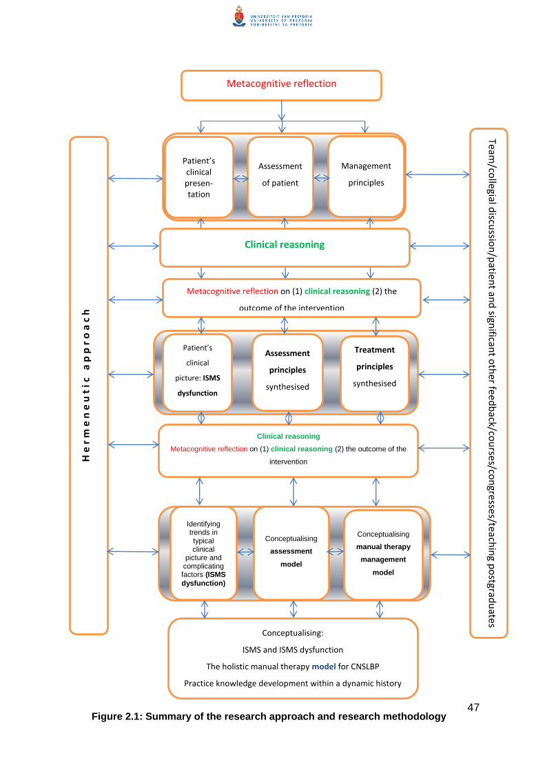
47
Metacognitive reflection
Patient’s clinical presen- tation
Assessment
of patient
Management
principles
Clinical reasoning
Metacognitive reflection on (1) clinical reasoning (2) the
outcome of the intervention
Patient’s
clinical
picture: ISMS
dysfunction
Assessment
principles
synthesised
Treatment
principles
synthesised
Clinical reasoning
Metacognitive reflection on (1) clinical reasoning (2) the outcome of the
intervention
Identifying trends in typical clinical
picture and complicating factors (ISMS
dysfunction)
Conceptualising
assessment
model
Conceptualising
manual therapy
management
model
Conceptualising:
ISMS and ISMS dysfunction
The holistic manual therapy model for CNSLBP
Practice knowledge development within a dynamic history
H e
r m
e n
e u
t i
c a
p p
r o
a c
h
Team/co
llegial discu
ssion
/patien
t and
significan
t oth
er feed
back/co
urses/co
ngresses/teach
ing p
ostgrad
uates
Figure 2.1: Summary of the research approach and research methodology

48
2.3.1 The hermeneutic process as knowledge-generating process
Hermeneutics is defined by Polit and Beck (2007 p 755) as a Qualitative research
tradition drawing on interpretive phenomenology that focuses on the lived
experiences of humans and on how they interpret those experiences. Fonteyn and
Ritter (2010 p 237) states that: „Hermeneutics is based on the phenomenological
tradition that meaning is subjective and contextually constructed. The term
‗hermeneutic circle‘ is used to describe ‗the experience of ‟moving‟ one‟s thinking
dialectically between the parts and the whole‟ to understand the phenomenon
(Paterson and Higgs, 2010 p 182-183). Loftus and Higgs (2010) call hermeneutics
the ‗art and study of interpretation‟.
In this study the researcher reflected on human actions and situations that she was
involved in herself. During this process she shifted her thinking repeatedly between
all dimensions of clinical reasoning and metacognitive reflection to understand the
patient‘s problem and their response to treatment as well as her own therapeutic
actions. This process was repeated until a perspective of the development of the
clinical picture of a typical patient diagnosed with CNSLBP and his/her response to
treatment became clear. This repeated thinking and the reflective processes
revealed the fact that she treated the ISMS dysfunction of a patient with CNSLBP
and his/her low back pain problem as a person: in other words from an interpretive
paradigm she developed in an intuitive way and based on observation and
interpretation, developed an understanding of the patient‘s complex clinical picture
and how to address it effectively.
Various schools of thought exist on the process of analysis of hermeneutic data (also
called phenomenological data). The characteristic of analysing hermeneutic data
(also called interpretive phenomenology) ‗is the notion of the hermeneutic circle.
[Researcher‘s emphasis in bold] The circle signifies a methodological process in
which, to reach understanding, there is continuous movement between the parts and
the whole of the text being analysed‘ (Polit & Beck, 2008 p 521) as mentioned
above. This process is also relevant for analysing data from any human action or
situation.

49
The multidimensional manual therapy model for managing patients with CNSLBP
discussed in this thesis is the result of the ‗analysis of the parts in the context of the
whole‘ and a synthesis of a new holistic approach to the management of these
patients.
2.3.1.1 Metacognitive reflection as knowledge-generating process
Metacognition is described by the integrative link between knowledge and
cognition in the clinical reasoning process (Higgs, 2004), and as the self-
monitoring employed by the therapist in order to detect links or
inconsistencies between the current situation and expectations based on
learning from past clinical experience (Higgs and Jones, 2000).
Metacognition may involve reflecting on and critiquing data collection
processes and results, considering different strategies of reasoning and
reviewing personal biases or limitations in knowledge depth, breadth or
organization (Christensen, Jones, Higgs and Edwards, 2010).
Metacognition is also called ‗reflective self-awareness‘ (Christensen et al., 2010).
This ability in a clinician enables him/her to:
Identify limitations in the quality of the information that she/he gathered;
Identify inconsistencies or unexpected findings;
Monitor his/her own practice or clinical performance and clinical reasoning by
seeking errors and credibility;
Recognise when their knowledge or skills are insufficient and when remedial
action is needed;
Recognise, analyse and discuss their thinking processes and, as such,
develop their thinking processes
Self-modify their actions, and by practising metacognition develop into ‗a
thinking‘ professional (Cahill & Fonteyn, 2010).
50
2.3.1.2 Clinical reasoning as knowledge-generating process
The clinical reasoning process that clinicians engage in during patient care is a
complex process in which various thinking strategies can be identified. Clinical
reasoning is inherent in patient care and therefore played a major role in generating
the researcher‘s perspectives of the development of ISMS dysfunction and the way
in which the multidimensional manual therapy management model presented in this
study was developed.
Christensen et al. (2010) distinguish four dimensions in clinical reasoning: reflective
thinking, critical thinking, dialectical thinking and complexity thinking.
(1) Reflective thinking as dimension of clinical reasoning
Reflective thinking by clinicians can take place over a period of time when the
clinician reflects on his/her past performance with a particular patient. It can also
take place while the clinician is treating a patient, in which case it is called ‗reflection
in action‘ and ‗reflection on action‘ (reflection in the midst of action without
interrupting the action) as well as reflection on action which entails reflection after
treatment/intervention (Christensen et al., 2010). ‗Reflection on action‘ refers to
thinking back on experiences „to discover how our knowing in action may have
contributed to an unexpected outcome‟ (Schon, 1987 p 28). ‗In this sense reflection
becomes a way of cognitively organizing experience through construction of a sense
of coherence, and facilitating planning for future action‘ (Forneris, 2004, in
Christensen et al., 2010 p 105). Reflection on action and reflection in action
specifically create an awareness of critiquing one‘s own thinking and other actions
(‗metacognition‘) and is an essential element of sound clinical reasoning
(Christensen et al., 2010).
Reflective self-awareness associated with concurrent learning is viewed the same as
metacognition (Christensen et al., 2010). Reflection is also intimately linked with
‗critical thinking‘, although reflection is by definition not ‗critical‘. Reflection as part of
critical thinking may bring to light the underlying assumptions that people use to
justify their beliefs and, as such, contribute to a new perspective based on
experience (Christensen et al., 2010).

51
(2) Critical thinking as dimension of clinical reasoning
Critical thinking is defined as:
… the intellectually disciplined process of activity and skillfully
conceptualizing applying, analysing, synthesizing and/or evaluating
information gathered from or generated by observation, experience,
reflection, reasoning or communication as a guide to belief and action. It is
a skill that can be applied when developing an understanding of a
particular situation or context, and can also be applied to the examination
of thinking (one‟s own or that of others) in the context of particular
situations (Christensen et al., 2010 p 105).
Critical thinking is also defined as the ‗art of analysing and evaluating thinking with
the view to improve it and to learn from it‘ (Paul & Elder, 2004 in Christensen et al.,
2010 p 105).
In this context critical thinking links with metacognition. Metacognitive reflection is
described in Section 2.3.1.1.
The outcome of thinking critically in practice is the achievement of ‗a coherence of
understanding‟ (Forneris, 2004 in Christensen et al., 2010 p 105). Forneris also
identified four core attributes to critical thinking: reflection, context, dialogue and
time. When the four attributes are applied in clinical reasoning they form a useful
framework within which all the different elements of practice and the factors
influencing collaborative clinical reasoning are linked to critical thinking.
Through reflection the researcher can attach meaning to information and illuminate
‗the why and the reason for what we do and how we critically discriminate what is
relevant‘ (Forneris, 2004 in Christensen et al., 2010 p 105). Mezirow (2000) explains:
… „reflection‟ allows for interpretation of experience; as part of reflexion the
thinker comes to know the „why‟ of a situation by subjectively and objectively
reframing the context to bring to light the underlying assumptions used to justify
beliefs. New knowledge may then be produced if a new perspective on
experience is achieved. (Christensen et al., 2010 p 105)

52
The context in which the researcher developed a new understanding of the problem
of how and why ISMS dysfunction develops and forms a major part of patients with
CNSLBP‘s clinical picture was her own clinical practice in which she has treated and
still treats out-patients and in-patients (patients admitted to hospital) as an
independent practitioner (individually) as well as in the context of a multidisciplinary
team. In the same context she performs the clinical training of postgraduate
students.
The researcher has a client/patient-centred approach to clinical practice. In a
client/patient-centred approach to clinical practice patients are active participants in
their problem identification, and multidimensional management plan. Person-centred
care is grounded in a particular philosophical tradition in which the practitioner draws
on non-propositional knowledge of various kinds such as aesthetic and ethical
patterns of knowing, professional craft knowledge and the personal knowledge of the
patient/client/family member to manage each individual‘s (patient‘s) problem in a
unique way that will suit the individual‘s needs and preferences.
In a client-centred approach practitioners use a unique blend of propositional and
non-propositional knowledge to treat the patient in his/her unique context. This
unique blending of the different kinds of knowledge is intermingled with the
practitioner‘s qualities, intelligence, practical wisdom, practical skills and therapeutic
use of self. The unique blend of all these practical and ‗wisdom‘ ‗skills‘ in the ‗hot
action‘ of practice is called ‗professional artistry‟ (Paterson & Higgs, 2010).
Dialogue as inherent process in critical thinking
‗Dialogue‘ refers to the ‗interactive process of evaluating perspectives and
assumptions within context, in order to develop an understanding‘ (Forneris, 2004 in
Christensen et al., 2010 p 105). ‗Dialogue involves an ongoing evolving exploration
of how the context of a situation influences the way in which that situation is
understood‘. The interactive ongoing discussion or constructive conversation
(dialogue) can take place with oneself, patients, peers and/or team members to
reflect on or share ideas on an experience, or to assess reasons and justifications for

53
assumptions in order to reach new insight. The purpose of the constructive
conversation is to get ideas from different perspectives that will facilitate experiential
learning.
Time taken to reflect on experience is an absolute necessity to obtain a deeper
understanding of a phenomenon. During this reflection, insight gained from past
experiences can be integrated into present experiences and may inform future
action.
(3) Dialectical thinking as dimension of clinical reasoning
Dialectical reasoning during clinical reasoning is typical of the clinical reasoning of
experts. Clinicians who are able to do dialectical reasoning can shift their thinking
between two potentially opposing ways of thinking; namely, from empirico-analytical
thinking to interpretive thinking. The clinicians are therefore able to collaborate with
their patients to get a holistic understanding of the ‗lived experience‘ (in the
interpretive paradigm) of the patient as well as a biomedical experience (in the
empirico-analytical paradigm) of the patient‘s problem. Dialectic thinking therefore
also includes both deductive and inductive thinking.
The dialectic thinking process enables a clinician to get an understanding of the
complexity of the patient‘s problem. Dialectic thinking is also an integral part of
complexity thinking. Edwards and Jones (2007) describe a dialectical model of
clinical reasoning which reflects the clinical reasoning that the researcher has done
during the diagnosis and management of patients with CNSLBP. The dialectical
model is displayed in Figure 2.2.

54
Figure 2.2: Dialectical reasoning in the diagnosis and management of patients
(Edwards & Jones, 2007 in Jensen, Gwyer, Hack & Shepard, 2007 p 210)
(4) Complexity thinking as dimension of clinical reasoning
Complexity thinking is characterised by contemporary systems thinking. It recognises
the complex relationship between the many elements and influences in a given
situation. It entails forward reasoning (induction) based on specific cues toward a
general judgment, deduction which entails reasoning from a general premise toward
a specific conclusion, and dialectical thinking.
55
In the context of this study an example of complexity thinking (systems thinking)
would entail understanding the complexity of human behaviour (thoughts, beliefs,
emotional arousal, communication) integrated with a dysfunctional ISMS in which
neuromusculoskeletal dysfunction is integrated with pain processing and
psychological responses which develops into characteristic adaptive behavior in
which each of these components can be a driving force behind the patient‘s clinical
picture influenced by external as well as internal conditions (Stephenson, 2002).
By implementing the various forms of thinking and reasoning discussed in the
preceding paragraphs, the researcher was able to generate knowledge and insight
into the management of patients with CNSLBP from the clinical perspective that was
not yet part of the declarative knowledge of the profession.
2.3.1.3 Knowledge-generation process through clinical reasoning
Edwards and Jones (2007) describe a process through which clinical knowledge is
generated and conceptualised through clinical reasoning. This process (Figure 2.3)
reflects the generic process that the researcher used to conceptualise the
components of the multidimensional manual therapy model for managing patients
with CNSLBP.
The process displayed in Figure 2.3 was developed on research conducted to
identify the processes that clinical experts use to generate new clinical knowledge

56
Figure 2.3: The contributions of empirico-analytical and interpretive reasoning
paradigms to the formation of clinical knowledge
(Edwards & Jones, 2007 in Jensen, Gwyer, Hack & Shepard, 2007 p 204)
57
2.3.2 Model development
Model development as a research design and as the outcome of this study was
chosen because in a model the relationship between the different components and
concepts related to the development and management of CNSLBP could be
illustrated instead of extensively described in terms of management processes,
guidelines, services to patients and the identification of new fields for further
research. When the relationships between the components and concepts are
illustrated they can be tested with empirico-analytical research (Higgs et al., 2010).
The basic function of a model is to promote, explain and define relationships,
structure, and linkages between concepts to enhance understanding of a
phenomenon: in other words it is ‗heuristic, i.e. discovering or “exposing” certain
relationships between concepts‟ (Mouton & Marais, 1990 p 60).
The process for developing a model (Polit & Beck, 2007 p 85) will therefore be the
same as the process for developing a theory. This process is described by Walker
and Avant (1995) as:
Select a topic of interest (may be one concept / variable or a framework of
several concepts)
Conduct a review of the literature or use field observations and note related
variables
Organize relational statements in terms of patterns of relationships amongst
the variables. Diagrams may be used to express relationships amongst
concepts and to organize the components of the theory.
The study is divided into three main sections: the conceptualisation of the concept of
the ISMS; the discussion on the development of ISMS dysfunction integrated with
pain processing and psychological responses which develops into characteristic
adaptive behaviour. The management (assessment and multidimensional manual
therapy) of the patients is displayed in a model.
58
2.3.3 The role of literature in the development of a holistic manual therapy
model for managing patients with CNSLBP
Owing to the fact that the research topic (CNSLBP) is such a major clinical problem,
a dearth of literature is available on the topic. Because the researcher presents a
different framework (model) on the manual therapy management of patients with
CNSLBP, very little literature relevant to the topic could be found. The main focus of
the literature review in this study was therefore to search for and where possible to
provide evidence for the observations, statements (inductive reasoning statements)
and conclusions (deductive reasoning statements) made by the researcher in the
discourse throughout the study.
The fact that the literature was used extensively in the text to support the
researcher‘s arguments, statements and conclusions, and served as a contribution
to the trustworthiness of the observations, arguments, statements and conclusions
conceptualised and presented in this study that were not based on evidence based
literature.
The literature consulted in this study included textbooks, research articles,
systematic reviews and review articles.
2.4 Trustworthiness of the conceptualisation of ISMS dysfunction and
the development of the multidimensional manual therapy model
The phenomenon of ISMS dysfunction, the problem of the patient with CNSLBP from
a biomedical as well as a lived experience perspective, and the holistic manual
therapy management of the problem are discussed extensively in this thesis so that
readers are able to follow the processes and arguments.
The discussion in this thesis as described in Chapters 3, 4 and 5 can be considered
to be a ‗thick description‘ of the researcher‘s manual therapy management of
patients with CNSLBP as it developed from metacognitive reflection on clinical
reasoning during the treatment of patients from a hermeneutic approach (Polit &
Beck, 2007). Denzin and Lincoln (2011) call thick descriptions ‗describing a line of

59
argumentation‘ to make the researcher‘s thoughts visible. In the context of the
empirico-analytical and interpretive paradigms of patient management, the
researcher is bound to ensure that the statements and conclusions are in line with
the latest research evidence published in the literature (Polit & Beck, 2008). By doing
this she has shown the trail of evidence-based clinical reasoning throughout the
thesis.
The researcher is an experienced manual therapist who has kept abreast with the
development in the field of manual therapy over the years. She is therefore
experienced in practising the theoretical and clinical principles of manual therapy and
the dimensions of clinical reasoning. The clinical reasoning and metacognitive
reflection on the clinical reasoning presented in this study are therefore embedded in
the practice of manual therapy, which contributes to the validity of the arguments and
statements made in this thesis.
The researcher has developed the multidimensional manual therapy over the past 40
years and although the basic principles of the multidimensional manual therapy
approach to manage patients with CNSLBP, stayed the same it expanded over the
years as new knowledge was generated.
2.5 Significance of the study
The present Clinical Practice Guidelines for the management of patients with
CNSLBP (European Guidelines for the Management of Chronic Non-specific Low
Back Pain, 2004; National Collaborating Centre for Primary Care, 2009) do not give
a clear understanding of the causes of or the reasons for patients developing
CNSLBP. Manual therapy based on these Clinical Practice Guidelines also does not
lead to satisfactory clinical outcomes for the patients suffering from CNSLBP.
The model developed from this study was grounded in clinical practice supported by
research evidence from the literature and therefor brings a different approach to the
clinical and research area focusing on CNSLBP which may stimulate further
research in this area.
60
The model developed in this study can also shed new light on other aspects of
neuromusculoskeletal physiotherapy, which can contribute to further research to
expand the evidence-based (propositional) knowledge in the profession of
physiotherapy.
The model addresses the limited understanding of the biomechanical origin of
CNSLBP within the complexity of the heterogenetic nature of CNSLBP and the
manual therapy management of the problem. From this model new research
protocols can be developed not only to test the model empirically but also to develop
research protocols in areas in which research evidence is not yet available. The
clinical reasoning in this model leans heavily on research from the basic sciences
and because this basic scientific knowledge has been applied in clinical practice, it
may open up new understanding of and avenues for research in the basic sciences
such as physiology, pathology, endocrinology and genetics.
The research process in this study can serve as a basis for the development of new
knowledge from clinical practice (practice-theories) in other complex conditions
treated by physiotherapy.
2.6 Ethical considerations
This study is based on a hermeneutic enquiry approach, which focuses on the
researcher‘s reflection on the ‗parts‘ within the holistic clinical picture of the patient
as well as the holistic clinical picture and the patient‘s response on treatment by
analysing the particular ‗parts‘ of the patient‘s problem/response The researcher
reflected on her own clinical procedures, actions and thinking during clinical
reasoning (problem-solving strategies). The only ethical consideration relevant to the
research in this study was related to recognising authorship and avoiding plagiarism.
The researcher‘s reflection on her own thinking/clinical reasoning during the
treatment of patients did not involve the patients at all and no patients‘ identity was
ever revealed.

61
2.7 Summary of the chapter
In this chapter the research methodology for developing a multidimensional manual
therapy model through metacognitive reflection in a hermeneutic approach on the
researcher‘s clinical reasoning during clinical practice is discussed. The research
process is graphically displayed in Figure 2.1 and a summary of the components of
the model is given in Table 1.1 (Section 1.7). The nature of qualitative research is
such that the research methodology is not usually reproducible - which is the case in
the research methodology of this study. However, the researcher has constructed
this thesis on the principles discussed in this chapter to optimise the trustworthiness
of this work.

62
CHAPTER 3
Mechanisms generating the development of a dysfunctional
integrated spinal movement system
3.1 Introduction
Although much is written about the different aspects of the heterogenic clinical
picture with which patients with CNSLBP present, an understanding of the close
interactive relationship between the biomechanical, neurological and psychological
systems as part of the complex clinical picture of these patients has not been found
in the literature. A detailed description of how the complex clinical picture originates
or might develop is also not discussed in the literature. Parts of the biomechanical
origin of CNSLBP are discussed in the literature by Panjabi (2003) on the role of the
neutral zone in motion segment dysfunction. O‘Sullivan (2000 p 2) discussed a
lumbar segmental instability model based on the movement dysfunction within the
neutral zone resulting in excessive intervertebral motion at the symptomatic lumbar.
In 2007 Zusman states that validation for a motion segment instability model due to
muscular insufficiency is currently been sought. The lack of a comprehensive
understanding of the role of the motion segment dysfunction in relation to the whole
spine as a kinetic chain may be a contributing factor in varying outcomes in the
management of these patients on the basis of the biomedical model (O‘Sullivan,
2011). The management of patients with CNSLBP should be based on the
biopsychosocial model for the understanding and management of the individual
variations in patients with the clinical picture of CNSLBP (O‘Sullivan, 2011; Fourney
et al., 2011; Fersum et al., 2012; O‘Sullivan 2005) because it is such a complex
interaction between various physical, behavioural, lifestyle, neurophysiological,
psychological and cognitive factors (O‘Sullivan, 2011).
In this chapter the concept ‗integrated spinal movement system‘ (ISMS) is discussed
as the premise for the researcher‘s hypothesis on the development of ISMS
dysfunction and how it integrates with the patient‘s pain processing and
63
characteristic adaptive behaviour to culminate in the clinical picture of the patient
with CNSLBP.
The researcher first discusses how she has conceptualised the ‗ISMS‘, which serves
as the premise for the deduction of principles for the assessment and treatment of
the lumbar spine as a part of a functional biomechanical system. The discussion is
based on a literature review on functional biomechanics and on the generalisation of
observation of the most common clinical features in the presentation of the clinical
picture of patients with CNSLBP. The researcher‘s explanation of how ISMS
dysfunction develops and culminates in the clinical picture of patients with CNSLBP
is based on her clinical observations and reasoning over a period of 40 years. During
this time the researcher worked as a member of the multidisciplinary team in a spinal
unit and interacted with colleagues on the holistic management of the patients of that
unit.
The researcher agrees with Waddell (2004) that CNSLBP develops primarily or
secondarily in the low back. The low back pain (regardless of whether it is primary or
secondary) sensitises the pain mechanisms to the higher centres with its modulatory
influences. From here a dynamic self-perpetuating cycle develops, from which any
component (biomechanical, higher centres of the brain under the influence of
psycho-social stress) of the cycle can become a dominant driver of the CNSLBP.
This cycle implies that low back dysfunction may heal after an initial incident or may
become vulnerable to recurrences, which may maintain the spinal dysfunction. From
an alternative perspective the researcher assumes that despite clinical healing of the
‗back pain‘ the altered pathways in the higher centres may perpetuate the (phantom)
pain perception. The researcher concludes that regardless of the primary component
that drives the CNSLBP, patients will always present with a biomechanical
component and pain processing cycle and will display characteristic adaptive
behaviours (Norkin & Levangie, 2005; Waddell, 2004).
3.2 Conceptualisation of the ISMS
The ISMS is a complex integration between the articular, ligamentous, muscular,
neural and connective tissue systems of the motion segment, which is a component

64
of the entire spine. The entire spine is composed of a series of motion segments and
therefore functions as a kinetic chain but functions also inseparably with its adjacent
connections: the head, shoulder girdles and pelvis.
The spine is characterised as a series of 24 integrated and interconnected motion
segments that function as a closed kinematic chain. The term ‗motion segment‘
refers to the intervertebral disc and its articulations with the adjacent vertebral bodies
below and above. The integrated interconnected series of motion segments, which
forms the vertical axis of the body, has to reconcile stability and mobility as two
opposing mechanical requirements for the vertebral column (core segment) of the
body (Middleditch & Oliver, 2005).
Stability of the chain of motion segments is achieved by a series of segmental and
longitudinal ligaments and muscles (local and global stability muscles), which
stabilises the vertebral column from the pelvis to the occipit (Sahrmann, 2002;
Middleditch & Oliver, 2005; Comerford & Mottram, 2012). Muscular stability is
achieved by the segmental musculature between the motion segments as well as the
global musculature (quadratus lumborum and psoas major between the pelvis and
the ribcage), muscles between the shoulder girdle and pelvis (latissimus dorsi) and
postural muscles (trapezius and rhomboid minor and major), which stabilise the
vertebral column to the shoulder girdle (Kapandji, 2008).
Mobility of the spine is controlled by the global mobilisers in coordination with local
and global stabilising muscles. The design for the stability of the spinal column
therefore is characterised by mobility and stability. The muscular system under
control of the CNS, PNS and ANS controls the muscle tension and contraction and
adapts automatically to maintain or restore equilibrium and to achieve voluntary
functional movement (Kapandji, 2008).
3.2.1 The articular components of the ISMS
The motion segment is the traditional unit of study in spinal kinematics and is
therefore used as the premise for discussing normal and abnormal functional
biomechanics of the spine (Middleditch & Oliver, 2005). At a segmental level the
vertebral body has six degrees of freedom: forward and backward gliding (anterior
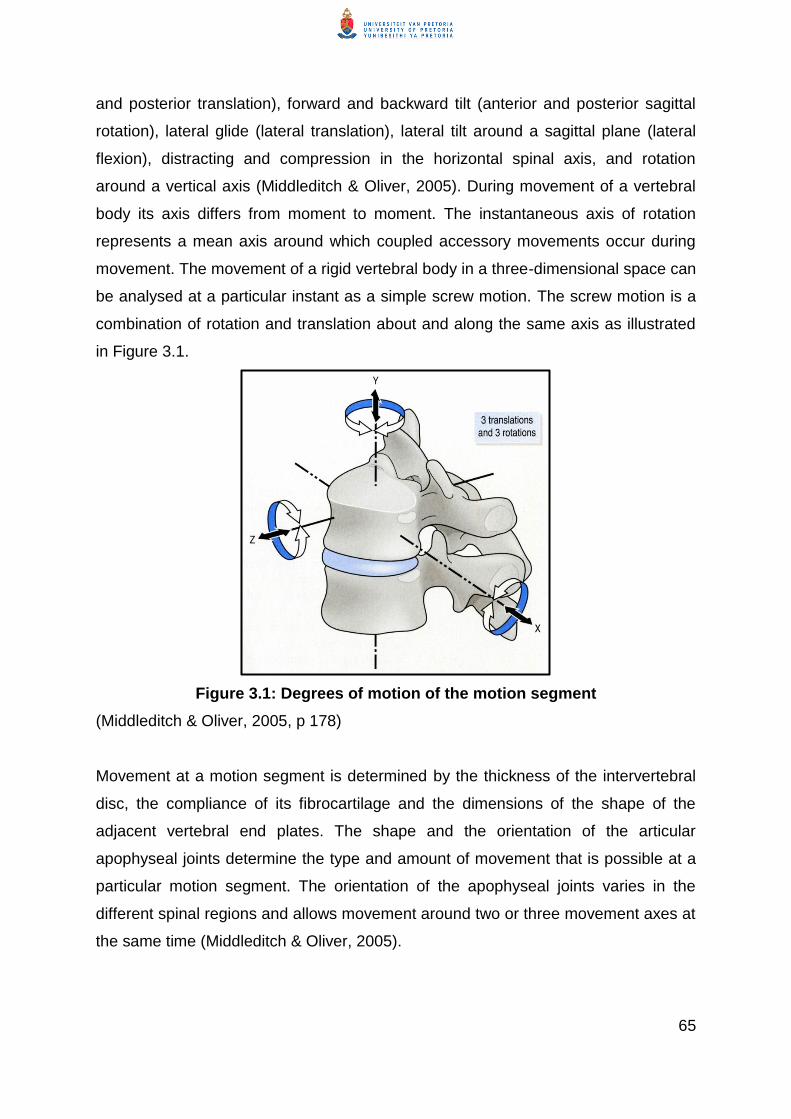
65
and posterior translation), forward and backward tilt (anterior and posterior sagittal
rotation), lateral glide (lateral translation), lateral tilt around a sagittal plane (lateral
flexion), distracting and compression in the horizontal spinal axis, and rotation
around a vertical axis (Middleditch & Oliver, 2005). During movement of a vertebral
body its axis differs from moment to moment. The instantaneous axis of rotation
represents a mean axis around which coupled accessory movements occur during
movement. The movement of a rigid vertebral body in a three-dimensional space can
be analysed at a particular instant as a simple screw motion. The screw motion is a
combination of rotation and translation about and along the same axis as illustrated
in Figure 3.1.
Figure 3.1: Degrees of motion of the motion segment
(Middleditch & Oliver, 2005, p 178)
Movement at a motion segment is determined by the thickness of the intervertebral
disc, the compliance of its fibrocartilage and the dimensions of the shape of the
adjacent vertebral end plates. The shape and the orientation of the articular
apophyseal joints determine the type and amount of movement that is possible at a
particular motion segment. The orientation of the apophyseal joints varies in the
different spinal regions and allows movement around two or three movement axes at
the same time (Middleditch & Oliver, 2005).

66
In terms of physiological spinal movements, rotation of the spine takes place
simultaneously with lateral flexion and vice versa and is known as coupling
movements. Tripled movements take place when movement occurs in three
dimensions around three axes. The implication of the phenomena of coupled or
tripled movements is of importance in the interpretation of abnormal movement
patterns in relation to pathology or abnormal spinal biomechanics as a result of
abnormal spinal loading (Middleditch & Oliver, 2005).
The global mobility of the spine as a functional biomechanical chain of motion
segments is enhanced by the fact that it has two mobile lordotic spinal curves (the
cervical and lumbar spines) on either side of the thoracic kyphosis, which is
stabilised by the ribcage. The sacrum as a rigid kyphosis links to the pelvis through
the two sacro-iliac joints and is stabilised by the pelvis. Body weight from the
vertebral column is transmitted via the 5th lumbar vertebra to the sacrum through the
SI joints along the alar of the sacrum and through the ischial tuberosities towards the
acetabulum (Middleditch & Oliver, 2005 p 215).
The spinal curves are interdependent and if the face is kept vertical the head has to
remain balanced over the sacrum: the relationship between the head and the pelvis
behaves as if it is part of a closed kinematic chain.
Owing to the differences in the size of the vertebral bodies, intervertebral discs and
the orientation of the zygapophyseal joints in the cervical spine together with the
presence of ribs and sternum in the thoracic region, the regions of the spine respond
differently to abnormal loading. The L4-S1 motion segments are especially
vulnerable for abnormal loading due to the forward tilt of the sacrum in standing.
With a decreased anterior stabilising force of transvers abdominus, the L5 vertebra
Changes in the position of any one motion segment will result in changes in
position of the adjacent superior and inferior [motion] segments and as such
will affect the cervical, thoracic and lumbar spinal curves (Norkin & Levangie,
1992).

67
has a tendency to slip anterior on the sacrum and the L4 has a lesser tendency to
slide anteriorly on the L5 motion segment. When the abdominal muscles are weak,
particularly the transvers abdominus muscle, the lumbar lordosis increases and also
the strain on the L4-S1 segments (Kapandji, 2008; Middleditch & Oliver, 2005).
Mobility at the lumbosacral joint is limited by the iliolumbar ligaments, which unite the
L4 and L5 lumbar vertebrae directly to the ilium. The superior band of the iliolumbar
ligament between the tip of the L4 transvers process and the iliac crest and the
inferior band of the iliolumbar ligament runs between the tip of the lower border of
the transvers process of L5 and the iliac crest to insert anterio-medially to the
superior band. Thus, the two iliolumbar ligaments stabilise the L4-S1 motion
segments during movement (Kapandji, 2005; Bogduk, 2011). The implication of this
stabilisation of the L4 and L5 motion segments by the superior and inferior bands of
the iliolumbar ligaments is that one can logically assume that during lumbar flexion
most strain will occur between L3 and L4. However, the L3 and L4 motion segments
are stabilised by the lumbar fibres of the longissimus longus that run from the ilium to
the transvers process of L3 and the fibres of the interspinalis thoracis muscles from
the thoracic spine and insert on the spinous process of L3.
The clinical implication of this mechanism is that the L3 vertebra is the first true
mobile lumbar segment above the stabile L4-L5 and is therefore vulnerable to being
pulled posteriorly by the longissimus longus as well as the fibres of the interspinalis
thoracis (Kapandji, 2008). The largest amount of intersegmental movement takes
place between L4 and L5 while the greatest amount of variability occurs at the L5-S1
level (Middleditch & Oliver, 2005).
It is important to realise also that during a change in the person‘s basic position; i.e.
from standing to unsupported sitting with the hips and knees in 90ᵒ flexion, the
mobile lumbar spine is flexed into a similar position as during maximal lumbar flexion
in standing. In the unsupported sitting, 60% of flexion occurs at the hip joint and 30%
of flexion occurs in the lumbar spine. Of this 30% lumbar flexion that occurs, 80% to
90% (of the lumbar flexion) takes place at L4 and L5. This allows one to conclude

68
that the largest amount of intersegmental flexion occurs at L4-L5 (Middleditch &
Oliver, 2005).
During a prolonged standing position, the impacted joints at each level of the spine
bear an average axial load of 16%. In people with a lumbar lordosis, the lumbar
joints between L3 and S1 bear approximately 19% of the axial load (Middleditch &
Oliver, 2005). However, from a sagittal view, the apophyseal joints do not impact on
each other but rather slide upon each other because the joint surfaces run more
parallel to each other. Because of this sliding action, they may not play a role in load-
bearing (Twomey & Taylor, 1987).
In the upright posture of humans, especially, the lower segments of the lumbar spinal
column are exposed to considerable pressure. The weight of all body parts above
these lumbar segments is borne on a small area of only a few square centimetres.
Shifting of the trunk out of midline increases pressure on the lumbar segments
considerably. As much as 70% of all movements in flexion and extension of the
entire spinal column takes place in these segments (Middleditch & Oliver, 2005;
Kapandji, 2005).
The clinical implication of the discussion is that the lumbar area should always be
assessed and treated by taking into consideration the alignment of the thoracic and
cervical curves and the position, control and range of movement of the scapulae and
gleno-humeral joints as well as the pelvis and hip complex.
In clinical practice, patients with CNSLBP treated by the researcher commonly
present with L4-S1 dysfunction, although involvement of L3 is not always excluded.
Based on the discussion in the preceding paragraphs, the researcher argues that
because the largest amount of intersegmental movement takes place between L4
and L5 because the greatest amount of variability occurs at the L5-S1 level, these
segments are most vulnerable to any abnormal loading forces (Kapandji, 2008;
Middleditch & Oliver, 2005; Moore, Dally & Agur, 2006).

69
3.2.2 The muscle system of the ISMS
Similar to the spinal ligaments that provide local and global stability to the
intervertebral column, the muscular system of the trunk also provides segmental,
local and global stability to the trunk (intervertebral column) (Comerford, Mottram
and Gibbens, 2008).
The intersegmental and multisegmental muscle attachments shown in Figure 3.2(a)
and 3.2(b) illustrate the complex and integrated connectivity of the posterior spinal
muscle system as a holistic muscle system.
Figure 3.2(a): Intersegmental posterior spinal muscles
(Travell & Simons, 1983)

70
Figure 3.2(b): Multisegmental posterior spinal muscles
(Travell & Simons, 1983)
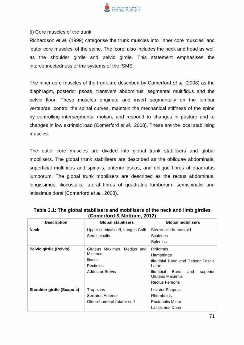
71
(i) Core muscles of the trunk
Richardson et al. (1999) categorise the trunk muscles into ‗inner core muscles‘ and
‗outer core muscles‘ of the spine. The ‗core‘ also includes the neck and head as well
as the shoulder girdle and pelvic girdle. This statement emphasises the
interconnectedness of the systems of the ISMS.
The inner core muscles of the trunk are described by Comerford et al. (2008) as the
diaphragm, posterior psoas, transvers abdominus, segmental multifidus and the
pelvic floor. These muscles originate and insert segmentally on the lumbar
vertebrae, control the spinal curves, maintain the mechanical stiffness of the spine
by controlling intersegmental motion, and respond to changes in posture and to
changes in low extrinsic load (Comerford et al., 2008). These are the local stabilising
muscles.
The outer core muscles are divided into global trunk stabilisers and global
mobilisers. The global trunk stabilisers are described as the obliquae abdominals,
superficial multifidus and spinalis, anterior psoas, and oblique fibres of quadratus
lumborum. The global trunk mobilisers are described as the rectus abdominus,
longissimus, iliocostalis, lateral fibres of quadratus lumborum, semispinalis and
latissimus dorsi (Comerford et al., 2008).
Table 3.1: The global stabilisers and mobilisers of the neck and limb girdles (Comerford & Mottram, 2012)
Description Global stabilisers Global mobilisers
Neck Upper cervical cuff, Longus Colli
Semispinalis
Sterno-cleido-mastoid
Scalenes
Splenius
Pelvic girdle (Pelvis) Gluteus Maximus, Medius and Minimum
Iliacus
Pectinius
Adductor Brevis
Piriformis
Hamstrings
Ilio-tibial Band and Tensor Fascia Latae
Ilio-tibial Band and superior Gluteus Maximus
Rectus Femoris
Shoulder girdle (Scapula) Trapezius
Serratus Anterior
Gleno-humeral rotator cuff
Levator Scapula
Rhomboids
Pectorialis Minor
Latissimus Dorsi

72
(ii) Local and global muscle roles
The local (stabilising) muscles are responsible for increasing the segmental stability
of the spine, decreasing excessive intersegmental motion and maintaining muscle
control (stabilisation) of the spine during low load tasks and activities They contract
regardless of the direction of loading or movement that is taking place and are prone
to be activated during low load functioning. During their functioning, the local
muscles do not significantly change length but work mostly isometrically and
therefore do not primarily contribute to range of motion. During functional activities,
they maintain their stabilising function during all ranges of motion.
Global stabilising and mobilising muscles (Table 3.1) are responsible for the
production of movement. They control movements that require high physiological
load and therefore work against the direction of the loading. These muscles contract,
depending on the demands of the environment/task at hand and the load. The global
muscles change length significantly (concentric and eccentric muscle contraction)
and therefore are the muscles that control the range of motion of a joint or series of
motion segments in the case of the spinal column. These global muscles may have a
primary global trunk stability or trunk mobility role during activities of daily living.
For normal efficient functional activity both the local and global muscles should work
in an integrated way. Neither the local nor the global muscle ‗systems‘ can control
functional stability during body movement that demands trunk mobility. Trunk stability
is controlled by the local stabilisers. The global muscles influence a patient‘s postural
alignment and contribute to the production and control of range of motion of the
spine (trunk).
(iii) Diaphragms attached to the spine
At each junction of the spinal curves C7/T1, T12/L1 and L5/S1 there are three very
important circular muscle planes: the cervical or thoracic inlet, the respiratory
diaphragm and the pelvic diaphragm. These circular muscle planes function as
transverse diaphragms to separate the three chambers of the trunk and permit
transmission of vital structures such as vascular and neural structures. These

73
diaphragms can be areas of major dysfunction and mechanical stress due to their
insertion on the spinal vertebrae at the appropriate levels.
The biomechanics of breathing is of vital importance to spinal functioning due its
influence primarily on the thoracic area and the cervical and lumbar spinal regions
(Kapandji, 2008).
The respiratory diaphragm as the main inspiratory muscle is a musculotendonous
dome that attaches to the lower thoracic outlet and separates the thorax from the
abdomen. Posteriorly the dome specifically attaches to the deep surfaces of the
costal cartilages, the costal arches, the tips of the eleventh and twelfth ribs and the
T12-L1 vertebral bodies. The elevation of the ribs by the inspiratory muscles and the
depression of the ribs and sternum by the expiratory muscles require mobility of the
costovertebral, costochondral and costosternal joints. Any dysfunction that can affect
the mobility of these joints will have an effect on the person‘s breathing. These joints
can also be a major source of nociception, which can refer pain to the lumbar spine
and/or restrict the patient‘s breathing pattern.
Other inspiratory muscles are the accessory inspiratory muscles; for instance, the
sternocleidomastoid, scalenes, pectoralis major and minor, and the lower fibres of
serratus anterior and latissimus dorsi, serratus posterior superior and the iliocostalis
thoracis. All these muscles are prone to developing trigger points and can refer pain.
Overuse of the accessory muscles results in elevation of the shoulder girdles, which
can affect the alignment of the cervical and thoracic spinal curves, which in turn will
influence the lumbar curve.
The primary expiratory muscles such as the internal intercostals and the accessory
expiratory muscles are involved in forced expiration (valsalva manoeuvre). These
muscles are the rectus abdominus, external oblique and internal oblique muscles
and they strongly depress the abdominal outlet. Other accessory expiratory muscles
are the iliocostalis thoracic, longissimus, serratus posterior inferior and the quadratus
lumborum.
74
Of clinical importance is the fact that muscle imbalance, trigger points in these
muscles and dysfunction of the costochondral, costovertebral and costo sternal joints
can affect not only breathing but also spinal dysfunction.
Further, the interaction between the stabilisation of the spine by the local and global
trunk muscles (in which abdominal muscle control plays an important role), the pelvic
floor and the respiratory diaphragm is of vital importance for optimal alignment of the
spine as a closed kinematic chain.
(iv) The head as part of the ISMS
The weight and position of the head have a profound influence on the alignment of
the cervical spine. Stability and mobility of the cervical spine are of utmost
importance because the spine has to move the head in space on a relative stabile
base. Owing to the interaction of the vestibular ocular reflex, the position of the head
is usually vertical (eyes are horizontal) and the cervical spine adapts continuously to
maintain the head‘s vertical position. Clinically, it is observed that the junction
between the mobile cervical spine and the stabile thoracic spine places the transition
vertebrae at the cervico-thoracic junction under great strain (Middleditch & Oliver,
2005).
The design of the musculature of the cervical spine is specialised to move the ‗head
on the cervical spine‘ (‗head on neck‘), to move the ‗cervical spine‘ as such and to
move the ‗head with the cervical spine‘ (Middleditch & Oliver, 2005). As the head‘s
stability and movement depend on the cervical spine, it is seen as part of the
functioning of the ISMS.
During functional activities the forces generated in the local and global trunk
muscles are transmitted across the cervical spine to the head and vice versa
and the shoulder and pelvic girdles.
So in the context of the biomechanical part of the ISMS, postural adjustments of
the trunk include the head, spine, shoulder girdle and pelvic girdle control.
75
Muscles anterior to the cervical area include muscles that ‗flex the neck‘ (longus colli,
sternocleidomastoid and scalenus anterior), muscles that ‗flex the neck and head‘
(sternocleidomastoid and longus capitis) and the muscle that flexes the ‗head on the
neck‘ (rectus capitis anterior).
Muscles that laterally flex the neck include the scalenus anterior, scalenus medius,
scalenus posterior, splenius cervices, levator scapulae and sternocleidomastoid.
Muscles that laterally flex the head and the neck include the sternocleidomastoid,
splenius capitis, trapezius, and erector spinae. The muscle that laterally flexes the
head on the neck is the rectus capitis lateralis.
In similar fashion the muscles that ‗extend the neck‘ are the levator scapulae and
splenius cervicis; the muscles that ‗extend the head and neck‘ are the trapezius,
splenius, and erector spinae; the muscles that ‗extend the head on the neck‘ are the
rectus capitis posterior major, rectus capitis posterior minor, and obliquus capitis
superior (the last-mentioned three muscles are also called the suboccipital muscles).
The muscles that rotate the neck are the semispinalis cervices, multifidus, scalenes
anterior and splenius cervicis; the muscles that rotate the neck and the head are the
sternocleidomastoid and splenius capitis; the muscles that rotate the head on the
neck are the obliquus capitis inferior and rectus capitis posterior major.
An important clinical implication is that the trapezius upper fibres and levator
scapulae elevate the scapulae. Longstanding elevation of the scapulae due to
prolonged periods of poor sitting posture is associated with an increase in the
thoracic kyphosis and cervical extension. The change in both these curves will flatten
the lumbar lordosis and put strain on the thoracolumbar fascia (TLF) and result in
abnormal muscle activation.
(v) The shoulder girdle as part of the ISMS
The position of the shoulder girdle (scapulae, clavicle and gleno-humeral joints)
strongly influences the alignment of the cervical and the thoracic regions of the spine
owing to its muscle attachments to the spine.

76
The medial borders of the scapulae can be in adduction (associated with a flat
thoracic curve) or in abduction (associated with an increased thoracic kyphosis). The
stabilisation of the scapulae is controlled by the trapezius upper, middle and lower
fibres, the rhomboid major and minor, latissimus dorsi, levator scapulae, and
serratus anterior. The global mobilisers of the shoulder girdle are the levator scapula,
rhomboids, pectoralis minor, and latissimus dorsi muscles.
The rotator cuff, together with the deltoid muscle and supraspinatus, controls all six
degrees of freedom of the gleno-humeral joint but needs a dynamic but stabile
scapula to control the full range of motion of the gleno-humeral joint.
With an increased thoracic kyphosis and winging of the scapulae (weak rhomboid
major and minor as well as serratus anterior and middle and lower trapezius
muscles) the rotator cuff muscles tend to shorten and the patient‘s posture becomes
one of an elevated abducted scapulae and anterior (medially) rotated gleno-humeral
joints, associated with an increased cervical and lumbar lordosis or a lumbar
kyphosis. The upper trapezius and levator scapulae contribute to the increased
cervical lordosis with shortening. Simultaneously the anterior pectoralis major and
minor shorten and also contribute to scapulae abduction. This muscle imbalance
results in a generally ‗poor posture‘ typical of a sedentary lifestyle and/or of a person
working in a prolonged poor sitting posture (Middleditch & Oliver, 2005).
When latissimus dorsi is shortened by unilateral muscle spasm and/or as part of
stiffening (due to guarded movement) it can create anterior shoulder pain at its
insertion on the intertubercular groove of the humerus. When the latissimus dorsi
performs its action of adduction, medial rotation and extension, it contributes towards
the abduction of the scapulae. This is because it pulls the inferior angle of the
scapula into abduction (one of the origins of the muscle) when it contracts and as
such contributes to an increased thoracic kyphosis.
Of clinical significance is the fact that the latissimus dorsi inserts on the
intertubercular sulcus of the humeral head shoulder joint of the spinous process of
77
the T7-T12 vertebrae, the ribs, and through the TLF, which attach to the spinous
processes of all the lumbar and sacral vertebrae, supraspinous ligaments and iliac
crests. It plays an enormous role in controlling a person‘s posture and through the
TLF plays an enormous role in lumbar pain and dysfunction. It is innervated by the
thoraco-dorsal nerve (C6-C8). As such it can become involved with lower cervical
pathology (Middleditch & Oliver, 2005).
Another muscle of clinical significance is the trapezius muscle, which attaches the
occipit, the scapulae (shoulder girdle) and thoracic vertebrae down to the T12
spinous process. It receives the motor innervated from the eleventh cranial nerve
and sensory branches from the ventral rami of C3-C4 (Middleditch & Oliver, 2005).
The position of the shoulder girdle in relation to the spine is therefore greatly
influenced by the directional forces or lack of these forces exercised by the
latissimus dorsi and trapezius muscles.
The anatomy and function of the latissimus dorsi and trapezius muscles reveal that
the head, cervical thoracic and lumbar spinal regions are intimately connected to the
shoulder girdle, trunk and pelvis. This is of significance in the clinical application of
manual therapy for CNSLBP.
(vi) The pelvis as part of the ISMS
Because the lumbar spine articulates directly with the sacrum, any movement
(anterior, posterior or lateral tilt) of the pelvis will affect the (depth/angulation) lumbar
lordosis. A posterior tilt flattens the lumbar curve, an anterior tilt increases the lumbar
lordosis and a lateral tilt results in lumbar sideflexion. The muscles that control the
anterior-posterior pelvic tilt are the erector spinae and psoas major, rectus
abdominus, oblique internus and externus, gluteus maximus, and the hamstrings
(semitendonosus, semimembranosus and biceps femoris) (Middleditch & Oliver,
2005).
The muscles acting as mobilisers of the pelvis on the hip and secondarily on the
lumbar spine are piriformis, hamstrings, ilio-tibial band and tensor fascia latae,
superior gluteus maximus and rectus femoris because they control the tilting of the

78
pelvis on the hip joint and therefore in nutation and counter-nutation (Middleditch &
Oliver, 2005; Kapandji, 2008; Comerford & Mottram, 2008). The control of nutation
and counter-nutation takes place through the interaction among the abdominal,
lumbar-thoracic and the pelvis-hip stabilisers and mobilisers. One can therefore talk
about the ‗spine-pelvis-hip complex‘ (Middleditch & Oliver, 2005).
Muscle imbalance and motor recruitment result in muscle stiffness and as such the
‗spine-pelvis-hip complex‘ during physiological movements of the lower limb and
functional activities such as gait. Boyling et al. (2004) state that control of pelvis
stability and balance is essential for pain-free functioning of the spine.
The clinical implication of this interaction among the spine and head; spine and
shoulder girdle and upper limbs; and spine and pelvis and lower limbs is that manual
therapy for the lumbar spine should include the release of soft tissues and/or muscle
recruitment around the shoulder girdle and gleno-humeral joints, pelvis and hip joints
as distal as the hamstring muscles and tensor fascia latae (Middleditch & Oliver,
2005).
3.2.3 The neural components of the spine
Under this heading a brief explanation of the link between the gross anatomy of the
nervous system – peripheral (PNS), central (CNS) and autonomic nervous systems
(ANS) – is given. The aim of providing this explanation is to indicate how interlinked
the neural system related to the motion segment is in terms of the mechanical and
physiological responses that can occur at the level of the motion segment. The
mechanical and physiological neural components at the level of each motion
segment are also related to each other as well as to the brain and the filum terminale
in the sacrum.

79
Figure 3.3: The interconnectedness between the spinal cord, the dorsal and
ventral rami of the spinal nerve and the sympathetic chain adjacent to the
vertebral column
(Marieb, 2004 p 539)
Understanding the mechanical unity among the different parts of the total nervous
system is central to understanding the effect of spinal torsion that may occur during
the development of ISMS dysfunction pain processing. In Figure 3.3 one can see the
interconnectedness between the three nervous systems: the PNS, ANS and CNS, at
the level of a motion segment. Each of these systems is a continuum in its own right.
Each of the peripheral nerves/nerve root trunks and autonomic sympathetic chain is
ensheathed by fascia. (The dura, arachnoid and pia mater (the meninges) are all
part of the connective tissue (fascia) system of the body.) The nerve root complex
consists of the root sleeve; the motor and ventral roots; the dorsal root ganglion,
which is highly vascularised; and the spinal sleeve. The epineurium in general is
continuous with the dura mater while a few layers of the peripheral nerve‘s
epineurium and the endoneurium are continuous with the pia mater.
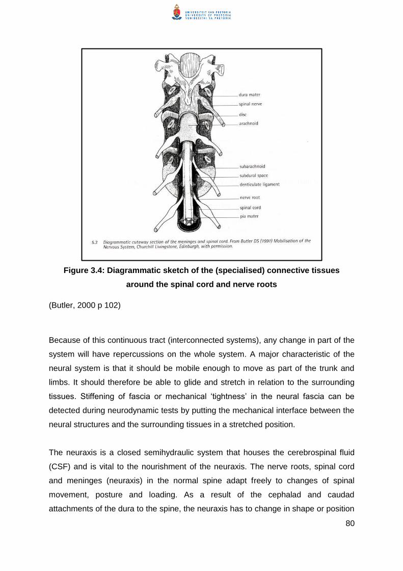
80
Figure 3.4: Diagrammatic sketch of the (specialised) connective tissues
around the spinal cord and nerve roots
(Butler, 2000 p 102)
Because of this continuous tract (interconnected systems), any change in part of the
system will have repercussions on the whole system. A major characteristic of the
neural system is that it should be mobile enough to move as part of the trunk and
limbs. It should therefore be able to glide and stretch in relation to the surrounding
tissues. Stiffening of fascia or mechanical ‗tightness‘ in the neural fascia can be
detected during neurodynamic tests by putting the mechanical interface between the
neural structures and the surrounding tissues in a stretched position.
The neuraxis is a closed semihydraulic system that houses the cerebrospinal fluid
(CSF) and is vital to the nourishment of the neuraxis. The nerve roots, spinal cord
and meninges (neuraxis) in the normal spine adapt freely to changes of spinal
movement, posture and loading. As a result of the cephalad and caudad
attachments of the dura to the spine, the neuraxis has to change in shape or position

81
to accommodate and adapt to changes in posture and has to adapt to loading
(Middleditch & Oliver, 2005; Brieg, 1978). These changes in the neuraxis entail
changes in length, diameter, shape and direction during movement and occur
because the collagen network of the pia mater has a rhomboid nature, which offers
the neuraxis its mobility when deformed by movement. The extensibility of the filum
terminale allows it to elongate in a linear fashion when it is stretched in a caudad
direction. The changes in the neuraxis and meninges that take place during
movement can entail ventral displacement, lengthening, axial sliding and angulation
of the nerve roots (Middleditch & Oliver, 2005). During flexion of the spine, the
cervical and lumbar regions can increase up to 28 mm each while the thoracic region
only increases about 3 mm in length.
The increased tension in the neuraxis and meninges during flexion of the spine is
caused by the fact that it attaches cranially to the foramen magnum, which causes
rostral pulling on the pons and medulla, and caudally it attaches to the sacrum. In the
cervical region the greatest strain occurs at the level of C5-C6 while in the lumbar
spine the area of greatest strain occurs on the level of L5-S1. During flexion, caudal
sliding of 3 mm in the dura at the levels of C6 and L4 occurs. At the level of L5
rostral displacement of about 3 mm takes place while at the level of T5 rostral
movement occurs above T5 and caudal movement occurs below the T5 level. The
ability to undergo longitudinal extensibility protects the neuraxis from positions of
high tension and from positions of low tension. The neuraxis is relaxed in the neutral
position, which is midway between flexion and extension (Middleditch & Oliver,
2005).
The change in dural length is smaller than the change in the bony canal during
flexion (Breig, 1978). The clinical implication of this fact is that any change in the
curves of the spinal canal will necessitate adaptability of the cord and its dural
covering. Depending on where the adaptation has to occur, interfacing between the
cord/dural sac and the bony canal occurs. With torsioning of the canal at any level,
the appropriate nerve roots will be torsioned accordingly. The torsioning can create
changes in alignment of the intervertebral canal and these changes are therefore

82
possible areas of interfacing of the nerve root sleeve. Central interfacing and lateral
interfacing are both potent sources of nociception.
Another factor that contributes to the stability of transitional areas ~ C6-T3 and ~
T10-L2 is the fact that the plexuses originate in these areas from the spinal cord. The
spinal canal also narrows slightly in these areas, which influences the mobility of the
spine (Middleditch & Oliver, 2005).
The continuum between the three aspects of the nervous system (PNS, ANS and
CNS) is the key concept in neurodynamics. The study of neurodynamics of the
nervous system is based on the interconnectedness between these three systems
because it can reveal the site of restrictions between the nervous system and any
other surrounding somatic tissues, in particular the different types of fascia. Fascial
restrictions are the most likely source of soft-tissue restriction, pain and dysfunction
of trunk and limb movement revealed through adverse neural tension, which can
restrict movement.
Neurodynamics indicates adverse neural tension, in particular of the PNS, which is
revealed by interfaces frictioning because of a lack of glide or restriction of
movement at interfaces due to fibrosis or points of tension within the system.
Restriction of soft tissue surrounding pain-sensitive structures, such as the nerve
root sleeve (the classic example) at the local lumbar motion segment, can occur as a
result of pathology that develops from strain typically at the L4-S1 motion segments.
Structures that are typically strained are the synovium of the facet joint capsules or
ligamentous structure of the annular disc or any of the insertions of the muscle
(multifidi) local ligaments (for example, anterior or posterior sacro-tuberous ligaments
or ligamentum flavum). Healing by fibrous tissue and shortening due to local spasm
enhance this restriction and pain (Butler, 2000).
The clinical implication of the neural system as integral part of the ISMS is given at
the end of Section 3.2.4.
83
3.2.4 The connective tissues in the trunk
Apart from the interconnectedness of the systems mentioned in the preceding
paragraphs, the muscular, skeletal and ligamentous systems as well as the neural
systems are infiltrated and surrounded by organised specialised and loose
unspecialised connective tissue (Langevin & Sherman, 2006). Langevin and
Sherman (2006) list 14 specific terms that are used to describe fascia but which also
indicate specific aspects of connective tissue. These are ‗dense connective tissue‘,
‗areolar connective tissue‘, ‗superficial fascia‘, ‗deep fascia‘, ‗intermuscular septa‘,
‗interosseal membrane‘, ‗periost‘, ‗neurovascular tract‘, ‗intra- and extramuscular
aponeurosis‘, ‗epimysium‘, ‗perimysium‘, ‗endomysium‘ and the ‗dura‘.
The function of fascia is to link all body systems and give structure and support to all
anatomical structures of the body, protect and separate specialised organs and
contribute to cellular respiration, elimination of toxins, metabolism and fluid and
lymphatic flow. Fascia contributes to the energy storage in the body, acts as a
passive force transmission and is a major source of proprioceptive feedback to the
CNS in the control of movement through the mechanoreceptors found in the fascia
(Cantu & Grodin, 2001). Trauma or malfunction of the fascia can result in poor
cellular efficiency, necrosis, disease, pain and dysfunction throughout the body
because it restricts the environment in which other structures function.
Langevin and Sherman (2006) confirm the fascial network as a continuous web
throughout the whole body. The implication of this continuous web of connective
tissue is that stiffness of the fascia at one end of the system will cause pain and
dysfunction at a distant point in the system.
Connective tissue is an integral part of each muscle, which is classically organised
into (1) the epimysium, which encloses the muscle; (2) the perimysium, which binds
groups of muscle fibres into fascicles (bundles); and (3) the endomysium, which
ensheathes individual muscle fibres (Middleditch & Oliver, 2005). The organised
fascial layers in intra-muscular tissue (epimysium, perimysium and endomysium) are
classified as part of the deep fascial layers (Findley, Chaudhry, Stecco & Roman,
2012).

84
Specialised as well as unspecialised loose connective tissue also surrounds and is
an integral part of the PNS, ANS and CNS, making up on average 50% of these
systems (Coppieters & Nee, 2012; Langevin & Sherman, 2006). The fascial network
acts as a passive force transmission and is a major source of proprioceptive
feedback through the mechanoreceptors found in fascia. to the CNS in the control of
movement (Cantu & Grodin, 2001).
A specialised fascial structure, the TLF, plays an important role in the functional
stability of the lumbar-thoracic area, the development of ISMS dysfunction and, as
such, CNSLBP. The TLF is a complex fascial structure and consists of posterior,
middle and anterior layers of dense connective tissue that are also separated from
each other by loose areolar connective tissue that allow the adjacent layers to glide
past each other (Langevin, 2011; Middleditch & Oliver, 2005). The posterior layer is
also called the superficial layer of the TLF.
Most of the fibres of the posterior TLF derive from the aponeurosis of the latissimus
dorsi and attach superior (cranial) to L4 to the supraspinous ligaments. Inferior to L4,
the superficial laminae of the posterior TLF attach to the sacrum, posterior superior
liac spine of the iliac crest on the contralateral side. The superficial part of the
posterior TLF blends with the fascia of the gluteus maximus muscle and
superolaterally with the fascia of the contralateral latissimus dorsi. Because these
two muscles are mechanically linked through the TLF they increase the tension in
the posterior layer of the TLF when they contract or are in spasm (Middleditch &
Oliver, 2005).
The deep layer of the posterior TLF medially to the interspinous ligaments, posterior
superior iliac spine, iliac crest and long dorsal sacroiliac ligament and some fibres
attach to the deep fascia of the erector spinae. The deep laminae also attach to the
lateral raphae and as such are indirectly linked to the internal oblique and
transversus abdominus muscles. At the sacral level the superficial and deep layers
of the TLF blend together.

85
The middle layer of the TLF attaches medially to the lumbar transvers processes and
is continuous with the intertransvers ligaments. Laterally it gives rise to the
aponeurosis of the transversus abdominus muscle. It lies posterior to the quadratus
lumborum muscle and compartmentalises the erector spinae muscles (Kapandji,
2008).
The anterior TLF layer attaches medially to the lumbar transvers processes and the
intertransvers ligaments. Inferiorly it attaches to the iliolumbar ligament and adjacent
iliac crest. Superiorly it forms the lateral arcuate ligament. It covers the anterior
surface of the quadratus lumborum and laterally it blends into the other layers of the
TLF.
The visible areas of superficial and deeper fascia on the posterior spine are shown in
Figure 3.6(a) and 3.6(b)
Figure 3.5(a) and 3.5(b): The visible areas of superficial and deeper fascia on
the posterior spine
(Netter, 2011 plates 168 and 169)
Fascia is densely innervated by myelinated sensory nerve endings, which are
assumed to serve a proprioceptive function. These include Pacini (and paciniform)
corpuscles, Golgi tendon organs and Ruffini endings (Stecco et al., 2010). In addition
86
they are innervated by free endings (nociceptors). When including periosteal,
endomysial and perimysial tissues as part of a body-wide interconnected network,
the fascial network can be seen as the human body‘s largest sensory organ,
particularly proprioceptive sensation (Scleip, 2003).
Of clinical implication is the fact that the CNS, PNS and ANS are interconnected at
segmental and global levels throughout the spine and continuous with the brain. That
connective tissue is an integral part of the nervous system might relate to the fact
that shortening/stiffening of the fascia within and around the total nervous system
can limit neural mobility and be a powerful source of nociception. As the fascial
system in the human body is a global network, stiffening of the fascia in the limbs
may not only affect the mobility of the ISMS but also refer pain to the lumbar spine.
Any imbalance or shortening of the muscles that insert onto the fascial sheet of the
TLF will probably result in an uneven pull on the TLF, causing pain in the lumbar
area because it is so rich in proprioception and nociception (Stecco, 2010).
Imbalance/dysfunction in the TLF will not only affect the lumbar area but, if
shortened, will affect the fascial running up in the spine and cause headache.
3.3 Postural control of the ISMS
Posture and coordinated movement are the result of integration between the multiple
inputs from spinal, medullary, midbrain and cortical levels (Horak & Macpherson,
2006). The result of the input from multiple levels is: (1) voluntary activity; (2)
adjustment of the body (posture) to provide a stable background for movement; and
(3) coordination of various muscle activities so that smooth and accurate movement
can take place. Horak and Macpherson (2006), Shumway-Cook and Woollacott
(2007) and Ganong (2003) describe postural control as the ability to control a stabile
position of the body in space while maintaining an appropriate relationship between
the body segments and between the body and the environment relevant to the task
that the person is performing.
The ability to maintain stability in a standing posture is, according to Levangie and
Norkin (2005), a learned skill based on the sensory impulses that the brain receives
87
from the skeletal, articular system; the sensory systems; and the muscular
(myofascial), fascial system; and also cognitive input or higher centre input.
The continuous integrated activity of the posture-regulating systems results in
continuous adjustment of posture before and during movement. These posture-
regulating systems (Shumway-Cook & Woollacott, 2007) include the integration of
the musculoskeletal components, the neuromuscular synergies, individual sensory
systems, sensory strategies, anticipatory mechanisms, adaptive mechanisms, the
respiratory system and internal representation.
Impaired postural control can result in biomechanical malalignment and dysfunction
and, as such, cause mechanical pathology (wear and tear on joints). This implies
that musculoskeletal pain syndromes are seldom caused by isolated events and that
movement systems should be assessed holistically in the context of the person‘s
psycho-social influences as well. Habitual movements and sustained postures also
play a role in the development of dysfunction (Sahrmann, 2002).
A painful stimulus originating from the musculoskeletal system stimulates the
appropriate nociceptors of the sensory system and results in a particular sensory
strategy conducted by the nervous system and integrated with the higher centre
control. Through the neuromuscular system the person‘s muscle recruitment
changes and movement patterns adapt to avoid the painful stimuli or to ‗hold the
body still‘ (fixation) to avoid the painful stimulus. This response in a person to adapt
their movement will affect their adaptive control (balance) and anticipatory control.
The person‘s breathing control or breathing pattern will change if the fixation affects
the trunk muscle control (abdominal and erector spinae cocontraction). If the
breathing pattern is limited due to the fixation it might lead to a decrease in
oxygenation and to tiredness. The fixation of the trunk muscles will also lead to an
increase in the muscle metabolism and a feeling of tiredness. Other internal organs
such as the heart and blood pressure might also be affected by the adapted postural
control. The adapted postural control will probably cause the person to decrease
their general level of activities of daily living (ADL) and adapt their lifestyle (if their
lifestyle has not been adapted already before the painful incident).

88
The ISMS is graphically displayed in Figure 3.6.
Figure 3.6: The integrated spinal movement system
The functioning of the ISMS at the local and global levels is influenced by the
patient‘s genetic inheritance of their morphology (body build) (Porterfield & DeRosa,
1992), which determines the integrity of the neuromusculoskeletal tissues and of the
process of degeneration that may be present in the zygapophyseal joints and
intervertebral discs. Because movement is strongly influenced by sensory input,
nociceptive stimuli will affect the functioning of the ISMS. The influence of
nociceptive stimuli on the ISMS is discussed in Section 3.4.
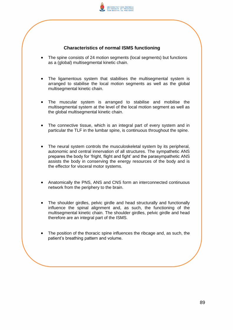
89
Characteristics of normal ISMS functioning
The spine consists of 24 motion segments (local segments) but functions as a (global) multisegmental kinetic chain.
The ligamentous system that stabilises the multisegmental system is arranged to stabilise the local motion segments as well as the global multisegmental kinetic chain.
The muscular system is arranged to stabilise and mobilise the multisegmental system at the level of the local motion segment as well as the global multisegmental kinetic chain.
The connective tissue, which is an integral part of every system and in particular the TLF in the lumbar spine, is continuous throughout the spine.
The neural system controls the musculoskeletal system by its peripheral, autonomic and central innervation of all structures. The sympathetic ANS prepares the body for ‗fright, flight and fight‘ and the parasympathetic ANS assists the body in conserving the energy resources of the body and is the effector for visceral motor systems.
Anatomically the PNS, ANS and CNS form an interconnected continuous network from the periphery to the brain.
The shoulder girdles, pelvic girdle and head structurally and functionally influence the spinal alignment and, as such, the functioning of the multisegmental kinetic chain. The shoulder girdles, pelvic girdle and head therefore are an integral part of the ISMS.
The position of the thoracic spine influences the ribcage and, as such, the patient‘s breathing pattern and volume.

90
3.4 Patho-physiological responses underlying the development of
ISMS dysfunction
The pathophysiological responses of muscle spasm, overuse and disuse and trigger
points that occur in muscular tissue, connective tissue stiffening, and the effect of
nervous tension on muscle tissue are processes integrated in the development of
ISMS dysfunction. These processes accumulatively over time contribute to the
clinical picture of CNSLBP.
For the sake of systematic discussion on the development of ISMS dysfunction, an
overview of these processes is given based on the synthesis of the researcher‘s
clinical experience and an overview of the relevant literature.
3.4.1 The effect of muscle spasm, overuse and disuse on muscular tissue
Muscle spasm is a response to injury to the muscle itself or a reflex response to
nociceptor irritation of joints and associated tissues. It is a protective mechanism to
splint a painful lesion and may be present segmentally or span a whole spinal region.
If short-lived, muscle spasm may be asymptomatic. Once it is present, spasm
increases the compressive forces on the structures of the motion segment, in
particular the intervertebral disc, which results in greater pain and dysfunction.
Muscle spasm can also cause pain through the tension on its attachment to the
periosteum.
Muscle spasm might be the primary mechanism in referred pain arising from the
spinal segment. Persisting muscle spasm results in decreased blood flow to the
muscle, leading to anoxia and accumulation of metabolic waste products in the
muscle, which would normally be dispersed during relaxation. With muscle fatigue,
pain may occur within the muscle and internal changes in the muscle occur, leading
to a contracture (Middleditch & Oliver, 2005).
Overuse or hypertrophy is the result of excessive use of a muscle where lack of use
(disuse) leads to atrophy. Excessive use of a particular muscle or altered motor
recruitment, which results in the habitual activation of certain muscle groups within

91
small and abnormally restricted amplitude of their available extensibility ranges, can
lead to a state of overactivation in these muscles and eventually they shorten. The
antagonists of the shortened muscles respond by inhibition, weakness and
lengthening (Janda, 1996). Postural muscles shorten or tighten while their
antagonistic phasic muscles become weakened and tend to lengthen.
Muscle atrophy as a result of disuse clinically presents as a decrease in the cross-
section area (CSA) of the muscle. Other clinical signs of muscle atrophy are
decreased muscle strength and endurance. Reduced muscular support will increase
the load on the joints and lead to abnormal movement patterns. A decrease in the
CSA of lumbar multifidus has been demonstrated in both acute and chronic LBP
pathology (Hides, Richardson & Jull, 1996;). Changes have also been reported in the
CSA of the psoas muscle in a patient with chronic LBP (Cooper, St Clair Forbes &
Jayson, 1992).
3.4.2 The development of trigger points
Trigger points are sustained contractions (previously known as fibrositis) of isolated
groups of muscle fibres. The most likely aetiology of trigger points, according to
Middleditch and Oliver (2005), is irritation of the nerve supply to the muscle, which
causes localised muscle spasm or a reflex response to irritation of deeper structures
supplied by the same segmental innervation. Trigger points are clinically tender on
palpation and can refer pain to other areas of the body (Middleditch & Oliver, 2005).
Kostopoulos and Rizopoulos (2001) do not relate the development of trigger points
only to the irritation of neural tissue. The authors illustrate the activation of
myofascial trigger points in Figure 3.7.
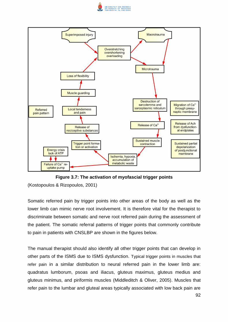
92
Figure 3.7: The activation of myofascial trigger points
(Kostopoulos & Rizopoulos, 2001)
Somatic referred pain by trigger points into other areas of the body as well as the
lower limb can mimic nerve root involvement. It is therefore vital for the therapist to
discriminate between somatic and nerve root referred pain during the assessment of
the patient. The somatic referral patterns of trigger points that commonly contribute
to pain in patients with CNSLBP are shown in the figures below.
The manual therapist should also identify all other trigger points that can develop in
other parts of the ISMS due to ISMS dysfunction. Typical trigger points in muscles that
refer pain in a similar distribution to neural referred pain in the lower limb are:
quadratus lumborum, psoas and iliacus, gluteus maximus, gluteus medius and
gluteus minimus, and piriformis muscles (Middleditch & Oliver, 2005). Muscles that
refer pain to the lumbar and gluteal areas typically associated with low back pain are

93
iliocostalis lumborum, psoas and iliacus muscles and quadratus lumborum muscles.
Referred pain from trigger points is characteristic of somatic referred pain: when the
trigger points are released the referred pain disappears.
Figure 3.8(a): Anatomy of the quadratus lumborum muscle trigger point; Figure 3.8(b):
Area of referral of the quadratus lumborum muscle (Kostopoulos and Rizopoulos,
2001).
Figure 3.8(c): Anatomy of trigger points of the psoas and iliacus muscles; Figure
3.8(d): Area of referred pain of psoas and iliacus muscles (Kostopoulos and
Rizopoulos, 2001).

94
Figure 3.8(e): Anatomy of the trigger point of the gluteus maximus muscle; Figure
3.8f): Area of referred pain of the gluteus maximus muscle (Kostopoulos and
Rizopoulos, 2001).
Figure 3.8(g): Anatomy of the gluteus medius trigger muscle; Figure 3.9(h): Gluteus
medius muscle point referral pain pattern (Kostopoulos and Rizopoulos, 2001).
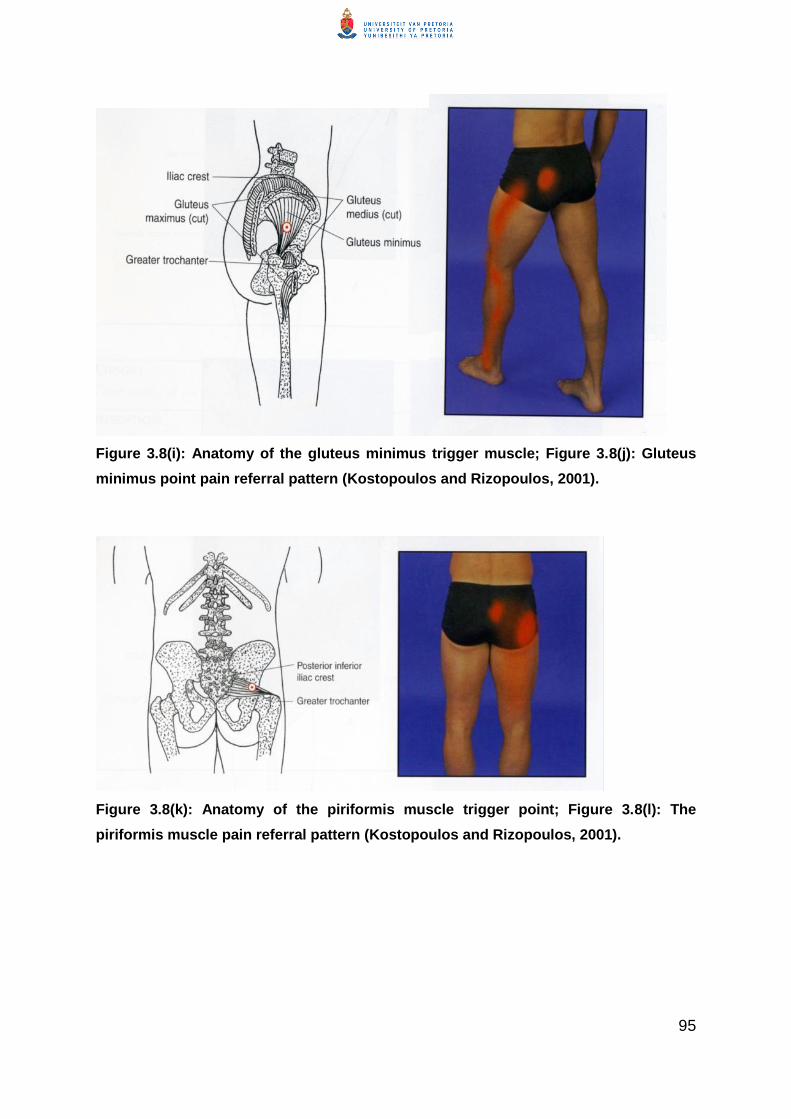
95
Figure 3.8(i): Anatomy of the gluteus minimus trigger muscle; Figure 3.8(j): Gluteus
minimus point pain referral pattern (Kostopoulos and Rizopoulos, 2001).
Figure 3.8(k): Anatomy of the piriformis muscle trigger point; Figure 3.8(l): The
piriformis muscle pain referral pattern (Kostopoulos and Rizopoulos, 2001).

96
Figure 3.8(m): Anatomy of the trigger point in the iliocostalis lumborum muscle;
Figure 3.8(n): Pattern of pain referral of the iliocostalis lumborum muscle
(Kostopoulos and Rizopoulos, 2001).
It is of utmost importance that the manual therapist distinguish between various
possible origins of the referred pain to accurately identify the variety of structures
that can contribute to the complex pain syndrome of the patient with CNSLBP.
3.4.3 The process of connective tissue stiffening in patients with CNSLBP
As connective tissue surrounds and is inherently a part of the structure of muscles, it
is subject to mechanical stress as a result of overuse, disuse, repetitive movement
and/or hypermobility. Owing to the fact that it is not always possible to determine the
tissue pathology in a clinic, the researcher has treated ‗soft tissue restrictions‘ as
shortening or stiffness in the connective tissue and muscular system, which have
possibly developed as a result of fibrosis. Fibrosis in connective tissues can develop
in two ways: (1) chronic local increase in stress (contraction of myofascial tissues),
which may be due to overuse, might cause microinjury and inflammation; and (2)
there may be a concurrent presence of inflammation, tissue hypo-oxygenation and
cytokines, such as TGFβ-1, during immobility or lack of stress on connective tissues
(Langevin & Sherman, 2006).

97
A consistent presence of hypomobility or immobilisation leads to atrophy of
connective tissue, architectural disorganisation, fibrosis, adhesions and contractures
(Langevin & Sherman, 2006). During the early phase of immobilisation the muscle-
associated connective tissue shortens before actual shortening of the muscle fibres
occurs. The presence of (myofascial) trigger points, taut bands in muscles and
muscle spasm may also contribute to connective tissue remodelling and fibrosis.
Shah, Phillips, Danoff and Gerber (2005) found a decrease in tissue pH and
increased levels of inflammatory cytokines in trigger points in the presence of pain.
The authors therefore argue that the presence of painful muscle contraction or
tender foci within the perimuscular fascia may be a contributing factor that promotes
hypomobility and tissue fibrosis.
Langevin and Sherman (2006) propose that connective tissue fibrosis in patients
with CNSLBP occurs as a result of one or a combination of the following factors:
decreased activity (sedentary lifestyle), which is characteristic of patients with
CNSLBP; changes in the patterns of muscle activation, which can result in muscle
cocontraction; muscle spasm or tissue microtrauma; and neuro-mediated
inflammation.
The fact that the TLF is so closely integrated with the muscles inserted onto it means
that muscle imbalance or spasm in one of these muscles such as latissimus dorsi
and the opposite gluteus maximus can result in a change in the biomechanics of the
TLF and can be the origin of severe pain due to the stimulation of the rich nociceptor
innervation of the TLF.
3.4.4 Effects of nervous tension (stress) on the musculature of the body
Nervous tension results in major chemical changes and alterations in the
musculature of the body. The causes of nervous tension can be anxiety, depression,
frustration or general stressors of life and can manifest as muscular tension, more
commonly seen in the cervical than the lumbar muscles. Elevation of the scapulae
results in an increased thoracic lordosis and can either lead to an increased or a
flattened lumbar lordosis (sway back) (Norkin & Levangie, 2005). So over time the
98
tension in the cervical muscles can lead to spinal imbalance and thoracic and lumbar
strain, as indicated in Section 3.4.
The researcher observed that the effect of emotional stress on muscle activity results
in shortening of soft tissues. Patients who exhibit emotional stress show an overall
pattern of shortening which differs from the pattern of shortening which originated
from a localised lumbar motion segment(s).
Prolonged muscle tension results in pain produced by the accumulation of
metabolites in the muscles. The metabolites become a source of irritation, setting up
a vicious cycle that eventually sustains the contraction and leads to joint restriction
(Middleditch & Oliver, 2005). Sustained contraction in the whole spinal system
becomes self-perpetuating and leads to widespread symptoms that pertain to
CNSLBP.
3.5 Development of integrated spinal movement system (ISMS)
dysfunction
The exact nature of the mechanisms at work during movement of the human ISMS
as well as the development of abnormal function of the ISMS is neither well
understood nor well defined. Zusman (2007, p 2) states that researchers are
presently looking for validation of a motion segment „instability‟ model that revolves
around pain-induced muscular insufficiency with continuing vulnerability to tissue
„injury‟. O‘Sullivan (2005) describes eight models or different approaches for the
diagnosis and classification of patients with CNSLBP to assist clinicians to
understand the complex heterogenetic condition of CNSLBP. One of these
classification models described by O‘Sullivan (2005) is the mechanical loading
model. In the following paragraphs the researcher starts the discussion on her
multidimensional explanation of the multiple components that can drive the clinical
picture of CNSLBP from the perspective of the abnormal biomechanical loading on
the lower lumbar segments. This is followed by a discussion on the effect of the
abnormal biomechanical loading on the soft and neural tissues, nociceptors

99
(peripheral and central sensitisation and altered pathways in the brain) and the
influence of the latter on the patients‘ psychosocial behaviour and vice versa.
The discussion in the following paragraphs is based on the fact that characteristic of
the normal ISMS is that the spine functions as a multisegmental kinetic chain, which
functions as a total system. The head, shoulder and pelvic girdles influence and are
influenced by the alignment of the spinal curves. The interconnectedness between
the systems around and within the motion segments, as well as the anatomical
muscle configuration of the deep z-shaped intersegmental spinal muscles found
bilaterally throughout the spine, attach all the motion segments of the lumbar,
thoracic and cervical regions of the spine and the O1. This deep z-shaped
configuration of the intersegmental spinal muscles lies at the core of the
development of the rotational strain typically found in the spine of a patient
diagnosed with CNSLBP. The rotational strain initiated by these z-shaped
intersegmental muscles when they contract unilaterally is enforced throughout the
spine by the global mobilisers and stabilisers. The opposite is also true: that change
in the position of the shoulder and pelvic girdles and the head can initiate change in
the spinal alignment and biomechanics of the spine to result in an asymmetrical
(rotational) strain on the z-shaped intersegmental muscles.
The characteristic configuration of each vertebra (motion segment) in the spinal
kinetic chain determines its specific action in posture and movement. The areas of
transition in the spine are the occipito-cervical, cervico thoracic, thoraco-lumbar and
lumbar-sacral areas. These transition areas undergo strain when a person‘s posture
changes or due to external forces working within and onto the spinal system and
create conflict between the demands for mobility and stability of the entire spinal
structure (Middleditch & Oliver, 2005).
Adams, Bogduk, Burton and Dolan (2002) postulate that relatively small forces if
concentrated onto a motion segment can produce pain. These small forces can
result in sudden strain or sprain of the soft tissues of the motion segment due to
bending and twisting of the vertebral column during lifting or other occupational,
The discussion in the following paragraphs is therefore based on the researcher‘s
clinical observation, clinical reasoning integrated with a review of the literature.
100
recreational or home environment activities (Frymore et al., 1983; Troup, Martin &
Lloyd, 1996; Radebould, Cholewicki, Panjabi & Patel, 2000). Zusman (2007)
confirms that the validation is being sought for the argument that muscular
insufficiency can be the origin of pain-induced ‗motion segment instability‘ which
increases the soft tissues vulnerability to injury.
It is therefore logical to assume that small changes in posture can also result in
major increases in spinal loading, depending on the spinal region (Omino & Hayashi,
1992). Over time, the spinal loading brought about by any of the potential causes
can generate a concentration of strain in innervated tissues, the annulus of the disc
and the periosteum (which is part of the deep fascial connective tissue). The
innervated soft tissues in the motion segment are rich in nociceptors and when
provoked, through a process of adaptation, initiate the process of peripheral
sensitisation (Brumagne, Cordo, Lysens, Verschueren & Swinnen, 2000). The
abnormal spinal loading that causes the periosteal or soft tissue strain is not
detectable on CT and MRI scans and may therefore easily not be identified.
The soft tissue adaptation is driven by pain processing, which is discussed in Section
3.5, and characteristic adaptive behaviour, which is explained in Section 3.6.
Radebould et al. (2000) found that during an experimental spinal loading incident
patients‘ with chronic LBP‘s muscular response pattern of their trunk muscles
differed from the muscular response pattern of healthy control subjects. Patients with
low back pain maintain their agonistic trunk muscle contraction while the antagonistic
muscles become concurrently activated. The healthy control subjects did not show
the same muscle cocontraction as the subjects with CLBP. The authors (Radebould
et al., 2000) concluded that patients with CLBP stabilise their lumbar spine in
response to sudden loading and that their response lasted longer than healthy
control subjects. The muscles that were monitored with surface EMG electrodes
were rectus abdominus, external oblique, internal oblique, latissimus dorsi, thoracic
and lumbar erector spinae. The deep trunk muscles were not monitored.

101
3.5.1 Musculoskeletal adaptation to unilateral abnormal spinal loading
In the case of strain on one or more of the L4-S1 motion segments, nociceptors are
stimulated in the area of strain by the abnormal loading. This results in unilateral
muscle spasm to protect the segment(s). The deep short powerful z-shaped
intersegmental muscles multifidus, rotatores and intertransversarius, which are
innervated by the posterior rami of the spinal nerves, are probably the first muscles
to respond to the strain by a muscle spasm (this is assumed by the researcher
based on the fact that it will be a protective reflex contraction in the area of abnormal
spinal loading to stiffen or immobilise the affected motion segments) (Middleditch &
Oliver, 2005). The severity of the spasm will depend on the severity of the irritation of
the nociceptors (intensity of the pain response) and primarily on the extent of the soft
tissue strain due to the abnormal spinal loading. The muscle spasm occurs as a
protective splinting response of the painful motion segment. If the pain intensity is
not severe, the muscle spasm is short lived. If it is more severe, it may involve the
deep intersegmental muscles over several segments and may include the entire
spine from sacrum to occiput (Middleditch & Oliver, 2005).
Because the protective muscle spasm is unilateral, clinically it appears that the
synergistic multifidus, rotatores and intertransversarius on the contralateral side are
less active. The principle of reciprocal innervation will probably further result in the
inhibition of the deep anterior postural muscles, in this case especially the
transversus abdominus (Middleditch & Oliver, 2005; Hodges, 2013).
The effect of psycho-social stressors on a locally provoked pain response is
discussed in Section 3.8.3 but it is important to realise that when the nociceptors are
activated the pain impulses feed into the central pain mechanisms. The central pain
mechanism activates the higher centres, which can be reinforced by emotional
stress, fear avoidance and guarded movements. The researcher hypothesises that
pain provoked by the abnormal loading response is increased by the presence of
psycho-social factors, which may influence the patient‘s pain processing (perception)
from the beginning (Adams et al., 2002; Waddell, 2004).
102
Biomechanically the unilateral muscle spasm will cause an increased compression
force on the zygapophyseal joint on the ipsilateral side while at the contralateral side
a distraction force will be created at the zygapophyseal joint and, as such, increase
the abnormal spinal loading on the primary affected side. These compression and
distraction forces will reinforce the peripheral sensitisation (reinforcing pain
processing as discussed in Section 3.8) as a result of nociceptive provocation of
structures such as the synovial membrane and joint capsules of the zygapophyseal
joints and the annuli of the intervertebral discs, which might also be affected by the
strain (Middleditch & Oliver, 2005).
The quadratus lumborum and the psoas major muscles, which are two posterior
global muscles (innervated by the anterior rami of the spinal nerves T12-L3/4 and
L1-L3 respectively), will most likely contract in a protective compensatory ‗stiffening‘
of the trunk (called cocontraction between agonists and antagonists by Radebould et
al., 2000). The global abdominal oblique external muscles (innervation anterior rami
of T7-T12) on the contralateral side, together with the intermediate internal oblique
muscles (innervation anterior rami of T7-T12 and L1) on the ipsilateral side, which
contract in synergy with the quadratus lumborum and the psoas major muscles, will
most probably also contract. The rectus abdominus muscle (innervation ventral rami
of T6/7-T12) will probably contract asymmetrically more on the ipsilateral side of the
posterior muscle spasm due to the sideflexion caused by the spasm of the unilateral
muscles. These global muscles, which should be participating in the physiological
movements of the spine, now become ‗fixators‘ of the trunk (Middleditch & Oliver,
2005). The muscle activation pattern of the trunk muscles therefore changes.
The contraction of the quadratus lumborum on the painful side elevates and rotates
the pelvis posteriorly on the ipsilateral side. The posterior rotation of the pelvis on the
painful side is reinforced by the rotational action of the internal oblique on the
ipsilateral side and the external oblique on the contralateral side. The sideflexion on
the painful side, which occurs as a result of the spasm in the quadratus lumborum, is
reinforced by the psoas major muscle, which also exerts a rotational force on the
vertebral bodies of the spine onto which it inserts towards the contralateral side
(Middleditch & Oliver, 2005).
103
The effect of the unilateral muscle spasm by the deep unilateral intersegmental
muscles as well as the global mobilisers of the trunk (latissimus dorsi and psoas
major), resulting in sideflexion of the lumbar vertebral column, tilts the vertebrae
inferiorly on the ipsilateral side and causes the intertransverse ligament to slacken.
The articular processes in the lumbar region glide relative to each other, with the
superior articular process of the lower vertebra on the ipsilateral side gliding upwards
while the inferior articular process of the upper vertebra glides inferiorly. On the
contralateral side the inferior articulating processes of the upper vertebra glide
downwards and the corresponding articular process of the inferior vertebra glides
upwards. The implication of this gliding in opposite directions is that the vertebra and
its zygapophyseal joints that were originally affected by the abnormal loading do not
stay local but spread to the adjacent vertebrae via the inferior and superior
zygapophyseal joints and the discs, which become wedge shaped to the side of
lateral flexion and thicker on the contralateral side.
The muscular component exerts a rotational force on the lumbar zygapophyseal
joints in addition to the superior and inferior gliding of the articular surfaces because
the superior zygapophyseal joints of the lumbar spine face posteriorly and medially.
The inferior zygapophyseal joints of the lumbar motion segment face laterally and
anteriorly. The articular surfaces are concave in the transvers plane and are
vertically oriented, which limits rotation. The total range of axial rotation in the lumbar
region is therefore approximately 10ᵒ with 2ᵒ of bilateral segmental (1ᵒ unilateral)
axial rotation (Middleditch & Oliver, 2005). When rotation occurs around the inferior
lumbar vertebra, the rotational movement is associated with a gliding movement of
the upper (superior) vertebra in relation to the lower (inferior) vertebra. This gliding
movement results in the disc also being subjected to gliding and shear force. The
disc limits rotation of the lumbar spine at segmental and global levels. The rotational
strain that is imposed on the vertebrae by the muscle spasm in the deep z-shaped
intersegmental muscles, and the global muscles described in the preceding
paragraphs, will create additional loading force on the zygapophyseal joints and the
annuli of the intervertebral discs, aggravating pain provocation and, as such, muscle
spasm.

104
In the upper four segments of the lumbar spine, lateral flexion is normally
accompanied by axial rotation of the vertebrae to the opposite side and rotation is
accompanied by lateral flexion to the opposite side. Conversely at the joints of the
fifth lumbar vertebra and the sacrum, axial rotation of the vertebra is accompanied by
lateral flexion to the same side and lateral flexion of the joint is accompanied by
rotation to the same side (Bogduk & Twomey, 1987). Clinically, because of the strain
caused by abnormal spinal loading in the upper lumbar vertebrae, the manual
therapist could find more muscle spasm on the contralateral side than on the
ipsilateral side. In an acute episode of NSLBP superimposed on CNSLBP, clinically
the manual therapist may find that longissimus thoracis spasm can flatten the upper
lumbar lordosis, which puts strain on the thoracolumbar junction.
In strain on the L5-S1 segments the manual therapist may find muscle spasm more
on the ipsilateral side. With these anatomical features it is also possible to get
fluctuating muscle spasms and flattening of the lumbar lordosis as part of the
patient‘s pain response. The malalignment of the joints described above results in
strain on the annulus fibrosis, which plays a stabilising and restricting role during all
the movements of the interbody joint. All collagen fibres of the annulus fibrosis are
involved in weight bearing and resist distraction. When the collagen fibres of the
annulus fibrosis are separated, they are stretched and resist movement. In
movements other than distraction, the collagen fibres‘ oblique orientation will
determine their participation or restriction in the movement (Kapandji, 2008).
The straining force on the zygapophyseal joints involves the ligaments as well. The
ligaments under direct strain because of the rotational force created by the unilateral
muscle spasm will be the ligamentum flavum and the anterior and posterior
longitudinal ligaments (Middleditch & Oliver, 2005). Owing to the unilateral
(side)flexion and rotation of the motion segments, these longitudinal ligaments will
experience unilateral strain. The superior/inferior displacement of the zygapophyseal
joints, which occurs as a result of the sideflexion and rotation, might cause a
rotational/unilateral strain on these longitudinal ligaments, which might not be
localised to the lumbar region only but extend to the sacrum and cervical regions

105
where it might also put strain on the nuchal line (Bogduk, 2011). The TLF plays an
important role in extending the strain from the lumbar region to the thoracic and
cervical regions to the nuchal line.
Another ligament that will also be strained by the lateral flexion and rotational force
created by the muscle spasm is the interspinous ligament, which runs between
adjacent spinous processes. The extent to which the supraspinous ligament will be
strained depends on the level of the abnormal spinal loading and the muscle spasm
because the supraspinous ligament is well developed in the upper lumbar area but
terminates at L3 or L4 in most individuals and is present in only 5% of individuals
and mostly lacking in L5-S1 (Middleditch & Oliver, 2005). Strain on the interspinous
and supraspinous ligaments will therefore depend on the level of the original
abnormal spinal loading that resulted in the muscle spasm that caused the
sideflexion and rotational strain.
The iliolumbar ligament, which connects the transverse process of L5 to the ilium
bilaterally, will be affected by the unilateral sideflexion and rotational force created by
the muscle spasm because, apart from preventing forward sliding of L5 on the
sacrum, it further resists twisting, flexion, extension and lateral bending. The
iliolumbar ligament may therefore be strained unilaterally.
The rotational strain that can develop due to the unilateral muscle spasm and
rotation of the pelvis will not only have an upward effect via the superior
zygapophyseal joints but also a downward effect via the inferior zygapophyseal joints
of the same vertebra. As such it can also result in strain on the sacro-iliac joints and
the iliolumbar ligaments.
The T12 vertebra is regarded as a true swivel joint between the thoracic kyphosis
and the lumbar lordosis. The superior articular facets have the characteristics of the
thoracic vertebrae while the inferior articular surfaces have the characteristics of the
lumbar vertebrae (Middleditch & Oliver, 2005). As a transition area where one spinal
curve changes into the next opposite curve (like the cervico-thoracic junction), the
joints and disc at these junctions take more strain.
106
Movement in the thoracic spine is limited and complicated by the costotransvers,
costovertebral and sternocostal joints; the ribcage and sternum as well as the shape
and size of the thoracic discs are all articular components that contribute to the
stability of the thoracic spine.
The shape of the superior zygapophyseal joints is almost flat and oval and faces
posteriorly and slightly laterally and superiorly. The inferior articular surfaces face
superiorly, slightly inferiorly and slightly medially (Middleditch & Oliver, 2005).
Rotation of the thoracic spine to one side accentuates the concavity of the rib to the
side of the rotation. On the opposite side the rib-concavity is flattened; the
costochondral rib angle is flattened on the same side of rotation while the
costochondral rib angle on the opposite side to the rotation is accentuated.
Owing to the cylindrical shape of the zygapophyseal joints, the rotational force that
was limited in the lumbar region will extend superiorly and be exaggerated in the
thoracic region and result in axial rotation. Axial rotation results in rotation-torsion of
the annulus fibrosis of the disc, which can be at least three times greater than the
axial rotation that occurs in the lumbar spine. Although rotation is the largest range of
movement that can take place in the thoracic spine, it is limited by the resisting
forces of the corresponding pair of ribs, which resultantly distort the corresponding
ribs and their cartilages (Middleditch & Oliver, 2005; Kapandji, 2008).
The capsular ligaments of the zygapophyseal facet joints are very short but strong
and provide strong resistance to flexion movements. They also resist extension and
may resist lateral flexion. This means that they appear to be stabilisers of the spine
and protect the disc from excessive bending in any direction (Middleditch & Oliver,
2005). During the torsional strain of the spine these joints will also be strained and
become a source of nociception.
The over-activation of the quadratus lumborum muscle spasm with its origin and
insertion on the pelvis and the ribcage could cause thoracic sideflexion strain. During
sideflexion the articular surfaces on the contralateral side glide upwards (superiorly)
107
while the articular surfaces on the ipsilateral side glide downwards (inferiorly).
Sideflexion is limited by the impact of these articular processes and by the tension
on the ligamenta flava and the intertransvers ligaments.
During sideflexion of the thoracic spine the contralateral side of the thorax elevates,
the intercostal spaces widen, the costochondral angle of the rib tends to gape and
the thoracic cage is enlarged. On the ipsilateral side as the thorax moves inferiorly
and inwards, the intercostal spaces are narrowed and the costochondral angles
decrease. Rotation and some sideflexion strain in the thoracic spine are associated
with the flattening of the ribcage on the one side and accentuation of the ribcage on
the other side, which will affect the patient‘s breathing pattern as well as forced
expiratory volume (FEV) and tidal volume (Middleditch & Oliver, 2005). Depending
on the increase or the decrease in the curve of the thoracic spine, the ribs will
accommodate by changing the articulation and therefore the angulation of the ribs at
these joints. Changes in the articulation of the costovertebral, costotransvers, and
sternocostal angulation will result in additional strain on these synovial joints, which
are rich in peripheral nociceptors. Because the thoracic spine is stabilised by the
ribcage, thoracic pain and breathing dysfunction are often a result of the changed
angulation. The nociception in the soft tissues around and within the costovertebral,
costotransvers, and sternocostal joints is irritated or stimulated and not necessarily
at the zygapophyseal joints. Overactivation (spasm) of the quadratus lumborum will
also limit lateral costal and diaphragmatic breathing (Kapandji, 2008).
The thoracic kyphosis is influenced by the position of the scapulae and the shoulder
girdle. Abduction and elevation of the scapulae are associated with an increased
thoracic kyphosis, resulting in an increase in the cervical and lumbar lordoses and
strain at the transitional junctions. If elevation of the scapulae occurs in conjunction
with a flattened thoracic region, it will most probably be associated with a flattened
cervical and lumbar lordoses (flat back).

108
3.5.1.1 Somatic pain referral patterns of the musculoskeletal system
Figure 3.9(a): A presentation of the somatic referred pain from thoracic
zygapophyseal joints;
Figure 3.9(b): A presentation of the somatic referred pain from lumbar
zygapophyseal joints
(Middleditch & Oliver, 2005 p 259)

109
3.5.1.2 The importance of the dysfunctional diaphragms in ISMS dysfunction
ISMS dysfunction is intimately associated with dysfunction in the diaphragms. The
functioning of the respiratory diaphragm is compromised by the torsioning of the
spine and especially the thoracolumbar junction, where the posterior part of the
respiratory-dome specifically attaches to the deep surfaces of the costal cartilages,
the costal arches, the tips of the eleventh and twelfth ribs and the T12-L1 vertebral
bodies. Torsioning at the T12/L1 segmental level might result in a pulling force of the
diaphragm on the T12/L1 segment and on the costal cartilages, the costal arches,
and the tips of the eleventh and twelfth ribs, which may become a source of
nociception.
At a more multisegmental level, unilateral spasm in the longissimus longus and
latissimus dorsi might reinforce the torsional force on the thoracolumbar junction.
The torsioning action of the local and global muscles will probably lead to an
increase in the thoracic kyphosis as well.
With the torsioning and increased tension in the musculature of the spine, the
circular muscles of the cervico-thoracic junction might also incriminate the lower
cervical structures and the shoulder girdle. Owing to the biomechanical dysfunction
of the trunk and pelvis musculature, patients might be involved in pelvic floor
dysfunction. Patients‘ inspiratory capacity might be affected by stiffening and torsion
of the thoracic spine and by the involvement of the costovertebral, costochondral and
costosternal joints. The fact that torsioning might be more explicit in the thoracic area
due to the alignment of the apophyseal joints might mean that the traction effect of
the ANS results in symptomsa general feeling of unwellness.
3.5.1.3 The role of the thoracolumbar fascia in the development of ISMS
dysfunction
If the transvers abdominus, internal oblique abdominus latissimus dorsi and gluteus
maximus contract unilaterally as described in the typical scenario in the preceding
paragraphs, these muscles will create a unilateral ‗pulling force‘ distribution on the
TLF. This force will add to the abnormal unilateral spinal loading not only on the
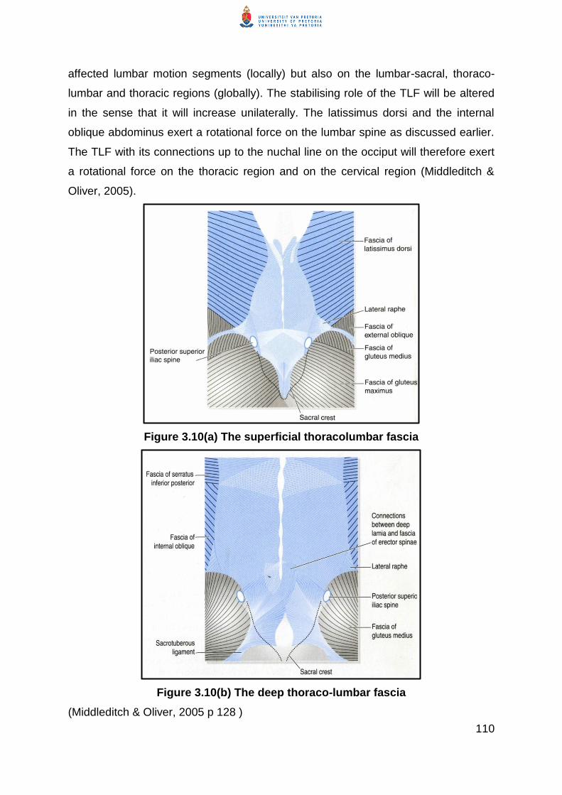
110
affected lumbar motion segments (locally) but also on the lumbar-sacral, thoraco-
lumbar and thoracic regions (globally). The stabilising role of the TLF will be altered
in the sense that it will increase unilaterally. The latissimus dorsi and the internal
oblique abdominus exert a rotational force on the lumbar spine as discussed earlier.
The TLF with its connections up to the nuchal line on the occiput will therefore exert
a rotational force on the thoracic region and on the cervical region (Middleditch &
Oliver, 2005).
Figure 3.10(a) The superficial thoracolumbar fascia
Figure 3.10(b) The deep thoraco-lumbar fascia
(Middleditch & Oliver, 2005 p 128 )
111
The envisaged effect of the rotational force initiated in the lumbar spine by multifidus
rotatores and intertransversarius has been enforced and magnified in the thoracic
spine and is continued by these deep z-shaped muscles of the core axial skeleton
throughout the cervical spine. This is because they have the same anatomical
configuration and innervation by the posterior primary rami throughout the spine.
At the cervico-thoracic junction the stiff thoracic spine articulates with the relatively
mobile lower cervical spine. At the transitional junction the intervertebral discs and
apophyseal joints are particularly subject to stress and strain. Owing to the
transitional strain, the C6-T1 segments are commonly subject to degeneration
(Middleditch & Oliver, 2005). The spinous processes of C7 and T1 may become
prominent and are often tender to palpation.
The uncovertebral joints of Luschka between C3 and C6 provide the cervical spine
with additional mobility because they are synovial joints. Because of the mobility of
the cervical spine, the effect of the rotational force by the TLF, which extends to the
nuchal line, is that the cervical spine is vulnerable to absorb the rotational force that
extends from the lumbar spine upwards. The apophyseal joints in the cervical spine
C3-C5 have a superior orientation (oval and facing posterior and superior (upwards))
while the inferior facets face forward (anterior) and inferior (downwards) (Middleditch
& Oliver, 2005). The orientation of these apophyseal joints allows flexion, extension,
rotation and sideflexion to occur. A clinical observation by the researcher is that
when the posterior cervical muscles stiffen, the cervical lordosis flattens. The whole
cervical spine is incriminated when the flattening of the cervical lordosis occurs. The
researcher hypothesises that this stiffening is not necessarily due to the spasm but
to shortening of the myofascial system to limit the head movements on the C1 as a
compensatory mechanism and also to limit consequential spinal movement. The
cervical column does not necessarily react with sideflexion or rotation like the lumbar
and thoracic regions but the asymmetrical muscle tension can be palpated and
directly related to the patient‘s headache or cervical symptoms in the presence of a
primary lumbar response to abnormal loading (Middleditch & Oliver, 2005).
112
The condyles of the occiput articulate with the kidney-shaped articular surface of the
C1 vertebra. The orientation of these facets and their high lateral margins ensure
that movement at the atlanto-occipital joint is mainly flexion and extension. The atlas
articulates with the axis below and provides a pivot around which the atlas and the
head rotate (Middleditch & Oliver, 2005).
The spinous process of the axis is large, usually bifid and normally provides a
prominent bony landmark for palpation. The suboccipital muscles attach onto the
spinous process of the axis, which provides powerful leverage for the muscles‘
actions. The asymmetrical pull of the extension of the TLF on the nuchal line can
explain why patients with CNSLBP also complain of headache.
The abnormal biomechanical loading in the lumbar spine also spreads inferiorly
towards the lower limb. The stability of the lumbo-pelvic region is dependent on the
interplay between the TLF, tensor fascia latae, and the abdominal fascial systems.
The TLF covers the muscles in the sacral region and then extends through the
thoracic region to the nuchal line. In its course on the posterior trunk, several
muscles attach to it, which results in adjusting the tension in the TLF and, as such,
contributes to transferring the load from the trunk to the pelvis and lower limbs
(Middleditch & Oliver, 2005). The sacral end of the torsioned pelvis initiates the pull
on the filum terminale as it winds up within the spinal canal towards the cranium.
3.5.1.4 Effect of ISMS dysfunction on gait
When the pelvis is in elevation and backwards rotation due to the unilateral muscle
spasm in the lumbar spine and the spasm in the quadratus lumborum and psoas
major, the muscle imbalance that results might cause somatic referred pain in the
gluteus medius and maximus. As a result of the distortion of the pelvis on the hip
joint, the patient bears weight on a slightly flexed, abducted and slightly laterally
rotated hip joint instead of on a hip joint that moves in the habitual movement, which
is in and out of the closed packed position. The patient will therefore present with an
asymmetrical gait pattern (a shorter step on the ipsilateral side of the muscle
spasm).

113
Weight bearing on a hip joint in slight flexion, abduction and lateral rotation places
abnormal loading on the hip joint during the weight-bearing phase of gait. The weight
bearing on the hip in slight flexion, abduction and lateral rotation reinforces the
asymmetry in the spine that originated from the unilateral muscle spasm in the z-
shaped segmental muscles (core axial muscles) around the spine. The asymmetry
was also caused by the rotational force created by the additional latissimus dorsi,
internal and external oblique, thoracis and longissimus lumborum and the TLF. The
asymmetrical gait pattern (decreased weight bearing on the affected side and limited
swing through) will therefore reinforce the rotational strain in the spine.
3.6 Soft tissue plasticity as an inherent process in the development of
ISMS dysfunction
Plasticity as part of the adaptation of soft and neural tissues (including brain) to
continuous stressors is argued to be one of the main reasons for the development of
CNSLBP. The fact that plasticity can be reversed (remodulated) (because it is an
inherent characteristic of soft and neural tissues (LAngevin & Sherman, 2006) is one
of the pillars on which the researcher‘s multifaceted manual therapy model for the
management of patients with CNSLBP is based.
Connective tissue changes its consistency depending on the condition that it is
under. When it is put under stress, it maintains its mobility. However, in the presence
of inflammation (e.g. due to microtrauma) and immobility it will shorten or stiffen due
to atrophy of connective tissue, architectural disorganisation, fibrosis, adhesions and
contractures (Langevin & Sherman, 2006) through a process called ‗plasticity‘.
It has been reported that shortened or stiffened connective tissues can be reversed
through a process of stretching these tissues; the gel-like ground substance can be
reversed by manual stretching or by stretching these tissues during exercise to
become viscous again (Shah, Phillips, Danoff & Gerber, 2005; Langevin & Sherman,
2006; Stecco et al., 2004). According to Schleip et al. (2006), proteoglycans (which
form the gel of the ground substance) play a major role in the absorption and
distribution of the compressive forces. When these tissues are stretched during the

114
myofascial release, the ground substance becomes viscous again. The phenomenon
of mobilising the connective tissues (fascia) through stretching is also known as
tissue creep.
Langevin and Sherman (2006 p 3) write:
A hallmark of connective tissue is its plasticity or „remodelling‟ in response
to varying levels of mechanical stress.
On the basis of this assertion by Langevin and Sherman (2006), plasticity can be
considered the process of shortening or stiffening of the connective tissue and
‗remodelling‘ indicates the reversing of the process of plasticity with the aim of
restoring tissue mobility.
Distinguishing between fibrosis and scar tissue is very difficult to determine clinically.
For this reason the researcher suggests that myofascial stretching on
shortened/stiffened connective tissue be undertaken on a symptomatic basis for the
patient with CNSLBP. Based only on the patient‘s tissue responses, a therapist can
clinically judge whether the ‗soft tissue restriction‘ offers a great deal of resistance.
This gives the therapist an idea of the type of tissue restriction, for example a
ligamentous, tendonous, facet joint capsule and potentially nerve root sleeve or a
combination of nociceptive tissues, involved in the patient‘s symptoms.
Post-treatment response will vary from treatment to treatment in one patient and
across patients, depending on the stage of healing and the skill of the applied
techniques. The researcher hypothesises that only in the case of scar tissue will the
patient respond with limited improvement in soft tissue mobility. This implies that soft
tissue restrictions, whether they are early adhesions or later fibrosis or even scar
tissue, may respond with a predictable outcome to manual therapy, which can be
identified based on the patient‘s post-treatment response. The process of mobilising
soft tissue restrictions (early adhesions, fibrosis or scar tissue) to restore ISMS
alignment and function occurs through a process of remodelling.

115
Contradictory reports exist in the literature on whether fascia can be over stretched
by manual therapy or not. Schleip et al. (2006) are of the opinion that it is not
possible to over-stretch fascia by means of therapeutic manual stretching (Schleip et
al., 2006). Langevin and Sherman (2006), however, caution that connective tissue
remodelling through stretching can potentially have a harmful as well as a beneficial
effect. Direct stretch to ligaments and joint capsules needs a careful approach to
avoid tissue inflammation. The challenge to the therapist is to reason clinically how
much force is enough to be beneficial and how much is harmful.
Release of muscle shortening is achieved by releasing muscle spasm, taut bands
and trigger points. Although the principles for the release of muscle spasm, taut
bands and trigger points differ from those for the release of connective tissues,
stretching plays an important role as well. The same principles of caution are
relevant to the release of muscle spasm, taut bands and trigger points as those
applicable to release of shortened/stiff connective tissues.
3.6.1 Plasticity and postural control
It is well known that motor skill can be relearned. In the presence of abnormal
muscle recruitment, guarded movements, and adaptation or compensation as a
result of malrecruitment and guarded movement, it is vitally important to plan the
relearning of normal muscle recruitment and movement patterns after soft tissue
release. This is so that mobility and alignment that were achieved through the
passive soft tissue release can be maintained.
As a result of the cortical changes that took place and resulted in or contributed to
the development of abnormal movement patterns (guarded movement and abnormal
muscle recruitment), normal movement has to be relearned through a goal-directed
process of motor learning (Shumway-Cook & Woollacott, 2007). The process of
motor relearning of movement patterns is closely associated with cognitive control
over habitual movement patterns to change/adapt them purposefully to prevent and
manage recurrences, avoid harmful behaviour or movement and introduce beneficial
movement patterns, health behaviour and lifestyle changes. The last mentioned can
116
only occur through appropriate health education and empowerment of the patient to
take control of their condition.
3.7 Factors that can influence/adapt the typical pattern of ISMS
dysfunction
A number of factors can influence the development of the typical pattern of ISMS
dysfunction, as described in the preceding paragraphs, resulting in differences in the
clinical presentation of the ISMS dysfunction. Dankaerts, O‘Sullivan, Burnet and
Straker (2006) found that patients with CNSLBP although there is no observable
difference in seating posture compared to normal healthy subjects, on sEMG is
became clear that there is not a homogenous trunk muscle activation pattern in the
patient group. These results may therefore indicate that there are different underlying
mechanisms for the abnormal trunk muscle activation. The researcher describes
various reasons/factors that may cause the differences, based on clinical experience
and the literature reviewed.
3.7.1 Differences in response of the lower and upper lumbar motion segments
The motion segments in the upper lumbar spine (L1-L4) respond differently on
rotation than the lower lumbar spine (L5-S1). This means that abnormal loading on
the L1-L4 motion segments, resulting in sideflexion due to unilateral muscle spasm,
will result in axial rotation of the motion segments to the contralateral side of
sideflexion while, in the L5-S1 motion segment, axial rotation towards the ipsilateral
side of lateral flexion will take place (Twomey & Bogduk, 1978). This difference in
response of the lower and upper lumbar motion segments to sideflexion caused by
the unilateral muscle spasm can result in differences in axial rotation that will torsion
(corkscrew) upwards in the spine or downwards to the L5-S1 motion segment and as
such have an effect on the rest of the spinal regions‘ response to lumbar dysfunction.
3.7.2 Poor posture and postural control
Poor posture can be the cause or a result of CNSLBP (Sahrmann, 2002). If
extension of the lumbar spine increases (as a result of muscle imbalance between
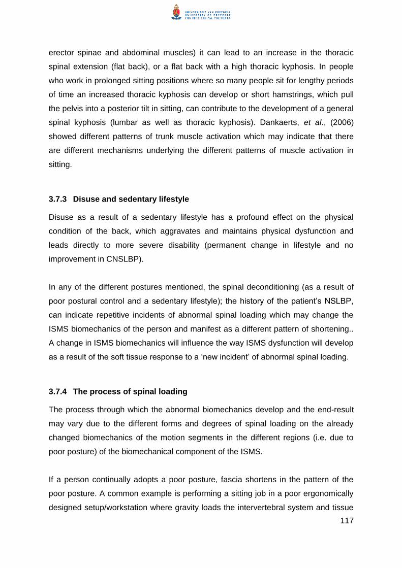
117
erector spinae and abdominal muscles) it can lead to an increase in the thoracic
spinal extension (flat back), or a flat back with a high thoracic kyphosis. In people
who work in prolonged sitting positions where so many people sit for lengthy periods
of time an increased thoracic kyphosis can develop or short hamstrings, which pull
the pelvis into a posterior tilt in sitting, can contribute to the development of a general
spinal kyphosis (lumbar as well as thoracic kyphosis). Dankaerts, et al., (2006)
showed different patterns of trunk muscle activation which may indicate that there
are different mechanisms underlying the different patterns of muscle activation in
sitting.
3.7.3 Disuse and sedentary lifestyle
Disuse as a result of a sedentary lifestyle has a profound effect on the physical
condition of the back, which aggravates and maintains physical dysfunction and
leads directly to more severe disability (permanent change in lifestyle and no
improvement in CNSLBP).
In any of the different postures mentioned, the spinal deconditioning (as a result of
poor postural control and a sedentary lifestyle); the history of the patient‘s NSLBP,
can indicate repetitive incidents of abnormal spinal loading which may change the
ISMS biomechanics of the person and manifest as a different pattern of shortening..
A change in ISMS biomechanics will influence the way ISMS dysfunction will develop
as a result of the soft tissue response to a ‗new incident‘ of abnormal spinal loading.
3.7.4 The process of spinal loading
The process through which the abnormal biomechanics develop and the end-result
may vary due to the different forms and degrees of spinal loading on the already
changed biomechanics of the motion segments in the different regions (i.e. due to
poor posture) of the biomechanical component of the ISMS.
If a person continually adopts a poor posture, fascia shortens in the pattern of the
poor posture. A common example is performing a sitting job in a poor ergonomically
designed setup/workstation where gravity loads the intervertebral system and tissue

118
creep occurs so that the upright posture is difficult to maintain. Owing to the tissue
creep, shortening of the fascia will occur at the anterior thoracic spine (anterior
shoulder girdle), with resultant aggravated thoracic kyphosis and decreased lumbar
lordosis. Continuous strain on fascia in the ISMS due to poor posture will result in
fascia shortening because fascia shortening occurs earlier than muscle fibres
through the process of ‗negative‘ plasticity (Langevin & Sherman, 2006), which
contributes to ISMS dysfunction.
Panjabi (2003) states that people with suboptimal neuromuscular control for some or
other reason and who perform dynamic activities are more likely to develop
CNSLBP. The implication of this hypothesis is that the pattern of change in the trunk
muscle activation might depend on the state of the person‘s neuromuscular control
before abnormal spinal loading took place.
Strain will occur at the transitional intervertebral junctions C7/T1, T12/L1 and L5/S1,
which may also result in malalignment of the biomechanical aspect of the ISMS. In
this case the shoulder and pelvic girdles are also involved due to the effect of the
abnormal biomechanics of the biomechanical ISMS on these girdles and vice versa.
3.7.5 Cervical and thoracic dysfunction
Anecdotally, clinicians observed that patients with neck pain commonly develop LBP.
Researchers (Hodges & Richardson, 1996) found that there is an association
between neck pain and LBP. Patients who suffer from LBP also suffer from neck
pain. Both conditions are associated with dysfunction of the trunk muscles,
especially loss of control of the transvers abdominus muscle, which has led to the
conclusion that in both cases abdominal muscle dysfunction (which is inherent part of
poor posture) may compromise ISMS control.
In a specific study conducted by Hodges et al. (1996) their first main finding was that
people with sub-acute neck pain have a reduced capacity to perform the abdominal
drawing-in task (ADIT). The second main finding was that reduced performance in
the ADIT was associated with increased risk of LBP over the following two years, for
both patients and control subjects.
119
The conclusion was made that suggested that maintenance of normal trunk muscle
control should be a goal of therapy in patients with sub-acute neck pain as well as in
patients with CNSLBP.
3.7.6 Association of chronic unilateral low back pain with disruption of tactile
input
Moseley, Gallagher and Gallace (2012) found that chronic unilateral LBP is
associated with spatially defined disruption of tactile input. The shortening of fascia,
which is known to be a source of nociception, might limit proprioceptive feedback to
conscious awareness of the strain on the TLF and other soft tissues and articular
structures (ligaments and joint capsules), which can lead to a decrease in tactile
discrimination and spatial awareness. It may be possible that a patient with CNSLBP
experiences spatial disruption of tactile input that their muscle recruitment may adapt
due to the changed (limited or abnormal) sensory input or interpretation of sensory
input in the brain. Abnormal muscle recruitment will probably lead to adapted
movement.
3.7.7 Neural referred pain through torsioning of the biomechanical ISMS
The researcher hypothesises that in the torsioning of the biomechanical ISMS the
disc can be forced against the soft tissue structures to give rise to tension on the
nerve root sleeve and can result in neural-referred pain (nerve root sleeve pain)
(discussed in Section 3.6) and spasm in the muscle innervated by the affected nerve
roots. The relevant muscles in patients with CNSLBP are, for example, the gluteus
maximus, medius and minimus; piriformis; quadratus lumborum; iliacus; and psoas
major muscles. These muscles may in themselves refer pain to the lumbar area and
pelvis and lower limb as well as to the shoulder girdles and upper limb(s). The pain
referred from these muscles is somatic-referred pain (discussed in Section 3.5.1.1).

120
3.7.8 The effect of stress on spinal dysfunction
The researcher observation of patients in clinical practice has revealed that patients
may present with soft tissue shortening due to biomechanical dysfunction as well as
the generalised soft tissue shortening seen typically in patients with emotional stress.
How the two combinations of the pattern of soft tissue shortening relates to the fact
that Brooks & Tracey (2005) found that anxiety induced pain results in a different
increase in brain activity is still not known (Section 3.8.4.1).
3.7.9 The influence of underlying degeneration in the synovial joints of the
spine
Adams et al. (2002) argue that a link exists between back pain, mechanical loading,
ageing, dysfunction and degeneration. The links between all these factors are
complex and justify further research based on clinical data. The researcher
hypothesises that the presence of degenerative changes in the spine before an
incident of ANSLBP can render the spine vulnerable for episodes of abnormal spinal
loading. Abnormal spinal loading on degenerative zygopophyseal joints can result in
an aggravated pain response, which can together with psychological responses such
as catastrophisation, fear of pain and guarded movement contribute to the
development of ISMS dysfunction.
From the moment the abnormal loading occurs and affects a degenerative joint(s)
the healing process sets in. Any of the tissue systems (articular, myofascial,
connective tissue and neural tissues) in the low back and therefore in the spine can
be affected by the wear and tear, strain or microtrauma as a result of the abnormal
spinal loading. Each of these tissue types has the „potential to be repaired by
mechanisms unique to its cellular composition and individual biochemistry‟
(Porterfield & DeRosa, 1991 p 4).
The healing process can be one of two types: regeneration of the injured tissue or
replacement of the injured tissue by dissimilar tissue, i.e. connective scar tissue. The
latter occurs when the injury exceeds the tissue‘s capacity to regenerate.

121
Factors that influence the type of tissue repair that will take place are the availability
of vascular supply and the extent of the injury.
3.7.10 Previous history of back pain and response to health care management
One of the best predictors for the development of CNSLBP is the patient‘s previous
history of back pain and his response to the management by health care
professionals. Waddell (2004) and Field (2009) are of the opinion that health care
professionals‘ management of patients with acute NSLBP in general can also
influence the development of characteristic behaviour in patients with acute/sub-
acute NSLBP by the way they manage patients. Doctors and therapists may be
treating patients with sub-acute or CNSLBP as if they still had acute pain and, as
such, contribute to the establishment of chronic pain and illness behaviour in patients
(Waddell, 2004).
3.8 Pain processing as integral component driving the development of
ISMS dysfunction
The International Association for the Study of Pain (Merksey & Bogduk, 1994)
defines pain as ‗an unpleasant sensory and emotional experience associated with
actual or potential tissue damage, or described in terms of such damage‘. Pain is a
multidimensional ‗sensation‘ with sensory-discriminative, motivational-affective and
cognitive-evaluative dimensions (Melzack- & Casey, 1968).
The ‗sensory-discriminative dimension‘ refers to the location, intensity,
duration and quality of pain.
The ‗motivational-affective dimension‘ refers to the unpleasant experience of
pain such as a feeling of nausea or a sickening feeling.
The ‗cognitive-evaluative dimension‘ refers to a patient‘s beliefs, which may
arise from their previous pain perceptions and, as such, have a negative or
positive effect on the present pain experience.

122
3.8.1 The biomechanical origin of pain processing in the development of ISMS
dysfunction
There is a wealth of experimental data that confirms that ligaments, muscles, neural
tissue, joints, annulus of the discs, as well as connective tissue in general, including
the specialised TLF of the lumbar spine, are all potential sources of nociceptive
stimuli (Langevin & Sherman, 2006). The researcher distinguishes between primary
and secondary ANSLBP. Primary ANSLBP can be caused by the tissues directly
within or surrounding the lumbar (L5-S1 or other) motion segments. In contrast,
secondary ANSLBP might be the result of (often longstanding) forces working within
and surrounding the lumbar motion segments but which are created by
biomechanical malalignment of the ISMS elsewhere such as the thoracic or cervical
spinal regions often associated with poor posture (Porterfield & DeRosa, 1991).
The researcher hypothesises that the unrelenting pain experienced by patients with
CNSLBP can be due to, amongst other causes, low grade persistent mechanical
strain on nerve, nerve roots and ANS chain. As the biomechanical rotational force on
the ISMS dysfunction involves more and more tissue structures or increases, the
ongoing irritation of nociceptors causes enduring sensitisation of the peripheral nerve
as well as in the dorsal horn (central sensitisation) and altered pathways in the brain.
The researcher further reasons that although there is no structural damage or tissue
pathology identified in patients with CNSLBP, the unrelenting pain that they
experience could possibly be due to constant irritation in the tissues and the neural
system as a result of the rotational force.
When stimuli from the various origins (joints, synovia, muscles, ligaments, loose and
specialised connective tissue such as the TLF) become more longstanding,
peripheral and central sensitisation at the level of the dorsal horn occurs due to the
biomechanical rotational strain of the ISMS. These stimuli are received and also
interpreted in the sensory-discriminative area (thalamus) as well as by the limbic or
emotional aversive area in the brain (Figure 3.11) (Kuner, 2010). Pain perception in
this instance is the result of physiological conduction of impulses along a structural
network (Figure 3.11).
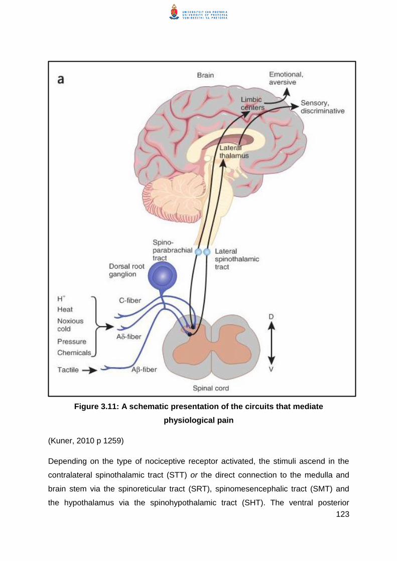
123
Figure 3.11: A schematic presentation of the circuits that mediate
physiological pain
(Kuner, 2010 p 1259)
Depending on the type of nociceptive receptor activated, the stimuli ascend in the
contralateral spinothalamic tract (STT) or the direct connection to the medulla and
brain stem via the spinoreticular tract (SRT), spinomesencephalic tract (SMT) and
the hypothalamus via the spinohypothalamic tract (SHT). The ventral posterior

124
nucleus of the thalamus receives input from the low threshold and wide dynamic
range (WDR) nociceptors via the laminae IV-V, which in turn projects input to the
somatosensory cortex (S1) (Figure 3.12).
Spinal projections to the ventrolateral medulla, parabrachial nucleus, periaqueductal
grey (PAG) and brain-stem reticular formation are also found. Cortical and
subcortical areas found to be commonly activated by the nociceptive stimulation are
the anterior cingulate cortex (ACC), insula, frontal cortices, the primary
somatosensory cortex (S1), secondary somatosensory cortex (S2) and the
amygdala. These areas are also referred to as the ‗pain matrix‘ (Brooks & Tracey,
2005). The pain matrix is subdivided into two parts: a medial and a lateral pain
system. This distinction between the two parts of the thalamus is a gross distinction
based on the projection sites from the thalamus to the cortex and on grouping the
brain regions with similar roles in pain perception.
It is thought that the S1 and S2 (lateral pain system) play a role in discriminating the
location and intensity of painful stimuli while the ACC is involved in the affective
(cognitive-evaluative) component of pain (Brooks & Tracey, 2005).

125
Figure 3.12:Thalamus (Th), the amygdala (Amyg), the insula cortex (Insula), the
supplementary motor area (SMA), the posterior parietal cortex (PPC), the prefrontal
cortex (PFC), the cingulate cortex (ACC), the periaqueductal grey (PAG), the basal
ganglia and cerebellar cortex (not shown) and the primary (S1) and secondary (S2, not
shown) sensory cortex
The insula also plays a role in affective pain processing and encodes the intensity
and laterality of painful stimuli and thermal non-painful stimuli. The insula therefore
integrates stimuli from both the medial and lateral pain systems in the thalamus
(Brooks & Tracey, 2005).
Activation of the operculum, which includes the S1 and the insula, is strongly
implicated in studies on pain and these are the only cortical areas that, when they
are stimulated by direct electrical stimulation, result in pain perception. If the stimulus
is short lived, the neuronal response of pain perception will be short lived (Brooks &
Tracey, 2005).
The neural adaptive changes in the amygdala (area involved in the emotional state
of pain perception) can also exert a powerful inhibitory influence on the prefrontal
cortex, which results in interference in a person‘s cognitive and decision-making
ability. If the inhibitory effect of the amygdala on the prefrontal cortex has been
established, it will negatively influence the understanding and management of pain
and quality of life of the patient with chronic pain. The amygdala is seen as part of
126
the limbic system, in which the emotions of a person are controlled. Bombardment of
the limbic system results in hypervigilance of the limbic system, which is
characteristic of patients with chronic pain.
In the presence of hypervigilance, CNS sensitisation because of anxiety and/or
emotional stress, beliefs and altered pathways in the brain enhances peripheral and
central sensitisation (Field, 2009).
Pain stimuli are caused by chemical irritation due to inflammatory reactions following
tissue damage. While it is very difficult to validate experimentally, the mechanism
seems to involve the direct stimulation of nerve endings by chemicals, such as
hydrogen and potassium ions, or proteolytic enzymes that are liberated from
inflammatory cells or damaged tissue cells.
Immune cells and microglia interact with neurons to alter pain sensitivity and to
mediate the transition from acute to chronic pain (Ren & Dubner, 2010). When an
injury (strain or sprain/microtrauma) occurs, local immune cells are activated and
blood borne immune cells, which sensitise the peripheral nociceptors, are recruited
to the area.
Through the synthesis and release of inflammatory mediators and
interactions with neurotransmitters and their receptors, the immune cells,
glia and neurons form an integrated network that coordinates immune
responses and modulates the excitability of pain pathways. The immune
system also reduces sensitization by producing immune-derived analgesic
and anti-inflammatory or pro-resolution agents. (Ren & Dubner, 2010 p
1267)
Langevin and Sherman (2006) found that activation of nociceptors by itself can
contribute to the development or worsening of an inflammatory response in
connective tissues. In the presence of inflammation hyper-excitability of the peripheral
nociceptors occurs, which may result in the patient presenting with primary hyperalgesia
(Brooks & Tracey, 2005).

127
When the central sensitisation occurs, painful stimuli are amplified (due to factors
that are not clear yet) and cause peripheral nerves (not normally associated with
evoking painful stimuli) to evoke pain. This centrally evoked pain response is called
secondary hyperalgesia and is a phenomenon where mechanical non-painful stimuli
on the normal skin around the site of primary injury cause a painful stimulus (Brooks
& Tracey, 2005). According to Kuner (2010), hyperalgesia is associated with
expansion of the peripheral receptive field of neurons, which may cause hyperalgesia to
spread to originally unaffected regions.
In a process similar to secondary hyperalgesia, damage to the peripheral nerve
induces plastic changes in the CNS, which are maintained by continuous discharge
from the damaged afferent, and recruitment of low-threshold mechanoreceptors such
as the Aβ fibres, resulting in allodynia (Figure 3.13). Allodynia is associated with
withdrawal behaviour in the presence of innocuous stimuli (Brooks & Tracey, 2005).
As the ISMS dysfunction progresses over time so that chronicity sets in, all soft
tissues (under strain including the myofascia, TLF and the peripheral dorsal
segmental nerve roots and the ANS ganglia; the joint capsules and ligaments of the
synovial joints and the annulus of the discs) are compromised in the rotational strain
occurring in the ISMS. The repetitive nociceptor drive in the pain cycle causes the
loose unspecified and specialised connective tissue structures to stiffen and muscles
to shorten as a result of muscle spasticity, taut bands and trigger points. As fascia
within and around the nerve roots and sympathetic ganglia of the ANS chain might
also stiffen and result in autonomic symptoms, the patient might complain of local
lumbar as well as widespread pain and a sickening feeling.
3.8.2 The neuromatrix as part of the ISMS dysfunction
In an ongoing stimulation of the nociceptors (such as in patients with ISMS
dysfunction) of the different types of tissue within and surrounding the motion
segment(s), neural pathways in the brain change as a result of the process of neural
plasticity. When somato-sensory representations in the somato-sensory cortex are
stimulated continuously, over time this generates altered processing, which could
128
lead to the spread of pain and cause various abnormal motor perturbations (Butler,
2000). Abnormal motor perturbations occur probably due to the excessive
somatosensory representation of muscles such as erector spinae and multifidus to
involve the motor representation of these muscles in the motor cortex through a
process termed ‗smudging‘. Smudging is associated with compromised activity of
these muscles.
Compromised movement (deep segmental as well as multisegmental muscular
system dysfunction) will lead to compromised muscle recruitment and fatigue and
generalised adaptation of spinal malalignment. On the basis of clinical reasoning the
researcher hypothesises that paraspinal muscles have discrete representation at the
motor cortex, which may be compromised in patients with LBP (Hodges, 2013).
Smudging may be even more likely in the case of patients with CNSLBP.
When the neural pathways in the brain (neuromatrix) change, patients complain of
spontaneous ongoing pain, which is initiated by these altered pathways in the brain.
These altered pathways can play a key role in the development of chronic pain and
in the clinical manifestation of chronic pain (Kuner, 2010; Flor et al., 1997). So, the
process of plasticity through which the altered pathways in the brain and nervous
system has developed is a powerful mechanism for manual therapists to use in
dealing with CNSLBP (Butler, 2000).
The macroscopic anatomical changes in the brain (ACC, orbitofrontal cortex, insular
cortex, dorsal pons) associated with chronic pain entail a decrease in the grey matter
of the brain. What is not clear in the literature yet is whether the changes in the
macroscopic grey matter are due to the chronic pain state of the patients or whether
the patients‘ chronic pain state causes the macroscopic anatomical changes in the
brain (Kuner, 2010). Of clinical importance in patients with CNSLBP is the fact that
decreased brain volume (grey matter) is also associated with depression, which may
be an explanation for the frequent reports that patients with CNSLBP suffer from
depression.
129
Field (2009) states that:
Hyper-vigilance in the limbic system has been described as a partial
explanation of chronic pain … patients with chronic pain have a
heightened sensitivity to pain (lower threshold and tolerance) because of
increased attention to external stimulation and a preoccupation with pain
sensations; states that are mediated through limbic activity. Limbic
dysfunction also manifests as an abnormal efferent innervation of
musculature, both visceral and somatic. The musculature undergoes tonic
contraction as a result of limbic efferent stimulation which may generate a
further sensation of pain. (Field, 2009 p 48)
3.8.2.1 Plasticity in the neural system
The development of chronic pain occurs through a process of plasticity that takes
place in the neural and the soft tissues. Plasticity in the neural system occurs in
different forms at different levels: the molecular, synaptic, cellular and network levels.
These changes are graphically illustrated in Figure 3.13.
When the physiological pain perception persists or endures, changes take place at
various levels of the nervous system; i.e. peripheral sensitisation, central
sensitisation and altered pathways in the brain.
A change in the strength of synaptic input is mediated by probable changes in
neurotransmitter release. Long-term potentiation of nociceptive transmission at all
levels of the nervous system (peripheral and central sensitisation as well as the
altered pathways in the ACC) is found in patients with CNSLBP, resulting in the
affective (cognitive evaluative) component of the pain experience associated with the
patient‘s beliefs, which may arise from their previous pain perceptions. The long-term
potentiation at a molecular level is found to be similar to the processes that are
probably involved in the formation of memories. Formation of memory (for instance
the memory of previous pain, especially if it is associated with unpleasant emotional
experiences such as catastrophisation, as well as the fear of pain) is associated with
sensitised neurons in the amygdala and the ACC (area in the brain involved in the
affective pain control) in patients with CNSLBP (Kuner, 2010).

130
At the molecular level the molecules may change in an ‗activity dependent‘ manner
(i.e. phosphorylation) and in this way alter molecular function (i.e. by a decrease in
the activation threshold of an ion channel) or localisation (endocytosis or trafficking)
(Kuner, 2010). These changes are illustrated in Figure 3.13.

131
Figure 3.13: Disease-induced functional and structural plasticity in neural
substrates of pain on molecular, synaptic, cellular and network levels
(Kuner, 2010 p 1260)

132
Through a complex process, functional plasticity in the nociceptive pathways gives
rise to a process of structural changes, which is detected by a ‗change in the
increase or decrease in the density of the synaptic spines, degeneration or
regeneration of axons leading to aberrant connectivity, degeneration of neurons, and
proliferation of asterocytes and microglia which influence nociceptive processing by
releasing modulatory substances‘ (Kuner, 2010 p1259).
In the end the physiological changes in pain processing lead to structural changes
that can become the origin of persistent pain as a result of neural plasticity.
Plasticity at the level of the neurons in the nociceptive pathways is seen
as an increase in the magnitude of responses to a defined sensory
stimulus, an increase in the level of spontaneous activity, or after
discharges, which represent continued activity after the termination of a
nociceptive stimulus, leading to central amplification of pain (central
sensitization). Furthermore, the peripheral receptive field of neurons can
expand, allowing hyperalgesia to spread to uninjured regions (Kuner, 2010
p 1259) [bold added by researcher]
The researcher hypothesises that synaptic plasticity and functional plasticity are very
important processes in the conceptualisation of the multidimensional management of
patients with CNSLBP because they imply that the strengthening of the synaptic
connections can be remodulated by decreasing nociceptive stimulation and
bombardment of the dorsal horn in a process of desensitisation.
3.8.3 Characteristic adaptive behaviour in patients with ISMS dysfunction
Waddell (2004) links the development of CNSLBP to a timeline in which people who
suffer from NSLBP after 12 weeks become chronic. Field (2009), however, states
that it is not only a time factor (Waddell, 2004) that determines the development of
CNSLBP, but that it depends on the variation of an individual’s response to an
episode of acute NSLBP: … ‗some people are set up for chronicity before the pain
starts; others develop it soon after the ANSLBP; a third and possibly the largest
group drift into it after suffering for a month or so‘ (Field, 2009 p 47).

133
From the earlier discussion in this section it is clear that pain is a multidimensional
sensation, which consists of sensory-discriminative, motivational-affective and
cognitive-evaluative dimensions (Melzack- and Casey, 1968). The intensity duration
and quality of nociceptor stimuli are therefore not only localised in the brain but also
associated with a sickening feeling or a feeling of nausea due to the motivational-
affective dimension and are cognitively interpreted based on previous pain
experiences.
In Section 3.5 discusses the development of ISMS dysfunction from a biomechanical
perspective and indicates a possible mechanism through which the torsional strain,
fixation of the trunk (guarded movement) and adaptive and/or compensatory
movement patterns may develop. These patterns are caused by the neuromuscular
responses to pain and the stiffening of the fascia as well as by the fear of pain and
injury (fear avoidance behaviour) associated with the motivational affective
dimension of pain or are driven by cognitive control of the patient (O‘Sullivan 2005).
Guarded movements are identified by the fact that the patient holds their
body/posture rigid to prevent movement as far as possible to the extent that even
their breathing pattern is restricted and shallow (Langevin & Sherman, 2006). The
presence of guarded movements is closely associated with psychological processes.
Guarded movements may start as a reflex physiologic response to injury or as a
primary dysfunction, but may persist due to psycho-physiologic rather than
physiologic processes alone. Guarded movements become a learned, protective
habit and then persist as physiologic dysfunction associated with abnormal patterns
of muscle activity, movement and neurophysiologic activity (Figure 3.14).
Catastrophising is defined by Field (2009 p 47) as the: „development of
inappropriately negative beliefs and understanding relating to pain and its possible
consequences‟. Catastrophisation ‗…. is felt to be a precursor to pain related fear;
with fear going on to cause avoidance behaviour, reducing activity and resultant
disability‘.

134
The development of guarded movement and fear avoidance behaviour can set off
the development of unwell/illness behaviour (Field, 2009; Waddell, 2004).
Figure 3.14: The fear avoidance model
(Waddell, 2004)
Hadjistavropoulos and Craig (1994) state that 10% to 15% of patients develop
emotional and behavioural problems out of proportion to their physical problem at an
early stage after the acute phase of NSLBP followed by CNSLBP.
Emotional and behavioural problems culminate in clinical psycho-social
characteristics, such as older patients (50% to 55%) who have a previous history of
back pain and show signs of catastrophisation and fear avoidance behaviour. They
have a poor relation between pain intensity and functional ability (i.e. low pain level
with severe disability); poor health perception; psychological distress and

135
depression; signs of decreased mental health, musculoskeletal conditions; and other
comorbidities.
Emotional distress further heightens (lowers the threshold for pain impulses) the
patient‘s pain perception because it increases the patient‘s awareness of bodily
sensations and lowers pain tolerance.
Socio-demographic characteristics include: time since last worked; occupational
status (no longer employed); employment rate; educational level; expectations about
return to work; job (dis)satisfaction; and duration of sickness absence. Underlying
these psycho-social and socio-demographic characteristics are the patient‘s beliefs
about hurting and disease and the fear of experiencing them, personal responsibility
and taking control of circumstances in their life and self-sufficiency. The patient‘s
beliefs and expectations regarding the treatment of their condition have a major
influence on the successful outcome of the treatment. The patient‘s experience of
their own condition is influenced by stories told about the condition by their peer
group or people who have a similar condition.
Psychological factors are seven times more predictive that symptoms will last for a
long time than physical tests or other factors identified from the patient‘s case
history. Catastrophisation also influences the patient‘s coping mechanisms and
beliefs on issues that affect health care.
Illness behaviour is an indication of the severity of the patient‘s problem but it may
also reflect the psychological factors mentioned above to such an extent that it is
more important than the physical signs and symptoms. Physical function,
performance and illness behaviour are inseparable.
Social behaviour and social factors that influence the development of CNSLBP are
closely interrelated. The patient‘s sick role and illness behaviour are typical social
phenomena. The patient‘s social networks influence their beliefs as well as their
coping strategies and illness behaviour. The nature, strength and availability of a

136
patient‘s social networks either reinforce or discourage illness behaviour (Field,
2009). Pain and pain behaviour are a strong way of communicating with other people
and with health care professionals (Waddell, 2004).
Social factors that play a role in the chronicity of a patient‘s clinical picture are listed
by O‘Sullivan (2005) as the patient‘s relationships in all spheres of life (family, friends
or at work), work structure, support structures, cultural factors, medical advice,
compensation and socio-economic factors.
One of the strongest influences on return to work and work status at six to 12 months
post the episode of acute LBP is the patient‘s own perceptions of their pain. Return
to work is determined by the patient‘s beliefs about what has happened to their
backs, by whether they think that their back pain was originally a work-related injury,
and about their fear of re-injury if they return to work (Waddell, 2004).
The development of a ‗dysfunctional ISMS‘ based on the process of plasticity in the
soft and neural tissues as discussed in the preceding sections implies that ISMS
dysfunction is potentially reversible because of the inherent plasticity in the systems
of the ISMS. Apart from this physical or physiologic loop, feedback and
reinforcement of behaviour play a role in the condition. What we do, our activity level
and illness behaviour all reinforce our beliefs about the pain and the coping
strategies we use to deal with it. Illness behaviour, disability and sickness absence
reinforce distress and depression, which increase illness behaviour and reduced
activity, and aggravate and perpetuate physiologic dysfunction and deconditioning.
Field (2009) describes a conceptual framework of the interrelated factors involved in
maintaining LBP. The conceptual framework displayed below is adapted from Field‘s
original framework to include the role of social interaction as an important factor in
CNSLBP.
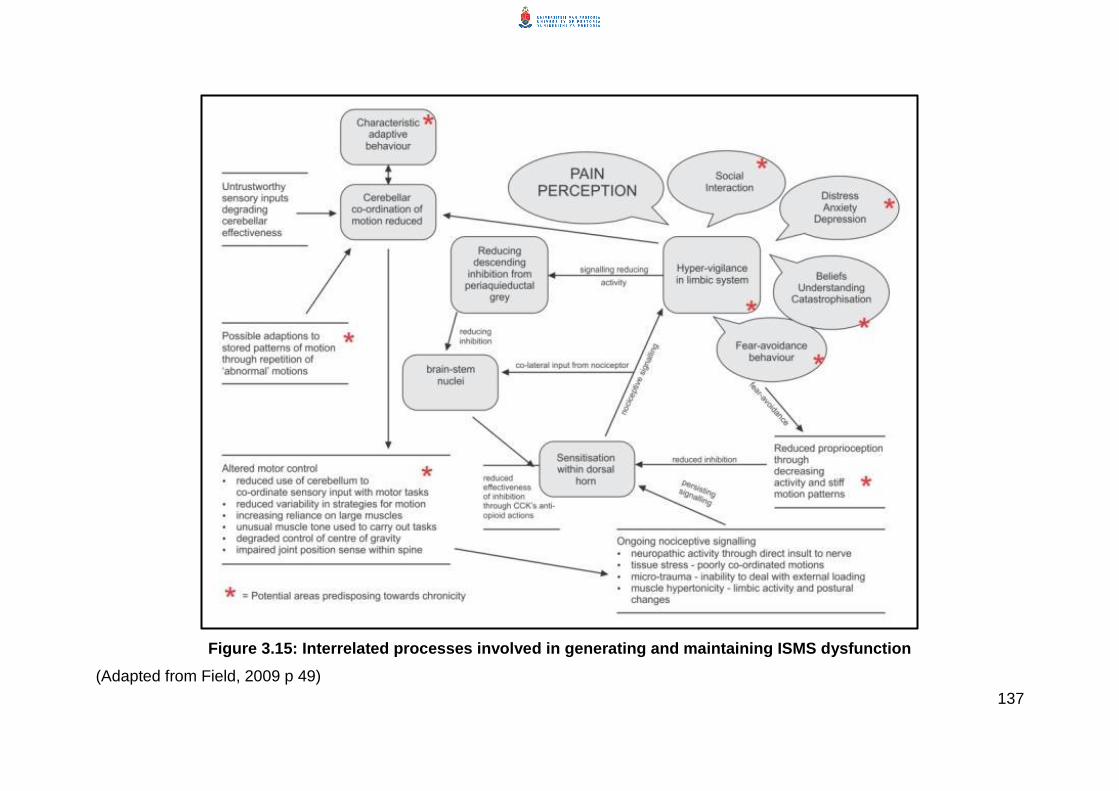
137
Figure 3.15: Interrelated processes involved in generating and maintaining ISMS dysfunction
(Adapted from Field, 2009 p 49)

138
3.8.4 Pain modulation
Pain modulation can entail either pain-enhancing mechanisms or descending pain
inhibition (modulation).
3.8.4.1 Pain-enhancing mechanisms
The perception of pain intensity in relation to the peripheral stimulus that causes the
pain depends on many factors such as the level of arousal, anxiety (in which fear
avoidance behaviour plays a major role), depression, attention and expectation or
anticipation and guarded movement. ‗These “pyschological” factors are in turn
regulated by overt and covert information, as well as more general contextual cues
that establish the significance of the stimulus and help determine an appropriate
response to it‟ (Brooks & Tracey, 2005 p 24). Studies found that anxiety-induced
(anticipation of) pain resulted in a different increase in brain activity than the brain
activity generated by a large nociceptive drive. Similar results were shown in studies
during which it was found that attention to and anticipation of an upcoming painful
stimulus activated the anterior insula (AI) (Brooks & Tracey, 2005).
An inflammatory process plays a major role in pain enhancing as is described in
Section 3.8.1.
A person‘s genetic make-up, previous experience and learning determine how pain
stimuli are filtered and modulated through an individual‘s whole being, current
physiological status, emotional state and socio-cultural environment (Turk, 2001).
„Sensitization may be both neurophysiologic and psychological‟ (Eriksen & Ursin,
2002. There is growing evidence that inherited (genetic) factors play a role in the
increased incidence of individuals that develop severe or enduring CNSLBP (Mishra
et al., 2007). Current research implies that there is a genetic link that predisposes
vulnerable individuals towards the production or not of an endogenous muscle
relaxant, and a predisposition towards the development of major depression when
back pain occurs. There is also convincing work on the role of genetic influences
affecting disc degeneration (Battie, Videman, Levalahti, Gill & Kaprio, 2007).
139
Another contributing factor to the persistence of pain and the widespread nature of
CNSLBP is a change in the descending pain modulation mechanism. The
parabrachial nucleus, PAG, the brain stem and reticular formation, which are known
as the descending pain control pathways, can either inhibit or facilitate (modulate)
nociceptive transmission and subsequent pain perception (Brooks & Tracey, 2005).
In an enduring pain state, as indicated above, the gene expression of the CCK and
its receptor protein within the dorsal horn increases. The increased CCK inhibits the
effectiveness of opiates released by the descending pathways and, as such,
decreases the effectiveness of second order pain inhibition, resulting in the
persistence of pain perception (Field, 2009).
The functional changes at the molecular, synaptic, cellular and network levels due to
peripheral and central sensitisation as well as hyperactivity in the neural pathways in
the cortex midbrain and brain stem result in altered pathways in the higher centres.
Over time, as chronicity sets in, these altered pathways become structural changes
at these levels.
In a chronic pain state brain areas involved in emotion such as the ACC, insula, and
the amygdala also become hyperactive. When this hyperactivity in these areas
occurs, peripheral noxious stimuli will not only activate the sensory areas in the brain
but also activate in the brain all the areas involved in emotion (ACC, insula, and the
amygdala). The pain responses in the ACC are powerfully modulated by a person‘s
mood, placebo and hypnotic suggestion, which indicates that the ACC plays a role in
the integration of sensory input with a person‘s emotional state (Brooks & Tracey,
2005; Field, 2009). The memories of previous pain episodes if reinforced through
beliefs and thinking can easily evoke pain (Bouton, 2002). Memories of previous
episodes of back pain can lead to chronicity when previous pain memories are
reinforced.
Catastrophising, which is the person‘s expectation, or fear, that pain will be severe
and unmanageable, is the likely reason for the development of the affective
disturbances associated with chronic pain (Waddell, 2004). Brooks and Tracey
(2005) report that the anticipation of pain activates the rostral anterior insula and

140
medial prefrontal cortices, in contrast to the fact that during nociceptive pain
stimulation activation of the insula is more caudal and the expected activity in the
prefrontal focus is replaced by activity within the ACC.
Cognitive factors such as fear of pain seem to play a greater role in the development
of chronic pain than factors such as pain intensity. It has also been established that
psycho-social factors play an important role in chronic pain disorders such as
CNSLBP. On the other hand, personality disorders may play a role in a person‘s
ability to develop coping skills to deal with pain and, as such, a lack of developing
coping skills can be an antecedent to chronic pain.
Kuner (2010) also explains that cortical thinning due to loss of cortical grey matter
occurs in patients with chronic pain, which results in a person experiencing problems
with attention and concentration. Cortical thinning is associated with depression in
patients with CLBP. Whether it can be viewed as a pain-enhancing mechanism is not
clear but one can argue that because depression is strongly associated with the
hyper-vigilant limbic system it will enhance pain perception. Wand, Parkitny,
O‘Connell, Luomajoki, McAuley, Thacker and Moseley (2011) state that (chronic)
pain is maintained by changes within the brain of a person with chronic
musculoskeletal pain. This finding is in line with the fact that Ploghouse, Narain,
Beckmann et al. (2001) found that pain induced by mood, emotional stress and/or
depression activates different areas in the brain than pain perception from a
nociceptive drive.
3.8.4.2 Pain-inhibitory modulation
The ascending STTs send impulses to the PAG via the SMT. When the PAG matter
in the midbrain is activated, enkephalin-releasing neurons that project to the raphe
nuclei in the brain stem are activated. The nuclei raphae release serotonin to the
dorsal horn, where it has an excitatory connection with the inhibitory interneurons in
the substantia gelatinosa. When the inhibitory interneurons are activated they
release enkephalin or dynorphin (endogenous opioid neurotransmitters), which bind
to the mu-opioid receptors on the axons on the A-delta and C fibres that carry
incoming nociceptive impulses. The activated mu-opioid receptors inhibit the release

141
of substance P, which in turn inhibits the activation of the neuron from where the
pain impulses are transmitted via the STT to the ventroposteriolateral nucleus of the
thalamus. The nociceptive impulse is therefore inhibited (because the pain threshold
for mechanical pain is increased) before it can reach the cortical areas responsible
for the interpretation of pain.
The researcher associates this change in the role of the ACC, insula and amygdala
with the development of characteristic adaptive behaviour displayed in patients with
CNSLBP.
3.9 Conclusion
In this chapter the characteristics of the normal ISMS were deduced based on an
overview of the functional biomechanics of the spine and head, shoulder and pelvic
girdles as discussed in Section 3.6.
The rationale behind the conceptualisation of the characteristics of the normal ISMS
is to give an overview of the functional biomechanics of the spine, head and pelvis
as a close kinematic chain with the shoulder girdle that can function as a closed or
an open kinematic chain (ISMS) and to deduce the principles that should guide the
management of patients with CNSLBP. The discussion of the normal ISMS is
followed by a presentation of the pathophysiological processes closely associated
with CNSLBP. From the discussion of the possible mechanism of the development of
ISMS dysfunction it is clear that three components of ISMS dysfunction in patients
with CNSLBP can be identified: biomechanical, pain processing and characteristic
adaptive behaviour. Characteristic adaptive behaviour is strongly associated with a
biomechanical and neurological component inherently part of the pain processing
which culminate in, or can be driven by psychosocial factors. Characteristic of ISMS
dysfunction is that the biomechanical component can be driven by pain processing
and/or the characteristic adaptive behaviour component of ISMS dysfunction.
In concluding the discussion on pain processing as a driving factor in the
development of ISMS dysfunction, the preceding paragraphs indicate that pain

142
processing is a complex combination of neurophysiological processes that take
place at peripheral and central levels as well as in the higher centres in the brain.
These neurophysiological processes result in pathological functioning of the CNS,
ANS and PNS in driving the pain processing and abnormal movement, the biological
component of the ISMS dysfunction and the characteristic adaptive behaviour.
Clinically it is apparent that some people are set up for the development of chronic
pain before the pain starts, while others develop it soon after the acute episode of
NSLBP and others drift into it (Field, 2009). Typically the largest group of patients
with CNSLBP drift into chronic pain after trying various available treatment options
which do not address their problem adequately.
The researcher hypothesises that the process of ISMS dysfunction, which is
characteristic of CNSLBP, develops over a period of time. This hypothesis correlates
with Waddell‘s (2004) view that ANSLBP develops progressively over a time period
of 12 to 15 weeks into CNSLBP (Figure 3.16) in a process marked by recurrences.
Figure 3.16: Three stages in the development of chronic non-specific low back
pain
(Waddell, 2004 p 123)
The development of ISMS dysfunction occurs through a process of plasticity, which
over time results in a chronic biomechanical, neural (as a result of pain processing

143
and/or cortical thinning) and psychological state that is characterised by behaviour
typical of a chronic condition. This complex neural network in patients with chronic
pain results in altered pathways in the brain that become sensitised to the extent that
they can initiate a chronic pain state. The processes are influenced by anxiety,
depression, anticipation (fear of pain/fear avoidance and guarded movement), and
cognition.
By the time that a patient is diagnosed with CNSLBP a varying degree of chronicity
has set in, which depending on the patient can be reversed to a great extent with
cooperation from the patient through intensive multidimensional manual therapy
management.
Patients who present with a severe degree of chronicity require a specialised
aggressive multidisciplinary approach to management by all relevant disciplines to
assist them in coping with their condition (Buchner et al., 2007).
The rationale for the multidimensional manual therapy management of patients with
CNSLBP therefore would be to desensitise the peripheral and, as such, also central
nociceptive activation through a process of release of soft tissues and joint
restrictions. Deactivation of the central nervous system in patients in whom the
learned memory for pain and its behavioural responses has become sustained,
manual therapy together with verbal instructions/ explanation/ education can
extinguish the associated learned pain memory (Zusman, 2007). Deactivation of
nociceptive stimulation through soft tissue and joint restrictions will also result in
decreased sensitisation of the neuromatrix in the brain. A decrease in the
hypervigilance in the neuromatrix is achieved by influencing the patient‘s negative
cognitive beliefs to becoming more positive beliefs and by decreasing the influence
of pain-enhancing emotional factors by decreasing the effect of the patient‘s fear
avoidance and hypervigilance through education and retraining of motor control. The
manual therapist can use relevant education, reassurance and retraining of motor
control to empower the patient to manage their condition through cognitive and
behavioural control. In this way the effect of the negative pain memories and fears
can be addressed by the therapist. This finding of the researcher coincides with the
144
findings from the randomised clinical trial by Fersum et al. (2012). Although the
altered pathways in the brain cannot be extinguished through treatment, they can be
made less active. At the same time conduction through alternative pathways
becomes more dominant through the process of peripheral desensitisation of the
nociceptor drive as well as higher centre (cognitive) control over behavioural aspects
based on knowledge and understanding of the condition.
The characteristics of ISMS dysfunction typical of patients with CNSLBP is
summarised in the following text box to serve as principles for the discussion of the
assessment and management of patients with CNSLBP in Chapters 4 and 5
respectively.
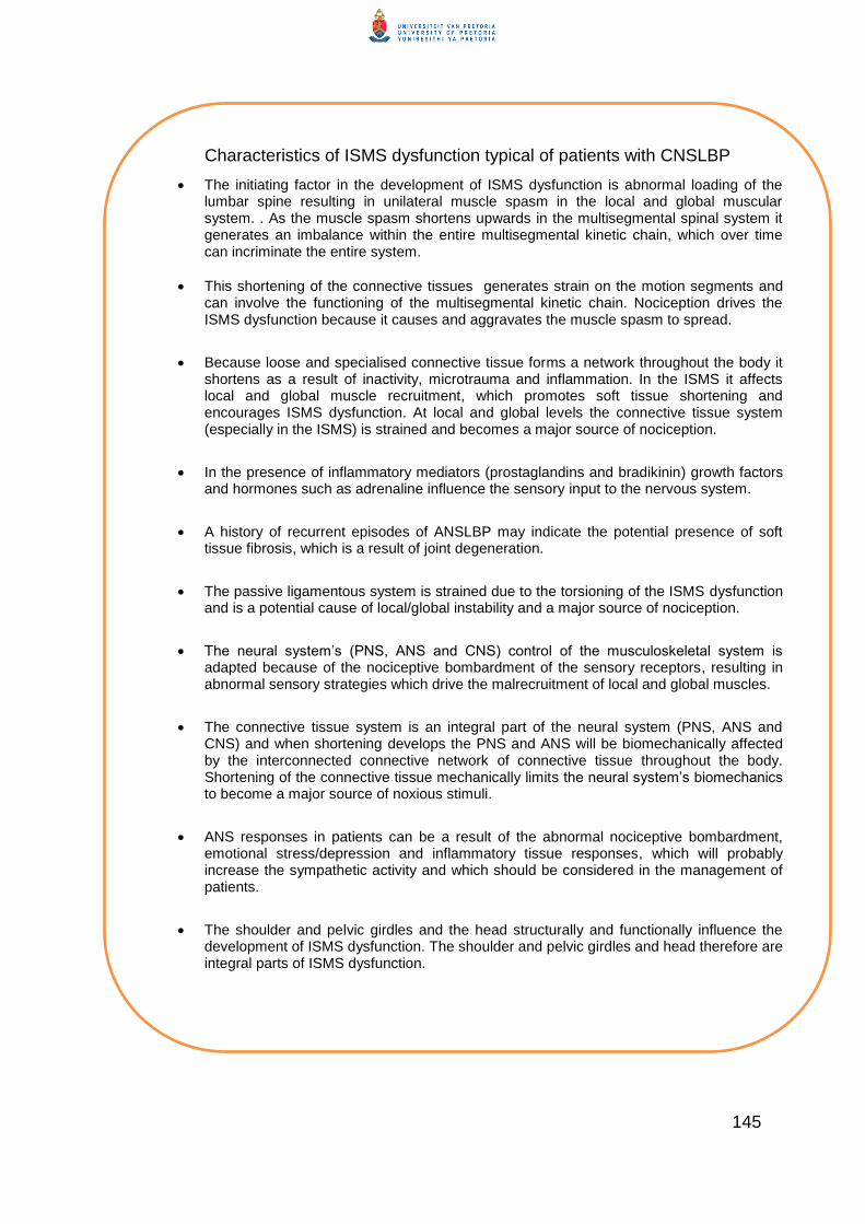
145
Characteristics of ISMS dysfunction typical of patients with CNSLBP
The initiating factor in the development of ISMS dysfunction is abnormal loading of the lumbar spine resulting in unilateral muscle spasm in the local and global muscular system. . As the muscle spasm shortens upwards in the multisegmental spinal system it generates an imbalance within the entire multisegmental kinetic chain, which over time can incriminate the entire system.
This shortening of the connective tissues generates strain on the motion segments and can involve the functioning of the multisegmental kinetic chain. Nociception drives the ISMS dysfunction because it causes and aggravates the muscle spasm to spread.
Because loose and specialised connective tissue forms a network throughout the body it shortens as a result of inactivity, microtrauma and inflammation. In the ISMS it affects local and global muscle recruitment, which promotes soft tissue shortening and encourages ISMS dysfunction. At local and global levels the connective tissue system (especially in the ISMS) is strained and becomes a major source of nociception.
In the presence of inflammatory mediators (prostaglandins and bradikinin) growth factors and hormones such as adrenaline influence the sensory input to the nervous system.
A history of recurrent episodes of ANSLBP may indicate the potential presence of soft tissue fibrosis, which is a result of joint degeneration.
The passive ligamentous system is strained due to the torsioning of the ISMS dysfunction and is a potential cause of local/global instability and a major source of nociception.
The neural system‘s (PNS, ANS and CNS) control of the musculoskeletal system is adapted because of the nociceptive bombardment of the sensory receptors, resulting in abnormal sensory strategies which drive the malrecruitment of local and global muscles.
The connective tissue system is an integral part of the neural system (PNS, ANS and CNS) and when shortening develops the PNS and ANS will be biomechanically affected by the interconnected connective network of connective tissue throughout the body. Shortening of the connective tissue mechanically limits the neural system‘s biomechanics to become a major source of noxious stimuli.
ANS responses in patients can be a result of the abnormal nociceptive bombardment, emotional stress/depression and inflammatory tissue responses, which will probably increase the sympathetic activity and which should be considered in the management of patients.
The shoulder and pelvic girdles and the head structurally and functionally influence the development of ISMS dysfunction. The shoulder and pelvic girdles and head therefore are integral parts of ISMS dysfunction.

146
In conclusion ISMS dysfunction is dynamic continuous and over time is progressive
without treatment because it is a self-perpetuating cycle within the systems due to
the continuous process of plasticity as long as all the components of ISMS
dysfunction don‘t change. Depending on the stage of chronicity, different degrees
ISMS dysfunction can be identified: mild ISMS dysfunction can be reversed easier
than ingrained longstanding ISMS dysfunction.
ISMS dysfunction is the term used to indicate the dysfunction of the integrated
neuromusculoskeletal and psychological systems that forms the essence of the
clinical picture of patients with CNSLBP regardless of the origin of the pain.
The clinical picture of a patient with CNSLBP will therefore include ISMS dysfunction
as well as other associated signs and symptoms.
Characteristics of ISMS dysfunction typical of patients with CNSLBP (continued)
ISMS dysfunction affects the strain on the structures of the thoracic spine and ribcage and as such affects the breathing pattern and tidal volume. The costovertebral, costosternal and costochondral joints are major nociceptive sources in patients with ISMS dysfunction.
Torsioning of the ISMS can be driven by the psychological components (fear avoidance, emotional stress, anxiety, and depression) as well as by configuration of the individual motion segments and the ISMS as a whole and may result result in an incident of unilateral spinal loading causing pain and muscle spasm.
Chronic pain is generated and driven by a dynamic integrated interaction between the biological, pain processing and the CNS. This interaction is influenced by social stressors.
A person‘s sedentary lifestyle reinforces his/her/their lack of movement due to general stiffness and muscle spasm as well as fear avoidance.
Genetic predisposition influences a person‘s physical and psychological response to mechanical and environmental influences, which are strong predictors for the development of CNSLBP.
Patients with CNSLBP present with a combination of ISMS dysfunction, pain processing and characteristic adaptive behaviour and any one of these components can become the primary driver of CNSLBP.
147
CHAPTER 4
The principles of a multidimensional assessment model for
patients with chronic nonspecific low back pain
4.1 Introduction
In Chapter 4 the researcher discusses the assessment of the patient with CNSLBP,
based on the principles of the multidimensional clinical presentation of a patient with
CNSLBP as discussed in Chapter 3.
During the assessment the manual therapist should continuously keep in mind that
the interaction between the ISMS dysfunction, pain processing and characteristic
adaptive behaviour is a dynamic self-perpetuating cycle in which any of the three
components can drive the clinical picture of CNSLBP. The main focus but also the
challenge of the assessment, therefore, is that the primary and secondary drivers of
the patient‘s CNSLBP as well as the complex interaction between all three
components should be identified in order to plan an appropriate multidimensional
management plan for the patient suggested by the researcher.
The premise for the assessment of a patient with NSLBP (it can be acute or chronic)
is the bio-psycho-social model (BPS model) (Waddell, 2004; Hodges, 2013).
However, the BPS model only gives an indication of the categories in which the
patient may experience symptoms. The BPS model serves as a generic model for
the approach to management of a patient that presents with (acute or chronic)
NSLBP but does not indicate or explain how some people can cope with back pain
while others become severely disabled. It is further a model of human illness rather
than wellness (Waddell, 2004 p 271).
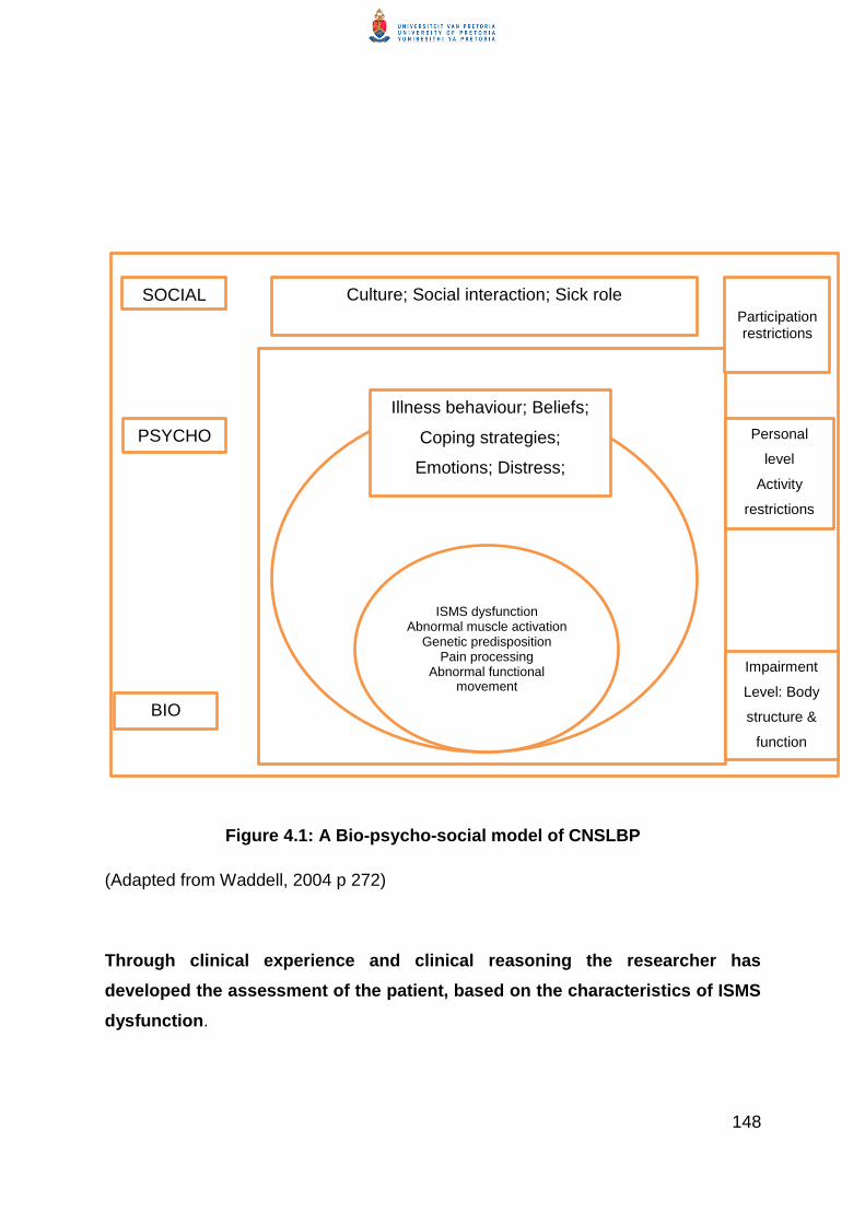
148
Figure 4.1: A Bio-psycho-social model of CNSLBP
(Adapted from Waddell, 2004 p 272)
Through clinical experience and clinical reasoning the researcher has
developed the assessment of the patient, based on the characteristics of ISMS
dysfunction.
Culture; Social interaction; Sick role
ISMS dysfunction Abnormal muscle activation
Genetic predisposition Pain processing
Abnormal functional movement
Illness behaviour; Beliefs;
Coping strategies;
Emotions; Distress;
Lifestyle, Catastrophisation
BIO
PSYCHO
SOCIAL
Impairment
Level: Body
structure &
function
Personal
level
Activity
restrictions
Participation restrictions
Culture; Social interaction; Sick role

149
Typically the history of the patients with (chronic and/or acute NSLBP, posture,
functional movement and anatomical structures palpated and emotional, pain or
behavioural responses during discussion of the history of the condition, posture and
functional assessment as well as the palpation of the structures should assist the
manual therapist to identify the primary and secondary drivers of the condition. The
therapist is also able to establish the complex interaction between all the contributing
factors and ‗hidden agendas‘.
The differentiation between the signs and symptoms of the primary and secondary
drivers of the condition and the interaction between all the factors are not always
clear in the case of a patient with CNSLBP and can sometimes only be clarified after
observation of the patient‘s progress, based on management over time rather than a
final conclusion after the initial assessment. The conclusion following the
assessment of the patient with CNSLBP is a clinical diagnosis and not an evidence-
based diagnosis (European Guidelines for the Management of Chronic Non-specific
Low Back Pain, 2004).
4.2 Typical clinical appearance of a patient with CNSLBP
Clinically there is no specific distinction between acute and chronic NSLBP (Waddell,
2004). A patient that presents with NSLBP can present with varying degrees and
combinations of the following signs and symptoms that may indicate that the patient
is developing chronicity. The clinical appearance of the patient described below is
based on the researcher‘s observations during the management of patients in clinical
practice.
Typical telltale non-verbal signs that give an indication that the patient has a
tendency to develop CNSLBP or has already developed CNSLBP include:
The patient‘s posture is ‗droopy‘ (generalised flexion posture), the patient
takes small almost guarded steps or lacks the associated arm swing or trunk
movements during gait;
The patient complains of pain in the back and generally elsewhere in the
musculoskeletal system; and

150
The patient‘s movements during gait in general are not ‗free‘ and s/he has lost
the normal associated flowing movement during gait.
As chronicity becomes embedded in the patient‘s ISMS towards the more advanced
stages of chronicity their clinical appearance becomes one of withdrawal (social
withdrawal should be established during the history taking). Advanced stages of
chronicity are also revealed in the following symptoms:
A stooping posture and a facial expression that may indicate a gloomy or
negative mindset and/or pain;
Movement can become so guarded that it is ‗locked in‘ because the patient is
terrified to move as it provokes pain. Waddell (2004) calls this disuse
syndrome;
The patient‘s posture and movement described above may be associated with
depression;
Patients present with a posture that displays a clear torsioning of the trunk
that may result in an appearance that the one leg is shorter than the other
The patient displays feelings of helplessness and hopelessness;
Owing to the chronicity of the pain and possible involvement of the ANS, the
patient can complain of excessive intermittent sweating, nausea, poor
sleeping pattern and quality of sleep, light headedness and dry mouth typical
of feeling ill. The patient may lose their appetite and show altered breathing
due to fear and resulting in decreased lung excursion. The patient often
appears generally ill, with limited functional movement such as walking and
change in position;
The patient‘s complaints of pain do not correlate with the excessive
disturbance in their guarded movement. Complaints of pain may decrease
although their level of disability may increase;
The patient‘s voice may be laboured and monotonous;
The patient often does not want to be touched because of fear of pain; and
This patient is full of fear and distrust; they never come to physiotherapy alone
but ask someone to bring them to treatment.
151
4.2.1 History taking
The purpose of the history taking is to distinguish between the dominance of the
ISMS dysfunction, characteristic adaptive behaviour, and pain processing as the
primary and secondary drivers of the patient‘s CNSLBP. This approach is in line with
the recommendations by O‘Sullivan (2011) on the management of patients with
CNSLBP.
Specific and non-specific LBP are differentiated through a system of triage. The
triage serves as a screening process to distinguish between specific serious spinal
pathology or nerve root compression or other ‗red flags‘) and non-specific LBP. If the
patient‘s condition is diagnosed as non-specific LBP, the second challenge is to
identify the stage of NSLBP (acute/sub-acute or chronic NSLBP).
4.2.1.1 Triage: Screening of the patient for NSLBP
The Clinical Practice Guidelines on the management of LBP published by the Royal
College of Physicians in 2009 recommend an initial triage to facilitate effective
assessment and management of the patient‘s problem. The researcher finds the
diagnostic triage described by Waddell (2004) a useful tool to apply in screening the
patient to ‗identify the red flags‘ and differentiate between the patient with acute or
sub-acute LBP at risk for developing CNSLBP and the patient that presents with
CNSLBP (Figure 4.2).
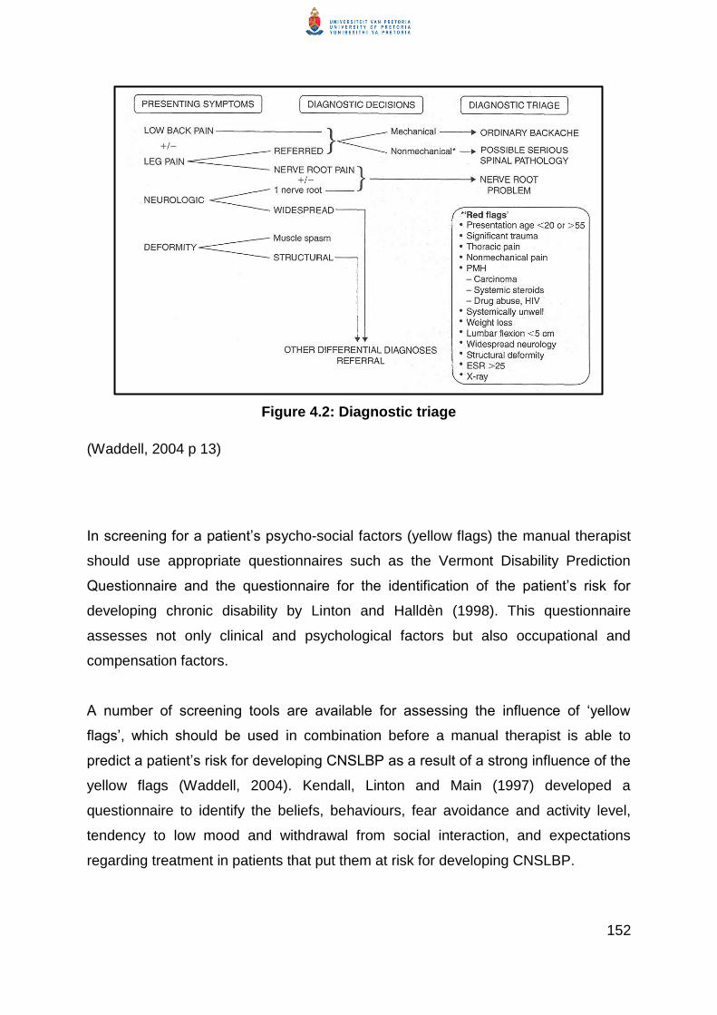
152
Figure 4.2: Diagnostic triage
(Waddell, 2004 p 13)
In screening for a patient‘s psycho-social factors (yellow flags) the manual therapist
should use appropriate questionnaires such as the Vermont Disability Prediction
Questionnaire and the questionnaire for the identification of the patient‘s risk for
developing chronic disability by Linton and Halldèn (1998). This questionnaire
assesses not only clinical and psychological factors but also occupational and
compensation factors.
A number of screening tools are available for assessing the influence of ‗yellow
flags‘, which should be used in combination before a manual therapist is able to
predict a patient‘s risk for developing CNSLBP as a result of a strong influence of the
yellow flags (Waddell, 2004). Kendall, Linton and Main (1997) developed a
questionnaire to identify the beliefs, behaviours, fear avoidance and activity level,
tendency to low mood and withdrawal from social interaction, and expectations
regarding treatment in patients that put them at risk for developing CNSLBP.
153
Waddell (2004) warns that even applying a combination of outcome measures to
assess the extent or the role of psycho-social factors as a potential driving factor in
the patient‘s pain perception/pain processing is not an accurate predictor and that
each patient should be managed individually, based on their clinical presentation. If
positive predictors are identified during the screening process, the therapist should
investigate those particular aspects (e.g. fear avoidance) further.
Waddell (2004) describes a combination of socio-demographic, clinical and psycho-
social predicting factors that predicts the development of CNSLBP and disability.
These factors are listed in Table 4.1.
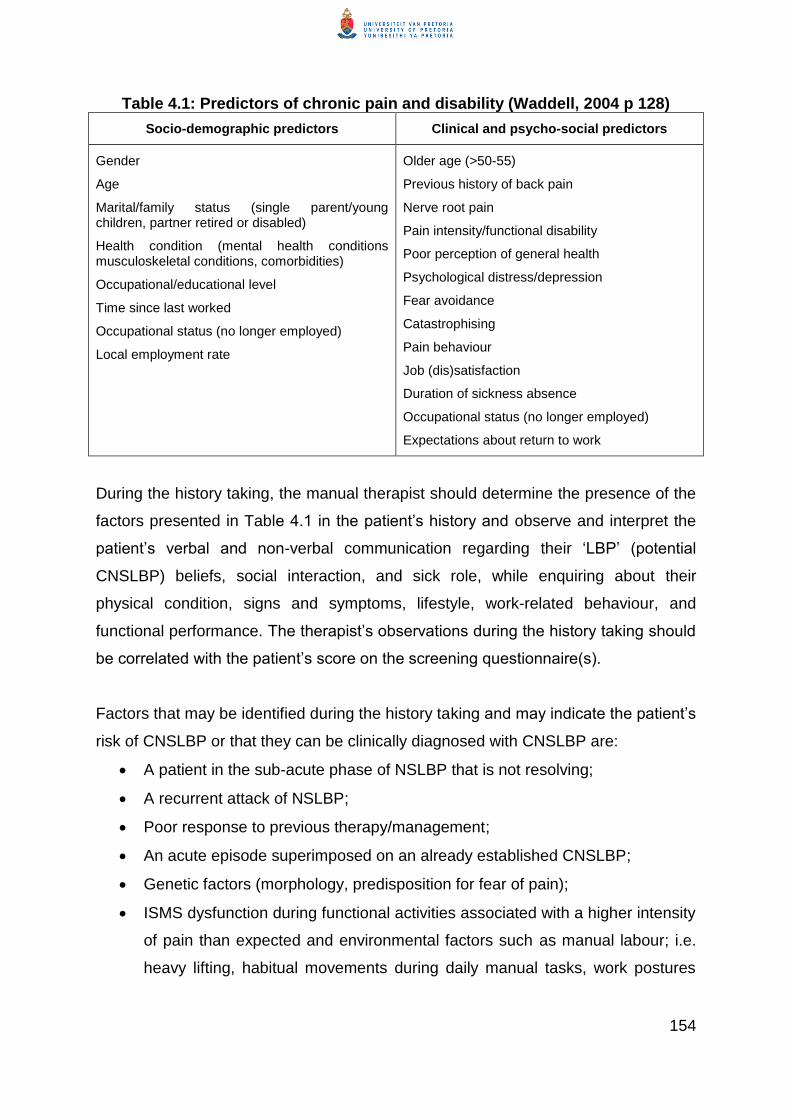
154
Table 4.1: Predictors of chronic pain and disability (Waddell, 2004 p 128)
Socio-demographic predictors Clinical and psycho-social predictors
Gender
Age
Marital/family status (single parent/young children, partner retired or disabled)
Health condition (mental health conditions musculoskeletal conditions, comorbidities)
Occupational/educational level
Time since last worked
Occupational status (no longer employed)
Local employment rate
Older age (>50-55)
Previous history of back pain
Nerve root pain
Pain intensity/functional disability
Poor perception of general health
Psychological distress/depression
Fear avoidance
Catastrophising
Pain behaviour
Job (dis)satisfaction
Duration of sickness absence
Occupational status (no longer employed)
Expectations about return to work
During the history taking, the manual therapist should determine the presence of the
factors presented in Table 4.1 in the patient‘s history and observe and interpret the
patient‘s verbal and non-verbal communication regarding their ‗LBP‘ (potential
CNSLBP) beliefs, social interaction, and sick role, while enquiring about their
physical condition, signs and symptoms, lifestyle, work-related behaviour, and
functional performance. The therapist‘s observations during the history taking should
be correlated with the patient‘s score on the screening questionnaire(s).
Factors that may be identified during the history taking and may indicate the patient‘s
risk of CNSLBP or that they can be clinically diagnosed with CNSLBP are:
A patient in the sub-acute phase of NSLBP that is not resolving;
A recurrent attack of NSLBP;
Poor response to previous therapy/management;
An acute episode superimposed on an already established CNSLBP;
Genetic factors (morphology, predisposition for fear of pain);
ISMS dysfunction during functional activities associated with a higher intensity
of pain than expected and environmental factors such as manual labour; i.e.
heavy lifting, habitual movements during daily manual tasks, work postures

155
such as prolonged sitting, and specific job-related demands, which may be
mechanical;
Lack of or limited participation in leisure activities and sports;
Lack of or limited emotional and physical support, which contributes to
inappropriate psychological and social behaviour;
Cognitive behaviour such as stress responses, provocative coping strategies,
and emotional factors such as hypervigilance;
The underlying dynamic tissue repair process (the patient‘s response to the
tissue repair process is influenced by their psychological frame of mind)
(Field, 2009; Waddell, 2004; Adams et al., 2002); and
Symptoms lasting longer than three months.
4.2.1.2 Principles for the (objective) assessment of ISMS dysfunction
The aim of the assessment of a patient at risk of developing CNSLBP or diagnosed
with CNSLBP is to determine any malalignment of the spine due to imbalance in the
neuromusculoskeletal systems. In essence the objective assessment entails the
identification of malalignment to eliminate structural causes such as scoliosis,
compensatory postures and movement displayed as guarded movements and pain-
provoking coping strategies, and the clinical determination of the possible reasons
for and causes of it. The assessment of ISMS dysfunction is totally integrated with
assessment of the patient‘s pain processing and characteristic adaptive behaviour.
This behaviour should be correlated with the psycho-social factors such as fear of
movement, beliefs and illness behaviour that were identified during the screening of
and history taking with the patient.
Finally the aim is to determine the potential reversibility of the local and global
shortening and stiffness of the soft tissues that have resulted through a process of
plasticity. The patient‘s neural response on initial palpation to determine the degree
of soft tissue stiffness and, potentially, fibrosis and the patient‘s ability to adapt to
correction of postural control may indicate the reversibility of the condition through
the process of plasticity.
156
(1) Observation of the patient‘s ISMS dysfunction
Observation of ISMS dysfunction entails the integration of the observation of the
patient‘s posture and postural control during functional movement, the patient‘s pain
perception during static posture as well as during functional movement, and the
patient‘s characteristic behaviour during static posture and functional activities.
Assessment of the patient‘s ISMS dysfunction should also include the identification
or confirmation of the risk factors or factors that might have been already identified
during the history taking that influence the patient‘s condition.
Assessment of the patient‘s clinical picture from the perspective of ISMS dysfunction
starts by observation and analysis of the patient‘s posture to determine baseline
markers, from where the patient‘s movement during functional activities (i.e. gait) can
be interpreted. The patient‘s posture in standing will give the therapist an indication
of their postural asymmetry, postural control at the level of the motion segment (local
alignment and muscle activation) and global alignment (and global muscle activation
that contributes to guarded movements/provocative and avoidance behaviour or
strategies). Assessment of posture also includes the limb girdles, shoulder and
pelvic girdles, and the position of the head on the cervical spine. All the limb girdles
and the head on the cervical spine are inseparably connected to in every aspect of
posture and movement.
A patient‘s posture is seen as an image and expression of the whole patient as
described during their history, so the therapist can relate his/her observations to the
patient‘s story of anxiety, frustration, or sheer desperation.

157
Knowledge block
Grieves (1981) points out that different muscles contain varying proportions of
slow and fast muscle fibres. Slow fibres maintain posture; fast fibres give dynamic,
voluntary movement (see Section 3.2.1 of the current study). Muscle imbalance
gives typical patterns of postural disturbance, which cause abnormal loads on
joints and soft tissue structures, abnormal patterns of movement, muscle fatigue,
and loss of coordination. In the patient with CNSLBP, the paraspinal muscles are
atrophied and contain an increased percentage of fat (Cooper et al., 1992;
Mooney et al., 1997). These paraspinal muscles are weaker and fatigue more
easily
Hides et al. (1994) found local wasting in the multifidus muscle, which is the
largest and most medially situated muscle of the erector spinae. The human
lordosis is unique because of our upright posture and this involves specific
development of the multifidus muscle. The changes were segmental and
unilateral, and corresponded to the level and side of symptoms. Hides et al.,
(1994) found about 30% reduction in the cross-sectional area of multifidus.
Because this wasting was so localised and developed so rapidly, they suggested
that it was due to segmental inhibition rather than to a general effect of disuse.
Even when symptoms settle, multifidus wasting may not recover spontaneously,
and this may predispose to recurrent attacks. Hides, Richardson and Jull (1996)
showed that specific, localised exercises for the multifidus may not make much
difference to symptomatic recovery from the acute attack but do produce better
muscle recovery.

158
(i) Observation of posture
Posture is the alignment of the body based on muscle activity to counteract the force
of gravity. In a supported position, such as sitting, posture is assessed during muscle
inactivity.
During observation of posture the manual therapist analyses the alignment of the
biomechanical component of the ISMS during static posture and postural
adaptations during dynamic movement. In essence the assessment of dynamic
posture comprises the assessment of the integrated muscle activity that counteracts
the force of gravity. In stable posture, regardless of the position, the combined centre
of gravity of the various body parts should fall within the base of support. Alignment
of the body parts should be maintained to ensure continuous stability in static and
dynamic posture. Maintenance of posture can create stress in malaligned joints and
poor recruitment of muscle activity. It can also cause muscle atrophy. The influence
of the alignment of the limbs on posture, as briefly discussed in Chapter 3, should be
taken into account and determined during the assessment of the patient‘s biological
component of ISMS dysfunction. The patient‘s frame of mind can also be reflected in
their posture.
The position of the head on the cervical spine, shoulder girdles in relation to the
thoracic spinal region, and the pelvis in relation to the lumbar spine, as well as the
position of the hips, knees and feet (in particular the foot arches), should be
evaluated (Middleditch & Oliver, 2005; Norkin & Levangie, 2008).
In Figure 4.3 the key points in the observation of a patient‘s posture are highlighted
to illustrate the assessment of posture in the patient with CNSLBP.
The images of the people in Figures 4.3, 4.4 and 4.5 show typical fascial
malalignment with dominance in the thoraco-lumbar area. Myers (2011) used the
photographs of the people displayed in Figures 4.4 and 4.5 to demonstrate particular
patterns characteristic of postural compensations. No history of pathology or signs
and symptoms that the people in the photographs may have experienced is given by
the author. However, these pictures display the typical postures found in patients

159
with CNSLBP and are therefore used here to explain the postural observations
during the assessment of a patient with CNSLBP.
Observation of posture (1)
Figure 4.3: An axial view of the position of the shoulder girdle relative to the
pelvis due to ISMS dysfunction (observation 1)
(Myers, 2011 p 241)
Figure 4.3(b): An anterior, posterior and lateral view of the patients posture
typical of ISMS dysfunction
(Myers, 2011, p 241)
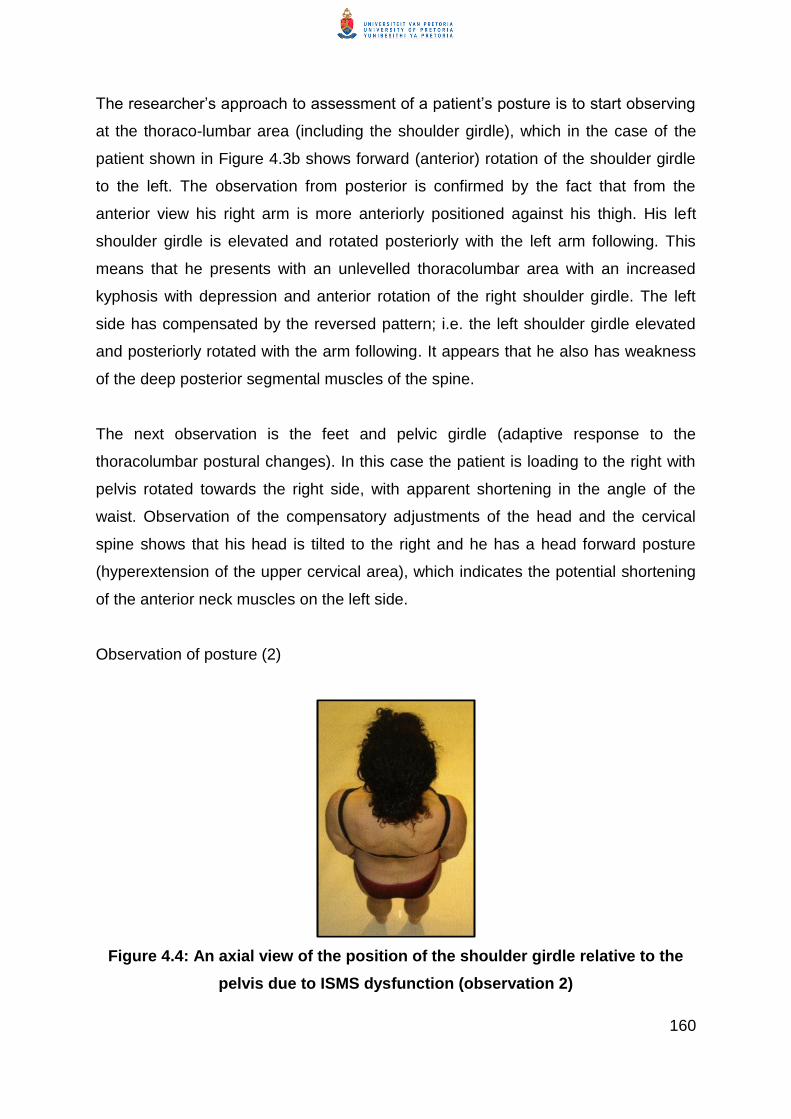
160
The researcher‘s approach to assessment of a patient‘s posture is to start observing
at the thoraco-lumbar area (including the shoulder girdle), which in the case of the
patient shown in Figure 4.3b shows forward (anterior) rotation of the shoulder girdle
to the left. The observation from posterior is confirmed by the fact that from the
anterior view his right arm is more anteriorly positioned against his thigh. His left
shoulder girdle is elevated and rotated posteriorly with the left arm following. This
means that he presents with an unlevelled thoracolumbar area with an increased
kyphosis with depression and anterior rotation of the right shoulder girdle. The left
side has compensated by the reversed pattern; i.e. the left shoulder girdle elevated
and posteriorly rotated with the arm following. It appears that he also has weakness
of the deep posterior segmental muscles of the spine.
The next observation is the feet and pelvic girdle (adaptive response to the
thoracolumbar postural changes). In this case the patient is loading to the right with
pelvis rotated towards the right side, with apparent shortening in the angle of the
waist. Observation of the compensatory adjustments of the head and the cervical
spine shows that his head is tilted to the right and he has a head forward posture
(hyperextension of the upper cervical area), which indicates the potential shortening
of the anterior neck muscles on the left side.
Observation of posture (2)
Figure 4.4: An axial view of the position of the shoulder girdle relative to the
pelvis due to ISMS dysfunction (observation 2)

161
This person shows a dominant posterior rotation of the right shoulder girdle and a
reduced thoracic curve. The scapulae are not symmetrical, which may be indicative
of a muscle imbalance between internal oblique abdominus on the left and external
oblique abdominus on the right; the latissimus dorsi on the right side seems to be
shortened. The lower trapezius, serratus anterior and rhomboids appear to be
atrophied and the levator scapulae and middle trapezius appear to be bilaterally
overactive.
In this case the pelvic rotation appears to be more anterior on the right side, which
results in the patient being loaded more on the right foot.
Her centre of gravity seems to be more posterior, which is probably why she
presents with an increased lumbar lordosis.
The compensatory response (position) of the cervical spine and neck is not
observable from this view and is not available in the text (Myers, 2011).

162
Knowledge block
Myers (2011) describes global fascial lines, which indicate not only that fascia may be shortened
(stiff) at a local level but that stiffness at a local level can incriminate the whole fascial system.
This fact explains why lumbar fascia micro-trauma leads to shortening and fibrosis, which may
cause distal signs and symptoms; for example, cervical involvement (immobility and/or
headache), restriction of shoulder girdle and gleno-humeral joint involvement (pain and
restriction through latissimus dorsi), restriction of breathing movements, and transferring the load
from the trunk to the pelvis and lower limbs.
Figure 4.6: Rotational and posterior longitudinal fascia lines
(Myers, 2011 p 72)
163
(ii) Observation of functional activities
Assessment of the patient‘s motor control is achieved by assessing their functional
activities, such as gait, sitting to standing and/or other functional tasks that will
reproduce the patient‘s symptoms. During functional activities the manual therapist
should identify abnormal movement patterns (due to abnormal muscle activation,
guarded movement patterns and compensatory mechanisms of movement).
Compensatory movement patterns are characterised by motion segments or spinal
regions carrying out a compensatory movement to accommodate the lack of
movement (due to stiffness) in another segment or region. Abnormal postural control
is identified by abnormal sequence of movement, limited or distorted equilibrium
reactions, and a small range of global movement patterns (due to guarded
movements). It is also characterised by asymmetry in bilateral cyclical movement
patterns (reciprocal trunk rotation and arm swing in, for example, gait), and lack of or
limited segmental stability and mobility, which results in abnormal or compensatory
movement patterns.
During all functional activities or physiological movements performed by the patient
the manual therapist should identify the patient‘s pain responses. Observation of gait
and other functional activities is based on the parameters of gait, and movement
analysis (Perry & Burnfield, 2010).
The manual therapist needs to observe the complex, dynamic patterns of spinal
movement during functional activities (Esola et al., 1996; Steffen et al., 1997) to
identify a change in the sequence of movement in order to determine the
coordination between lumbar and pelvic movements, or between spinal flexion and
extension. The relationship of the shoulder girdle and arm in relation to the pelvis
and leg during functional activities should be observed. A difference may exist
between the mobility of the upper and the lower lumbar spine during simple
functional activities and when the upper and/or lower limb movements are part of
more complex functional activities.

164
Pure physiological movements in the spine do not occur because spinal movements
are coupled movements and occur in three dimensions (coronal, sagittal and
transvers planes). Typical coupled movements are disturbed in a patient with ISMS
dysfunction. During observation (assessment) it is essential for the manual therapist
to determine how the components of the coupled movements interact during spinal
movements without or associated with upper and lower girdle and limb function. Lack
of components of coupled movements is an indication of dysfunction in the motion
segment and in global mobility and stability of the trunk. This is further investigated
by the manual therapist by using passive accessory movements during palpation.
Observation of abnormal movement patterns includes the ‗pain-spasm-pain‘ cycle
(reflex sustained co-contraction between agonistic and antagonistic muscles
(Langevin & Sherman, 2006)) and ‗pain adaptation‘ (slowing and decreased range of
motion due to selective increased activation of antagonists). Altered muscle
activation patterns in CNSLBP can stabilise the spine during movement (owing to
fixation), to prevent further injuries. This adaptation comes at the cost of restricted
range of motion. Patients with CNSLBP appear to have many motion-limiting muscle
activation patterns that may be initiated or aggravated by emotional factors (fear and
anxiety).
Abnormal movement patterns can have important influences on the connective
tissues that surround and infiltrate muscles. Both increased stress due to overuse
(repetitive movement) and decreased stress due to immobilisation or hypomobility
can cause changes in connective tissue (Langevin & Sherman, 2006).
Within this complex cycle of events, the manual therapist should be able to identify
how to interpret components of movement caused by disinhibition, disuse, atrophy
and hypertrophy, with the view to isolating the probable source of dysfunction and
putting it functionally to the test during activities such as walking and sitting to
standing. For example, if the latissimus dorsi is in spasm or hyper active it will fixate
the scapula and the trunk and limit arm swing during gait.
Isometric muscle testing is appropriate in the case of severe pain.

165
(2) Palpation guided by observations
The aim of palpation is to determine specifically (1) the alignment of the palpable
components of the vertebral column (spine), pelvic girdle and lower limb and
shoulder girdle and upper limb; (2) the mobility of all soft tissue; and (3) the analysis
of the texture of the soft tissues.
During palpation of the soft tissues and skeletal landmarks and zygapophyseal
joints, rib angles and costal junctions, the manual therapist should correlate with the
patient‘s pain responses. The pattern of soft tissue tension in the ISMS upper and
lower limb should be identified. The musculature of the rotator cuff may develop
major anterior shortening in response to a thoracic kyphosis and poking chin.
The patient should be asked to report any awareness of abnormal sensation during
the palpation. The patient‘s responses could be malinterpreted because sensory
awareness and spatial orientation are inhibited due to the bombardment of
nociceptive stimuli from various structures within and around one or several motion
segments. Moseley, Gallagher and Gallace (2012) and Langevin and Sherman
(2006) have found that tactile discrimination and spatial orientation on the spine are
poorly represented in the brain of normal subjects and even more so in patients with
CNSLBP.
The manual therapist should be aware that the muscular-tendonous junction,
intermuscular septum, ligaments or capsules, junction with the periosteum and bone
are more sensitive than other areas and should be taken into account during the
interpretation of the patient‘s response to palpation. Identification of soft tissue
abnormality should include the factors outlined below.
Muscle tone (spasm or atrophy) taut bands and trigger points
The manual therapist should be sensitive to detecting any changes in muscle tone
and areas of muscle activity or loss of activity. Presence of areas of muscle spasm
and tender spots (i.e. trigger points) indicates an active nociceptive source. These
observations through palpation should be correlated with the observations of
166
inactivity, muscle imbalance or malrecruitment, which all contribute to the decreased
postural control observed during functional activities.
Taut bands and trigger points in the muscles
On palpation active trigger points give rise to local and referred pain (Travell &
Simons, 1983). The presence of painful muscle contraction or tender foci within
perimuscular fascia may add to the factors that promote hypomobility and tissue
fibrosis because increased muscle tension and contractures may place strain on
nociceptive sensitive soft tissue areas.
Referred pain to the lower quadrant due to nerve root involvement should be
identified during the triage because it is a red flag and falls under the category of
specific LBP (see Section 4.2.1.1). Any other referred pain down the leg is typically
due to the activation of trigger points (refer to Sections 3.4.2 and 3.5.1.1). Taut
bands and trigger point activity due to L4-S1 dysfunction commonly affect the
gluteus maximus, medius and minimus, quadratus lumborum, piriformis, iliacus and
psoas muscles. The distribution of the pattern of pain can mimic nerve root pain
down the lateral aspect of the thigh and into the lateral lower limb. With careful
localisation and successful de-activation of active trigger points, this referred pain
should subside if it is somatic (i.e. no nerve root involvement) in origin.
Fascial stiffness (shortening) and fibrosis
‗Fascial stiffness‘ refers to the general fascia as well as the thoraco-lumbar fascia,
which includes the gluteus maximus, medius and minimus, quadratus lumborum,
piriformis, and iliacus and psoas muscles as well as the internal and external oblique
muscles and transversus abdominus.
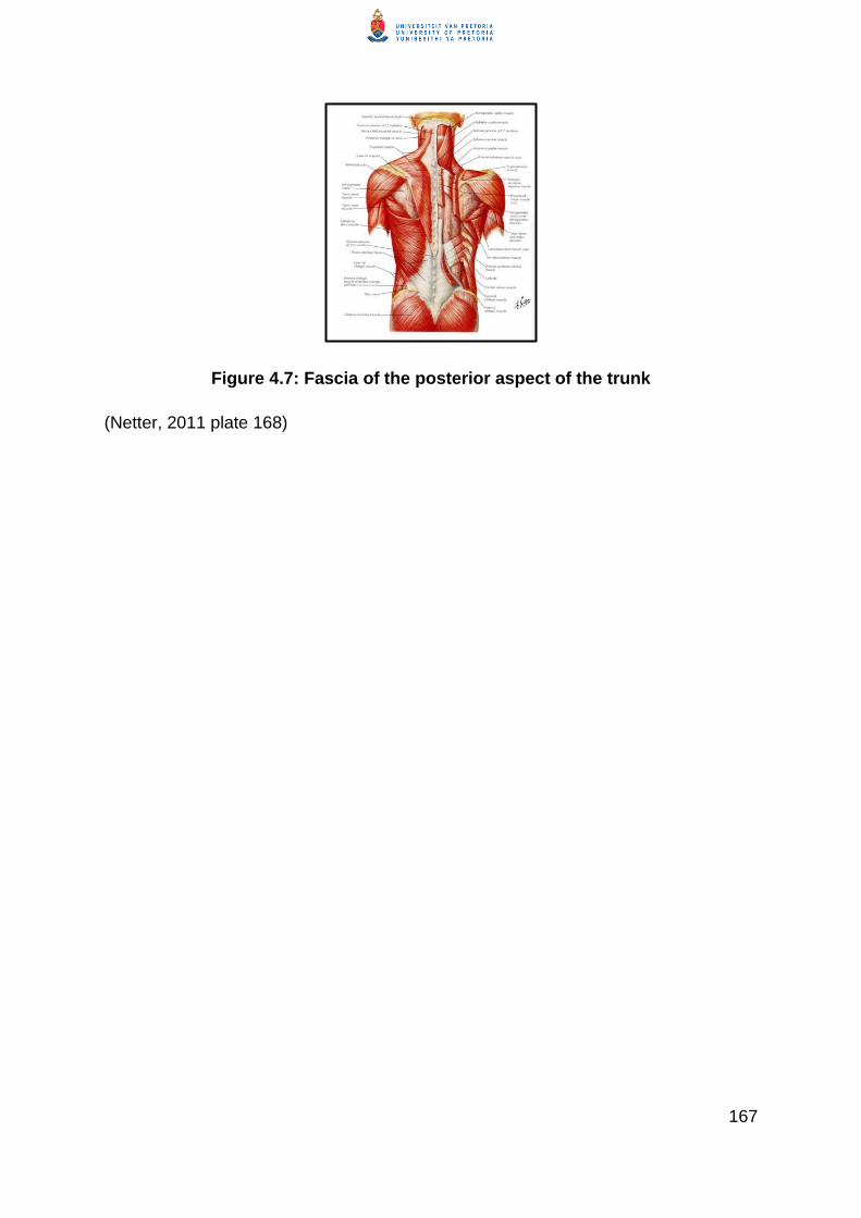
167
Figure 4.7: Fascia of the posterior aspect of the trunk
(Netter, 2011 plate 168)

168
Knowledge block: Inflammation and tissue stiffness
Chronic, local increase in mechanical stress on the connective tissues (fascia)
can lead to micro-injury and inflammation (overuse injury, cumulative trauma
disorder). A consistent absence of mechanical stress on connective tissue
leads to connective tissue atrophy, architectural disorganisation, fibrosis,
adhesions and contractures. Factors that influence whether atrophy or fibrosis
predominates during stress deprivation include the concurrent presence of
inflammation, tissue hypo-oxygenation and cytokines such as TGFb-1 that
promote fibrosis. Fibrosis therefore can be the direct result of hypomobility or
the indirect result of hypermobility via injury and inflammation (Langevin &
Sherman, 2006).
Tissue micro-injury, inflammation and fibrosis not only can change the
biomechanics of soft tissue (e.g. increased stiffness) but also can profoundly
alter the sensory and nociceptive input arising from the affected tissues.
Connective tissue is richly innervated with chemical, mechanical, and thermal
nociceptors and nociceptive neurons (Langevin & Sherman, 2006).
Modulation of nociceptor activity has been shown to occur in response to
changes in the innervated tissue (Section 3.8.2). The level of protons,
inflammatory mediators (prostaglandins, bradykinin), growth factors (NGFs)
and hormones (adrenaline) all have been shown to influence sensory input to
the nervous system (physiological pain resulting in hypersensitivity).
Conversely, nociceptor activation has been shown to modify the innervated
tissue (Langevin & Sherman, 2006).
In addition, the release of substance P from sensory C-fibres in the skin
enhances the production of histamine and cytokines from mast cells,
monocytes and endothelial cells. Increased TGFb-1 production, stimulated by
tissue injury and histamine release, is a powerful driver of fibroblast collagen
synthesis and tissue fibrosis (Langevin & Sherman, 2006). Thus, activation of
nociceptors by itself can contribute to the development or worsening of fibrosis
and inflammation, causing more tissue stiffness and movement impairment.

169
Regardless of its original cause, connective tissue fibrosis is detrimental, as it leads
to increased tissue stiffness and (further) movement impairment. The manual
therapist should correlate the tissue fibrosis with the (lack of) mobility of the
particular spinal regions during all the functional activities.
Interpretation of referred pain and asymmetry
If the patient complains of referred pain, the therapist should identify the origin of the
referred pain as somatic referred pain or neural referred pain. In relation to other
findings during palpation the therapist can perform neural tension tests, which in
themselves are not diagnostic tests for nerve root pain but involve the nerve due to
an adhesion in the neural sheath or nerve root compression (Butler, 2000).
Somatic referred pain patterns have been discussed under ‗Taut bands and trigger
points in the muscles‘ and Section 3.4.2. Reflexes such as the ankle (S1) and knee
jerk (L3/4), and elbow jerk (C6) should be tested to differentiate between somatic
and neural referred pain in order to confirm that referred pain is not neural in origin.
When assessed as part of a clinical picture, posture and functional activities
demonstrate disrupted biomechanics. These disruptions are most probably the result
of the influence of the shortened soft tissue due to an injury or mechanical failure of
one or more of the musculoskeletal structures, commonly due to involvement at the
level of L3-S1.
The manual therapist should determine the alignment of the trunk by specifically
looking out for the position of the shoulder girdles in relation to the pelvis and the
position of the pelvis, in particular, to identify a potential unilateral pull of the
quadratus lumborum and sometimes latissimus dorsi muscles that will cause trunk
torsion together with a lateral tilt of the pelvis that results in the corkscrew effect on
the biomechanical motion segments of the ISMS.
Knowing the patient‘s pain responses, the therapist should purposefully and carefully
palpate deeper to verify the location, level, and pain responses of areas with different
tissue textures. During the process of deeper palpation the manual therapist should

170
also palpate the deep intersegmental muscles of the whole spine to identify on a
deeper level the tissue tension, trigger points, taut bands, muscle spasm, differences
in tissue textures and, very specifically, the bony alignment of the motion segments.
In the thoracic areas the rib angles and costovertebral junctions are also determined.
Motion segment distortion may be detected in any motion segment in the spine but
especially at the lower lumbar, thoracolumbar, and cervico-thoracic regions because
these are areas of transition where great mechanical strain is placed on the
transitional vertebrae.
Palpation of the bony prominences of the ISMS
Palpation of the ISMS is performed by using accessory movements to determine the
alignment and quality of the motion segments of the whole spine. Alignment of the
motion segments of the spine and of the whole spine, including the coccyx, is
important to note because the coccyx can be tilted posteriorly or anteriorly due to the
potential disposition of the pelvis. If the spine is in a rotoscoliosis, the pelvis and the
sacrum will be torsioned. The result of the palpation of the motion segments may
reveal hyper- or hypomobility, lack of joint play or altered ‗end feel‘. These findings
should be interpreted in the context of the presenting signs and symptoms; findings
observed during the observation of the patient‘s habitual posture, gait and functional
activities; and the soft tissues.
Assessment of the integrity of the neural system
The neural system integrity is assessed as follows:
Integrity of the neural system is assessed by testing muscle strength,
sensation (discrimination between light touch and a pain response) and deep
tendon reflexes.
Assessment of functional muscle strength is based on the observations of the
patient‘s impaired alignment, mobility and stability during static and dynamic
postures; characteristic adaptive behaviour, which manifests during gait and
relevant functional activities of daily life; and, lastly, abnormal muscle
activation patterns observed as asymmetrical muscle bulk that indicates
hyper- or atrophy. The challenge to the manual therapist is to identify the
interlocking vicious circles between guarded movements, muscle wasting,
171
loss of strength and endurance, loss of neuro-muscular coordination, muscle
imbalance and physical dysfunction, all resulting in deconditioning and illness
behaviour. Disuse syndrome develops out of reduced activity and illness
behaviour (Bortz, 1984; Mayer & Gatchel, 1988). The effect on the physical
condition of the ISMS due to disuse is profound as it aggravates and
maintains physical dysfunction and leads directly to more severe dysfunction.
Autonomic nervous system involvement
ANS involvement can be observed and palpated by identification of the pilomotor
effect, sweating and decreased skin temperature, vaso-motor effects (such as
‗blanching‘ or excessive redness), pseudomotor effects, and sub-cutaneous skin
oedema – also called peau d‟orange (Waddell, 2004). As explained in Section 3.5
torsioning of the spine and increased thoracic kyphosis can mechanically involve the
ANS. With release of all the soft tissue structures involved and re-alignment of the
motion segments of the spine, this mechanical strain should be reversible.
The ANS may also be involved through emotional stress via the amygdala and the
rest of the limbic system via neurotransmitters. During assessment the patient‘s
awareness and understanding of the influence of emotional stress on their
(acute/sub-acute/chronic) NSLBP should be determined. The manual therapist‘s
management of the patient in a reassuring manner is the starting point in addressing
the patient‘s problem.
Altered breathing patterns
The adaptation in the pattern and depth of breathing due to the mechanical distortion
of the trunk and associated abnormal muscle activation patterns manifests in poor
breathing patterns and limited excursion of the ribcage and lungs. Mechanically and
anatomically the ANS is therefore vulnerable to becoming involved in potentially
contributing to the patient‘s poor breathing pattern (Hodges, 2013).
Throughout the observation (assessment) the manual therapist must take into
account the dynamics of the strain and pain of the ISMS dysfunction in terms of

172
inflammation and the repair processes, taking into account the time since the origin
and the subsequent course of the NSLBP.
In summary, observing skeletal landmarks, palpating soft tissues, determining ISMS
dysfunction and interpreting the patient‘s pain response (pain processing) and
characteristic adaptive behaviour to form a holistic clinical picture is a skill that a
manual therapist should acquire. All observations (visual as well as through
palpation and muscle testing) should be based on clinical reasoning to explain the
reason(s) for malalignment. The observation is therefore an interpretation of the
underlying mechanisms that drive the ISMS dysfunction, analysed in terms of the
underlying neuro-musculo-skeletal structures and functional activities related to the
patient‘s genetics, pain processing, lifestyle, other psycho-social-related issues, and
characteristic adaptive behaviour.
During the assessment the therapist should endeavour to evaluate the patient‘s
potential to respond to direct soft tissue handling to determine the texture and
condition of the soft tissues and the patient‘s pain responses during handling, as well
as their response to cognitive demands and instructions in terms of ability to respond
to changes in postural control. These responses will enable the therapist to make an
estimated judgement on the patient‘s likelihood of responding to treatment through a
process of remodelling of soft tissues and relearning postural control. The degree of
improvement is determined by the patient‘s level of chronicity. The success of the
treatment depends on the therapist‘s clinical judgement during assessment and the
selection and performance of the treatment techniques.
For most people LBP is a short-lived inconvenience. Even when severe, if
the patient can be given appropriate information to disarm or prevent fear
and anxiety, and if they can keep their motor systems functioning then the
chances for chronicity can be dramatically reduced. (Field, 2009 p 50)

173
4.3 A multidimensional model for the assessment of patients with
CNSLBP
On the basis of the discussion above the researcher has developed a model for the
multidimensional assessment of a patient who presents with CNSLBP of varying
degrees of chronicity (Figure 4.8). The model is based on the premise that plasticity
in both soft tissues and nervous systems affects pain processing and changes in
motor behaviour play a key role in the natural cause and course of CNSLBP.
The researcher hypothesises that with the first incident of ANSLBP, the sensory
afferent input system generates pain responses from the peripheral to the CNS,
which together with hypervigilance develop altered pathways in the brain. Pain
causes active inhibition of the slow motor unit recruitment, which results in
dysfacilitation. Even after the pain has been resolved, the dysfacilitation may persist.
The initiating response to pain is fear avoidance, which can also cause active
inhibition of the slow motor unit, and the central fatigue associated with the fear,
stress, or anxiety, all of which contribute to dysfacilitation. The dysfacilitation leads to
altered postural control (Comerford et al., 2008). In the presence of reduced physical
activity connective tissue is not remodelled after the process of inflammation and/or
inactivity and because it is so interconnected with all types of tissues it provides a
powerful nociceptive input to the CNS.
Through a process of plasticity, the fascial (myofascial and neurofascial) tissues
shorten and stiffen (fibrosis) (peripheral sensitisation). The continuous feed-in to the
dorsal horn (central sensitisation) and the higher centres of these tissues results in a
continuous pain processing within the whole nervous system within the neuromatrix.
The motor output effect of pain processing contributes to the development of the
biomechanical malalignment of the ISMS but also to ISMS stiffening (fixation), and
guarded movements. When influenced by the modulation of pain processing in the
neuromatrix the motor output effect results in characteristic adaptive behaviour which
will include the psychosocial responses and illness behaviour of the patients. .
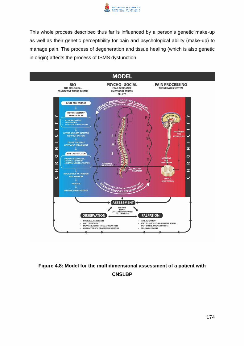
174
This whole process described thus far is influenced by a person‘s genetic make-up
as well as their genetic perceptibility for pain and psychological ability (make-up) to
manage pain. The process of degeneration and tissue healing (which is also genetic
in origin) affects the process of ISMS dysfunction.
Figure 4.8: Model for the multidimensional assessment of a patient with
CNSLBP

175
4.4 Summary of the chapter and discussion of the holistic integrated
model for the assessment of patients with CNSLBP
The model displays the assessment of a patient in the three aspects of ISMS
dysfunction: the dysfunction of the biomechanical component of the ISMS, pain
processing, and characteristic adaptive behaviour, which are characteristic of
patients with CNSLBP.
As discussed in Chapter 3, the biomechanical dysfunction of the ISMS is intimately
integrated with the pain processing and characteristic adaptive behaviour. Although
the three components of ISMS dysfunction are discussed separately a correlation
can be made among them. For this reason, the researcher discusses the
assessment from the biological perspective, pain processing perspective, and from a
characteristic behavioural perspective.
In this chapter assessment of a patient is discussed from the history taking stage,
which includes the patient‘s psycho-social factors that may contribute to their
condition, and the cause and course of the condition. From the history the therapist
should be able to evaluate and deduct the patient‘s ability to manage the condition.
The biological (clinical) manifestation of the signs and symptoms (local as well as
widespread) is the result of the processes that have worked within the ISMS over a
period of time. These biological processes (clinical manifestation) are reflected in the
patient‘s posture, gait and functional movement, which are strongly affected by the
current pain processing as well as the characteristic adaptive behaviour, which can
mask the biological origin (signs and symptoms) of the clinical picture. The challenge
to the manual therapist is therefore to identify the extent of the ISMS dysfunction in
relation to the pain processing and the characteristic adaptive behaviour.
It is possible that the primary driver of the patient‘s clinical picture cannot be
distinguished at the first assessment but may become apparent as the management
of the patient progresses and the patient relates to and trusts the therapist. Empathic
cognitive feedback from the therapist to the patient during the assessment is critical

176
for helping the patient understand their condition and address the fear avoidance
and characteristic adaptive behaviour from the initial assessment and management.
In Chapter 5 the manual therapy for a patient with the typical presentation of
CNSLBP is discussed.

177
CHAPTER 5
Principles of a multidimensional manual therapy approach to
patients with chronic non-specific low back pain
5.1 Introduction
In this chapter the manual therapy model for the management of patients with
CNSLBP is presented as a multidimensional approach. The multidimensional
manual approach, developed by the researcher over years of clinical experience,
takes into account the factors discussed in Chapter 3. Chapter 3 outlines the intimate
interaction between the biomechanical dysfunction, the neural changes that take
place at the peripheral, autonomic and central nervous systems as a result of pain
processing and the characteristic adaptive behavior. The biomechanical dysfunction
of the ISMS can be the primary reason for the abnormal sensory strategies, which
lead to guarded or compensatory movement patterns. The guarded or compensatory
movement patterns in turn can become part of the feedback mechanisms that
aggravate the pain processing, ISMS dysfunction and the development of
characteristic adaptive behaviour.
The pain and ISMS dysfunction can also be driven by the process of plasticity at the
peripheral, autonomic and central nervous system due to the effect of the pain
processing at these levels, which results in altered pathways in the brain or cortical
degeneration (Kuner, 2010). If the hypervigilance of the limbic system is increased
by a person‘s fear avoidance, health belief and illness behaviour (job-related
stressors and absence from work),which are all part of the patient‘s characteristic
adaptive behaviour, this facilitates the pain processing cycle, which results in
biomechanical dysfunction (Field, 2009).
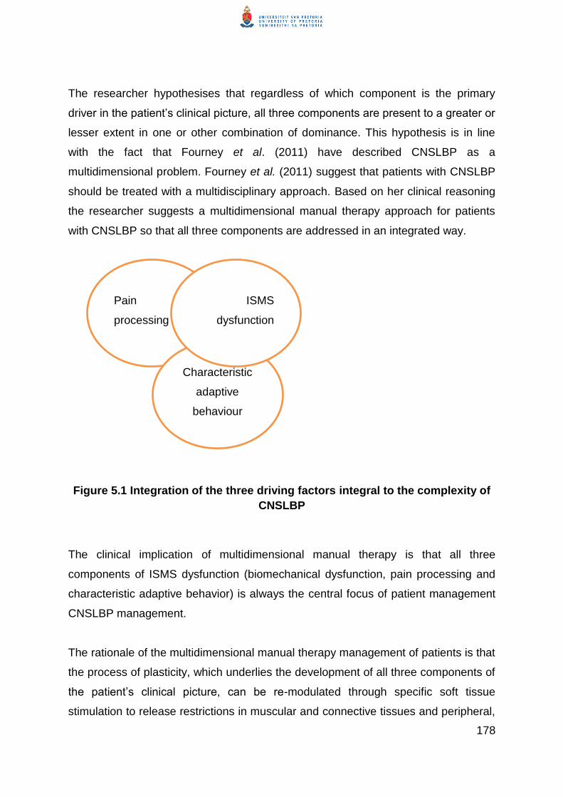
178
Pain
processing
Characteristic
adaptive
behaviour
The researcher hypothesises that regardless of which component is the primary
driver in the patient‘s clinical picture, all three components are present to a greater or
lesser extent in one or other combination of dominance. This hypothesis is in line
with the fact that Fourney et al. (2011) have described CNSLBP as a
multidimensional problem. Fourney et al. (2011) suggest that patients with CNSLBP
should be treated with a multidisciplinary approach. Based on her clinical reasoning
the researcher suggests a multidimensional manual therapy approach for patients
with CNSLBP so that all three components are addressed in an integrated way.
Figure 5.1 Integration of the three driving factors integral to the complexity of
CNSLBP
The clinical implication of multidimensional manual therapy is that all three
components of ISMS dysfunction (biomechanical dysfunction, pain processing and
characteristic adaptive behavior) is always the central focus of patient management
CNSLBP management.
The rationale of the multidimensional manual therapy management of patients is that
the process of plasticity, which underlies the development of all three components of
the patient‘s clinical picture, can be re-modulated through specific soft tissue
stimulation to release restrictions in muscular and connective tissues and peripheral,
ISMS
dysfunction
179
and autonomic neural systems. The hypervigilance of the central nervous system is
addressed by direct handling techniques to remodel soft tissues as well as
reassurance of the patient, implementing cognitive behavioural principles during the
handling techniques to educate the patient regarding his /her condition.
The guarded and maladaptive movement responses are influenced from a
biomechanical approach by the release of the soft tissue and joint restrictions at
segmental and multisegmental levels. The researcher‘s observation in clinical
practice is that by releasing the soft tissue and joint restrictions the muscle tone and
elasticity (length) is prepared for recruitment. The researcher hypothesises that by
releasing the soft tissues and joint restrictions, it is possible that the restoration of
disrupted spatial awareness and tactile processing can be facilitated (Moseley et al.,
2012). The guarded movements can also be prepared for realignment of the
segmental and multisegmental skeletal structures as well as from the perspective of
re-learning of postural control (Fersum et al, 2012)..
The maintenance of the released restrictions in the soft tissues and ISMS alignment
(through a specifically well-designed exercise programme and lifestyle) can only be
achieved through re-education of postural control and movement through
appropriate cognitive behavioural input. Based on her clinical experience the
researcher found that input regarding a specific exercise program needs to be
provided at a frequency that the patient can manage so that she/he is able to take
control of the behavioural and lifestyle changes that are needed to manage their
condition.
The multidimensional approach does not exclude simultaneous intervention by other
health professionals in a multidisciplinary approach but whether other interventions
are included will depend on the patient‘s responses to the manual therapy approach
and needs.
The multidimensional manual therapy approach as discussed in this chapter is in
keeping with a dialectic approach to the management of patients with CNSLBP. In a

180
dialectic approach the identification of the patient‘s problem requires an integrated
understanding of their characteristics (identified by the patient‘s stories or the
patient‘s lived experience) as well as the definable physical or other signs and
symptoms. In Section 5.2 the researcher discusses the principles of a
multidimensional manual therapy approach to patients with CNSLBP from the
perspective that the potentially strong emotional and behavioural factor in these
patients makes it necessary for them to have confidence and trust in the therapist to
address their problem. The manual therapist should establish an interactive
professional relationship with the patient during the assessment of the patient to
engender mutual trust and confidence. This professional relationship with the patient
forms the background as well as the channel through which the multidimensional
management takes place. The manual aspect of the treatment is based on
continuous interactive feedback between therapist and patient and for this reason is
an inseparable part of patient management from a dialectic perspective.
Pain management through facilitation of endogenous pain-inhibiting mechanisms
involves an integrated interaction between:
The therapist as a pain-inhibiting agent in a professional therapist-patient
relationship in which the therapist re-assures the patient regarding their
condition;
Manual therapy to:
o Inhibit ascending pain modulation by releasing soft tissue and joint
restrictions at segmental and multisegmental levels and re-aligning the
ISMS;
o Facilitate the restoration of disrupted tactile processing (Moseley et al.,
2012); and
o Facilitate descending pain modulation through the ventral
periaqueductal grey (vPAG) and dorsal periaqueductal grey (dPAG);
Pharmacology; and
Re-education of postural control.

181
Appropriate health education is given on adaptation of lifestyle to maintain
and re-enforce the effect of the pain modulation and ISMS dysfunction
intervention.
5.2 Principles of pain modulation
The neurophysiology of pain modulation is complex and is the subject of much
research at present. The principles of pain modulation discussed below have been
identified in the literature as significant in pain modulation in patients with chronic
pain (Bioloski et al., 2009).
5.2.1 The therapist as pain-inhibiting agent through a professional therapist-
patient relationship
Reflection on and discussion of the researcher‘s clinical experience and clinical
reasoning in this thesis have been undertaken from a dialectic perspective, which
implies that the patient‘s stories and participation in their assessment and treatment
are crucial.
Evidence is growing that the therapist-patient therapeutic relationship plays a role in
the management of pain disorders (Slade, Molloy & Keating, 2009). This suggests
that putting the patient at ease about how serious their condition is a very important
starting point for managing the patient with CNSLBP. This conversation can take the
form of an explanation of what the condition is and what the intervention will entail
following the outcome of the assessment.
By putting the patient at ease, explaining why they are experiencing consuming pain
and how it can be treated, the manual therapist aims to decrease the activation of
the amygdala and insula (hypervigilant limbic system) through decreasing the fear of
pain and catastrophising. At the same time the explanation gives the patient
information necessary for dealing with the pain and for making deliberate cognitive
(prefrontal cortex) decisions on how to understand the pain and manage it.

182
Empowering the patient to manage all three components of their condition, their
lifestyle and the environmental factors influencing the condition is crucial for the
success of the total management of the patient. By giving the patient essential,
coherent, accessible and valid information about their condition, the manual therapist
aims to correct any false beliefs about the condition and understand their role in the
holistic management.
5.2.2 The role of cognitive behavioural therapy in the multidimensional
manual therapy of patients with CNSLBP
In this thesis the researcher has indicated that cognitive behavioural principles are
essential in the multidimensional manual therapy management of patients with
CNSLBP. The fact that reassurance, presentation of information and appropriate
health education are integrated into the manual therapy is probably the reason why
patients are empowered to take control of their condition and manage it in
collaboration with the therapist. The appropriate behavioural responses (Buchner et
al., 2007; Gatchel &Turk, 2002) that a patient needs to be able to achieve include:
Developing confidence in their skills and ability to re-think and change their
beliefs about their pain to decrease the catastrophic thinking about their
predicament that if they move they will experience pain;
Acquiring mental techniques to reduce pain by getting rid of unhelpful
thoughts, changing the focus of their attention away from their pain and
redefining pain as a different sensation; and
Managing work-related and social stress more effectively and acquiring skills
to cope with the pain and/or recurrences of symptoms.
Relaxation techniques can form part of the cognitive behavioural approach to the
management of CNSLBP. The ultimate goal will be for the patient to take
responsibility to adapt their lifestyle and to manage their social stressors and achieve
job satisfaction.

183
If the manual therapist observes that the patient needs more intensive cognitive
behavioural therapy to achieve more appropriate behavioural responses and self-
management of their condition, the therapist can refer the patient to a psychologist
(Fourney et al., 2011; Buchner et al., 2007).
Health education during the handling techniques and directly afterwards should be
directed towards correcting the patient‘s health beliefs and perceptions and
addressing mal-perceptions the manual therapist identifies during the assessment.
Empowering the patient to change their lifestyle and manage environmental factors
cannot be separated from the segmental and multisegmental soft tissue release and
ISMS re-alignment, from re-education of postural control, and from kinetic handling.
Empowerment takes place as the patient is able to take control of their postural
control and maintain their lifestyle (Zusman, 2002).
5.2.3 Pain modulation through manual therapy
Manual therapy addresses pain modulation through soft tissue release and through
stimulation of the mechanoreceptors in the joints. Zusman (2010 p 109) states:
‗Constant mechanical stimulation is fundamental to the homeostasis of the
musculoskeletal system.‘ The mechanical stimulation Zusman (2010) refers to is
applicable to soft tissue and to mechanical stimulation of the mechanoreceptors in
the joints.
5.2.3.1 Inhibition of ascending pain modulation
Ascending pain modulation through manual therapy is achieved through release of
soft tissue and joint restrictions on segmental and multisegmental levels and through
re-alignment of the ISMS.
Muscle tone decreases (because ISMS dysfunction starts as a result of local reflex
muscle spasm at the L4-S1 motion segments) and muscle stiffness filters throughout
the entire muscular component of the ISMS as a result of nociceptive stimulation.
Muscle spasm, taut bands and trigger points are released sequentially through
184
stretching, myofascial release and trigger point release (deep myofascial release
techniques applied according to the patient‘s pain response).
The aim of the release techniques is to restore the viscosity of the ground substance
in fascia (actin and myosin filaments) in the muscle, which results in the decrease of
the sensory afferent input to the dorsal horn and higher centres. Added to this is the
fact that myofascial release stimulates the mechanoreceptors, which results in
deactivation of the gamma motor tone regulation system. The slow deep pressure
stimulation leads to the stimulation of mechanoreceptors and probably also the
slowly adapting Ruffini endings and some of the interstitial receptors, such as the
muscle spindles. This leads to an altered proprioceptive input to the CNS, which then
results in a changed tonus regulation of motor units (Schleip, 2003). If the functioning
of the motor unit is restored, muscle fibres can fire and recruit (Comerford, 2008).
Specialised and unspecialised loose connective tissue (fascia) stiffening, including
myofascial shortening within the entire ISMS (with emphasis on the TLF), occurs as
a result of a lack of movement (due to compensatory fixation and guarded
movements most probably as a result of fear of pain) and/or as a result of adaptive
characteristic behaviour and a passive lifestyle.
Poor posture can result in shortened fascia or can occur as a result of shortened or
dysfunctional fascia (especially TLF) that contributes to poor posture. Either way the
mechanism operates initially locally (L4-S1 motion segments) and then filters
throughout the entire ISMS.
When fascia is released in the dysfunctional ISMS through manual therapy, a major
source of afferent nociceptive input (Langevin & Sherman, 2006) to the dorsal horn
and higher centres is also decreased, which results in a decrease of sensitisation in
these areas. The decreased afferent input to the dorsal horn is further improved by
the fact that muscle ischaemia, which contributes to pain, is also reduced. As Sluka
and Milosavljevic (2009) point out, ‗Decreased muscle spasm would be expected to
decrease muscle ischemia and this reduces nociceptor sensitization and lessens
central input to the spinal dorsal horn.‟

185
The soft tissue release creates the space to restore alignment of the biomechanical
aspect of the dysfunctional ISMS through the joint system, by applying passive
intervertebral movements in required therapeutic directions with the aim of restoring
the dysfunctional ISMS joint mobility and the soft tissues (muscles, joint capsules,
ligaments and tendons) to their normal length. Mobilisation of the intervertebral (and
costovertebral, costosternal and costochondral joints in the thoracic region)
throughout the entire dysfunctional ISMS will also restore the patient‘s rib excursion
and improve their breathing pattern. When this happens, the afferent input via the
nociceptors to the dorsal horn and the higher centres is further decreased from this
nociceptive source.
Zusman (2010) states that the effect of passive movement procedures on pain
modulation is relatively small and changes in tissue position or movement are not
lasting. He reports conflicting evidence on the long-term changes in structure and
symptoms and disability following passive movement. In the multidimensional
manual therapy approach that is the subject of this thesis these limited responses do
not affect the short-term effect of the passive movement because they serve as a
preparatory technique for the implementation of treatment techniques such as active
re-education of postural control.
Pain modulation through manual therapy takes place through the direct muscular
reflexogenic response as a result of the effect of manual therapy at the spinal level
(Potter, McCarthy & Oldham, 2005). Because the motion segments, head and pelvic
girdles of the dysfunctional ISMS is re-aligned the spinal curves is restored and the
local reflexogenic muscular (myofascial) response on segmental and multisegmental
levels occur.
Stroking over the areas of the skin innervated by primary posterior rami of spinal
nerves leads to decrease in muscle tone (relaxation), which contributes to decrease
in pain (Schleip, 2003; Kostopoulos & Rizopolous, 2001).

186
On the basis of clinical reasoning and clinical experience, the researcher
recommends that in patients with major tissue stiffness and pain the treatment
technique of choice would be to release the motion segment (segmental level) to
address pain relief first. Thereafter, the soft tissue release should be performed. The
reasoning behind this recommendation is that the primary release (mobilisation) of
the motion segment (joints and soft tissues within and surrounding the motion
segment) releases the restricted tissues (containing the nociceptors) in response to
the movement.
For the lower lumbar motion segments to be released the patient should be in the
most neutral position as possible. This neutral position of the lower lumbar vertebrae
is reached the easiest in sidelying (Figure 5.1).
Figure 5.1: Lumbar rotation in sidelying (Maitland, 2004)
Segmental joint mobilisation, which should routinely be carried out in prone, is
passive accessory movements in all directions of each motion segment, especially in
the direction of segmental restriction. Segmental joint mobilisation does not mobilise
only the osseus structures but also the deep fibrous structures, such as the
ligaments and facet joint capsules (Maitland, 2004; Richardson, Jull, Hodges &
Hides, 1999). Graded tensile loading by passive movement onto healing or unhealed
soft tissues could facilitate optimal repair and tissue integrity (Zusman, 2010).
187
Tendons, joint capsules and ligaments are also classified as specialised connective
tissue and are also mobilised (Middleditch & Oliver, 2005; Porterfield & DeRosa,
1991; Kapandji, 2008).
Release of segmental joint restrictions in the lower lumbar area will, by definition, be
the area of focus for initiating the treatment, followed by releasing the soft tissues
and fascia that attach to the pelvis and in the lower limb. The thoracolumbar junction,
thoracic spine, and soft tissues that attach to the scapulae and relevant soft
structures in the upper limb and the cervico-thoracic junction, cervical region and
head on the 01 are also released.
The researcher found clinically that sidelying is the key position for initiating the
release of the segmental restrictions in patients who present with a torsioned lower
quadrant. The release of the motion segments and the soft tissues is achieved when
the range of motion increases and the patient experiences less pain with less
somatic referred pain.
Mobilising the motion segments and soft tissue at and around the transitional areas
results in elongation of the spine and in some release of the transverse pelvic and
respiratory diaphragm and the thoracic inlet. The relevance of mobilising these
transition areas is that it is an essential part of the restoration of the patient‘s
breathing pattern.
The soft tissue release includes the muscles (myofascia) (through release of muscle
spasm, trigger points and taut bands), TLF and other connective tissue structures
such as loose unspecialised connective tissue, joint capsules and, finally, the highly
sensitive neural and dural tissues.
Because specialised and unspecialised connective tissue depends on movement
and stretch to maintain its mobility, it should be put under lengthening strain such as
during normal full range movement (Langevin & Sherman, 2006). Myofascial
techniques to remodel connective tissues are therefore skilled applications of manual
stretching, trigger point release and positioning to release trigger points, taut bands,
188
muscle spasm and shortened soft tissue in general (Mannheim & Lavett, 1989;
Schleip et al., 2012; Langevin & Sherman, 2006).
Myofascial release creates mobility of soft tissues because the mechanical effect of
the stretching of the deeper muscle fibres (elasto-collagenous component of the
muscle) changes the ground substance from a solid block to its state of viscosity.
Fascia is the only tissue that modifies its consistency when under stress (plasticity)
and which is capable of regaining its elasticity when subjected to manipulation
(malleability) (Stecco et al., 2004).
Treatment techniques for releasing soft tissues used by the researcher include
myofascial release (Barnes, 1997), trigger point therapy (Dommerholt, 2012), Rolfing
structural integration (Caspari & Massa, 2012) myofascial induction approaches
(Pilat, 2012), and neural release (Wander & Weinschenk, 2012). In this thesis these
techniques are defined as direct manual mechanical tissue release techniques. The
manual therapist should implement the most appropriate techniques for achieving
the required outcome.
Other techniques in the literature include osteopathic manipulative therapies (King,
2012), connective tissue manipulation (Prendergast & Rummer, 2012) fascial
manipulation (Stecco & Stecco, 2012), accupuncture as a fascia-oriented therapy
(Irnich & Fleckenstein, 2012), and stretching of fascia (Myers & Frederick, 2012).
Stretching exercises, which are inherently part of yoga and pilates, are also
described as techniques to mobilise fascia and are an important part of the
discussion on exercise for maintaining soft tissue and joint mobility. In sidelying,
release of the soft tissues at segmental level is also achieved more easily (Figure
5.2).
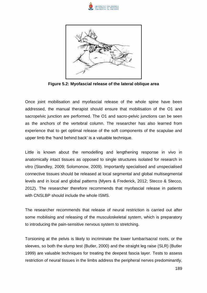
189
Figure 5.2: Myofascial release of the lateral oblique area
Once joint mobilisation and myofascial release of the whole spine have been
addressed, the manual therapist should ensure that mobilisation of the O1 and
sacropelvic junction are performed. The O1 and sacro-pelvic junctions can be seen
as the anchors of the vertebral column. The researcher has also learned from
experience that to get optimal release of the soft components of the scapulae and
upper limb the ‗hand behind back‘ is a valuable technique.
Little is known about the remodelling and lengthening response in vivo in
anatomically intact tissues as opposed to single structures isolated for research in
vitro (Standley, 2009; Solomonow, 2009). Importantly specialised and unspecialised
connective tissues should be released at local segmental and global multisegmental
levels and in local and global patterns (Myers & Frederick, 2012; Stecco & Stecco,
2012). The researcher therefore recommends that myofascial release in patients
with CNSLBP should include the whole ISMS.
The researcher recommends that release of neural restriction is carried out after
some mobilising and releasing of the musculoskeletal system, which is preparatory
to introducing the pain-sensitive nervous system to stretching.
Torsioning at the pelvis is likely to incriminate the lower lumbar/sacral roots, or the
sleeves, so both the slump test (Butler, 2000) and the straight leg raise (SLR) (Butler
1999) are valuable techniques for treating the deepest fascia layer. Tests to assess
restriction of neural tissues in the limbs address the peripheral nerves predominantly,
190
while passive neck flexion (PNF) and slump movements are predominantly tests and
treatment (stretching techniques) for restrictions in the neural canal (CNS).
These peripheral and central neural tension techniques can be carried out at
different stages of the treatment, based on the stage at which the spinal and limb
movements can be stretched into the restriction. These technique(s) can also be
carried out together with breathing exercises to increase the range of movement.
Breathing exercises serve as a strategy to empower the patient to take control of the
movement and the discomfort that accompanies the stretching technique.
Although myofascial release should be achieved by stretching to change the ground
substance from a solid block to its proper viscosity, the force should be adapted on
the basis of the clinician‘s interpretation of the patient‘s response to the clinician‘s
handling skills.
It is well known in physical therapy for example that application of direct
tissue stretch to ligaments, joint capsules needs to be gauged carefully to
avoid causing increased tissue inflammation. To understand how much
force (or movement) is beneficial, and how much can be harmful is one of
the challenges of these clinical modalities (Langevin &Sherman, 2006 p
5).
The therapist should also apply the mobilisation of the peripheral and central neural
restrictions with caution, taking cognisance of the reasons for underlying strain on
the soft tissues and joints and potential underlying inflammatory responses, which
will sensitise the nervous system rather than mobilise it.
In addition, the continuum of the nervous system is proof of its potential to reach
beyond the physiological limits of the nervous system. This means that with
treatment the handling skill of the manual therapist is critical for gauging the extent of
the forces and angles applied to the nervous system with this technique. The
nervous system is naturally full of nociception but the other hidden problems such as
neural and dural adhesions and restrictive interfaces in general can also flare the
system to a painful state.
191
5.2.3.2 The facilitation of the restoration of disrupted tactile processing
The restoration of disrupted tactile processing and spatial discrimination (Moseley et
al., 2012) is probably facilitated throughout manual therapy when the manual
therapist receives feedback from the patient on the localisation of touch and the
response of pain during the handling techniques. Tactile processing is disrupted
probably due to the descending pain-enhancing mechanism by reducing the
effectiveness of the second order inhibitory pathways in the spinal cord (Field, 2009).
Schleip (2003 p 17) states that:
In the case of a slow deep pressure, the related mechanoreceptors are
most likely the slowly adapting Ruffini endings and some of the interstitial
receptors; yet other receptors might be involved too (e.g. spindle receptors
in affected muscle fibers nearby and possibly some intrafascial Golgi
receptors).
This statement adds to the support of the argument that manual therapy (myofascial
release) will contribute to the restoration of tactile processing and spatial
discrimination in addition to the cognitive awareness created by the localisation of
touch during manual therapy.
The researcher hypothesises that through the feedback during the manual therapy
the prefrontal and limbic centres are stimulated through the spinobrachial tract
(Kuner, 2010; Figure 5.3). The localisation of the touch and proprioceptive input
through direct handling techniques (deep pressure and stretch) is carried through the
lateral spinothalamic tract and posterior columns (together with the cognitive feed-in
to the prefrontal cortex and the limbic centre, which is the area for interpreting the
cognitive-emotional experience of pain).This enables the patient to regain any tactile
spatial orientation that may have been lost / diminished due to the pain and
dysfunction the patient has experienced and that overrides the proprioceptive and
tactile input. As the manual therapist guides the patient through the process of the
handling techniques she attempts to alert the patient to the site of the pain and depth
of the pain, and its relationship to other structures. The manual therapist should
explain to the patient throughout the treatment what is happening with the soft tissue

192
release and re-alignment of the motion segments of the spine as the treatment
progresses. Through this interaction of therapeutic feedback, the manual therapist
aims to facilitate the restoration of disrupted tactile processing by increasing the
patient‘s understanding of the origin of their signs and symptoms and how these
signs and symptoms are responding to manual therapy.
During the multidimensional manual therapy the patient is assisted towards taking
cognitive control of their emotions evoked by catastrophisation and towards adapting
their beliefs regarding their functional limitation caused by CNSLBP.
Figure 5.3: A schematic overview of the main circuits that mediate
physiological pain
(Kuner, 2010)

193
The process described above is preparation for active and loaded restoration of
muscle balance, which involves posture and movement retraining during activities.
5.2.3.3 Facilitation of descending pain modulation through the vPAG and the dPAG
Another aspect of endogenous pain-inhibiting mechanisms is descending pain
modulation through the vPAG and dPAG.
Through the direct stimulation of the intervertebral joints and the myofascial and
trigger point release, the ‗stimulus‘ reaches not only the sensory discrimination
centre and emotional aversive areas in the limbic centre (see Figure 5.4) (Kuner,
2010; Brooks &Tracey, 2005) but also the dPAG. From the dPAG the descending
pathways carry noradrenaline when it synapses with the dorsal horn and activates
the inhibitory interneurons.
When the inhibitory interneurons are activated, they release the endogenous opioid
neurotransmitters enkephalin or dynorphin, which bind to the µ-opioid receptors on
the axons of the A-delta and C fibres that carry afferent nociceptive impulses from
the periphery. The activated µ-opioid receptors inhibit the release of substance P,
which in turn inhibits the activation of the neuron from where the pain impulses are
transmitted to the brain. The nociceptive impulse is therefore inhibited before it can
reach the cortical areas responsible for the interpretation of pain and before the
limbic centre is activated. The dPAG inhibitory control on the dorsal horn is an
immediate hypoalgesic response and is part of the flight or fright reaction (Kuner,
2010).
The vPAG consists of the ventrolateral columns of the PAG and the dorsal raphae
nucleus, which is sympatho-inhibitory in action and involves serotonin as the
neurotransmitter.
The vPAG is stimulated via the stimulation of the sympathetic trunk when the
intervertebral and, especially, the costovertebral joints are being mobilised during
manual therapy.
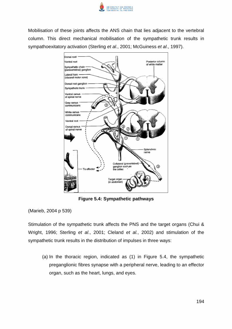
194
Mobilisation of these joints affects the ANS chain that lies adjacent to the vertebral
column. This direct mechanical mobilisation of the sympathetic trunk results in
sympathoexitatory activation (Sterling et al., 2001; McGuiness et al., 1997).
Figure 5.4: Sympathetic pathways
(Marieb, 2004 p 539)
Stimulation of the sympathetic trunk affects the PNS and the target organs (Chui &
Wright, 1996; Sterling et al., 2001; Cleland et al., 2002) and stimulation of the
sympathetic trunk results in the distribution of impulses in three ways:
(a) In the thoracic region, indicated as (1) in Figure 5.4, the sympathetic
preganglionic fibres synapse with a peripheral nerve, leading to an effector
organ, such as the heart, lungs, and eyes.
195
(b) The passive therapeutic mobilisation of the motion segments also leads to
stimulation of the sympathetic trunk in the adjacent vertebral levels higher
and lower than the stimulated level – [number (2) in Figure 5.4] which
synapses with neurons in other parts of the sympathetic trunk to join a
peripheral nerve. The effect of the stimulation of the sympathetic trunk at
one level therefore has a diffuse effect throughout the nervous system
because during the passive mobilisation of the motion segments is
performed on the entire ISMS system and will therefore mobilise the
sympathetic trunk at various levels and have a widespread effect on the
ANS.
(c) Mobilisation to the intervertebral and costovertebral joints (ISMS joints)
can also activate the preganglionic fibres directly (immediate hypoalgesic
effects following SMT, which is specific to mechanical nociception as
opposed to thermal nociception).It may be affected via the sympathetic
trunk, which can form part of the splanchnic nerve [(3) in Figure 5.4] that
synapses with the adrenal medulla in the kidneys. The result of the
activation of the adrenal medulla is the release of catecholamines into the
blood stream (Marieb, 2004 p 540), which reach the vPAG over time. The
release of serotonin from the vPAG to the dorsal horn results in a
hypoexitatory effect, known as a descending pain-modulating mechanism,
which results in decreased pain perception (Sterling et al., 2001; Vicenzino
et al., 1996).
Descending pain modulation through the dPAG has an immediate effect on
pain perception, whereas pain modulation through the vPAG has a delayed
pain-modulatory effect because the release of catecholamines from the
adrenal medulla stimulates the vPAG via the blood circulation to release
serotonin.
Not all researchers who discuss descending pain modulation distinguish very clearly
between the effect of pain modulation via the dPAG and the vPAG (Kuner, 2010;
196
Brooks &Tracey, 2005). Kuner (2010) states that much debate still takes place on
the noradrenergic and seronergic pain-modulatory mechanisms.
To conclude the discussion on the pain modulation through manual therapy, the
researcher presents an adapted version of the interrelations of processes involved in
generating and maintaining CNSLBP in Figure 5.5. This diagram is included to
indicate how manual therapy can contribute to the reversing of the interrelated
processes that generate and maintain pain processing as described by Field (2009).
The focus of the intervention starts with understanding the patient‘s ‗problem‘ from
their individual (usually ill-structured) perspective to determine in which way it
impacts their life during the assessment of the patient. This enables the manual
therapist to relate the patient‘s responses to the activities she/he experiences and
relates to his/her CNSLBP problem
Empowering the patient to participate in the management of their CNSLBP problem
is something that runs through every aspect of the management from assessment to
intervention through health education.
Releasing the soft and neural tissues, mobilising and re-alignment of dysfunctional
ISMS on segmental and multisegmental levels to achieve pain modulation on
peripheral, central and cognitive level, and
Re-education of postural control, adaptive and maladaptive movement during
functional activities
5.2.4 The role of pharmacology as part of the holistic approach to manual
therapy for patients with CNSLBP
Release of soft tissue restrictions and mobilising synovial joints in the ISMS may
result in inflammatory responses. The most effective way to treat this post-manual
therapy inflammation is through appropriate pharmacological agents. The reasoning
behind this is the promotion of quicker healing in the soft tissues and limitation of the
formation of adhesions. Pharmacological agents can include anti-inflammatories,

197
muscle relaxants and analgesics and/or anxiolytics to treat pain-induced fear of
movement (Langevin &Sherman, 2006).
Improving our understanding of therapeutic mechanisms of drug interactions is key
to developing more effective treatment strategies for CNSLBP with minimal adverse
effects.
5.2.5 Re-education of postural control
Re-education of postural control is introduced the moment that pain modulation
becomes effective. The re-education of movement as such then becomes another
pain modulation factor because it actively maintains the passive (manual) release of
segmental and multisegmental soft tissue and joint restrictions. General exercise and
activity are also known to have a pain modulatory effect due to mechanoreceptor
stimulation and result in endogenous opioid release that inhibits pain (Van Tulder &
Koes, 2002).
Direct comparisons of different exercises have failed to show that one is any more
effective than another or that changes in pain are non-specific and similar in all three
groups (Oldervoll et al., 2001; Petersen, 2002).
An explanation for the conflicting evidence on the effectiveness of exercise in the
management of patients with CNSLBP is that the specific exercises are not as
important as physical activity per sé (Van Tulder & Koes, 2002; Macedo, Latimer,
Maher, Hodges, Nichola, Tonkin, McAuley & Stafford, 2008; Middelkoop et al.,
2010). Exercises are aimed at specific (neuro)-musculoskeletal outcomes where
(functional) rehabilitation is aimed at restoring a patient‘s functional ability in their
environment on a daily basis and therefore need to be more patient specific than a
general exercise programme. Activities of daily life (which may include specific
exercises or functional activities to achieve a specific physical effect, i.e. muscle
activation) should therefore be adapted to each patient‘s needs and challenges in
their environment.

198
In keeping with the underlying reasoning in this thesis the researcher suggests that
re-education of postural control should aim to teach the patient to recruit appropriate
muscles and muscle groups to restore or optimise the patient‘s posture in the
functional positions that they adopt during activities of daily life (ADL). Muscle
recruitment should be carried out on appropriate segmental and multisegmental
levels as well as during functional movement to optimise segmental and
multisegmental stability and mobility. It is, however, also critical to optimise or restore
the patient‘s sequence of movement during functional activities with the aim of
addressing guarded, adaptive and provocative movements.
Van Tulder and Koes (2002) and Macedo, Latimer, Maher1, Hodges, Nichola,
Tonkin, McAuley and Stafford (2008) found no clear relationship between the type or
intensity of exercise and physical performance or improvement in pain and ability.
That is also true of rehabilitation and return to work.
O‘Sullivan (2011) concludes that contracting stabilising muscles (pelvic floor,
transverse abdominus and lumbar multifidus) prior to spinal loading exercises has
not addressed the disability associated with CNSLBP. Although randomised
controlled trials have shown the small effect of sizes in the treatment of patients with
CNSLBP, this is not a superior approach to other conservative approaches.
O‘Sullivan (2011) acknowledges that stabilising exercises for patients with CNSLBP
have some benefit but indicates that the underlying basis for this approach is
questioned.
In the RCT by Fersum et al. (2012) the results have shown that multidimensional
CB-CFT programme (cognitive behavioural education regarding their CNSLBP,
health education regarding lifestyle, combined with an exercise programme as well
as retraining adaptive and maladaptive movement including in the work place)
resulted in superior reduction of patients‘ pain, disability, fear, beliefs, mood and sick
leave at the 12 month follow-up. It therefore appears that exercise should not be
given without extensive health education and cognitive behavioural input by health
professionals and specifically manual therapists

199
Based on the multidimensional complexity of the clinical picture of patients with
CNSLBP in this study, the researcher suggests that the role of exercise in the
management of patients should include not only segmental stabilisation exercises
but specifically segmental mobility and multisegmental stability and mobility
exercises to address the ISMS dysfunction. However, restoring the patient‘s
segmental and multisegmental stability and mobility with specific exercises
addresses only the ISMS components of postural control and not the re-learning of
functional movement during ADL, which has also been impaired due to the abnormal
changes in the brain.
It is also important that segmental and multisegmental stabilising and mobilising
exercises should contribute to the patient‘s awareness of good posture in all
functional positions that they adopt during the day. The rationale behind this
statement is that the patient has to be aware of the strain that poor posture has on
the segmental and multisegmental levels of the ISMS and how it contributes to their
CNSLBP condition.
The researcher suggests that balance exercises and kinetic handling should be
included to address the patient‘s tendency to use guarded and adaptive movement
patterns to avoid pain. The patient has to re-learn to move with a normal sequence
of movement without fear avoidance. The therapist should therefore ensure
appropriate muscle recruitment during functional activities of daily living (FADL),
such as climbing stairs, walking, getting in and out of a car, and sitting to standing.
The researcher is of the opinion that muscle activation during normal physical activity
(ADL) is the key to successful intervention. However, this statement still needs to be
verified by research.
The manual therapist should therefore ensure that the patient‘s postural control is
restored by teaching them to normalise their anticipatory control (inhibit excessive
anticipatory movement during ADL) and movement strategies (i.e. through kinetic
handling) during performance of instrumental activities of daily living (IADL) and
general ADL.

200
These assumptions suggest that for retraining postural control it is essential to work
on identifiable functional tasks rather than on movement patterns for movement‘s
sake alone. A task-orientated approach to re-learning postural control during
functional activities assumes that patients learn by actively attempting to solve the
problems inherent in a functional task rather than repetitively practising normal
patterns of movement. Adaptation to changes in the patient‘s environment is a
critical part of recovery of function. In this context, patients are helped to learn a
variety of ways to solve a problem to perform a task rather than a single muscle
activation pattern through a process of external feedback.
Moseley (2005) describes graded motor imagery as a useful method to retrain
movement-related networks without eliciting pain in the CNSLBP population. The
researcher supports the concept of using graded motor imagery to retrain
movement-related networks (through motor re-learning).This is because, by
changing the patient‘s visual (visuo-motor) input to imitate more normally sequenced
movement (without causing pain) during activity, characteristic adaptive behaviour
can be changed/adapted. In addition more normally sequenced movement can be
systematically re-learned (unlearning maladaptive compensatory movement) by
utilising the role of higher centres in re-education of motor (postural) control
(Shumway-Cook & Woolacott, 2007).
5.3 Principles of multidimensional pain modulation in patients with
CNSLBP
To summarise the discussion on the multidimensional pain modulation discussed in
this chapter the researcher presents an adapted version of the interrelations of
processes involved in generating and maintaining CNSLBP in Figure 5.5. Figure 5.5
also indicates how the pain-contributing factors can be modulated by addressing
chronic pain on various dimensions (Field, 2009).
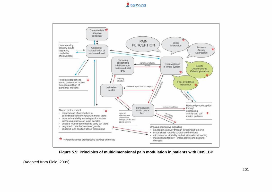
201
Figure 5.5: Principles of multidimensional pain modulation in patients with CNSLBP
(Adapted from Field, 2009)

202
In Sections 5.2 and 5.3 ascending pain modulation through a multidimensional
manual therapy approach in patients with CNSLBP is discussed. Ascending pain
modulation is achieved by decreasing the ongoing nociceptive signalling due to
ISMS dysfunction (peripheral desensitisation) and desensitising the dorsal horn
(central sensitisation). The hypervigilance in the limbic system is also changed
through peripheral and central desensitisation as well as cognitive behavioural input
to decrease the patient‘s fear avoidance, change their health beliefs and improve
understanding of their condition, and as such decrease catastrophisation. Through
decreasing the ascending pain modulation mechanisms the descending pain
modulation is also reduced because the hypervigilance of the limbic system
decreases the descending pain inhibition from the PAG. This process is now
reversed by the decreased signalling to the limbic system so that the effectiveness of
the neuropeptide cholecystokinin in inhibiting the descending pain control via the
PAG is decreased, resulting in pain modulation onto the dorsal horn.
Descending pain inhibition is also especially targeted by the segmental joint
mobilisation of the ISMS, which also affects the ANS chain and activates the pain
modulation process via the vPAG. By facilitating the restoration of disrupted tactile
processing, it is possible that the neuropeptide cholecystokinin, which plays a role in
inhibiting the descending pain control via the PAG, is reversed. This results in
enhancing the recovery of the descending pain inhibition onto the dorsal horn.
A decrease in the hyper-vigilant limbic system, which also plays a role in decreasing
cerebellar coordination and in altered motor control (impaired local and global
stabilisation and mobilisation), might contribute to the cognitive behavioural input to
re-educate postural control (restoring impaired local and global stabilisation and
mobilisation, kinetic handling and inhibiting guarded movement) during functional
activities through a process of motor re-learning.
5.4 Clinical principles for the treatment of patients with CNSLBP
The clinical principles discussed in this section are based on the researcher‘s clinical
experience. The management of a patient with CNSLBP does not only imply the

203
application of techniques but also requires that the therapist skillfully apply the
appropriate handling techniques based on the reasoning that should be based on the
driving factor(s) of the patient‘s condition and other complicating factors such as:
The stage (chronicity) of the CNSLBP condition and the presence of joint
degeneration (wear and tear on joints);
The stage of healing of tissues such as adhesion formation and fibrosis, which
will probably identify the stage of chronicity;
A recurrent (ANSLBP) strain on an existing chronic state; and
The patient‘s immune-neuronal interaction.
Buchner et al. (2007) report that patients with CNSLBP at all stages of chronicity
(including the more advanced stages) benefit significantly from a multidisciplinary
treatment approach to treatment. The more advanced stage of chronicity is usually
associated with adhesions in soft tissues, which became more fibrosed. The
researcher agrees with Buchner et al. (2007) but also found that patients in the
advanced stage of chronicity tissue response show more resistance to treatment and
may need more aggressive handling techniques. The frequency of the treatment in
the advanced chronic stage of the condition still depends on the patient‘s response
to treatment.
Recurrences are likely to occur in patients who are developing CNSLBP due to the
vulnerability of the tissues together with the changes that might have occurred in the
PNS, ANS and CNS due to chronic (recurrent) pain together with the associated fear
avoidance and other psychosocial factors.
Neuronal function can be dramatically altered by activated immune and immune-like
glial cells (Watkins, Hutchinson, Milligan & Maier, 2007). These authors found that
activated immune and immune-like glial cells affect the neural function at the levels
of the peripheral nerve, dorsal root ganglia and spinal cord and therefore have an
effect on patients‘ neuropathic pain and play a role in the development of chronic
pain. This mechanism has been found to disrupt the opioid tolerance and opioid
dependence/withdrawal. In a patient with CNSLBP who has altered immune
responses pain modulation may be decreased due to the effect on the neural

204
function. The therapist should be aware of this potential effect of the immune-neural
interaction on the patient‘s pain response during and after treatment.
5.5 Risk factors to take cognisance of during manual therapy
Throughout treatment, feedback to and from the patient is essential to identify non-
desirable effects of treatment. Undesirable effects of treatment can include biological
and psycho-social factors.
Biologically undesirable effects include the following:
Any centrally initiated symptoms such as nausea, sweating, light headedness
and any changes in sensory disturbance that the patient may have
experienced to the spine, head and limb girdles are biologically undesirable
effects. The manual therapist should be skilled in identifying these signs and
symptoms and make a decision on the progress of the treatment. These signs
and symptoms are usually caused by ANS responses. If these signs and
symptoms are present, the therapist should evaluate the situation and
determine the stage of response before either continuing or stopping the
treatment.
With the direct handling skills the challenge to the manual therapist is to know
how much treatment is enough as too much is detrimental to patient
compliance. Monitoring pain responses and adapting handling skills
accordingly are critical to patient compliance and trust. Excessive tissue
stretching or pressure actually causes inflammation and can conceivably
worsen the patient‘s condition.
The manual therapist‘s skills for addressing the patient‘s fear, encouraging
activity and for appropriately educating the patient to understand and
cooperate in the management of their condition also play a major role in the
successful outcome of the treatment.
The patient is encouraged to tell the therapist everything they experience
during the manual therapy and during the first 24 hours after treatment
(without putting words into their mouth) to enable the therapist to judge the
patient‘s response to treatment and estimate the patient‘s outcomes.
205
The patient‘s emotional response and mood may be reflected in their
willingness to undergo the treatment, positive involvement, and commitment to
participate/collaborate in the treatment.
At the conclusion of the first treatment the manual therapist should:
Encourage and guide the patient to continue or to resume usual activities,
remain as active as possible, including resuming work activities (remaining
active is the most widely respected clinical and scientific recommendation in
the world today) and avoid bed rest as much as possible (European
Guidelines for the Management of Chronic Non-specific Low Back Pain,
2004; National Collaborating Centre for Primary Care, 2009);
Explain to the patient that they may experience discomfort and why it may
happen; and
Should there be a flare-up of soft tissue reaction inform the patient that they
are at liberty to contact the therapist at any time.
The researcher does not give the patient specific exercises at this point in time
because she intends to identify the effect of the first treatment on the patient‘s
inherent capability to respond to the first treatment and not to teach the patient any
new activities to adapt to. After the first treatment the patient must be given time and
be encouraged to use the newly created movement gained during functional activity.
The therapist should know that the repair of tissue should occur within four to five
days. A follow-up treatment should only be carried out after a period of tissue repair
because of the inflammation of the myofascia that might have occurred as a result of
the myofascial release and the passive accessory movements to mobilise the spine.
This treatment should have given the patient new movement to work with during their
ADL. With the confidence of and encouragement by the manual therapist it is
expected that they will have used this movement during their ADL.
Ata the follow-up treatment sessions the manual therapist should re-assess the
patient to identify the effect of the treatment on the ISMS dysfunction and on the
206
patient‘s posture, muscle activation, adaptive and maladaptive movement during gait
and other functional activities that the patient might have engaged in.
If the probability of returning to usual activities is deemed to be low after the first
treatment, the manual therapist should seek to identify the reasons preventing the
patient from returning to their usual activities. These reasons should have been
identified during the assessment of the ‗yellow flags‘ (Waddell, 2004) on the:
Intensity of pain (Visual Analogue Scale);
Perceived disability (Quebec Back Pain Disability Scale or Roland-Morris
Disability Questionnaire or Oswestry Disability Questionnaire);
Symptoms (with no signs) of radiating pain below the knee (clinical
consultation);
Fears and beliefs (Tampa Scale for Kinesiophobia);
Patient‘s projection regarding return to work (three-month projection question
in the Fear-Avoidance Beliefs Questionnaire);
Catastrophising (Pain Catastrophising Scale); and
Absence from any type of work (employment status).
Such a patient may be at high risk for an emotional breakdown and should be
referred for multidisciplinary management.
Whatever documentation system the manual therapist uses, these findings must be
documented to assess future progress of the patient and for legal purposes.
Follow-up treatment should be scheduled for approximately four to five days after the
previous treatment. The second treatment will follow the principles discussed in the
preceding paragraphs. During the second treatment the therapist should be able to
apply the techniques at a deeper level.
At each follow-up treatment the therapist should assess/re-assess:
The salient warning signs and symptoms that the therapist recorded about the
patient‘s original complaints; and
The patient‘s response to the previous treatment.
207
The therapist would anticipate the leg and back pain to be easier. Should this be the
case, the soft tissues in the pelvis and lower limb should be more palpable, revealing
the presence of latent active trigger points. Owing to the release of the soft tissue
restrictions during the first and second treatments the therapist should be able to
palpate and release deeper structures. As the therapist is able to go deeper into the
soft tissue she/he will identify and stir up more restrictions in the soft tissues and
joints. These structures include releasing trigger points and taut bands in the
shoulder and pelvic girdles as well as the attachments between the spine and the
head. Other appropriate techniques for soft tissue release, joint mobilisation and
neural and dural release can be applied. As indicated earlier over treatment is as
bad as under treatment.
The skill of the manual therapist is to identify the dominant components during the
re-assessment and through therapist reflection during and after treatment
hypothesise what the persistent and recurring symptoms are so that they can plan
and perform treatment on the ISMS alignment in terms of posture, gait, abnormal
movement and muscle activation. Any abnormal change in posture, gait and muscle
activation is an indication that pain and restrictive processes are still active in the
ISMS.
As the therapist is able to apply soft tissue release on a deeper level, neural and
dural stretches are introduced manually and by using theraband. As the treatment
progresses it becomes increasingly complex because the repeated release reveals
the structures that may have been the root cause (hidden agenda) of the patient‘s
problem.
As the therapist releases and mobilises the deep lying structures (spine and
capsules of the apophyseal joints in the spine, shoulder (glenohumeral) and hip
joints, this can awaken emotional responses in the patient as a result of fear of the
original pain/catastrophising about the past pain and their unknown response to
treatment. In this case the manual therapist needs to deal with the patient tactfully at
all levels of fear and trust.
208
With the introduction of activity (exercises) and cognitive awareness and
(re)education of motor control, the mobility of the spine, including the lower and
upper quadrants, which have been passively mobilised at segmental and global
levels, is maintained.
Treatment can be a mix of several principles, depending on the patient‘s
presentation and response to treatment. Because of the variety of combinations of
treatment and the multidimensional nature of the condition, treatment competence of
the physiotherapist needs to be monitored over time.
Treatment is not a cure but the improvement of the patient‘s quality of life in terms of
pain responses, which vary from being pain free to experiencing manageable pain
and optimised function during daily life.
The multidimensional model for the manual therapy management of patients with
CNSLBP is presented in Figure 5.6.
5.6 A multidimensional manual therapy model for management of
patients with CNSLBP
The model for the multidimensional manual therapy for management of patients with
CNSLBP is an extension of the model for the assessment of these patients
presented in Chapter 4.

209
Figure 5.6: A Multidimensional manual therapy model for management of
patients with CNSLBP
The focus of the multidimensional manual therapy for patients with CNSLBP is to
optimise the re-alignment of the dysfunctional ISMS. Patients should re-learn
postural control in order to regain and maintain the optimal ISMS function. The aim
of the multidimensional manual therapy is achieved by first releasing soft tissue
210
restrictions in the muscles, connective tissues (including ligaments, joint capsules
and neural and dura tissue) at segmental and multisegmental levels. Soft tissue
release is achieved through the process of modulation of these tissues as a result of
the initiation of the process of plasticity.
Secondly, the realignment of the ISMS is optimised by segmental and
multisegmental mobilisation of the apophyseal and costovertebral, costosternal and
adjacent joints in the ISMS. Optimising the realignment of the ISMS is mainly
possible due to the release of the soft tissue restrictions.
Thirdly, through the release of the soft tissue restrictions and optimisation of the
realignment of the ISMS, pain modulation occurs at peripheral and central levels of
sensitisation and by decreasing nociceptive input to the ‗over-used‘ neuromatrix in
the brain through which the hypervigilance of the limbic system is activated. De-
activating of the ‗over-used‘ pain-generating neuromatrix is suggested in this thesis
as achieved by reassuring the patient regarding their condition, enabling the patient
to change their beliefs and providing appropriate education regarding self-
management and re-education of postural control.
In Chapter 6 an evaluation of the model is presented, limitations of the study are
outlined and suggestions for further research offered.
211
CHAPTER 6
Conclusion, discussion, limitations and recommendations
6.1 Introduction
The core problem in the limited success that has been reported in the treatment and,
specifically, the manual therapy for patients with CNSLBP has been identified as a
lack in the understanding of the complex clinical picture and therefore a lack of
multidimensional management of the patient‘s problem.
The research questions of this study were:
Can the concept of an ‗integrated spinal movement system‘ ISMS be
conceptualised based on the anatomy of the trunk?
What are the underlying systems, processes and influences that result in
ISMS dysfunction and contribute to the clinical picture of patients with
CNSLBP?
What contribution can the professional craft knowledge and the personal tacit
knowledge acquired by the researcher over many years of clinical practice,
make towards the declarative professional knowledge of manual therapy?
The primary aim of this study was to make a contribution to the theoretical
foundation which may help clinicians to understand the biomechanical development
of the complexity of the clinical picture in patients with CNSLBP and the role of
multidimensional manual therapy model in the management of these patients.
Through years of experience, metacognitive reflection on her clinical reasoning and
social construction of knowledge the researcher has developed a multidimensional
manual therapy model for managing the complex clinical picture of patients with
CNSLBP.
212
The development of the components of the model is discussed in Chapters 3, based
on the principles that have evolved from the researcher‘s metacognitive reflection on
her clinical reasoning during patient management and the validation of the
reasoning, arguments, statements and conclusions with published research
evidence. In Chapter 5 the multidimensional manual therapy approach to managing
patients with CNSLBP is discussed and the chapter concludes with the presentation
of the multidimensional conceptualised practice model (theory).
A multidimensional manual therapy model for managing patients with CNSLBP has
been developed and described in three different phases. The phases have not been
developed in sequence, but emerged as the researcher performed metacognitive
reflection on her (1) clinical reasoning and (2) outcomes of her practice and (3)
identified commonalities between patients‘ with CNSLBP‘s clinical presentation and
their (4) responses to the way she practice manual therapy. This process of
interpreting the lived experiences of humans and to generate new knowledge from it
is a subjective and contextually research process which falls within the qualitative
research tradition drawing on interpretive phenomenology and grounded theory (Polit
and Beck, 2007)
The researcher‘s thinking process to and fro (dialectically) between these aspects to
understand and verbalise the whole process/concept of the development of CNSLBP
is called the hermeneutic circle. This process was inherently the researcher‘s
process of creation of meaning (learning and gaining experience in clinical practice
as clinician) and was expanded by the courses and congresses she attended to
broaden her understanding of the research at the time, as well as through the social
learning environment in which she worked by discussing and interpreting her views
against the meanings of colleagues. This process isinherently based on the
phenomenological tradition that meaning is subjective and contextually constructed
(Higgs et al., 2010).
The process of moving between the ‗parts‘ of the multidimensional manual therapy
approach to understand and conceptualise the ‗whole‘ model resulted in
213
metacognitive reflection and culminated in the formulation of the general manual
therapy principles characteristic of the multidimensional manual therapy approach.
6.2 Evaluation of the multidimensional manual therapy model for the
treatment of patients with CNSLBP
6.2.1 Summary of the multidimensional manual therapy model
In Chapter 3 (Section 3.5) the researcher has explained that motion segment
instability does not only affect the lumbar spine only but the ISMS as a whole and
therefore over time ISMS dysfunction becomes the core element of the clinical
picture in patients with CNSLBP. The conceptualisation of the ISMS and
consequently the discussion on the development of ISMS dysfunction, based on the
functional anatomy, is based on the researcher‘s observation and in clinical practice
that patients diagnosed with CNSLBP‘s whole spine and head position, shoulder and
pelvic girdles are affected.
Based on functional anatomy the researchers discussed the possible mechanism
through which ISMS dysfunction can develop as the core element of the clinical
picture of patients with CNSLBP. Characteristic of this ISMS dysfunction is that it
shows the integrated interaction between the neuromusculoskeletal systems and the
patient‘s psychological response to pain and the mechanism through which cognitive
beliefs, social and work-related stressors influence patient‘s pain perception and
ISMS dysfunction. The discussion also indicates that due to hypervigilance in the
brain, social stressors can result in increased muscle tone and shortening of soft
tissues in particular fascia, which triggers ISMS dysfunction (although the
mechanism is different from the motion segment instability approach) and can result
in CNSLBP.
For this reason the researcher advocates that ISMS dysfunction in patients with
CNSLBP should be treated by multidimensional manual therapy regardless of the
mechanism through which it developed. Treatment of ISMS dysfunction entails the
release of soft and neural tissue shortening and realignment of the spine, position of

214
the head and shoulder and pelvic girdles by remodelling the soft and neural tissues
through a process of plasticity.
Remodelling of the soft and neural tissues within and around the spine, head,
shoulder and pelvic girdles and realignment of these structures, pain modulation
takes place through a process of activation of the local and central endogenous pain
modulatory mechanisms.
The multidimensional manual therapy, pain modulation and re-education of tactile
discrimination take place within a dialectic approach to patient care.
Multidimensional manual therapy prepares the patient as a person and his/her
neuromusculoskeletal system for appropriate re-education of postural control and
functional rehabilitation.
Pharmacology is administered to treat soft tissue swelling, pain and to maintain
muscle relaxation.
6.2.2 Evaluation of the multidimensional manual therapy model against other
models used in the management of CNSLBP
6.2.2.1 The lumbar segmental instability concept versus the concept of ISMS
dysfunction
O‘Sullivan (2000) describes lumbar segmental instability as the origin of CNSLBP
and also the cause of recurrences of ANSLBP superimposed on CNSLBP. Although
the researcher agrees in principle that lumbar segmental instability due to the neutral
zone dysfunction can be the primary origin of CNSLBP and that the flexion pattern,
extension pattern, lateral shift pattern and multidirectional pattern can be associated
with some of the variations in ISMS dysfunction (Section 4.2.1.2).
However in ISMS dysfunction as the core contributing factor to the development of
CNSLBP discussed in this thesis the concept of ISMS dysfunction differs from the
lumbar segmental instability concept in the sense that:

215
(1) ISMS dysfunction was observed clinically and then substantiated theoretically
based on functional anatomy. There are therefore similarities between ISMS
dysfunction and the lumbar segmental instability concept. The difference between
the two concepts behind the development of CNSLBP is that the lumbar instability
concept is limited to the lumbar spine while the ISMS dysfunction concept includes
the whole spine, the position of the head, shoulder and pelvic girdles. The
researcher believes that the rotation of the pelvis due to the shortening of unilateral
segmental multifidus on the affected lumbar level together with the shortening of
quadratus lumborum which will result in pelvic torsioning (Section 3.5) which will
spiral up the spinal column to the head and can result in ISMS dysfunction. The
researcher hypothesises that the torsioning of the spine involving the
neuromusculoskeletal and fascia systems especially the thoracolumbar fascia may
be the cause of the widespread signs and symptoms and the heterogenetic nature of
CNSLBP.
Lumbar segmental instability due to neutral zone dysfunction is discussed only in
terms of the effect on the lumbar spine and does not include the effect of the .
(2) ISMS dysfunction involves the position of the head, shoulder and pelvic girdles in
relation to the spine based on the fact that in the presence of ISMS dysfunction,
other areas of the spine can refer pain to the low back and as such it can be
interpreted as symptoms in the low back while the origin is not in the low back.
Hence the motivation behind the multidimensional manual therapy model is to treat
the patient in all dimensions of CNSLBP and not only the low back in patients with
CNSLBP.
(3) The authors Panjabi (2003) and O‘Sullivan (2000) have not reported on the
change in the flexion, extension and lateral shift and multidirectional patterns but
only mention that the patients‘ were treated successfully.
Based on the researcher‘s experience when these flexion, extension and lateral shift
and multidirectional patterns observed in patients in the motion segment instability

216
concept are treated through multidimensional manual therapy the soft and neural
tissues can be released, the tactile awareness can be ‗alerted‘ and the motion
segments can be realigned in preparation to activate the motor units for better
recruitment. The muscle recruitment (re-education of postural control) can then be
done in functional movement patterns during activities as well as work and
recreational environments.
6.2.2.2 Subgrouping models versus ISMS dysfunction
O‘Sullivan (2005) describes eight subgrouping models for sub-classification of
patients with CNSLBP in order to administer patient-centered targeted intervention.
The mechanical loading model described by O‘Sullivan (2000) is also the basis of
the abnormal loading on the lumbar spine as the origin of ANSLBP which according
to the researcher‘s observation, clinical reasoning and discussion based on the
functional anatomy slowly develops over time (more or less 12 weeks) into ISMS
dysfunction.
ISMS dysfunction as it is described in Section 3.5 encompasses the following:
peripheral pain generator model, neurophysiological model, psychosocial model,
signs and symptoms model, motor control model and biopsychosocial model. Over
time as ISMS dysfunction becomes chronic the characteristics of the different
models start to manifest in different combinations. As ISMS dysfunction merges from
acute to chronic (CNSLBP progresses from the early to the late stage) so the
characteristics of the various models manifest in the patients clinical picture. The
longer chronicity persists, the more complex the patient‘s clinical picture becomes
because more of the characteristics of the different models become integrated into
the patients clinical picture. The clinical picture can be further complicated by
superimposed recurrences of acute on chronic to the point where the strain on all the
systems reach a point of break down to the point of failure of adaptive capacity to
cope
The models described by O‘Sullivan (2005) were designed to guide clinicians on
which dimension (model) they should focus their treatment. The multidimensional
manual therapy model presented in this thesis was developed on the inseparable
interaction between the various systems. Therefore the researcher is of the opinion

217
that because of the heterogeneity of CNSLBP as a condition, the clinician should
identify the dominant drivers of the condition but also address the less dominant
drivers of the condition otherwise residual mechanisms will remain to perpetuate the
problem (hence the development of the multidimensional manual therapy model).
6.2.2.3 Manual therapy in clinical trials versus the multidimensional manual
therapy model
Randomised clinical trials in which manual therapy was compared to various forms
of exercise, were conducted by Fersum et al. (2012) and Kääpä et al. (2006). Kääpä
(2006) compared a multidisciplinary rehabilitation which consisted of a combination
of (1) General fitness exercise, (2) Muscle strengthening exercise for all main muscle
groups in the trunk and the lower limbs, (3) Special exercises to correct mobility of
the spine and hip joints, activate the stabilising muscles of the spine, and increase
flexibility of the lower limb muscles. (4) Functional exercises to improve postural
control dynamic body balance, and coordination; (5) Progressive relaxation
exercises to normalise muscle tension, with an individual physiotherapy intervention
group. These different types of intervention was presented by a multidisciplinary
team consisting of a physiotherapist, occupational therapist, rehabilitation physician,
The individual physiotherapy group received (1) Massage, (2) Spine traction, (3)
Manual mobilisation of the spine and (4) Therapeutic ultrasound as well as (5) 15-20
minute light active exercise (muscle stretching, spine mobilisation and deep trunk
muscle exercises), only from a physiotherapist.
The outcome of the study showed that there were no statistically significant
difference between the two groups after the rehabilitation (6-8 week programme), at
6, 12 and 24 months follow-ups.
The difference between the manual therapy that the patients in this individual
physiotherapy group in the trial by Kääpä (2006) received and the multidimensional
manual therapy model in this thesis, is that the aims of the massage, spine traction
and manual mobilisation was not clear from the publication. The aims of the manual
therapy which is part of the multidimensional manual therapy model are clearly
stated as the soft and neural tissues (which include fascia) modulation, the re-

218
education of tactile discrimination, and the re-alignment of the ISMS. The soft and
neural tissue modulation as well as the realignment of the ISMS results in pain
modulation on spinal as well as central level by activating the endogenous pain
modulation process in the body.
The individual physiotherapy explained by Kääpä (2006) as well as the
multidimensional manual therapy model includes exercise as integral part of the
intervention. In the multidimensional manual therapy model the manual therapist
also acts as a pain modulating agent in the sense that she/he reassure the patient
and give him/her relevant patient-education regarding his/her condition. These
aspects were not mentioned as part of the individual physiotherapy intervention by
Kääpä et al. (2006).
The overall encompassing aim of the multidimensional manual therapy model is to
reverse the ISMS dysfunction that occurred in the patients‘ neuromusculoskeletal
systems including the altered pathways in the brain and the patient‘s thinking
patterns through a process of plasticity.
It seems that the individual physiotherapy intervention in the trial by Kääpä et al.,
(2006) was not specifically designed to address the heterogenetic nature of patients
with CNSLBP.
Fersum et al. (2012) also conducted a randomised clinical trial to compare the
outcome of a CB-CFT programme with MT-EX intervention. The CB-CFT
programme seemed to address most of the characteristics of patients with CNSLBP.
However the MT-EX with which it was compared, was also not reported to be
designed to address the heterogenetic characteristics of the clinical picture of
patients with CNSLBP.
The MT-EX intervention was according to the publication not designed to modulate
the dysfunctional systems in patients with CNSLBP through a process of plasticity on
peripheral, central (spinal cord level) and the brain levels. It is therefore not
surprising that CB-CFT results in superior outcomes above the MT-EX intervention.
219
The multidimensional manual therapy model presented in this thesis was specifically
designed to address the heterogenetic characteristics of patients with CNSLBP from
a manual therapy perspective and can therefore not be compared to the outcome of
manual therapy combined with exercise in the trials that was discussed.
6.2.2.4 The mechanisms of a comprehensive manual therapy model for
musculoskeletal pain versus the mechanisms of the multidimensional manual
therapy model for patients with CNSLBP
Bialosky, Bishop, Price, Robinson and George (2009 p 532) propose a
comprehensive model which suggest that a mechanical stimulus initiates a number
of potential neurophysiological effects which produce the clinical outcomes
associated with manual therapy in the treatment of musculoskeletal pain.
The model indicates that the effect of a mechanical stimulus results in a
biomechanical mechanism, neurophysiological mechanism, peripheral mechanism,
spinal mechanisms and supraspinal mechanisms.
Although lasting changes of the biomechanical mechanism have not been identified,
the authors (Bialosky et al., 2009) suggest that the outcomes associated with manual
therapy are a result of the additional mechanisms initiated by the mechanical
stimulus.
Although the authors state that the clinical effectiveness of manual therapy are not
established their model serves as a guide for clinicians as well as further research.
An important aspect of their conclusion for this thesis is that the effect of manual
therapy should not be studied in isolation but that clinicians and researchers should
be aware of the other potential non-specific effects of manual therapy through other
mechanisms.
The comprehensive model by Bialosky et al. (2009) coincides with the
multidimensional manual therapy model developed by the researcher based on
clinical observation and clinical reasoning. Where Bialosky et al. (2009) only

220
presents the model, the researcher has applied the same principles and explained
the outcome thereof in the treatment of the heterogenetic clinical picture of patients
with CNSLBP.
6.2.2.5 Evaluation of the multidimensional manual therapy model compared to
the challenges for the future management of CSNLBP
O‘Sullivan (2011) formulated 12 criteria for the future management of patients with
CNSLBP. The researcher evaluates her multidimensional model for the management
of patients with CNSLBP against these criteria to establish the extent that the model
presented in this thesis met these criteria.
To date there is no clear understanding of the complex heterogenetic nature of
CNSLBP. The multidimensional manual therapy model presented in this thesis
provides the link between the biomechanical, physiological and behavioural
processes inherent in ISMS dysfunction which is suggested by the researcher as the
preceding processes to the development of CNSLBP influenced by social factors.
The multidimensional manual therapy model for the management of patients with
CNSLBP therefore serves as a point of departure from where patients with CSNLBP
can be managed/or managed regardless of the main driver of the condition (ISMS
dysfunction, pain processing and/or characteristic adaptive behaviour) because the
patients will probably present with a varying combination of these three components.
The multidimensional manual therapy model in this thesis further provides clinicians
with a combination of mechanisms within the musculoskeletal, neuromuscular,
neural (brain and ANS), sensory receptors (systems), and behavioural systems
culminating in adaptive and maladaptive postural control within which the main or
multiple driver(s) of chronic pain and the interaction between the systems (and pain
driving factors) can be identified.
The multidimensional manual therapy model has been developed from a dialectic
perspective and as such the interpretation of information from two different
paradigms (psychosocial and biomedical paradigms) is inherently part of the model.
In the discussion on the model the researcher has indicated the role of the therapist

221
as a pain-modulating agent which implies that she/he has to have a thorough
understanding of and ability to interpret the neurophysiological responses (sensory-
discriminative, peripheral and central pain processing as well as altered pathways in
the brain) associated with the biomechanical and social stressors to generate the
patients clinical picture and responses and stressors on his/her condition. With a
dialectic thinking process the clinician has to interpret information from various
domains to synthesise the patient‘s specific problem which may lie mainly in the
biomedical and/or the psychosocial domain. From this synthesis the therapist must
be able to plan and conduct a management plan that should address the patient‘s
heterogenetic problem.
Due to the dynamic interaction between the components that can drive the patient‘s
clinical picture the driving component may change as the patient responds to
multidimensional manual therapy. Recurrent episodes of ANSLBP superimposed on
CNSLBP can therefore be initiated by either social stressors or another
biomechanical strain. The researcher has indicated that regardless of what the main
driving factor of the patient‘s recurrent pain episode is, the patient should always be
expected to present with a combination of ISMS dysfunction, pain processing and
characteristic adaptive behaviour which should be managed to a greater or lesser
extent at each treatment.
The researcher has indicated that referral to other health care practitioners is not
excluded from the multidimensional manual therapy model. She is in favour of a
multidisciplinary approach to management of these patients if and when the need
arises.
The distinction between the three components (ISMS dysfunction, pain processing
and characteristic adaptive behaviour) which can be single or multiple drivers of the
patient‘s CNSLBP and the various combinations between them make it possible for
future research to use the details of this model to refine screening tools or broad
categories (subgroups) which will make it possible to identify and manage these
patients more effectively.
222
6.3 Limitations of this study
Limitations in this study are the factors which are inherent in any qualitative research
study.
The main limitation in studies from an interpretive paradigm is that the researcher is
involved in the data-gathering and data-interpretation processes. In the case of this
study the data-generation and interpretation were conducted on the basis of the
metacognitive reflection on the researcher‘s own clinical practice and clinical
reasoning processes. A specific limitation with such an approach is that the
researcher can ‗make up‘ the clinical reasoning as the study progresses because it is
difficult to recall clinical reasoning on aspects of patient management that have taken
place quite some time ago. It is also possible that the researcher‘s clinical reasoning
could have been based on her personal opinion at the time of the conceptualisation
of the components of the model. These limitations have been limited by the fact that
the researcher‘s conceptualisation of the basic principles of the model was published
in 1995 and still remains the same. She continues to treat patients with (C)NSLBP
and reflect on her treatment approach all the time while writing up this thesis to
confirm the clinical reasoning, statements, arguments, and development of the
concepts. These mental activities have also been supported by evidence from the
literature to provide a sound foundation and research evidence for her clinical
reasoning, statements, arguments and concept development.
The researcher‘s curriculum vitae is attached to provide evidence of her continuing
professional development and participation in courses and congresses at national
and international level. She works as a member of a multidisciplinary team where
she shapes her ideas on management of patients regularly during discussions.
Patient feedback occurs continuously in a patient-centred approach to the
management of their CNSLBP. The researcher has also taught postgraduate
students and colleagues on continuing professional development courses. Social
construction of knowledge therefore still plays a major role in her knowledge base
and clinical reasoning during clinical practice.

223
The diagram on the formation of clinical knowledge from an interpretive and from an
empirico-analytical paradigm (Section 2.3.1.2) has been published almost a decade
after the researcher conceptualised the ISMS and multidimensional manual therapy
management model for patients with CNSLBP. The diagram (Edwards & Jones,
2007), displayed as Figure 2.3, explains the generation of clinical knowledge of
clinical experts based on clinical reasoning. This diagram also reflects the knowledge
generation of the researcher based on her clinical reasoning and which culminated in
the multidimensional manual therapy model for managing patients with CNSLBP
conceptualised in this study.
A limitation that could have occurred during writing up the thesis is that the
researcher could have implied conclusions without explaining them explicitly
because they are part of her intuitive tacit knowledge and could have been overseen
in the preparation of this thesis.
This research thesis has been based on a grounded practice theory development in
which the results of the research is qualitative conceptualisation based on
observations and interpretation of clinical phenomena
6.4 Recommendations
6.4.1 Recommendations for further research
Because the model is dialectic in nature any research that is done to validate the
model should take the dialectic nature of the model into consideration. The
recommendations for further research therefore include suggestions for research on
the ISMS dysfunction as one of the main constructs within the clinical picture of
patients with CNSLBP. The main recommendation for further research is that the
multidimensional manual therapy model must be empirically validated to be
incorporated into the declarative knowledge of Physiotherapy.
The researcher recommends that the different biomechanical manual applications
(stimuli) recommended in this multidimensional manual therapy model, be studied by

224
a multidisciplinary team such as manual therapists to apply manual therapy within
the principles of manual therapy, endocrinologists to monitor the effect of manual
therapy on inflammatory mediators, neurophysiologists to monitor potential CNSL,
PNS and ANS mechanisms, psychologists to monitor patient‘s non-specific effects
such as fear, expectations and catastrophising.
Current research in the process of plasticity in remodelling soft and neural tissues by
Langevin (2011) should be followed up and its effect of manual therapy on plasticity
should be clinically investigated. The current research looks so promising and should
be followed-up clinically and theoretically to enhance the understanding of soft tissue
release.
The clinical application of manual therapy should be critically evaluated by manual
therapists to develop baseline expertise handling skills and the effects thereof.
Research from an empirico-analytical paradigm should be conducted to analyse and
validate the development of ISMS dysfunction and its interrelated processes of pain
processing and characteristic adaptive behaviour.
Research on the role of manual therapy on the immediate-, medium- and long-term
pain modulation should be further investigated. The biochemistry of peripheral and
central sensitisation and altered pathways in the brain in patients with CNSLBP and
its response to manual therapy is not well understood.
Research on the role of the immune system and tissue irritation (Watkins,
Hutchinson, Milligan & Maier, 2007) in pain processing specifically in patients with
CNSLBP is suggested.
The skilful application of the manual therapy techniques and the clinicians clinical
judgement should also be investigated to determine how ‗much is enough‘ and ‗how
much is too much‘ in terms of the restoration of the process of plasticity to restore
intervertebral alignment and mobility and tactile discrimination.

225
Finally the cost-effectiveness of the multidimensional manual therapy management
model for patients with CNSLBP should be assessed against the cost-effectiveness
of other approaches to treatment such as CB-CFT (Fersum et al., 2012).
The risk factors that contribute to the development of CNSLBP, should be
investigated so that the high prevalence of CNSLBP can be addressed and the
burden on health care services and absenteeism from work be decreased. Research
on the risk factors for the development of CNSLBP should have a ‗bench to clinic‘
(laboratory to clinic) approach to include the potential influence of factors from
genetic predisposition to psycho-social factors and cognitive behaviour on the
development of CNSLBP.
6.5 Summary
This thesis was initiated by the researcher‘s realisation and the confirmation in the
literature that CNSLBP is still a major problem that has not been addressed
sufficiently by the health care professions. A review of the literature has revealed
opposing and complementary viewpoints on the management of patients with CLBP
– all of them between 2007 and 2012.
How it should all be integrated in clinical practice is still not well conceptualised and
discussed in the literature. The core of the problem has been identified as the lack of
a multidimensional conceptualised framework to explain the possible causes of and
contributing factors to the heterogeneous nature of CNSLBP and the principles of
management of these patients with CNSLBP.
Based on her experience in clinical practice and ongoing continuing professional
education and her approach of working in a team as well as educating postgraduate
students, the researcher has developed a multidimensional approach to the
management of patients with CNSLBP which addresses their problem(s) with
substantial success. Through a process of metacognitive reflection on her clinical
reasoning during the management of patients with CNSLBP within a hermeneutic
research approach and interpretive paradigm, the researcher aimed to verbally

226
express her tacit and clinical knowledge, supporting and adapting her observations
and reasoning based on evidence from the literature (propositional knowledge). The
principles of the research process are described in Chapter 2 and were found to be
similar to the principles of the processes used by expert clinicians to generate clinical
knowledge (Edwards & Jones, 2007).
As premise for the multidimensional manual therapy model for the management of
patients with CNSLBP the researcher substantiated the concept of the integrated
spinal movement system (ISMS) from a literature review based on her observations
in clinical practice. From her clinical experience the researcher observed that the
whole spine, position of the head, shoulder and pelvic girdles of patients with
CNSLBP were affected by the ‗low back pain‘ and discovered that pain is referred to
the ‗low back‘ from other areas in the spine, and that it was associated with neck
pain. From about 1993 the researcher started treating patients with CNSBP‘s ‗whole
back by releasing soft and neural tissues, including the position of the head,
shoulder and pelvic girdles and re-aligned of the spinal motion segments through
intervertebral mobilisation. She then realised the interaction between the ‗low back
pain‘ and the neck was probably through the deep structures of the thoraco-lumbar
fascia pulling on the base of the skull. The researcher conceptualised the ISMS
based on the clinical conclusion that patients who present with CNSLBP‘s whole
ISMS should be treated through manual therapy.
In addition to the conceptualisation of the ISMS in Chapter 3, the concept of ISMS
dysfunction was developed in this thesis through a literature review mainly on the
biomechanics of the spine as a result of pain processing due to abnormal spinal
loading. Further literature review revealed the inseparable link between the
psychological response typical in patients with CNSLBP and ISMS dysfunction.
ISMS dysfunction, pain processing and the development of characteristic adaptive
behaviour are discussed as the integrated components of the complex heterogenetic
clinical picture of patients with CNSLBP.

227
In Chapter 4 the assessment of patients, based on this complex heterogenetic
clinical picture, is discussed and displayed in the second stage of model
development as the ‗multidimensional integrated model‘ for the assessment of
patients with CNSLBP‘.
The principles of the multidimensional manual therapy management of the
typical patient with CNSLBP are discussed in Chapter 5. These principles
include the: Release of soft and neural tissues and realignment of all the
components of the ISMS by re-modulation of the soft and neural tissues.
Retraining of tactile discrimination;
Modulation of pain on peripheral, central (spinal cord) and brain through
cognitive behavioural education;
Relevant muscle recruitment within the patient‘s physical, psychological and
cognitive limitations/requirements;
Giving the patient appropriate patient-specific information / education on what
the condition entails, to address the patient‘s anxiety about his/her condition,
fear avoidance, catastrophisation healthy lifestyle. and
Retraining of postural control including during functional ADL to address
characteristic adaptive behaviour.
All these aspects are interrelated and are addressed in an interrelated way, which is
characteristic of a multidimensional manual therapy model for the management of
patients with the complex heterogenetic clinical picture characteristic of CNSLBP.
Finally the researcher evaluates the multidimensional manual therapy model in the
context of related models, clinical trials in which the effect of manual therapy was
compared to various forms and combinations of exercise therapy and education.
Suggestions for further research are briefly presented.
228
References
Adams, M., Bogduk, N., Burton, A.K,. & Dolan, P. 2002. The biomechanics of back
pain. Edinburgh: Churchill Livingstone.
Allan, D.B., Waddell, G., 1989. An historical perspective on low back pain and
disability. ActaOrthopScand Suppl. 234(60):1-23
Apkarian, A.V., &Scholtz, J., 2006. Shared mechanisms between chronic pain
neurodegenerative disease. Drug Discovery Today: Disease Mechanisms, 3(3), 319-
26.
Apkarian, A.V., Baliki, M.N., Geha, P.Y., 2009. Towards a theory of chronic pain.
Progress in neurobiology 87: 81-97
Assendelft, W.J., Morton, S.C., Yo, E.L., Suttorp, M.J., Shekelle, P.G. 2004. Spinal
Manipulative therapy for low back pain. Cochrane Database of Systematic Reviews;
2004 (1)
Barnes, J.F., 2006. The John F. Barnes Myofascial Release Approach. Part 1, Part
2, Part 3. Massage Magazine, December 2006.
Barnes, M.F., 1997. The basic science of myofascial release. Journal of
Bodyw.Mov.Ther, 1, 231-8.
Barnett, R., 1990. The idea of higher education. Buckingham:Society for Research
into Higher Education and Open University Press.
Battié, M.C., Videman, T., Levalahti, E., Gill, K., & Kaprio, J., 2007. Heritability of low
back pain and the role of disc degeneration. Pain, 131(3), 272-80.Epublication
Mar 1.
229
Bialosky, J.E., Bishop, M.D., Price, D.D., Robinson, M.E., & George, S.Z., 2009. The
mechanisms of manual therapy in the treatment of musculoskeletal pain: A
comprehensive model. Manual therapy 14 (531-538)
Bogduk, N., 2004. Management of chronic low back pain. Med J Aust. Jan
19;180(2):79-83.
Bogduk, N., 2011. The anatomy and pathophysiology of neck pain. Phys Med
RehabilClin N Am. Aug; 22(3):367-82, vii. doi: 10.1016/j.pmr.2011.03.008.
Bogduk, N., & McGuirk, B., 2002. Medical management of acute and chronic low
back pain: An evidence-based approach. Elsevier.
Bogduk, N., & Twomey, L., 1987. Clinical Anatomy of the Lumbar Spine. Churchill
Livingstone Edinburgh
Bortz, W.M., 1984. The disuse syndrome. Western Journal of Medicine, 141, 691-4.
Bouton, M. E., 2002. Context, ambiguity, and unlearning: Sources of relapse after
behavioral extinction. Biological Psychiatry, 52, 976-986
Boyling, J., & Jull, G., 2004. Grieve‘s Modern Manual Therapy, the vertebral column.
Chapter 33: A contemporary approach to manual therapy. Edinburgh: Longman,
Churchill Livingstone.
Breig, A., 1978. Adverse mechanical tension in the central nervous system.
Stockholm: Almqvist & Wiksell.
Brooks, J., & Tracey, I., 2005. From nociception to pain perception: Imaging the
spinal and supraspinal pathways. Journal of Anatomy, 207(1), 19-33.

230
Brumagne, S., Cordo, P., Lysens, R., Verschueren, S., & Swinnen, S., 2000. The
role of paraspinal muscle spindles in lumbosacral position sense in individuals with
and without low back pain. Spine, 25(8), 989-94.
Buchner, M., Neubauer, E., Zahlten-Hinguranage, A., & Schiltenwolf, M., 2007. The
Influence of the Grade of Chronicity on the Outcome of Multidisciplinary Therapy for
Chronic Low Back Pain. Spine, 32:26 p3060-3066
Bucy, P.,C. 1988. Sixty years of neurological surgery JAMA: 260:2264-2266
Bunzli, S., Gillham, D. & Esterman, A. 2011. Physiotherapy-provided operant
conditioning in the management of low back pain disability: A systematic review.
Physiotherapy Research International,16(1), 4-19.
Butler, D.S., 2000. The sensitive nervous system. Adelaide, Australia: Noigroup
publications.
Cahill, M., & Fonteyn, M., 2010. Using mind mapping to improve students‘
metacognition. In Higgs, J., Jones, M.A., Loftus, S,. & Christensen, N., eds. Clinical
reasoning in the health professions. 3rd ed. Chapter 47 Philadelphia: Elsevier.
Cantu, R.I., & Grodin, A.J., 2001. Myofascial manipulation: Theory and clinical
application Aspen publishers
Carper, B.A.,1978. Fundamental patterns of knowing. Advances in Nursing Science,
1,13-33.
Caspari, M., & Massa, H., 2012. Rolfing structural integration In: Schleip, R., Findley,
T.W., Chaitow, L., & Huijing, P.A., eds. Fascia: The tensional network of the human
body. Chapter 7.3 Churchill Livingstone Elsevier
Cervero, R., 1992. Professional practice, learning and continuing education: An
integrated perspective. International Journal of Lifelong Education, 10, 91-101.

231
Chanda, M.L., Alvin, M.D., Schnitzer, T.J., Apkarian, A.V., 2011. Pain characteristic
differences between subacute and chronic back pain. Journal of Pain. Jul;12(7):792-
800.doi:10.1016/j.jpain.2011.01.008.
Cholewiki, J., & McGill, S.M., 1996. Mechanical stability of the in vivo lumbar spine:
Implications for injury and chronic low back pain. Clinical Biomechanics, 11, 1-15.
Christensen, N., Jones, M.A., Higgs, J., & Edwards, I., 2010. Dimensions of clinical
reasoning capability. In Higgs, J., Jones, M.A., Loftus, S. & Christensen, N. eds.
Clinical reasoning in the health professions. 3rd ed. Chapter 9. Philadelphia:
Elsevier.
Comerford, M.J., & Mottram, S.L., 2008(a). Movement and stability dysfunction:
Contemporary development. Manual Therapy, 1, 15-26.
Comerford, M.J., & Mottram, S.L., 2008(b). Functional stability retraining. Principles
and strategies for managing mechanical dysfunction. Manual Therapy, 1,3-14.
Comerford, M.J., & Mottram, S.L., 2012. Kinetic Control: The Management of
Uncontrolled Movement. Elsevier Australia
Comerford, M.J., Mottram, S.L., & Gibbens, S.G.T., 2008. Understanding movement
and function: Concepts Course. Kinetic Control, United Kingdom
Cook, D.I., Gebski, V.J., Keech, A.C., 2004. Subgroup analysis in clinical trials. Med
J Aust 2004; 180 (6): 289-291
Cooper, R.G., St Clair Forbes, W., & Jayson, M.I.V., 1992. Radiographic
demonstration of paraspinal muscle wasting in patients with chronic low back pain.
Journal of Rheumatology, 31, 389-94.
232
Coppieters, M.W., & Nee, R.J., 2012. Neurodynamics: Movement for neuropathic
states. In Schleip, R., Findley, T.W., Chaitow, L., & Huijing, P.A., eds. Fascia: The
tensional network of the human body. Chapter 7.19. Churchill Livingstone Elsevier.
Crepeau, E.B.,1991. Achieving intersubjective understanding: Examples from an
occupational treatment session. American Journal of Occupational Therapy,
45,1016-25.
Creswell, J.W., 1998. Qualitative inquiry and research design: Choosing among five
traditions.ThousandOaks, CA:SAGE.
Creswell, J.W., 2007. Designing and conducting mixed methods research. Thousand
Oaks, CA: SAGE.
Croft, P., Papageorgiou, A., McNally, R., 1997. Low back Pain. In Stevens A,
Rafferty J, (eds) Health care needs assessment, second series. Radcliffe Medical
Press, Oxford 129-182.
Cyriax, J.H., 1982. Textbook of orthopaedic medicine: Vol 1: Diagnosis of soft tissue
lesions. Oxford: Bailliere Tindall.
Dagenais, S., Gay, E., Tricco, A.Z.C., Freeman, M.D., Mayer, J.M.N.A.S.S., 2010.
Contemporary concepts in spine care: spinal manipulation therapy for acute low
back pain. Spine Journal 10:918-940
Dankaerts, W., O'Sullivan, P.B., Burnett, A.F., Straker, L.M., Davey, P., Gupta, R.,
2009. Discriminating healthy controls and two clinical subgroups of nonspecific
chronic low back pain patients using trunk muscle activation and lumbosacral
kinematics of postures and movements. A statistical classification model. Spine
34(15):1610-8.

233
Denzin, N.K., & Lincoln, Y.S., 2011. The SAGE handbook of qualitative research.
London: SAGE.
Docking, R.E., Fleming, J., Brayne, C., Zhao, J., Macfarlane, G.J., & Jones, G.T.,
2011. Epidemiology of back pain in older adults: Prevalence and risk factors for back
pain onset. Rheumatology, 50, 1645-53.
Dommerholt, J., 2012. Triggerpoint therapy. IN: Schleip, R., Findley, T.W., Chaitow,
L., & Huijing, P.A., eds. Fascia: The tensional network of the human body. Chapter
7.2. Churchill Livingstone Elsevier
Dreyfus, H.L., & Dreyfus, S.L., 1980. A five-stage of the mental activities involved in
directed skill acquisition. In Higgs, J., Jones, M.A., Loftus, S., & Christensen, N. eds.
Clinical reasoning in the health professions. 3rd ed. Chapter 11. Philadelphia:
Butterworth Heinemann Elsevier.
Dreyfus, H.L., & Dreyfus, S.E., 1996. The relationship of theory and practice in the
acquisition of skill. In Tanner, C.A. &Chesla, C.A. eds. Expertise in nursing practice.
New York: Springer, 29-48
Dvorak, J., & Dvorak, V., 1990. Manual medicine: Diagnostics. 2nd ed. New York:
Thieme Medical Publishers.
Docking, R.E., Fleming, J., Brayne, C., Zhao, J., Macfarlane, G.J., & Jones, G.T.,
2011. Epidemiology of back pain in older adults: prevalence and risk factors for back
pain onset. Rheumatology, 50, pp.1645-1653.
Dorland‘s Illustrated Medical Dictionary, 2012. Philadelphia, Pa. : Elsevier/Saunders,
Edwards, I., & Jones, M.A., 2007. Clinical reasoning and expert practice. In Jensen,
G.M., Gwyer, J., Hack, L.M., & Shepard, K., eds. Expertise in physical therapy
practice. 2nd ed. London: Elsevier Saunders, 192-213
234
Elstein, A.S., Shulman, L.S., & Sprafka, S.A., 1990. Medical problem solving: A ten-
year retrospective. Evaluation and the Health Professions, 13, 50.
Elvey, B., O‘Sullivan, P.B., 2004. A contemporary approach to manual therapy
Chapter 33 in Grieves Modern Manual Therapy: The vertebral Column Ed Boyling,
J.D., & Jull, G.A. 3rd ed.
Eriksen, H.R., & Ursin, H., 2002. Sensitization and subjective health complaints.
Scandinavian Journal of Psychology, 43,189-96.
Esola, M.A., McClure, P.W., Fitzgerald, G.K., & Siegler, S., 1996. Analysis of lumbar
spine and hip motion during forward bending in subjects with and without a history of
low back pain. Spine, 21, 71-8.
European Guidelines for the Management of Chronic Non-specific Low Back Pain
(CNSLBP) 2004. [Internet] On behalf of the COST B13 Working Group on Guidelines
for Chronic Low Back Pain. [cited 2012 Jan]. Available from:
www.backpaineurope.org
European Guidelines for the management of chronic nonspecific low back pain.
2006. Eur Spine J 15 (Suppl. 2): S192–S300DOI 10.1007/s00586-006-1072-1
Ferreira, M.L., Ferreira, P.H., Latimer, J., Herbert, R.D., Hodges, P.W., Jennings
M.D., Maher, C.G., Refshauge, K.M., 2003. Comparison of general exercise, motor
control exercise and spinal manipulative therapy for chronic low back pain: A
randomized trial. Pain 131: 31-37.
Fersum, K.V., Dankaerts, W., O'Sullivan, P.B., Maes, J.X., Skouen, J.S., Bjordal,
J.M., Kvåle, A., 2010. Integration of subclassification strategies in randomised
controlled clinical trials evaluating manual therapy treatment and exercise therapy for
non-specific chronic low back pain: a systematic review. Br J Sports Med
2010;44:1054-1062 doi:10.1136/bjsm.2009.063289

235
Fersum, K.V., O‘Sullivan, P., Skouen, J.S., Smith, A., Kvåle, A., 2012. Efficacy of
classification-based cognitive functional therapy in patients with non-specific chronic
low back pain: A randomized controlled trial. European Journal of
Pain.doi:10.1002/j.1532-149.2012.00252.x
Field, J., 2009. Back pain: What is happening when it becomes chronic? Clinical
Chiropractic, 12, 45-51.
Findley, T., Chaudhry, H., Stecco, A., & Roman, M., 2012. Fascia research – A
narrative review. Journal of Bodywork and Movement Therapies, 16, 67-75.
Flor, H., Braun, C., Elbert, T., & Birbaumer, N., 1997. Extensive reorganisation of
primary somatosensory cortex in chronic back pain patients. Neuroscience Letters,
244, 5-8.
Fourney, D.R., Andersson, G., Arnold, P.M., Dettori, J., Cahana. A., Fehlings, M.G.,
Norvell, D., Samartzis, D., & Chapman, R., 2011. Chronic low back pain. A
heterogeneous condition with challenges for an evidence-based approach. Spine,
36(21Suppl),S1-9.
Frymore, J.W., Pope, M.H., Clements, J.H., Wilder, D.G., MacPherson, B., Ashikaga
T. 1983. Risk factors in low back pain. An epidemiological survey. J Bone Joint Surg.
65A, 213-18.
Ganong, W.F., 2003. Review of medical physiology. New York: Lange Medical
Books/McGraw-Hill.
Gill, K.P., & Callaghan, M.J., 1998. The measurement of lumbar proprioception in
individuals with and without low back pain. Spine, 23(3), 371-7.
Grieves, G.P., 1981. Common vertebral joint problems. Edinburgh: Churchill
Livingstone.112-21 p.

236
Hadjistavropoulos, H.D., & Craig, K.D., 1994. Acute and chronic low back pain:
Cognitive, affective and behavioural dimensions. Journal of Consulting and Clinical
Psychology, 62, 341-9.
Haynes, W., 2003. New strategies in the treatment and rehabilitation of the lumbar
spine. Journal of Bodywork and Movement Therapies. April.
Henchoz, Y., Kai-Lik, So, A., 2008. Exercise and nonspecific low back pain: a
literature review. Joint, Bone, Spine (Revue du Rhumatisme) 75:533-9.
Henrotin, Y., Moyse, D., Bazin, T., Cedraschi, C., Duplan, B., Duquesnoy, B.,
Laroche, F., Valat, J.P., Marty, M., 2011. Study of the information delivery by general
practitioners and rheumatologists to patients with acute low back pain. European
Spine Journal, May, 20(5), 720-30. [Doi: 10.1007/s00586-010-1612-6.] Epublication
2010 Nov 11.
Herbert, R., 2007. Dealing with heterogeneity in clinical trials. Manual Therapy,
12(1), 1-2.
Hides, J.A., Gilmore, C., Stanton, W., & Bohlscheid, E., 2008. Multifidus size and
symmetry among chronic LBP and healthy asymptomatic subjects. Manual Therapy,
13(1), 43-9.
Hides, J.A., Richardson, C.A., & Jull, G.A., 1996. Multifidus muscle recovery is not
automatic after resolution of acute, first-episode low back pain. Spine, 21(23), 2763-
2769.
Hides, J.A., Stokes, M.J., Saide, M., Jull, G.A., & Cooper, G.H., 1994. Evidence of
lumbar multifidus muscle wasting ipsilateral to symptoms in patients with acute/sub-
acute low back pain. Spine, 19, 165-72.

237
Higgs, J., Fish, D., & Rothwell, R., 2010. Knowledge generation and clinical
reasoning in practice. In Higgs, J., Jones, M.A., Loftus, S., & Christensen, N., eds.
Clinical reasoning in the health professions. 3rd ed. Chapter 14. Philadelphia:
Butterworth Heinemann Elsevier.
Higgs, J., & Jones, M.A., 2010. Clinical decision making and multiple problem
spaces. In Higgs, J., Jones, M.A., Loftus, S., & Christensen, N., eds. Clinical
reasoning in the health professions. 3rd ed. Chapter 1 Philadelphia: Butterworth
Heinemann Elsevier.
Higgs, J., Jones, M.A., & Titchen, A., 2010. Knowledge, reasoning and evidence for
practice. In Higgs, J., Jones, M.A., Loftus, S., & Christensen, N., eds. Clinical
reasoning in the health professions. 3rd ed. Chapter 13. Philadelphia: Butterworth
Heinemann Elsevier.
Higgs, J., & Titchen, A., 1995. The nature, generation and verification of knowledge,
Physiotherapy, 81, 521-30.
Huijing, P.A., 2003. Muscular force transmission necessitates a multilevel integrative
approach to the analysis of function of skeletal muscle. Exercise Sport Science
Review, 31, 167-75.
Hodges, P., 2011. Managing pain by changing the brain: Insights from research and
practice. StuVac Seminars 12 October 2011, Kenya.
Hodges, P., 2013. Lumbopelvic motor control: An integrated approach to clinical
assessment and treatment of motor control dysfunction in low back pelvic pain.
Centre of clinical research excellence. University of Queensland.
Hodges, P.W., Moseley, G.L., 2003. Pain and motor control of the lumbopelvic
region: effect and possible mechanisms. Journal of Electromyography and
Kinesiology Volume 13, Issue 4 , p 361-370, August

238
Hodges, P.W., & Richardson, C.A., 1996. Inefficient muscular stabilization of the
lumbar spine associated with low back pain. A motor control evaluation of
transversus abdominus. Spine (Phila Pa 1976), 21(22), 2640-50.
Hodges, P.W., & Richardson, C., 1997. Feedforward contraction of transversus
abdominus is not influenced by the direction of arm movement. Experimental Brain
Research, 114, 362-370.
Horak, F.B., & Macpherson, J.M., 2006. Postural orientation and equilibrium
handbook of physiology, exercise: regulation and integration of multiple systems,
Supplement 29. John Wiley and Sons, Inc. ONLINE LIBRARY 2011.
Janda, V., 1996. Evaluation of muscle imbalance. In Liebenson, C. ed. Rehabilitation
of the spine. Baltimore: Williams and Wilkins.
Jensen, G., Gwyer, J., Hack, L., & Shepard, K., 2007. Expertise in physical therapy
practice: Applications for professional development and lifelong learning.2nd ed. St
Louis, MO: Elsevier Saunders.
Jones, M.A., Edwards, I., Gifford L., 2002. Conceptual models for implementing
biopsychosocial theory in clinical practice. Manual Therapy 7:2-9
Kääpä, E.H., Frantsi, K., Sarna, S., and Malmivaara A., 2006,. Multidisciplinary
Group Rehabilitation Versus Individual Physiotherapy for Chronic Nonspecific Low
Back Pain: A Randomized Trial. Spine vol 31 number 4 pp 371-376
Kapandji, I.A., 2008. The Physiology of Joints: The spinal column, pelvic girdle and
head. Edinburgh : Churchill Livingstone/Elsevier
Karayannis, N.V., Jull, G.A. & Hodges, P.W., 2012. Physiotherapy movement based
classification approaches to low back pain: comparison of subgroups through review
and developer/ expert survey. BMC Musculoskeletal Disorders 2012, 13:24
239
Kellgren, J.H., 1939. On the distribution of pain arising from deep somatic structures
with charts of segmental pain areas. American Journal of Bone and Joint Surgery, 4,
35-46.
Kendall, N.A.S., Linton, S.J., & Main, C.J., 1997. Guide to assessing psychosocial
yellow flags in acute low back pain. Wellington, NZ: Accident Rehabilitation and
Compensation Insurance Corporation and National Advisory Committee on Health
and Disability. www.acc.org.nz.
Kent, P., Marks, D,. Pearson, W., Keating. J., 2005. Does clinician treatment choice
improve the outcomes of manual therapy for non specific low back pain? A meta-
analysis Journal of Manipulative and Physiological Therapeutics 28(5): 312-322.
Kostopoulos, D., & Rizopoulos, K., 2001. The Manual of Triggerpoints and
Myofascial Therapy. Slack Incorporated. USA
Kuner, R., 2010. Central mechanisms of pathological pain. Nature Medicine, 16(11),
1258-66.
Lamoth, C.J.C., Daffertshofer, A.D., Meijer, O.G., Moseley, G.L., Wuisman, P.I., &
Beek, P.J., 2003. Effects of experimentally induced pain and fear of pain on trunk
coordination and back muscle activity during walking. Clinical Biomechanics,
Elsevier.
Langevin, H.M., 2006. Connective tissue: A body-wide signaling network? Medical
Hypotheses, 66, 1074-7.
Langevin HM, Fox, J.R., Koptiuch, C., Badger, G.J., Greenan-Naumann AC,
Bouffard, N.A., Konofagou, E.E., Lee, W.N., Triano, J.J., Henry, S.M., 2011.
Reduced thoracolumbar fascia shear strain in human chronic low back pain. BMC
MusculoskeletDisord. 2011 Sep 19;12:203. doi: 10.1186/1471-2474-12-203.

240
Langevin, H., & Sherman, K.J., 2006. Pathophysiological model for chronic low back
pain integrating connective tissue and nervous system mechanisms. Medical
Hypotheses, article in press 2006, 2-7.
Langevin, H.M., Stevens-Tuttle, D., Fox., J.R., Badger, G.J., Bouffard, N.A., Krag,
M.H., Wu, J., & Henry, S.M., 2009. Ultrasound evidence of altered lumbar connective
tissue structure in human subjects with chronic low back pain. BMC Musculoskeletal
Disorders, 12, 203.
Lederman, E., 2010. The myth of core stability. Journal of Bodywork and Movement
Therapies, 14, 84-98.
Leinonen, V., Airaksinen, M., Taimela, S., Kankaanpaa, M., Kukka, A., Koivisto, T.
Airaksinen, O., 2007. Low back pain suppresses preparatory and triggered upper
limb activation after sudden upper limb loading. Spine, 32(5), E150-5.
Levangie, P.K., & Norkin, C.C., 2005. Joint Structure and Function: A
Comprehensive Analysis. Fourth Edition. F A Davis Company. Philadelphia
Liddle, S.D., Baxter, G.D. & Gracey, J.H., 2004. Exercise and chronic low back pain:
what works? Pain, 107, 176-190
Linton, S.J., & Halldèn, K., 1998. Can we screen for problematic back pain? A
screening questionnaire for predicting outcome in acute and subacute back pain.
Clinical Journal of Pain, 14, 200-15.
Loftus, S., & Higgs, J., 2010. Learning the language of clinical reasoning. In Higgs,
J., Jones, M.A., Loftus, S., & Christensen, N. eds. Clinical reasoning in the health
professions. 3rd ed. Chapter 31. Philadelphia: Butterworth Heinemann Elsevier.
Louw, Q.A., Morris, L.D., & Grimmers-Somers, K., 2007. The prevalence of low back
pain in Africa: A systematic review. BMC Musculoskeletal Disorders, 8(105), 1-14.

241
Maigne, R., 1980. Low back pain of thoracolumbar origin. Archives of Physical
Medicine and Rehabilitation. 61(9),389-95.
Main, C.J., & Watson, P.J., 1996. Guarded movements: Developments of chronicity.
Journal of Musculoskeletal Pain, 4,162-70.
Main, C., & Watson, P., 1999. Psychological aspects of pain. Manual Therapy, 4,
203-15.
Maitland, G.D., 2004. In Hengeveld, E., Banks, K., & English, K., eds. Maitland‘s
vertebral manipulation. Butterworth: Heinemann.488
Mannheim, C.J., & Lavett, D.K., 1989. The myofascial release manual. Slack
Incorporated.
Marieb, E. N., 2004. Anatomy and Physiology. Benjamin-Cummings Publishing
Company, Subs of Addison Wesley Longman, Inc.
Mayer, T.G., & Gatchel, R.J., 1988. Functional restoration for spinal disorders: the
sports medicine approach. Philadelphia: Lea Febiger.
McCarthy, C.J., Arnall, F.A., Strimpakos, N., Freemont, A., & Oldham, J.A., 2004.
The biopsychosocial classification of nonspecific low back pain: A systematic review.
Physical Therapy Reviews, 9, 17-30.
McEwen, M., & Wills, E., 2002. Theoretical basis for nursing. Philadelphia: Lippincott
Williams and Wilkins.
McKenzie, R., 2004. Lumbar spine mechanical diagnosis and therapy. The Spine
Journal, 4, 425-35.
McKenzie, R., & May, S., 2003. The lumbar spine: Mechanical diagnosis and
therapy. Vol. 1. Orthopedic Physical Therapy.
242
Melzack, R., Wall, P., 1996. The Challenge of Pain. Chapter 6. The Physiology of
prolonged pain after nerve injury. Penguin Books England.
Melzack, R., & Casey, K.L., 1968. Sensory motivational and central control
determinants of pain: A new conceptual model. In D. Kenshalo (Ed.), The Skin
Senses (pp. 423-443), Springfield: Thomas
Merskey, H. & Bogduk, N., 1994. Classification of chronic pain. Seattle: International
Association for the Study of Pain.
Mezirow, J., 2000. Learning to think like an adult: Core concepts of transformation
theory. In Mezirow, J., ed. Learning as transformation: Critical perspectives on a
theory in progress. San Francisco: Jossey-Bass.3-33 p.
Middleditch, A., & Oliver, J., 2005. Functional anatomy of the spine.2nd ed. Elsevier,
Butterworth Heinemann.
Middelkoop, M., Rubinstein, S.M., Kuijpers, T., Verhagen, A., Ostelo, R., Koes, B., et
al. 2011. A systematic review on the effectiveness of physical and rehabilitation
interventions for chronic non-specific low back pain. Journal of European Spine, 20,
19-39.
Mishra, B.K., Wu, T., Belfer, I., Hodgkinson, C.A., Cohen, L.G., Kiselycznyk, C.,
Kingman, A., Keller, R.B., Yuan, Q., Goldman, D., Atlas, S.J., & Max, M.B., 2007. Do
motor control genes contribute to inter individual variability in decreased movement
in patients with pain? Molecular Pain 2007, 3:20 doi:10.1186/1744-8069-3-20
Mooney, V., Gulick, J., Perlman, M., et al. 1997. Relationships between myo-electric
activity, strength and MRI of extensor muscles in back pain patients and normal
subjects. Journal of Spinal Disorders,10, 348-56.
Moore, K.L., Dally, A.F., & Agur, A.M.R., 2006. Clinically oriented anatomy.6th
ed.Wolters Kluwer / Lippencott Williams & Wilkens

243
Moseley, G.L., 2005. Is successful rehabilitation of complex regional pain syndrome
due to sustained attention to the affected limb? A randomised clinical trial. Pain,
114(1-2), 54-61.
Moseley, G.L., Gallagher, L., & Gallace, A., 2012. Neglect-like tactile dysfunction in
chronic back pain. Neurology, 79(24), 327-28.
Mottram, S.L., & Comerford, M.J., 2008. A new perspective in risk assessment.
Physical Therapy in Sport 9,1 40-51
Mouton, J., & Marais, H.C., 1990. Basic concepts in the methodology of the social
sciences. Pretoria, SA: Human Sciences Research Council Press.
Myers, T.W., 2009. Anatomy trains: Myofascial meridians for manual and movement
Therapists. 2nd ed. Edinburgh: Churchill Livingstone, Elsevier.
Nachemson, A., Schulz, A.B., & Berkson, M.H., 1979. Mechanical properties of the
human lumbar spine motion segments. Spine, 4, 1-8.
National Collaborating Centre for Primary Care (NCCPC). 2009. Low back pain:
Early management of persistent non-specific low back pain. Full guideline. [Internet],
May. [cited 2012 Jan]. Available from:
http://www.nice.org.uk/nicemedia/live/11887/44334/44334.pdf.
Netter, F.H., 2011. Atlas of human anatomy. 5th ed. Saunders Elsevier.
Omino, K., & Hayashi, Y., 1992. Preparation of dynamic posture and occurrence of
low back pain. Ergonomics, 35, 693-707.
Ostelo, R.W., van Tulder, M.W., Vlaeyen, J.W., Linton, S.J., Morley, S.J., Assendelft
W.J., 2000. Behavioural treatment for chronic low-back pain., Cochrane Database
244
Syst Rev. 2005 Jan 25;(1):CD002014. Review. Update in: Cochrane Database Syst
Rev. 2010;(7):CD002014.
O‘Sullivan, P.B., 2000. Lumbar segmental `instability': clinical presentation and
specific stabilizing exercise management. Manual Therapy 5(1), 2±12
O‘Sullivan, P., 2005. Diagnosis and classification of chronic low back pain disorders:
Maladaptive movement and motor control impairment as underlying mechanism.
Manual Therapy, 1-14.
O‘Sullivan P, Twomey L, Allison G, Sinclair J, Miller K, Knox J., 1997. Altered
patterns of abdominal muscle activation in patients with chronic back pain. Australian
Journal of Physiotherapy; 43(2):91–8.
O‘Sullivan, P., 2011. It‘s time for change with the management of non-specific
chronic low back pain. British Journal of Sports Medicine.
[Doi:1136/bjsm.2010.081638]
O‘Sullivan, P.B., Burnett, A., Floyd, A.N., Gadsdon, K., Logiudice, J., & Miller, D.,
2003. Lumbar repositioning deficit in a specific low back pain population. Spine,
28(10), 1074-9.
Panjabi, M.M., 1992. The stabilising system of the spine. Part I. Function dysfunction
adaptation and enhancement. Journal of Spinal Disorders, 5(4), 383-9.
Panjabi, M.M., 1992. The stabilizing system of the spine. Part II. Neutral zone and
instability hypothesis, Journal of Spinal Disorders, 5(4), 390-6.
Panjabi, MM., 2003. Clinical spinal instability and low back pain. JElectromyogr
Kinesiol. 2003 Aug;13(4):371-9.
245
Paterson, M., & Higgs, J., 2010. Professional practice judgement artistry. In Higgs,
J., Jones, M.A., Loftus, S. and Christensen, N. eds. Clinical reasoning in the health
professions. 3rd ed. Chapter 16. Philadelphia: Butterworth Heinemann Elsevier.
Perry, J., & Burnfield, J.M., 2010. Gait analysis: Normal and pathological function.2nd
ed. Slack USA.
Pilat, A., 2012. Myofascialinduction approaches. In Fascia: The tension network of
the human body. Chapter 7.4 Churchill Livingston Elsevier.
Polit, D.F., & Beck, C.T., 2008. Nursing research: Generating and addressing
evidence for nursing practice. Philadelphia: Lippincott Williams and Wilkins.
Ploghaus A, Narain C, Beckmann, C.F. Clare, S., Bantick, S., Wise, R., Matthews,
P.M., Rawlins, J.N., Tracey I., 2001. Exacerbation of pain by anxiety is associated
with activity in a hippocampal network. J Neurosci 21:9896–9903
Porterfield, J.A., & DeRosa, C.P., 1991. Mechanical low back pain: Perspectives in
functional anatomy. WB Saunders.
Pransky, G., Borkan, L.M., Young, A. E., & Cherkin, D.C., 2011. Are We Making
Progress? Spine Volume 36, number 19 pp 1608-1614.
Putz, V., 1983. The motion segment as a gear system. In Vleeming, A., et al., eds.,
3rd Interdisciplinary World Congress on Low Back and Pelvic Pain. 31 p.
Radebould, A., Cholewicki, J., Panjabi, M.M., & Patel, T., Ch. 2000. Muscle
response pattern to sudden trunk loading in healthy individuals and in patients with
chronic low back pain. Spine, 25(8), 947-54.
Ren, K., Dubner, R., Interactions between the immune and nervous systems in pain.
2010. Nature Med. 16(11):1267-76. PMCID: PMC3077564

246
Richardson, C.A., Jull, G.A., Muscle control—pain control. What exercises would you
prescribe? Manual Therapy 1995;1(1):2–10.
Richardson C, Jull G, Hodges P, & Hides, J., 1999. Therapeutic exercise for spinal
segmental stabilization in low back pain. London: Churchill Livingstone.
Rubinstein, SM, van Middelkoop M, Assendelft, WJJ, de Boer MR, & van Tulder
MW., 2011. Spinal Manipulative Therapy for Chronic Low-Back Pain. Spine 36: (13)
E825-E846
Sahrmann, S.A., 2002. Diagnosis and treatment of movement impairment
syndromes. Philadelphia: Mosby.
Schleip, R., 2003. Fascial plasticity – a new neurobiological explanation: Part 1.
Journal of Bodywork and Movement Therapies, 7(1), 11-19.
Schleip, R., Klingler, W., & Lehmann-Horn, F., 2005. Active fascial contractility:
Fascia may be able to contract in a smooth muscle-like manner and thereby
influence musculoskeletal dynamics. Medical Hypotheses, 65(2),273-7.
Schleip, R., Naylor, I.L., Ursu, D., Melzer, W., Zorn, A., Wilke, H.J., Lehmann-Horn,
F., Klingler, W., 2006. Passive muscle stiffness may be influenced by active
contractility of intramuscular connective tissue. Medical Hypotheses, 66(1),66-71.
Schleip, R., Findley, T.W., Chaitow, L., & Huijing, P.A., eds. 2012. Fascia: The
tensional network of the human body. Churchill Livingstone Elsevier
Scleip, R., 2003. Fascial plasticity – A new neurobiological explanation: Part 1.
Journal of Movement and Bodywork Therapies, 7(1), 11-19.
Schon, D.A., 1987. Educating the reflective practitioner. San Francisco:Jossey-Bass
247
Shah, J.P., Phillips, T.M., Danoff, J.V., Gerber, L.H., 2005. An in vivo microanalytical
technique for measuring local biochemical milieu of human skeletal muscle. J Appl
Physiol. July 21 [epub] 99:1977-1984. PMID 16037403
Shumway-Cook, A., & Woollacott, M.H., 2001. Motor control: Theory and practical
applications. Philadelphia: Lippincott Williams and Wilkins.
Shumway-Cook, A. & Woollacott, M.H., 2007. Motor control: Translating research
into clinical practice. Philadelphia: Lippincott Williams and Wilkins.
Slade, S.C., Molloy, E., & Keating, J.L., 2009. People with non-specific chronic low
back pain who have participated in exercise programs have preferences about
exercise: A qualitative study. Australian Journal of Physiotherapy, 55, 115-21.
Slater, S.L., Ford, J.J., Richards, M.C., Taylor, N.F., Surkitt, L.D., Hahne, A.J. 2012.
The effectiveness of sub-group specific manual therapy for low back pain: A
systematic review. Manual Therapy 17: 201-212
Sluka, K.A., & Milosavljevic, S., 2009. Manual therapy. In Sluka, K. ed. Mechanisms
and management of pain for the physical therapist. Seattle: IASP Press.
Smith, T.J., & Fernie, G.R., 1991. Functional biomechanics of the spine. Spine,
16(10) 1197-1203
Solomonow, M., 2009. Ligaments: A source of musculo-skeletal disorders. J. Bodyw.
Mov.Ther, 13(2), 136-54.
Standley, P., 2007. Biomechanical strain regulation of human fibroblast cytokine
expression: An in vitro model for myofascia; release? [DVD]Presentation at Fascia
Research Congress, Boston. [cited 2012 Aug]. Available from:
www.fasciaresearch.com.

248
Standley, P.R., 2009. In vitro modeling of repetitive motion strain and manual
medicine treatment: Fascia research: Basic science and implications for
conventional and complementary health care. Munich: Elsevier GmbhH.
Stanton, T.R., Latimer, J., Maher, C.G., & Hancock, M.J., 2010. A modified Delphi
approach to standardize low back pain recurrence terminology. European Spine
Journal, December.
Stecco, L., 2004. Fascial Manipulation for Musculoskeletal Pain. Piccin, Padova.
Stecco C., Macchi, V., Porzionato, A., Morra, A., Delmas, V., De Caro, R., 2010. The
ankle retinacula: morphological evidence of the proprioceptive role of the fascial
system. Cells Tissues Organs 192: 200-210.
Steffen, T., Rubin, R.K., Baranki, H.G., Antonio, J., Marchesi, D., & Aebi, M., 1997. A
new technique of measuring lumbar segmental motion in vivo. Spine, 22, 156-66.
Strutton, P.H., Theodorou, S., Catley, M., McGregor, A.H., & Davey, N.J., 2005.
Corticospinal excitability in patients with chronic low back pain. Journal of Spinal
Disorders Technology, 18, 420-4.
Tracey, I., & Bushnell, M.C., 2009. How neuroimaging studies have challenged us to
rethink: Is chronic pain a disease? Journal of Pain, 10,1113–20.
Travell, J.G., & Simons, D.G., 1983. Myofascial pain and dysfunction: The trigger
point manual. Baltimore: Williams and Wilkins.
Troup, J.D.G., Martin, J.W., & Lloyd, D.C., 1996. Back pain in industry: A prospective
survey. Spine, 1981(6), 61-9.
Turk, D.C., 2001. The Control of pain In: The Challenge of Pain. A modern medical
classic Melzack, R., Wall, .P., D Penguin Books Reprinted 1996

249
Twomey, L.T., & Taylor, J.R. 1987. Factors Influencing Ranges of Movement in the
Lumbar Spine. Modern Manual Therapy Edinburgh. Churchill Livingston
Van Dieen, J. H., Cholewicki, J., & Radebold, A., 2003. Trunk muscle recruitment
patterns in patients with low back pain enhance the stability of the lumbar spine.
Spine, 28(8), 834-41.
Van-Dieen, J., Selen, L., Cholewicki, J., 2003. Trunk muscle activation in low back
patients, an analysis of the literature. Journal of Electromyography and Kinesiology
13:333–51.
Van Korff, M., Deyo, R. A., Cherkin, D., Barlow, W., 1993. Back pain in primary care:
outcomes at one year. Spine 18: 855-862
Waddell, G., 2004. The pack pain revolution. 2nd ed. Edinburgh: Churchill
Livingstone.
Walker, L.O., Avand, K.C., 1995. Strategies for theory construction in nursing.
Norwalk: Appleton and Lange.
Wand, B.M., & O‘Connell, M.E., 2008. Chronic non-specific low back pain – sub-
groups or a single mechanism? BMC Musculoskeletal Disorders, 9, 11.
Wand, B.M., Parkitny, L., O‘Connell, N.E., Luomajoki, H., McAuley, J.H., Thacker, et
al. 2011. Cortical changes in chronic low back pain: Current state of the art and
implications for clinical practice. Manual Therapy, 16, 15-20.
Wander, R., & Weinschenk, S., 2012. Neural therapy. In: Schleip, R., Findley, T.W.,
Chaitow, L., & Huijing, P.A., eds. Fascia: The tensional network of the human body.
Chapter 7.12. Churchill Livingstone Elsevier

250
Watkins, L.R., Hutchinson, M.R., Milligan, E.D., & Maier, S.F., 2007. ―Listening‖ and
―talking‖ to neurons: Implications of immune activation for pain control and increasing
the efficacy of opioids. Brain Research Reviews, 56, 148-69.
White, A.H., Anderson, R., 1991. Conservative care of low back pain. Williams &
Williams Baltimore.
World Health Organization 2011. World report on disability
http://www.who.int/disabilities/world_report/2011/report.pdf
Yang, G., Im, H.J., & Wang, J.H., 2005. Repetitive mechanical stretching modulates
IL-1beta induced COX-2, MMP-1 expression, and PGE2 production in human
patellar tendon fibroblasts. Gene, 363, 166-72.
Kirkaldy-Willis, W.H. 1984. The relationship of structural pathology to the nerve root
Spine 9 (1) :49-52
Zusman, M., 2002. Forebrain-mediated sensitization of central pain pathways: 'non-
specific' pain and a new image for manual therapy. Manual Therapy 7(2): 80-88.
Zusman, M., 2007. Associative memory for movement-evoked chronic back pain and
its extinction with musculoskeletal physiotherapy. Physical Therapy Reviews, 12,1-
12. [DOI 10.1179/108331907X222994]
Yong-Hing, K., Kirkaldy-Willis, W.H., 1983. The pathophysiology of degenerative
disease of the lumbar spine Orthopaedic Clinics of North America Vol 14 Number 3.