a mutation in the receptor binding site of gdf5 causes mohr-wriedt
TRANSCRIPT
1
A mutation in the receptor binding site of GDF5 causes Mohr-Wriedt brachydactyly type A2
Klaus W. Kjaer1*, Hans Eiberg2, Lars Hansen1, Carl Birger van der Hagen3, Karen Rosendahl4, Niels Tommerup1, Stefan Mundlos5,6
1. Wilhelm Johannsen Centre for Functional Genome Research, Department of Medical Biochemistry and Genetics, University of Copenhagen, Denmark
2. Department of Medical Biochemistry and Genetics, University of Copenhagen, Denmark 3. Department of Medical Genetics, Ullevaal University Hospital, Oslo, Norway 4. Department of Radiology, Haukeland University Hospital, Bergen, Norway 5. Max Planck Institute for Molecular Genetics, Berlin, Germany 6. Department of Medical Genetics, Charité, Berlin, Germany Keywords: BDA2, Brachydactyly, GDF5, chondrogenesis * Corresponding author: Klaus W. Kjaer, Wilhelm Johannsen Centre for Functional Genome Research, Dept. of Medical Biochemistry and Genetics, Panum Institute 24.4, Blegdamsvej 3, 2200 Copenhagen N, Denmark.
JMG Online First, published on July 13, 2005 as 10.1136/jmg.2005.034058
Copyright Article author (or their employer) 2005. Produced by BMJ Publishing Group Ltd under licence.
group.bmj.com on April 2, 2018 - Published by http://jmg.bmj.com/Downloaded from

2
ABSTRACT Introduction: Brachydactyly type A2 (OMIM 112600) is characterized by hypoplasia/aplasia of the second middle phalanx of the index finger and sometimes the little finger. BDA2 was first described by Mohr and Wriedt in a large Danish/Norwegian kindred and mutations in BMPR1B was recently demonstrated in two affected families. Methods: We found and reviewed Mohr and Wriedt’s original unpublished annotations, updated the family pedigree and examined 37 family members clinically, and radiologically by constructing the metacarpo-phalangeal profile pattern (MCPP) in nine affected subjects. Molecular analyses included sequencing of BMPR1B, linkage analysis for STS markers flanking GDF5, sequencing of GDF5, confirmation of the mutation by a restriction enzyme assay, and localisation of the mutation inferred from the very recently reported GDF5 crystal structure, and by superimposing the GDF5 protein sequence onto the crystal structure of BMP2 bound to Bmpr1a. Results: A short middle phalanx of the index finger was found in all affected individuals, but other fingers were occasionally involved. The 4th finger was characteristically spared. This distinguishes Mohr-Wriedt type BDA2 from BDA2 caused by mutations in BMPR1B. An MCPP analysis most efficiently detected mutation carrier status. Molecular results: We identified a missense mutation, c.1322T>C, causing substitution of a leucine with a proline at amino acid residue 441 within the active signalling domain of GDF5. The mutation was predicted to reside in the binding site for BMP receptors type 1. Conclusion: GDF5 is a novel BDA2 causing gene. Mohr-Wriedt type BDA2 can be clinically distinguished from BDA2 caused by BMPR1B mutations. It is suggested that impaired activity of BMPR1B is the molecular mechanism responsible for the BDA2 phenotype.
INTRODUCTION Brachydactylies (BDs) constitute a group of inherited malformations characterized by shortening of the digits due to abnormal development of the phalanges and/or the metacarpals. They have been classified on an anatomic and genetic basis into five groups, A–E, including three subgroups (A1–A3) that usually manifest as autosomal dominant traits.[1] Three types of brachydactyly, A1, A2 and C, mainly affect the middle phalanges.
BDA1 shows hypoplasia/aplasia of all middle phalanges, sometimes with joint fusions between the middle and the proximal phalanx, and is caused by mutations in Indian hedgehog (IHH),[2] a signalling molecule with pivotal roles in the regulation of chondrocyte proliferation and differentiation. BDA2 patients display hypoplasia/aplasia of the middle phalanx of the second, sometimes the fifth fingers. It was described first by Mohr and Wriedt in a large Norwegian kindred of Danish descent [3] We recently showed that BDA2 is caused by mutations in Bone Morphogenetic Receptor 1b (BMPR1B),[4] a receptor with an important role in cell proliferation and differentiation in especially limbs and CNS. BDC is characterized by brachymesophalangia of the second, third, and fifth fingers, subsequently combined with hyperphalangia, usually of the second and third fingers. Heterozygous frameshift or nonsense mutations affecting growth/differentiation factor 5 (GDF5), a signal protein of the bone morphogenetic protein (BMP) family, have been identified in several individuals with BDC.[5] Thus, these types of brachydactyly have certain overlapping features including hypoplasia/aplasia of the second middle phalanx and abnormal interdigital joint formation in the index finger, suggesting that the formation of joints and phalanges in this region are linked on a developmental and molecular basis. We report on the updated kindred studied by Mohr and Wriedt, showing that this family constitute a distinct subtype of BDA2 caused by a mutation in GDF5.
group.bmj.com on April 2, 2018 - Published by http://jmg.bmj.com/Downloaded from
3
METHODS
Patients We studied 37 Norwegian members from a family with BDA2 segregating in 9 generations (Fig.1A) and one person from a Danish family with a similar phenotype. The study was approved by the Danish and Norwegian ethical committee systems and written, informed consent was obtained for all participants in accordance with the Helsinki II declaration. The genealogy was unravelled, helped by a family book containing information on family members with “crooked fingers” and information from living family members. All subjects were examined clinically. Fourteen out of twenty-three molecularly confirmed mutation carriers were examined in more detail, of which radiographs of the hands and feet were obtained in nine. Bone lengths were measured in 19 hand bones, and classified into normal or short according to population based references[6]. We then performed metacarpal-phalangeal profile (MCPP) analysis, according to Poznanski (1984)[7], and evaluated the shape, structure and position of the different bones. Molecular testing Mutation analysis of BMPR1B. DNA was extracted by a standard salt-out method from a peripheral venous blood sample in adults, and from a filter blot or a buccal swap in children. The BMPR1B gene was tested for mutations as previously described.[4] Linkage analysis. Linkage to the chromosomal region harbouring the GDF5 gene was tested with STS markers D20S847 and D20S106, located 827 Kb 5’ and 513 Kbp 3’ from the gene, respectively. Primers for STS markers were radioactively labelled using c-[33P]-dATP (Hartmann Analytic, Braunschweig, Germany) and T4-DNA polynucleotide kinase (Fermentas, Denmark) according to the manufacturer’s protocols. Polymerase chain reaction (PCR) was carried out using 50 ng DNA template and Taq- DNA polymerase (New England Biolabs, Beverly, MA, USA ) under standard conditions according to the manufacturer’s instructions, followed by separation by 5% acrylamide, 7 M urea, 1xTBE gel electrophoresis and overnight exposure to X-ray films. A two point linkage analysis on affected individuals was performed by using the computer program LIPED,[8] and LOD scores were calculated using MLINK and LINKMAP routines of the FASTLINK software version 2.2,[9] assuming an autosomal dominant mode of inheritance with a disease allele frequency of 0.0001 and equal female and male recombination rates. Mutation analysis of GDF5. Both exons and exon-intron border regions in the GDF5 gene were sequenced on both strands; primers were designed in the intron regions and amplified by PCR using Platinum Taq (Invitrogen, Carlsbad, CA, USA) or NEB Taq-DNA polymerase (New England Biolabs, Beverly, MA, USA). PCR conditions included an initial denaturation for 5 min at 94°C, followed by 40 cycles of 30 s at 94°C, 30 s at the individual annealing temperature determined for each primer set (table 1), 45 s at 72°C, and a final extension for 8 min at 72°C. PCR products were separated by 2% agarose, 1xTBE gel electrophoresis. Primers were removed by treatment with 1 U shrimp alkaline phosphatase (USB, Cleveland, OH, USA) and 10 U exonuclease I (New England Biolabs) followed by sequencing using the BigDye Terminator Kit (Applied Biosystems, Foster City, CA, USA) and analysed on an ABI 377 sequencer (Applied Biosystems). Table 1 Primers used for sequencing of GDF5. *Sequencing primer. Exon Forward Reverse DNA Poly-
merase Annealin
g
Product
length
group.bmj.com on April 2, 2018 - Published by http://jmg.bmj.com/Downloaded from

4
temp
(oC)
(bp)
1 CAGCATTACGCCATTCTTCCT
CCGGTGCCTCCCTTTG Platinum Taq 63 613
1 GGGACCAGGCCAGGATTG
GGGCCAGATGCACACAGTG
Platinum Taq 63 637
2 GACTGGCTCCCTTGGT
GGAATGCGGTTAGAGGTGAGC
Platinum Taq 63 1688
2 ATGAGATTAAGGCCCGCTCTG
TGTCTCCCTGGACCTGTGC
Platinum Taq 60 692
2 GGAGGCGGGCGGGCTGCCCA
GCAGAGTCAATGAAGAGGAT
NEB Taq 55 680
2 GGCCCCCTTTTATCCACAAGT*
GTCGAACACCTCCCAGCCAG*
Confirmation of the mutation. The mutation was confirmed by HpaII (NEB, Beverly, MA, USA) restriction enzyme digest of the PCR product generated by the primers 5’-ATGAGATTAAGGCCCGCTCTG-3’, 5’-GCAGAGTCAATGAAGAGGAT-3’. Fifty normal individuals (100 alleles) served as a control group. Digest of the PCR product from an unaffected resulted in a 379 bp product, whereas heterozygous mutation carriers had additional two products of 124 bp and 255 bp. The digested PCR products were separated on a 20% acrylamide 1xTBE gel.
RESULTS Mutation analysis of BMPR1B Initially, sequence analysis of the known BDA2 causing gene, BMPR1B, in the first identified affected family member, VI:1, did not identify any mutations. Linkage analysis The hands and feet were clinically examined in thirty-seven family members, of which 19 had short second middle finger as compared to the fourth middle fingers. In most affected cases this was obvious, even though the trait was not always recognized by the person himself. Affected individuals were then included in the linkage analysis. As GDF5 is the primary ligand for BMPR1B, we subsequently focused on this gene. Linkage analysis with STS markers D20S847 and D20S106 spanning a ~ 1.35 Mb region containing GDF5 showed a shared haplotype in all affected persons, and a two point LOD score of 3.98 was calculated in affected subjects only. Molecular analysis of GDF5 Bidirectional sequencing of GDF5 demonstrated heterozygosity for a single base pair substitution, c.1322T>C (Fig.1B), causing a change in amino acid residue 441 from a leucine to a proline. The mutation induced a novel cleavage site for the restriction enzyme HpaII, and a digest confirmed the mutation in 22 persons. Three of these (VI:3, VIII:2, VII:23, Fig.2C) were clinically unaffected (Fig.1C). Furthermore, the same mutation was identified in the Danish subject. The mutation was not observed in a panel of 100 Danish control alleles.
group.bmj.com on April 2, 2018 - Published by http://jmg.bmj.com/Downloaded from

5
Phenotypic spectrum in mutation carriers The hands were more constantly affected than the feet (Table 2). Relative shortening of the 2nd mesophalanx was the predominant trait in all affected individuals. In two of nine radiographic evaluated cases this bone was absent, and in five other individuals it was shortened by more than 2SD. In two cases (VI:15, VII:23) the 2nd mesophalanx was of normal length. Clinodactyly occurred in four cases. Occasionally, other bones were also affected. Short 3rd, 4th, or 5th mesophalanx, or 1st proximal phalanx was observed in three, two, two and two cases, respectively. Metacarpal 1 and distal phalanx 1 and 2 were each short in one case of nine, respectively. Three mutation carriers appeared clinically normal. Radiographs were only available for one of these (VII:23), and confirmed normal bone length. The MCPP demonstrated that the 2nd mesophalanx constantly (eight out of nine subjects) was relatively shorter than mesophalanges 3, 4, and 5, and the 4th was relatively longer than the others (Table 3, Fig.3). The 3rd mesophalanx was shorter than the 5th mesophalanx in half of the cases. Another common finding was a relatively short first proximal phalanx. The characteristic MCPP was evident in VI:15 who otherwise had bones of normal length, suggesting that this analysis is the most certain method for determining carrier status. The other phenotypically normal mutation carrier (VII:23) had an uncharacteristic profile pattern (Fig.3), demonstrating reduced penetrance. Table 2 Phenotype in 14 patients, determined by clinical examination, and an additional radiological examination in nine out of the 14.
Hands Feet ID
Right Left Right Left V:4 Clinically described in the original paper by Mohr and Wriedt V:5 Clinically described in the original paper by Mohr and Wriedt VI:8 Absent 2nd mesophalanx,
milder shortening of 3rd, 4th, and 5th mesophalanx with slightly camptodactyly in the 4th DIP joint. Radiographs showed short 1st and 2nd proximal phalanx bilaterally.
Absent 2nd mesophalanx, 3rd mesophalanx short with camptodactyly in the MP and DIP joints. Flexion was limited in the 4th DIP joint.
Hypoplastic middle phalanges 2-5
Similar to right foot
VI:10 Short 2nd mesophalanx and clinodactyly of the 5th DIP joint. Radiographs showed short 1st proximal phalanx bilaterally.
Short 2nd mesophalanx and clinodactyly of the 5th DIP joint
Broad foot, hallux overriding 2nd toe
Normal
VI:12 The 2nd mesophalanx slightly short compared to the 4th mesophalanx. Radiographs showed short 1st metacarpals.
Similar to right hand Normal Normal
VI:15 The 2nd mesophalanx slightly short compared to the 4th mesophalanx
Normal Normal Normal
VI:17 Short 2nd mesophalanx, limited flexion of the 4th DIP joint
Short 2nd mesophalanx. Short 2nd toe Normal
group.bmj.com on April 2, 2018 - Published by http://jmg.bmj.com/Downloaded from

6
VII:11 Absent 2nd mesophalanx and ulnar deviation in the 2nd DIP joint. The 3rd, 4th and 5th mesophalanx were short with clinodactyly and limited flexion in the 5th DIP joint. Radiographs showed long 2nd distal phalanges.
Similar to the right hand Hypoplastic middle phalanges 2, 4 and 5
Similar to the right foot
VII:18 Short 2nd and 5th mesophalanx with clinodactyly of the 5th DIP joint
Similar to the right hand Normal Normal
VII:19 The 2nd mesophalanx slightly short compared to the 4th mesophalanx. Radiographs in addition showed a short 3rd mesophalanx and short 1st distal phalanx bilaterally.
Similar to the right hand Normal Normal
VII:20 The 2nd mesophalanx slightly short compared to the 4th mesophalanx
Similar to the right hand Normal Normal
VII:22 Short 2nd mesophalanx and clinodactyly of the 5th DIP joint
Short 2nd mesophalanx Broad Broad
VIII:5 Short 2nd mesophalanx Similar to the right hand Normal 2nd mesophalanx hypoplastic
VI:3/4/19, VII:16/23/24, VIII:2/3/4/6 appeared clinically normal by clinical examination (KWK) VI:1, VII:4/5/9/13, and VIII:7 were described as affected by another physician VI:20, VII:1/6/7/25/26; and VIII:1/8 were described as unaffected by another physician Table 3 Measurements of hand bone length in nine mutation carriers, given by standard deviations (SD) from normal[6] in 19 hand bones (left hands, >2SD marked in grey). Mc: Metacarpal, Pp: proximal phalanx, Mp: mesophalanx, Dp: distal phalanx, nm: not measurable.
ID Sex Age Mc1 Mc2 Mc3 Mc4 Mc5 Pp1 Pp2 Pp3 Pp4 VI:8 M 76 -1.93 -1.24 -0.79 -1.17 -1.33 -2.63 -2.59 -1.35 -1.09
VI:10 M 76 -0.90 -1.5 -1.32 -0.60 -0.67 -3.16 -1.23 0.19 0.22 VI:12 M 67 -2.97 -1.24 -1.32 -1.46 -1.67 -1.58 -1.23 -0.58 -0.65 VI:15 M 65 -1.59 -0.18 0.26 -0.03 -0.33 0.53 0.14 0.19 -0.22 VII:11 M 57 -1.24 -0.97 0.53 -0.31 -0.33 -1.05 -0.32 -0.19 -0.22 VII:19 M 30 -1.93 -1.5 -1.32 -0.89 -1.67 -1.58 -0.77 -0.19 -0.65 VII:22 F 35 -1.62 -0.67 -0.65 -0.57 -0.53 -1.7 -1.30 -0.87 -0.75 VII:23 F 35 0.69 0.02 -0.65 -0.86 -0.25 -0.7 -1.30 -1.30 -0.75 VIII:5 F 12 0.8 -0.27 -0.50 -0.22 0.10 -1.33 -0.38 0.01 -0.31
ID Pp5 Mp2 Mp3 Mp4 Mp5 Dp1 Dp2 Dp3 Dp4 Dp5 VI:8 -1.15 miss -5.06 -4.125 -8.5 -1.57 0.86 nm -1.25 1.00
VI:10 -0.15 -5.06 -1.72 -0.38 -1.00 -0.86 0.14 -0.08 0.42 1.00 VI:12 -0.65 -2.56 -1.17 -0.38 -1.00 -0.14 0.14 -0.08 0.42 -0.54 VI:15 -0.15 -0.69 -0.61 0.88 0.25 -0.14 0.20 nm 1.25 0.23 VII:11 -0.65 miss -3.39 -2.25 -5.38 -1.57 2.29 -0.08 0.42 0.23
group.bmj.com on April 2, 2018 - Published by http://jmg.bmj.com/Downloaded from

7
VII:19 -0.65 -2.56 -2.83 -1.00 -1.00 -2.29 -1.29 -1.75 -1.25 -1.31 VII:22 -1.32 -3.25 -1.71 -0.82 -1.59 -1.31 -0.46 -1.31 -0.77 -1.83 VII:23 -0.79 -1.38 -1.12 -1.41 -0.41 -0.69 -0.46 -0.54 -0.77 -0.17 VIII:5 -0.23 -3.05 -0.63 0.17 -1.29 -0.53 -0.13 0.29 -0.07 0.00 Structure of GDF5 and localisation of the mutation The amino acid L441 in GDF5 is conserved in human, chimp, mouse, and chicken (Fig.4A) and resides in the active signalling domain of GDF5 (Fig.4B). Furtermore, a leucine corresponding to amino acid residue 441 is conserved in the paralogous proteins GDF-5/-6/-7 and BMP-2/-4 (Fig.4C), which suggests a crucial function of this residue. The corresponding leucine in Bmp2 is located in the receptor binding site as revealed by the the crystal structure of Bmp2 bound to the Bmpr1a receptor (Fig.4D). This suggests that the L441 residue in GDF5 is also located in the receptor binding site and may therefore interfere with the normal binding of BMP receptors of type 1. This was supported by the very recently reported crystal structure of GDF5.[10]
DISCUSSION The reported family is the original Mohr-Wriedt family Brachydactyly type A2 was first described by two pioneers in Human Genetics, O.L. Mohr and C. Wriedt, in a 5-generation Norwegian family of Danish descent.[3] We searched for their original material in the archive of Ullevaal Hospital, Oslo, and found an original letter sent by Mohr to Wriedt during their field study. Further a copy of a family book containing important phenotypic and genealogical notes was found. We were then able to update the pedigree and find the living family members (Fig.1A). Of the finally included persons, V:4 and V:5 had been examined by Mohr and Wriedt when they were children.
The identification of the same mutation in the Norwegian and Danish family suggests a founder effect. This is supported by the fact that the father of I:2 was a salesman from the city of Aarhus in Denmark. The single Danish affected person was living in this area, and his family had been living there for generations. His father was reported to have short index fingers and to have in possession a book with the pedigree of the family. However, he refused to participate in the study. GDF5 is the second BDA2 causing gene and a third locus exists We recently reported mutations in BMPR1B in two German BDA2 families.[4] However, a third family did not map to this locus suggesting genetic heterogeneity. The Danish/Norwegian family presented here did not have any mutation in BMPR1B either. Instead, our data showed linkage to the region containing the GDF5 gene, and subsequent sequencing demonstrated heterozygosity for a mutation c.1322T>C causing substitution of leucine 441 with proline. The third family from our previous study was also tested, but did not show linkage to the GDF5 region, suggesting that a third BDA2 locus exists. The family represents a specific Mohr-Wriedt subtype of BDA2 We found that the 2nd mesophalanx was the only constantly shortened bone, confirming the BDA2 phenotype. However, several findings distinguish the phenotype of the reported family from that of the families with BMPR1B mutations. The length of the second mesophalanx was apparently normal in 3 of 22 mutation carriers, and radiographs showed that in 2 of 9 cases the length was within the normal range. Furthermore, medial deviation of the index finger was only observed once. In the two previously reported BDA2 families caused by BMPR1B mutations, all affected individuals had medially deviated or shortened index fingers.[4] Furthermore, we observed
group.bmj.com on April 2, 2018 - Published by http://jmg.bmj.com/Downloaded from

8
shortened mesophalanx of finger 3, 4, and 5 in a limited number of patients, and the MCPP demonstrated relative short mesophalanx 2, relative long mesophalanx 4, and relative short first proximal phalanx. These findings suggest that the original Mohr-Wriedt family represents a distinct subtype of BDA2. Mohr-Wriedt type BDA2 is different from BDC The relatively “spared” fourth mesophalanx also occurs in another condition caused by GDF5 mutations, brachydactyly type C (BDC). BDC mainly affects the middle phalanges of the 2nd, 3rd, and 5th fingers and the 1st metacarpal bone. However, the constantly more severe affection of the second mesophalanx is a distinct feature of BDA2, whereas no specific mesophalanx could be identified as most severely affected in BDC.[11] The difference is further demonstrated by the limited number of cases with affected 3rd or 5th mesophalanx, short 1st metacarpal, or ulnar deviation of the index finger, and the absence of hypersegmentation or other defects commonly seen in BDC.
Three heterozygous BDC causing missense mutations have been reported in the mature domain of GDF5 (C400Y, R438C, C498S). They all removed or introduced a cysteine residue important for the normal folding of the protein, resulting in early degradation, which suggests that the molecular cause of BDC is haploinsufficiency.[5] [12] The phenotypic difference between BDC and BDA2 makes it highly unlikely that the BDA2 phenotype described here should be caused by simple haploinsufficiency for GDF5. The L441P mutation resides in the BMP receptor binding domain of GDF5 and likely impairs normal activation of BMPR1B GDF5 belongs to the TGFbeta superfamily and is most closely related to GDF6/7 and BMP2/4, which all can bind BMP receptors of type 1. The conservation of L441 in all these molecules suggests it has an important biological function. The crystal structure of Bmp2 bound to Bmpr1a is known: The BMP2 residue corresponding to L441 of GDF5 (Fig.4C) resides in the domain forming a pocket, which allows proper contact with BMP receptors of type 1 (Fig.4D). The very recently reported crystal structure of GDF5 confirmed that the homologue residue in GDF5 also is located in the binding site of BMP receptors type 1. Binding affinity for the Bmpr1a receptor can be altered by targeted mutagenesis in the receptor binding site of its ligands. [13] In contrast to the severe mutations reported previously, a change of a Leucine with a Proline within this site is expected to have a mild conformational effect as both are non polar amino acids. Gdf5 binds with high affinity to Bmpr1b (but not Bmpr1a) [14 15], and dominant negative mutations in BMPR1B were previously shown to cause BDA2. Thus, though unproved, it is very likely that the L441P mutation impairs the normal activation pattern of BMPR1B. Recessive conditions caused by GDF5 mutations Two recessive conditions, duPan syndrome and Hunter-Thompson type of chondrodysplasia (HTC), were reported to be caused by mutations in the mature domain of GDF5. Homozygosity for the L441P mutation which we found in our BDA2 family, caused duPan syndrome with aplasia of fibula and severe acromesomelic limb shortening with small, non-functional toes,[16][17] and a homozygous frameshift mutation (1475ins22) caused HTC.[18] Heterozygous L441P carriers in the duPan syndrome families were described as normal, but our study suggest that they may have BDA2, and heterozygous 1475ins22 carriers have not been described. Interestingly, duPan syndrome represents a milder phenotype than HTC, which is in accordance with the prediction that the L441P mutation only alters the protein to a very limited extent, whereas the 1475ins22 mutation is suspected to dramatically alter the normal properties of the mature protein.
group.bmj.com on April 2, 2018 - Published by http://jmg.bmj.com/Downloaded from

9
In conclusion, we have identified GDF5 as a novel BDA2 causing gene in the original BDA2 family reported by Mohr and Wriedt, and we have found evidence for the existence of a third BDA2 locus. The constantly shortened index finger and occasional involvement of other fingers, and the relatively spared 4th finger defines a specific Mohr-Wriedt subtype of BDA2. The mutation is suggested to affect the binding affinity of GDF5 to BMP receptors of type 1, and thereby possibly impair the normal activity of the BMP receptor 1B. ACKNOWLEDGEMENTS Wilhelm Johannsen Centre for Functional Genome Research was established by the Danish National Research Foundation. Klaus W Kjaer was supported by a grant from the IMK Almene Fond. LEGENDS Fig.1 A. Pedigree showing the clinically examined part of the family. The full pedigree is
not shown. B. Chromatogram showing the c.1322T>C mutation. C. Digest with HpaII results in a 379 bp fragment in unaffected persons, whereas heterozygous mutation carriers in addition have two additional fragments of 124 and 255 bp, respectively.
Fig.2 A-F. Different degrees of BDA2 observed in the family (A,D: VI:8; B,E: VI:10; C,F:
VI:15). G,H. Foot phenotype with shortened or absence of middle phalanges. Fig.3 Metacarpal-phalangeal profile analysis in 9 mutation carriers. Fig.4 A. Conservation of L441 in human, chimp, mouse, and chicken. B. GDF5 monomer
contains a signal peptide (black), a prodomain (grey), and an active signalling domain (white). The mutations reported in the active signalling domain are shown. C. Conservation of the mutated proline residue in human GDF-5/-6/-7 and BMP-2/-4. D. The mutations R438C and L441P superimposed on the crystal structure of Bmp2 bound to its receptor. Both mutations are located in the receptor binding site.
REFERENCES 1. Bell, J. (1951) in Treasury of Human Inheritance (Cambridge Univ. Press, London), Vol. 5,
pp. 1–31.
2. Gao B, Guo J, She C, Shu A, Yang M, Tan Z, Yang X, Guo S, Feng G, He L. Mutations in IHH, encoding Indian hedgehog, cause brachydactyly type A-1. Nat Genet 2001;28:386-8.
group.bmj.com on April 2, 2018 - Published by http://jmg.bmj.com/Downloaded from

10
3. Mohr OL, Wriedt C. A New Type of Hereditary Brachphalangy in Man (Institution of Washington, Washington, DC). 1919.
4. Lehmann K, Seemann P, Stricker S, Sammar M, Meyer B, Suring K, Majewski F, Tinschert S, Grzeschik KH, Muller D, Knaus P, Nurnberg P, Mundlos S. Mutations in bone morphogenetic protein receptor 1B cause brachydactyly type A2. Proc Natl Acad Sci U S A 2003;100:12277-82.
5. Polinkovsky A, Robin NH, Thomas JT, Irons M, Lynn A, Goodman FR, Reardon W, Kant SG, Brunner HG, van der Burgt I, Chitayat D, McGaughran J, Donnai D, Luyten FP, Warman ML. Mutations in CDMP1 cause autosomal dominant brachydactyly type C. Nat Genet 1997; 17:18-9.
6. Garn SM, Hertzog KP, Poznanski AK, Nagy JM. Metacarpophalangeal length in the evaluation of skeletal malformation. Radiology 1972; 105:375-81
7. Poznanski AK. The hand in radiologic diagnosis. With Gamuts and Pattern Profiles. 1984; W.B. Saunders Company, Philadelphia, PA, US. ISBN 0-7216-1324-1 pp.46-54
8. Ott J. A computer programme for linkage analysis of general human pedigree. Am J Hum Genet 1973;28:528–529.
9. Schäffer AA, Gupta SK, Shriram K, Cottingham RW. Avoiding recomputation in linkage analysis. Human Heredity 1994;44:225–237.
10. Schreuder H, Liesum A, Pohl J, Kruse M, Koyama M. Crystal structure of recombinant human growth and differentiation factor 5: Evidence for interaction of the type I and type II receptor-binding sites. Biochem Biophys Res Commun 2005;329:1076-86.
11. Galjaard RJ, van der Ham LI, Posch NA, Dijkstra PF, Oostra BA,Hovius SE, Timmenga EJ, Sonneveld GJ, Hoogeboom AJ, Heutink P. Differences in complexity of isolated brachydactyly type C cannot be attributed to locus heterogenity alone. Am J Med Genet 2001:98:256–262.
12. Everman DB, Bartels CF, Yang Y, Yanamandra N, Goodman FR, Mendoza-Londono JR, Savarirayan R, White SM, Graham JM Jr, Gale RP, Svarch E, Newman WG, Kleckers AR, Francomano CA, Govindaiah V, Singh L, Morrison S, Thomas JT, Warman ML. The mutational spectrum of brachydactyly type C. Am J Med Genet 2002;112:291-6.
13. Keller S, Nickel J, Zhang JL, Sebald W, Mueller TD. Molecular recognition of BMP-2 and BMP receptor IA. Nat Struct Mol Biol 2004;11:481-8.
14. Storm EE, Huynh TV, Copeland NG, Jenkins NA, Kingsley DM, Lee SJ. Limb alterations in brachypodism mice due to mutations in a new member of the TGF beta-superfamily. Nature 1994;368:639-43.
15. Nishitoh H, Ichijo H, Kimura M, Matsumoto T, Makishima F, Yamaguchi A, Yamashita H, Enomoto S, Miyazono K. Identification of type I and type II serine/threonine kinase receptors for growth/differentiation factor-5. J Biol Chem 1996;271:21345-52.
16. Ahmad M, Abbas H, Wahab A, Haque S. Fibular hypoplasia and complex brachydactyly (Du Pan syndrome) in an inbred Pakistani kindred. Am J Med Genet 1990;36:292-6.
17. Faiyaz-Ul-Haque M, Ahmad W, Zaidi SH, Haque S, Teebi AS, Ahmad M, Cohn DH, Tsui LC. Mutation in the cartilage-derived morphogenetic protein-1 (CDMP1) gene in a kindred affected with fibular hypoplasia and complex brachydactyly (DuPan syndrome). Clin Genet 2002;61:454-8.
group.bmj.com on April 2, 2018 - Published by http://jmg.bmj.com/Downloaded from

11
18. Thomas JT, Lin K, Nandedkar M, Camargo M, Cervenka J, Luyten FP. A human chondrodysplasia due to a mutation in a TGF-beta superfamily member. Nat Genet 1996;12:315-7.
The Corresponding Author has the right to grant on behalf of all authors and does grant on behalf of all authors, an exclusive licence (or non exclusive for government employees) on a worldwide basis to the BMJ Publishing Group Ltd to permit this article (if accepted) to be published in JMG and any other BMJPGL products and sublicences such use and exploit all subsidiary rights, as set out in our licence (http://jmg.bmjjournals.com/misc/ifora/licenceform.shtml)."
group.bmj.com on April 2, 2018 - Published by http://jmg.bmj.com/Downloaded from

A
C
Fig.1
100bp
300bp200bp
400bp
B
1322C/T 13321312
group.bmj.com
on April 2, 2018 - P
ublished by http://jm
g.bmj.com
/D
ownloaded from

A
per
thoreB C
D E F
G
H
Fig.2
group.bmj.com
on April 2, 2018 - P
ublished by http://jm
g.bmj.com
/D
ownloaded from
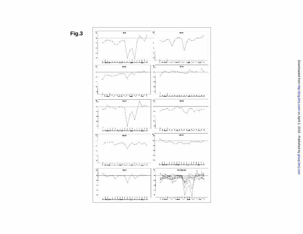
Fig.3
group.bmj.com
on April 2, 2018 - P
ublished by http://jm
g.bmj.com
/D
ownloaded from

GDF5 (NP000548) 430 EGLCEFPLRSH L EPTNHAVIQTP 452GDF6 (NP001001557) 384 EGVCDFPLRSH L EPTNHAIIQTL 406GDF7 (NP878248) 379 EGLCDFPLRSH L EPTNHAIIQTL 401BMP2 (NP001191) 325 HGECPFPLADH L NSTNHAIVQTL 347BMP4 (NP001193) 337 HGDCPFPLADH L NSTNHAIVQTL 359
Human (NP000548) 430 EGLCEFPLRSH L EPTNHAVIQTL 452Chimp (NP000548) 430 EGLCEFPLRSH L EPTNHAVIQTL 452Mouse (BC034546) 424 EGLCEFPLRSH L EPTNHAVIQTL 446Chicken (NP989669) 429 EGLCEFPLRSH L EPTNHAVIQTL 451
C400YC498S
1475ins22
R438CP441L
A
B
C
D
R438C
L441P
Fig.4
group.bmj.com
on April 2, 2018 - P
ublished by http://jm
g.bmj.com
/D
ownloaded from

causes Mohr-Wriedt brachydactyly type A2A mutation in the receptor binding site of GDF5
Rosendahl, Niels Tommerup and Stefan MundlosKlaus W Kjaer, Hans Eiberg, Lars Hansen, Carl Birger van der Hagen, Karen
published online July 13, 2005J Med Genet
http://jmg.bmj.com/content/early/2005/07/13/jmg.2005.034058.citationUpdated information and services can be found at:
These include:
MaterialSupplementary
http://jmg.bmj.com/content/suppl/2006/03/06/jmg.2005.034058.DC1Supplementary material can be found at:
serviceEmail alerting
box at the top right corner of the online article. Receive free email alerts when new articles cite this article. Sign up in the
Notes
http://group.bmj.com/group/rights-licensing/permissionsTo request permissions go to:
http://journals.bmj.com/cgi/reprintformTo order reprints go to:
http://group.bmj.com/subscribe/To subscribe to BMJ go to:
group.bmj.com on April 2, 2018 - Published by http://jmg.bmj.com/Downloaded from