a new method for myoelectric signal acquisition: preparing ... · a new method for myoelectric...
TRANSCRIPT
ROMANIAN JOURNAL OF INFORMATIONSCIENCE AND TECHNOLOGYVolume 20, Number 2, 2017, 115–123
A new Method for Myoelectric Signal Acquisition:Preparing the Patients to Efficiently Use an
Artificial Arm
Petru Lucian MILEA1, *, Adrian BARBILIAN2, *, Marius MOGA3, *, MarkEdward POGARASTEANU2, *, Ana Maria OPROIU6, *, Victor LAZO4, 5, and
Cristian Ioan STOICA2, *, **
1Politehnica University of Bucharest (PUB), Bucharest, Romania,2University of Medicine and Pharmacy ”Carol Davila” Bucharest, Romania
3Central Military Emergency Universitary Hospital (CMH), Bucharest, Romania4National Institute for Research and Development in Microtechnologies, Bucharest, Romania
5RACAI, Bucharest, Romania6University Emergency Hospital (UEH), Bucharest, Romania
*These authors contributed equally to this work.**Corresponding author: [email protected]
Abstract. This paper presents a new methodology in amputation surgery that is dedi-cated to patients who intend to use a myoelectric forearm prosthesis. The control signals for amyoelectric prosthesis are the surface EMG signals. In the case of an amputation stump, thesesignals are weak and difficult to use for effective control, due to the amputation methodologyand the lack of activity after amputation. The proposed method aims the osteomyoplastic su-turing of the muscles in circumferentially offset positions in order to obtain clear myoelectricsignals, non-overlapping and with less correlated information. Following the surgery, a spe-cific training is used, based on a biofeedback device, in order to help the patient to graduallyobtain better control of stump’s muscles. The proposed methodology was evaluated experi-mentally at CMH, in a stump retouch surgery (on a patient who lost his hand almost 20 yearsago), allowing comparison of the signals before surgery with ones after surgery and also withones obtained after completing the post-surgery training. While going through these stages,a constant improvement of amplitude and independence of EMG signals collected from thestump was found. In the end, the patient was able to control an experimental model of artifi-cial hand, by five different EMG signals, collected from stump’s muscles. The results appearto validate, in this case, the working hypotheses used as the base for the surgical method andbiofeedback training.
Key-words: amputation, CONM, artificial hand, prosthesis, myoelectric control.

116 P. L. Milea et al.
1. Introduction
Coordinated by half of the brain motor centers, for a healthy man, hands are indispensablefor most of his activities. Total or partial amputation of one or both hands is one of the mostdevastating situations that a human being could experience [2]. Statistics in recent years showthat, globally, the number of people with amputated hands is alarmingly high and continuallygrowing: 450,000 people in 2010 in the USA [3], 11,000 registered people in the UK [4] and35,000 registered people in Romania [5]. Traumatic injuries, cardiovascular diseases, malignantdiseases and birth defects are the most common causes that lead to hands amputation. Theonly chance for these patients to be able to recover at least some of the functions lost duringamputation is the acquisition and use of an artificial hand. In this context, it should be takeninto account that most of them have a modest financial condition [6] and, because the artificialhand market price is high or very high, the real situation of these millions of people is worryinglydifficult [7]. Neural control prostheses offer high-performance functions, but are inaccessible dueto high purchase price. The only solution more accessible in terms of price/performance ratio isthe use of myoelectric control prostheses, even if they have technological limitations.
One of the main limitations of myoelectric prostheses is the reduced number of movementsthat the patient can do, because of the small number of EMG control signals which can be col-lected from the patient stump [8]. Due to the difficulty (or even the impossibility) to collect fromthe forearm stump enough quality EMG surface signals (sEMG) to control the functions of theprosthesis, control signals from the arm or even shoulder muscles are often used, which greatlyhamper the development of skills by the patient [9].
Measurements that have been made in order to collect sEMG signals through various con-tractions of the stump’s muscle showed that not all the contractions from the stump were leadingto quality sEMG signals. According to intelligent prostheses theory, because the sEMG controlsignals are ”mixed”, there were revealed two main ways of diversifying the commands that thepatient can give to an artificial hand: recognition of signals/patterns of movement and their usefor making complex movements, respectively using ”unnatural” muscles to control the prosthesismovements [10]. sEMG measurements performed on the patient showed strong attenuation ofsEMG signals collected from the amputation stump compared to signals collected in similar po-sitions from the healthy hand. Possible causes leading to the attenuation of sEMG signals wereset as working hypotheses as follows:
1. Retraction of the muscles that were not appropriate sutured during the amputation;
2. Shrinking of transmission area of the nervous impulse to muscle fibers through motor endplates, due to the reduction of muscle mass after amputation;
3. Muscle atrophy due to their disuse;
4. Reduction of motor memory.
To remedy or at least reduce these causes or their effects, there were suggested solutions forsurgical correction of the first two and individual training and biofeedback for the other two. Inthe following we present the methodology that includes all.

A new Method for Myoelectric Signal Acquisition 117
2. Methods
2.1. Classical methodology of amputation
The usual medical methodology for forearm amputation involves suturing the muscles overthe bone stumps, without explicitly specifying a particular individual disposition of them. Al-though it is medically correct, the usual procedure makes the acquisition of surface EMG signals(sEMG) from different groups of muscles of the stump very difficult after amputation. Patientsundergoing a specific post-surgery training, even if they manage (with difficulty) to contract eachmuscle group from the stump separately, this success does not lead to the collection of distinctsEMG signals, due to the existence of four volar muscle plates. Thus, the number of commandsto control an artificial hand is limited to the small number of distinct sEMG signals.
2.2. The method of amputation/retouch of stump by circumferential os-teoneuromyoplasty (CONM)
The improvement of the methodology of amputation was aimed by suturing muscle groupsin appropriate positions in order to get clean and distinct myoelectrical signals, with independentinformation (low correlation) after the distinct contraction of each muscle groups. This methodaims to bring deep muscle plans to the surface, preserving the nervous distribution area in orderto obtain distinct EMG signals with low correlation. This methodology involves placing and su-turing the stump muscles by muscle groups and categories of motor functions, in order to allowcollection of several uncorrelated sEMG signals from the stump. This allows distinct and morenatural control (with signals from muscles that would have produced the same movements to ahealthy hand) of movement of the prosthesis components. Thereby it becomes justified to equipthe intelligent prosthesis with larger motor functions that the patient could use. The eligibilitycriteria for participants are: forearm amputees which needs forearm’s stump reshape and who arealso interested in using a noninvasive intelligent prostheses. Elbow disarticulation amputees areexcluded. During the surgery it is aimed to identify all muscle groups and each muscle with theircircumferential transosseous reinsertion, in order to avoid muscle retraction and increase the sur-face of muscle exposed to EMG measurements. Muscles will be well individualized, yet keepingthe topography of the main muscle groups: ventral muscles into ventral horn and dorsal musclesinto dorsal horn. In the classical method of amputation (Figure 1a) it can be observed that somemuscles of the stump, which might provide useful sEMG signals are superimposed on others,due to natural anatomical distribution. The osteoneuromyoplastic circumferential methodology(CONM) that was proposed (Figure 1b) aims to bring muscle groups in the stump to the surfacecircumferentially before anchoring. This placement of muscle groups in the stump allows alsocollection of sEMG signals from deep muscle groups with surface EMG electrodes. The CONMmethodology also involves the forearm fasciotomy at the amputation stump in order to reducethe electrical resistance of the tissue layer between muscles and electrodes, for increasing thelevel of collected sEMG signal. To improve the nervous impulse transmission to muscle fibers,the methodology involves nerves neurolysis followed by increasing the length of the motor nervebranches entering each muscle group.

118 P. L. Milea et al.
Fig. 1. Schematic section representation of partially amputated forearm muscles’ position: bythe classical method (a) and by the circumferential osteoneuromyoplastic method (b).
3. Training methods and techniques for increasing sEMGsignal
The main training method begins with isometric contractions of stump’s muscles in orderto increase both the patient’s ability to control these muscles, and muscle’s strength. In thenext stage, different movements are performed with stump’s muscles and healthy arm’s musclessuccessively and then simultaneously. Thus, certain movements made symmetrically and simul-taneously with both hands help to rehabilitate some affected features of the partially amputatedhand because the patient with an amputated hand and a healthy hand has an active brain map ofthe healthy hand’s muscles and a brain map that is intended to be reactivated for the amputatedhand. Finally, the patient learns again to use certain muscle groups on the stump by contract-ing them separately. This training is supported by biofeedback techniques but also by solutionsfor reactivating the psychomotor representation of the amputated forearm in the patient’s motorcortex. During the training workout, the patient is connected with the electrodes on the stumpto biofeedback device, first with each muscle separately, in order to improve his control over it,then with more muscles, in order to simulate the complex control contractions of a prosthesis.Thus, the patient sees directly on the biofeedback model, the straining effects of certain musclesfrom the stump. The patient will be able to command gradually the moving parts of prosthesis.
3.1. Evaluation equipment and biofeedback devices used
The EMG devices that was used were Contec CMS6600 (PUB) and Nicolet EDX (UEH),together with their dedicated EMG software. There were also developed a portable biofeedbackdevice and a model of intelligent prosthesis with independently actionable fingers, that were usedas a support for patient’s training. The biofeedback device is hand shaped and each finger canlight LEDs in three colors, depending on the control signal amplitude generated by the patient.The most complex device, developed in collaboration by UPB and IMT is an artificial handmodel that allows independent movements of the five fingers.

A new Method for Myoelectric Signal Acquisition 119
4. Discussion
Methodology and training techniques were experimentally evaluated on a patient with tran-sradial amputation of the right hand, who needed surgery to retouch the stump. Experimentalevaluation of CONM methodology for a retouching surgery allowed comparison of the myoelec-trical effects of the two methods of amputation.
4.1. Case presentation
The 37 year old patient had a post-traumatic amputation at the age of 9 and, due to muscleatrophy and retraction, experienced pain on amputation stump. Thus, the retouch surgery of thestump became necessary and the patient subscribed to the CONM method by self-selection (on04.04.2013). The recruitment was based on the patient’s interest in buying forearm prosthesiswith myoelectrical command.
Measurements done before surgery showed the fact that the sEMG signals collected fromthe patient’s stump at alternate contractions of different muscle groups had a low amplitudeand also were strongly correlated, thus being not useful for obtaining different commands of aprosthesis. Figure 2 shows such typical results (on the display in the background): sEMG signalare monitored on two different channels for two distinct muscle groups. Although the patientintends to contract only one group, the signals or both channels are similar (or, in mathematicalterms, are highly correlated). The amplitude of the signals has a mean value of around 10µVwith a statistical variance of about 15% (from 5 different measurements) and comparable withthe noise.
Fig. 2. sEMG measurements before retouch surgery.
120 P. L. Milea et al.
5. The retouch of the stump surgery performed by the CONMmethod
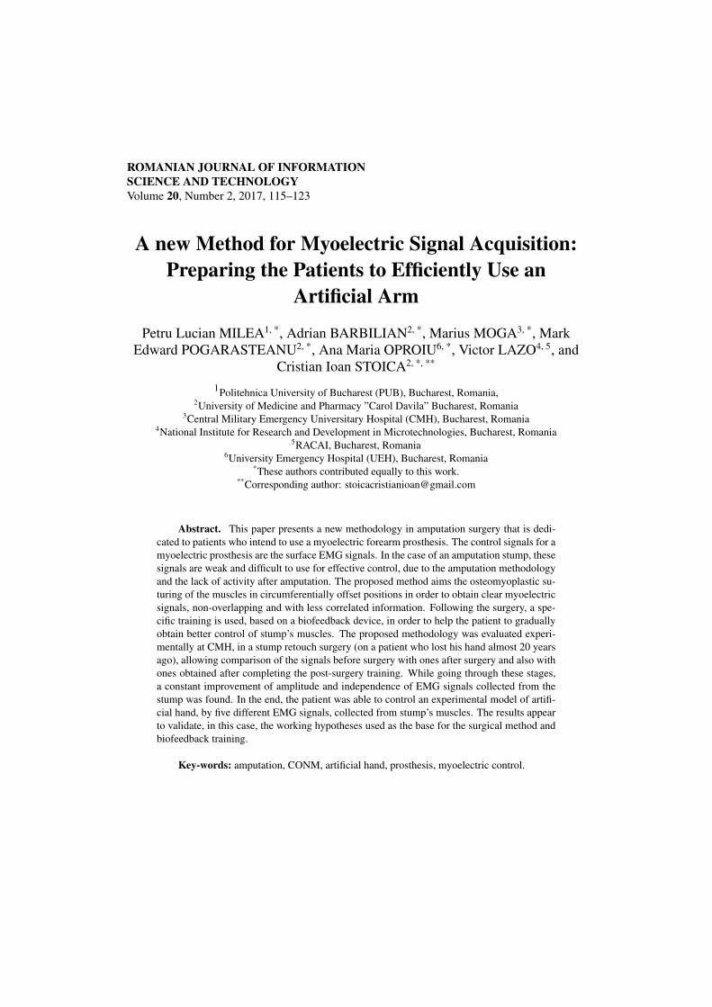
The surgery involved the excision of bone ends, the identification of all muscles followed bybringing muscles to the surface circumferentially, according to the presented methodology, inorder to increase muscle surface exposed to EMG measurements and to avoid muscle retraction.The intervention was done at Central Military Emergency Universitary Hospital by Prof. AdrianBarbilian and his medical team.
Muscles were placed individually, yet keeping the topography of the main muscle groups:ventral muscles into ventral horn and dorsal muscles into dorsal horn (see Figure 3).
Fig. 3. sEMG measurements before retouch surgery.
Additionally, in order to reduce the electrical resistance of the layer of tissue between muscleand EMG electrodes, the forearm fasciotomy was also performed near the amputation stump. Inorder to enhance nerve inflows transmission to muscles, motor nerves neurolysis was conductedas well. There were found residual nerve items: posterior interosseous nerve, median nerve-motor nerves, which were neurolysed.
5.1. The results achieved immediately after surgeryPost-surgery evolution was favorable with complete disappearance of painful symptoms within
two weeks. Then, after other two weeks, sEMG signals was collected from the stump. The newsignals had 20% higher amplitude (under the same statistical conditions) compared with thoseobtained from pre-surgery measurements.
A new Method for Myoelectric Signal Acquisition 121
5.2. Training program and results
In order to improve the capability to control a prosthetic device with myolectric stimuli, thepatient followed a specific training program that aimed the development of specific abilities.The program started one month after surgery, with a daily basic training, consisting in 4 hourssessions of isometric contractions of stump’s muscles. The sEMG signals collected after thesesessions showed a slight improvement in amplitude (10%) and control capability.
In the next stage, for a month, the patient conducted sessions of successive movements andthen synchronous with both hands performing contractions of the same muscle. Stum’s muscleshave generated sharper sEMG signals and with 80% higher amplitude for synchronous move-ments made by the patient, showing a better control (same statistical conditions).
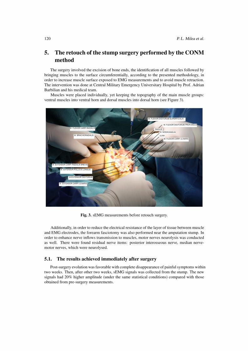
The results achieved in the post-surgery training phase were highlighted by simultaneousmeasurements of sEMG signals with two EMG devices. For the measurements pictured in figure4, the electrodes are placed on the stump on different muscle groups.
Fig. 4. Simultaneous measurements with two EMG devices.
The measurements have shown the fact that sEMG signals corresponding to different musclegroups’ contractions was distinct and less correlated than the previous ones. This was a signifi-cant improvement related to the initial situation and was due to the CONM retouch surgery. Thetwo EMG devices were used in order to certify that there are low or no interferences betweendifferent signals’ envelopes.
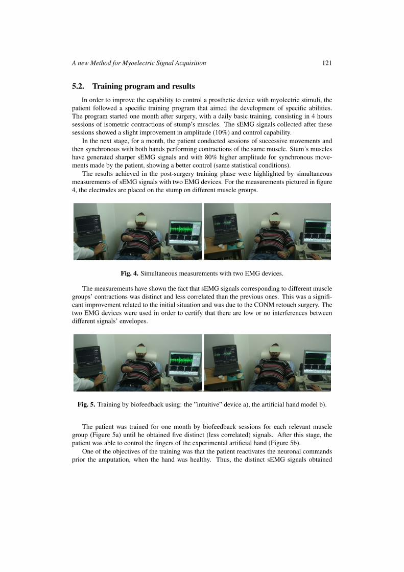
Fig. 5. Training by biofeedback using: the ”intuitive” device a), the artificial hand model b).
The patient was trained for one month by biofeedback sessions for each relevant musclegroup (Figure 5a) until he obtained five distinct (less correlated) signals. After this stage, thepatient was able to control the fingers of the experimental artificial hand (Figure 5b).
One of the objectives of the training was that the patient reactivates the neuronal commandsprior the amputation, when the hand was healthy. Thus, the distinct sEMG signals obtained
122 P. L. Milea et al.
correspond to commands already existing in the neuronal map of the patient. As the experimentsproved, this helps the patient to reactivate some motion reflexes at the level of the stump muscles.The results appear to validate, in this case, the working hypotheses used as the base for thesurgical method and biofeedback training. The interdisciplinary team continues the CONM studyon other cases and also the work to develop a new sEMG controlled artificial hand [11, 12].
6. ConclusionsThe proposed methodology (CONM) was evaluated experimentally on a patient with tran-
sradial amputation of the right hand. He received a retouch surgery of stump that removed thepainful symptoms on this level and brought deep muscles of the stump to the surface, in or-der to better highlight their EMG signals. After surgery and recovery period, the collection ofsEMG signals from the stump allowed the highlight of a better control and higher amplitude.The retouch surgery of stump that was conducted in this case allowed the comparison of myo-electrical effects of the two methods of amputation, and the highlight of the CONM method’sadvantages. The CONM method allowed the non-invasive collection of sEMG signals from allmuscle groups of the stump. The access with surfaces non-invasive electrodes at muscle groupsthat were brought to surface with CONM method made possible the collection of five differentsignals with low statistical inter-correlation. The training program helped the patient to improveboth the amplitude of sEMG signals on the stump and his ability to control them. The trainingwas assisted by biofeedback, allowing the patient to improve his performance and to evaluate hisprogress constantly. This led him to the individual control five different signals, allowing its con-nection to the experimental model of artificial hand. Taking into account the fact that the patientlost his hand almost 20 years ago, the results obtained are already impressive. The results areunder discussion and generalization and are to be updated following other patients enrollmentsinto the CONM trial and subsequent training.
Acknowledgements. This work was supported from Robocom++ FLAG-ERA JTC Project.
References[1] World Health Organisation – International Clinical Trials Registry Platform, The circum-
ferential osteoneuromyoplasty method of forearm amputation or stump retouch – CONM,http://apps.who.int/trialsearch/trial.aspx?trialid=DRKS00004868.
[2] JIBBY E. Kurichi, BATES E. Barbara, STINEMAN G. Margaret, Amputation, International Encyclo-pedia of Reabilitation, http://cirrie.buffalo.edu/encyclopedia/en/article/251/
[3] ZIEGLER-GRAHAM K., MACKENZIE E. J., EPHRAIM P. L., TRAVISON T. G., BROOKMEYERR., Estimating the prevalence of limb loss in the United States: 2005 to 2050, Archives of PhysicalMedicine and Rehabilitation, 89(3) , pp 422–429, 2008.
[4] University of Salford, Limbless Statistics Report – 2006/07, Manchester, http://www.limbless-statistics.org/documents/Report2006-07.pdf
[5] Romanian National Organization of Disabled People, http://www.onphr.ro/
[6] GRANT McGimpsey and BRADFORD C. Terry, Limb Prosthetics Services and Devices CriticalUnmet Need: Market Analysis, Bioengineering Institute Center for Neuroprosthetics Worcester Poly-technic Institution.

A new Method for Myoelectric Signal Acquisition 123
[7] HAITIAN J. Daniel Kelly Amputees – Lessons Learned from Sierra Leone, New England Journal ofMedicine 2010.
[8] CHIHARU Ishii, Recognition of Finger Motions for Myoelectric Prosthetic Hand via Surface EMG,Advances in Mechatronics, pp. 176 – 190, 2011.
[9] CASTELLINI Claudio, FIORILLA E. Angelo, SANDINI Giulio, Multi-subject/daily-life activityEMG-based control of mechanical hands, Journal of NeuroEngineering and Rehabilitation, 2009.
[10] LA Miller, KA Stubblefield, RD Lipschutz, BA Lock, TA Kuiken, Improved myoelectric prosthesiscontrol using targeted reinnervation surgery: A case series, IEEE Trans Neural Syst Rehabil Eng 2008.
[11] GOSCHIN S., FRANTI E., DASCALU M., PIETRAROIU M., Autonomous agents with control sys-tems based on genetic algorithms, in Proceedings of the 12th IASTED International Conference onRobotics and Applications, Book Series: IASTED International Conference on Robotics and Appli-cations, pp 49–54, 2006.
[12] GOSCHIN S., FRANTI E., DASCALU M., OSICEANU S, Combine and compare evolution-ary robotics, and reinforcement learning as methods of designing autonomous robots, 2007 IEEECONGRESS ON EVOLUTIONARY COMPUTATION, (1-10), IEEE Congress on EvolutionaryComputation, Singapore, pp. 1511-1518